AN INVITRO STUDY TO EVALUATE THE
ANTIBACTERIAL PROPERTY AND SHEAR BOND
STRENGTH OF AN ORTHODONTIC ADHESIVE
COMBINED WITH TWO DIFFERENT
NANOPARTICLES.
Dissertation submitted to
The Tamil Nadu Dr M.G.R. Medical University
In partial fulfilment of the degree of
MASTER OF DENTAL SURGERY
BRANCH V
ORTHODONTICS AND
DENTOFACIAL ORTHOPEDICS
ACKNOWLEDGEMENT
First and foremost, I would like to thank my Creator for giving me health to
live life and learn, and particularly to work on my dissertation project, as a part of
completing my Master’s studies.
I would like to give my highest, respectful gratitude to Dr. Anilkumar. P,
Professor and Head of the Department, Department of Orthodontics, Sree
Mookambika Institute Of Dental Sciences, Kulasekharam for his guidance,
understanding and most importantly his patience during my course. His mentorship
was paramount in providing a well rounded experience which helped me in attaining
my goals. Without his guidance and persistent help this dissertation would not have
been possible, I thank you, Sir.
I consider it a great privilege and an honour to express my profound gratitude
to Dr.Shino P.Mathew, Professor, Department of Orthodontics, SMIDS, Kulasekharam for his guidance. He encouraged me to try new innovative ideas and to not only grow
as an experimentalist but also as an independent thinker. I have benefited greatly from
his attention to the finer details.
It is with immense gratitude that I acknowledge the support and help of
Dr.Amal S.Nair, Professor, Department of Orthodontics, SMIDS, Kulasekharam for providing me his great insights, advices, help and kind support right from the
selection of my thesis topic through out the course of this study
I extend my sincere thanks to Dr.Antony Shijoy Amaldas, Senior Lecturer, SMIDS, Kulasekharam for his valuable help and support. I thank him for sharing his
I sincerely thank Dr. Anna Oommen, Senior Lecturer, SMIDS, Kulasekharam. She had always supported me whenever I was in need. I thank her for
the grammatical corrections she made in this work.
I would like to express my heart filled gratitude to Dr.Anjana S.Nair Senior Lecturer, SMIDS, Kulasekharam and also my senior who paved the path of guidance
and support throughout this project.
I would like to express my thanks to Dr. David T.Danny Senior Lecturer, SMIDS, Kulasekharam who offered collegial guidance and support.
I am fully indebted to my fellow post graduate student Dr.Anitha for helping me survey all the stress through these years. I thank her for sailing in the same path
with me without any egos and clashes.
I would also like to thank my seniors Dr. Rajesh, Dr. Rahul,Dr.Rajkumar, and my juniors Dr.Harsha, Dr.Thasneem , Dr.Surya and Dr.Chandana for their constant encouragement and help.
I also place on record my sense of gratitude to Dr.Roy Joseph, SCTIMST, TVM and members of CSIR, Karaikudi, and all who directly or indirectly lend their
helping hand in this project.
Finally, I would like to thank my better half, my soul mate, and my husband
Dr. Stalin Joseph who is the sole reason for me to persuade this degree. I would like to thank him for his never ending motivation, support and love. I would like to
express my eternal appreciation toward my kids and my parents who have always
SPECIAL ACKNOWLEDGEMENT
I take this opportunity to thank Dr.C.K Velayudhan Nair MS, Chairman, Dr.Rema.V.Nair MD, Director, Trustees Dr.R.V Mookambika MD,DM, and Dr.Vinu Kopinath MS,MCh and Dr.Elizabeth Koshy MDS, Principal Sree Mookambika Institute of Dental Sciences, Kulasekharam, Tamil Nadu for giving me an opportunity to utilize the facilities available in this institution for conducting
SL NO
INDEX
PAGE NO
1.
List of Abbreviations
i - ii
2.
List of Figures
iii-iv
3.
List of Tables
v-vi
4.
List of Graphs
vii
5.
List of annexure
viii
6.
Abstract
ix-x
7.
Introduction
1-5
8.
Aims and Objectives
6
9.
Review of Literature
7-29
10.
Materials and Methods
30-39
11.
Result and Observations
40-52
12.
Discussion
53-65
13.
Summary and Conclusion
66-67
14.
Bibliography
xi-xxiii
i
ACP Amorphous Calcium Phosphate
AgO Silver Oxide
ANOVA Analysis of Variance
BAC Benzalkonium Chloride
BAG Bioactive Glass
BHI Brain Heart Infusion
CDA Chlorhexidine Diacetate
CPC Cetylpyridinim Chloride
DCT Direct Contact Test
DNA Deoxyribose Nuclear Acid
ECA Experimental Composite Adhesive
EDAX Energy Dispersive X-ray Analysis
ENMs Engineered Nano Materials
GIC Glass Ionomer Cement
H2O2 Hydrogen Peroxide
ii
META/MMA-TBB MethacryloxyethylTrimellitic Anhydride/ Methyl
Methacrylate-Tri-N-Butyl Borane
MIC Minimum Inhibitory Concentration
MPa Mega Pascals
NPs Nano Particles
OH Hydroxyl
RMGI Resin-Modified Glass Ionomer
ROS Reactive Oxygen Species
SBS Shear Bond Strength
SD Standard Deviation
SEM Scanning Electron Microscopy
SFE Surface free Energy
TEM Transmission electron Microscope
TiO2-NPs Titanium dioxide Nanoparticles
WSL White Spot Lesions
iii
Figure Number Details
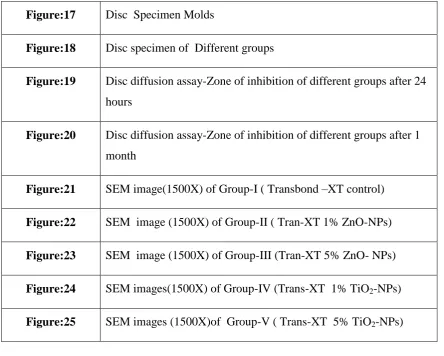
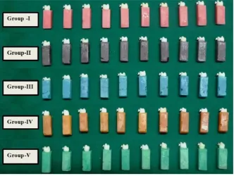
Figure:1 Different groups of premolar teeth mounted in color coded acrylic blocks
Figure:2 Representative sample of bracket
Figure:3 Armamentarium
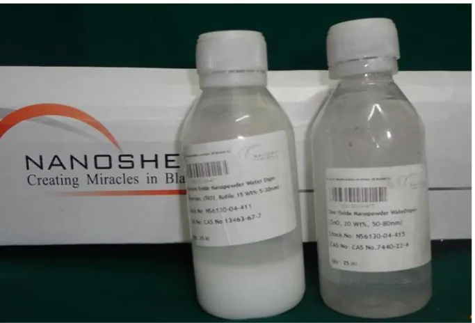
Figure:4 Nano particles dispersion
Figure:5 Modified orthodontic composite adhesive
Figure:6 Application of etchant gel-37% Phosphoric acid
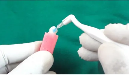
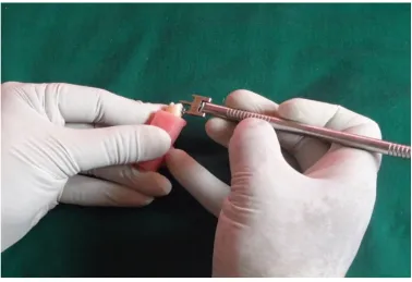
Figure:7 Bracket placement
Figure:8 Positioning of the bracket
Figure:9 Curing of brackets
Figure:10 Different groups after bonding brackets on the tooth
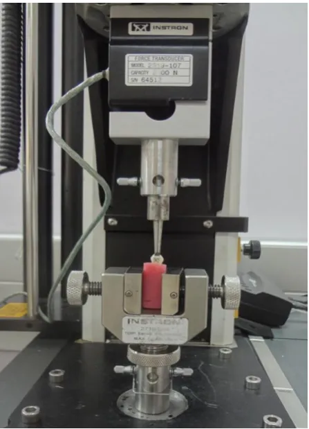
Figure:11 Instron universal testing machine
Figure:12 Instron universal testing machine-lateral view loaded with tooth specimen
Figure:13 Speed mixer
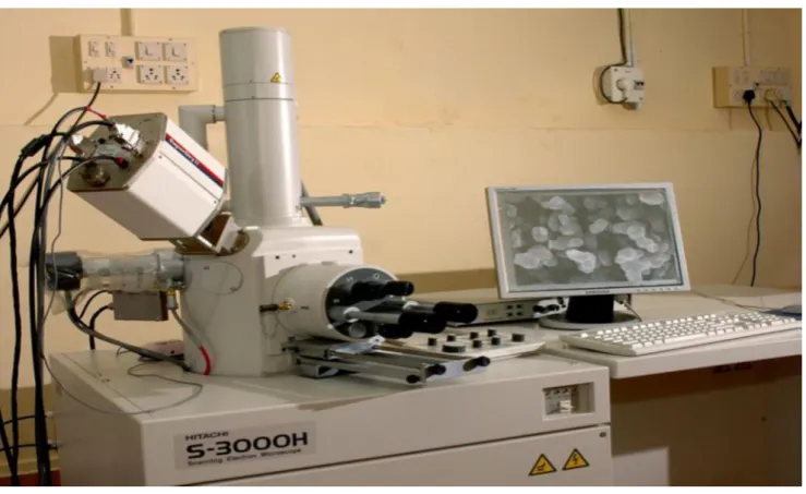
Figure:14 SEM with Energy-dispersive X-ray analysis unit
Figure:15 Sputter coating Machine
iv
Figure:18 Disc specimen of Different groups
Figure:19 Disc diffusion assay-Zone of inhibition of different groups after 24 hours
Figure:20 Disc diffusion assay-Zone of inhibition of different groups after 1 month
Figure:21 SEM image(1500X) of Group-I ( Transbond –XT control)
Figure:22 SEM image (1500X) of Group-II ( Tran-XT 1% ZnO-NPs)
Figure:23 SEM image (1500X) of Group-III (Tran-XT 5% ZnO- NPs)
Figure:24 SEM images(1500X) of Group-IV (Trans-XT 1% TiO2-NPs)
[image:11.595.75.520.71.427.2]v
Table Number Details
Table-1 Mean Shear Bond Strength (MPa) values of different groups
Table-2 Comparison of mean Shear Bond Strength values of Group-I with other groups
Table-3 Comparison of mean Shear Bond Strength values of Group-II with other groups
Table-4 Comparison of mean Shear Bond Strength values of Group-III with other groups
Table-5 Comparison of mean Shear Bond Strength values of Group-IV with other groups
Table-6 Comparison of mean Shear Bond Strength values of Group-V with other groups
Table-7 Multiple comparison of mean Shear Bond Strength values between the groups
Table-8 Mean Zone of inhibition diameter (mm) values of different groups at different time intervals
vi
Table-11 Comparison of mean Zone of inhibition diameter (mm) values Group-IV with other groups at different time levels
Table-12 Comparison of mean Zone of inhibition diameter (mm) values Group-V with other groups at different time levels
Table-13 Multiple comparison of mean Zone of inhibition diameter (mm) values between the groups at 24 hours
Table-14 Multiple comparison of mean Zone of inhibition diameter (mm) values between the groups after one month
[image:13.595.102.539.70.474.2]vii
Graph Number Details
Graph- I Mean Shear Bond Strength (MPa) values of different groups
Graph -II Multiple comparison of mean Shear Bond Strength values between the groups
Graph III Multiple comparison of mean Zone of inhibition diameter (mm) values between the groups at 24 hours
Graph IV Multiple comparison of mean Zone of inhibition diameter (mm) values between the groups after one month
Graph V EDAX graph of Group-I ( control)
Graph VI EDAX graph of Group-II
Graph VII EDAX graph of Group-III
Graph VIII EDAX graph of Group-IV
viii
LIST OF ANNEXURE
Annexure number Details
1 Institutional Research Committee Certificate
2 Institutional Human Ethics Committee Certificate
3 Nanoparticles-Safety Data Sheet
4 Test Report- SCTIMST,TVM
ix Introduction:
The purpose of this invitro study is to evaluate the antibacterial property and
shear bond strength of the modified orthodontic composite incorporated with
nanoparticles of Titanium dioxide (TiO2) and Zinc Oxide (ZnO) at two different concentrations and this study also compares which nanoparticle has better
antibacterial property without compromising the shear bond strength.
Materials and Methods:
ZnO nanoparticles and TiO2 nanoparticles in dispersion form were added to
commercially available orthodontic adhesive Transbond-XT at 1% and 5% concentration
separately by a speed mixer to form modified composite. In this study 50 human
extracted teeth were used which are divided into five groups of 10 tooth each and
were bonded with brackets using different group bonding adhesives Transbond-XT ,
Tran- XT 1% ZnO-NPs, Tran XT 5% ZnO-NPs, Tran- XT 1% TiO2-NPs and
Tran-XT 5% TiO2-NPs respectively. Shear bond testing was done using Universal testing machine.30 disc specimens were prepared using different group bonding
adhesive and one disc from each group were subjected to SEM-EDAX analysis and
the remaining were used for antibacterial analysis against Streptococcus mutans using
Bauer-Kirby disc diffusion assay.
Result:
Analysis of variance indicated a significant difference (P < 0.005) among the
groups. The mean shear bond strength of Transbond-XT (Group-I) control was found
to be 16.91±1.35MPa It was slightly decreased to16.13±1.42MPa in Group-II
x
(Tran-XT 5% ZnO-NPs). The mean shear bond strength was increased in Group-IV
(Tran-XT 1% TiO2-NPs) to 17.54±1.74MPa than the control but it shows a decrease in Group-V (Tran-XT 5 %TiO2- NPs) to 14.35±1.78 MPa. All groups except control
showed antibacterial effect. The biggest zone of inhibition was seen in Group-III
(Tran-XT 5 % ZnO- NPs) 18.08±0.14 mm. Smallest zone of inhibition is seen in
Group-IV (Tran-XT 1% TiO2-NPs) 5.94±0.15 mm. The antimicrobial effect is present
in the modified adhesive even after one month.
Conclusion:
The result showed that ZnO nanoparticles and TiO2 nanoparticles incorporated modified orthodontic adhesive showed added microbial property to the original
compound without adversely affecting the Shear bond strength. ZnO nanoparticles
showed the greatest antimicrobial activity. Antimicrobial property was seen even after
1 month.
Key Words:
White spot lesion, Shear bond strength, Antimicrobial activity, Nanoparticles,
1
“A thing of beauty is a joy forever’’ is a common saying. Facial appearance
plays the most important part in deciding the beauty of a person. A good smile with
well aligned teeth is considered aesthetic. Most patients view orthodontic treatment as
a means to improve dentofacial aesthetics1. Fixed orthodontic appliances offer a myriad of opportunities to improve smile aesthetics and occlusal relationships. In this
modern era teenagers and adult prefer to get esthetically bloomed than to get the
treatment certainly for the irregular teeth. But the presence of white spot lesions after
orthodontic appliances therapy is a disappointing finding to a specialty whose goal is
to improve facial and dental aesthetics. Decalcification of the enamel surface, known
as white spot lesions (WSL), adjacent to fixed orthodontic appliances is an important
and prevalent iatrogenic effect of orthodontic therapy2. Demineralization is an inevitable side-effect associated with fixed orthodontic treatment, especially when
associated with poor oral hygiene.
After the introduction of orthodontic fixed appliances into the oral cavity, a
rapid shift in the bacterial flora of plaque occurs. Higher levels of acidogenic bacteria
are present in the plaque, most notably Streptococcus mutans and Lactobacillus3. High levels of these bacteria are capable of decreasing the PH of plaque in orthodontic patients to a greater extent than in non-orthodontic patients. The acidic byproducts of
these bacteria in plaque are responsible for the subsequent enamel demineralization
and formation of white spot lesions. These can cause caries thereby leading to poor
aesthetics, patient dissatisfaction and legal complications. Clinically, formation of
white spots around orthodontic attachments can occur as early as 4 weeks into
2
including proper tooth brushing with a fluoridated dentifrice5. Decalcification occurs when specific bacteria are retained on the enamel surface for a long time and produce
organic acids; therefore, if a bonding agent could prevent bacterial growth, it could
inhibit demineralization adjacent to orthodontic brackets. For less compliant patients,
an antimicrobial bonding system around the bracket base would be advantageous6.
Different orthodontic bonding resins that provide antimicrobial protection
have been evaluated, but to date, none are commercially available. Fluorides play an
important role in the prevention of WSL.Bonding agents containing fluoride have the
potential for decreasing enamel decalcification. It was found that the fluoride release
is more with resin modified GIC, as compared with the fluoride containing
composites.Bonding agents that release fluoride shows rapidly decreasing level of
fluoride after the first 24 hours7.Combining chlorhexidine with the bonding primer or applying it after bonding resulted in no significant decrease in shear bond strength and
induced antiplaque benefits8.More recently, the use of an antimicrobial, Cetylpyridinim chloride (CTC), showed to inhibit bacterial growth9. An experimental antibacterial bonding system Methacryloxydodecyl-pyridinium bromide(MDPB), was incorporated
into the Transbond XT primer showed positive result against WSL10.Resin-modified glass-ionomer cement (Fuji Ortho LC) exhibit significant inhibition of adjacent
demineralization compared with the non-fluoride releasing adhesives11. However, traditional fluoride releasing cements, glass-ionomer cements and resin-modified GIC
have bond strengths that are substantially lower than those of conventional resins.
Bioactive glass (BAG)-Bond adhesives successfully releases ions (calcium,
phosphate and fluoride ions) and maintaining superficial enamel hardness
surrounding orthodontic brackets and decreased the chances of WSL around
3
The antibacterial properties of metals such as silver, zinc and copper have
been known for centuries and this has been exploited in modern medicine for
infection control. Recently the potential of engineered nanomaterials (ENMs) as
antibacterial agents for infection control in dentistry and the management of the oral
biofilm13 has been recognized and metal-containing nanomaterials available in chemical forms including solid nanoparticles (NPs) of metal or metal oxides
(e.g.,ZnO NPs, TiO2 NPs, AgO NPs) has been used in dentistry for infection control14. Nanoparticles are clusters of atoms in the size range of 1-100 nm. Nano as a word means one-billionth of a physical quantity. Nanoscale materials have very
different new properties which differ from materials in molecular form.
Nanomaterials have a much greater surface area to volume ratio than their
conventional forms15. Recent studies have demonstrated that specially formulated metal oxide nanoparticles have good antibacterial activity, and antimicrobial
formulations comprising of nanoparticles could be effective bactericidal materials16. The nanoparticles are highly reactive, as it binds to tissue proteins and brings
structural changes in the bacterial cell wall and nuclear membrane, leading to cell
distortion and death. It is believed that interaction with the three main components of
bacterial cells produce the bactericidal effect: the peptidoglycan cell wall and plasma
membrane, bacterial DNA, and bacterial proteins, especially enzymes involved in
vital cellular processes such as the electron transport chain17.The nanoparticles antibacterial property is induction of intracellular reactive oxygen species like
hydrogen peroxide, a strong oxidizing agent harmful to bacterial cells18.
In dentistry, Nano composites and orthodontic adhesives seem to be the major
applications for nanoparticles, with promising results for antibacterial properties.
4
demineralization without compromising physical properties. It has been shown that
Streptococcus mutans is sensitive to nanoparticles of silver, zinc oxide, and gold,
titanium which allows achieving important clinical effects19.The incorporation of silver nanoparticles into bonding adhesives was successful on both physical and
antimicrobial level20.
Among metal oxide nanoparticles, ZnO nanoparticles have many significant
features such as chemical and physical stability, high catalysis activity and effective
antibacterial activity. Zinc oxide has been shown to inhibit the growth of plaque and
shows antimicrobial activity when incorporated into dental composites21-23.Zinc oxide has been used in various ways, including pharmaceutical creams or ointments for the
treatment of leg ulcers, traumatic wounds and burns24. Dental materials with zinc oxide such as endodontic sealers and fixed restoration cements have been utilized for
this same reason. Antibacterial nanoparticles such as zinc oxide alone or the
combination of zinc oxide and chitosan nano-particulates shows reduction of
Erchersia faecalis adhered to the surface of the root canals25. Zinc, which serves as
an activator of enzymes can be toxic to microbes at concentrations as low as 0.5 ppm
Recently Titanium dioxide (TiO2) is widely utilized as a self-cleaning and self-disinfecting surface coating materials, titanium dioxide has a more helpful role in
environmental purification due to its photo induced super-hydrophobicity and
antifogging effect. These properties have been applied in removing harmful bacteria.
Titanium dioxide nanoparticles (TiO2-NPs) decompose organic compounds by the
5
containing TiO2 nanoparticles possessed the more potent antibacterial activity against
Streptococcus. mutans compared to unmodified glass ionomer27.
The direct bonding of orthodontic brackets with composite resin, described by
Newman, has been considered the most popular method and the clinical standard for
attaching orthodontic brackets to teeth28. Bonding of orthodontic brackets has become an essential procedure to accomplish the clinical treatment, and researchers have
worked hard to achieve the best qualities of bonding agents to maintain a sound
unblemished enamel surface after removing orthodontic bracket. Incorporating
nanoparticles of zinc oxide and titanium dioxide in orthodontic adhesive composite
can help prevent enamel demineralization around bracket surfaces. Zinc oxide and
titanium dioxide nanoparticles have well-known inhibitory and bactericidal effects.
This study is to incorporate nanoparticles of TiO2 and ZnO at different concentration into the commonly used orthodontic adhesive Transbond XT and to
evaluate the antibacterial property and the shear bond strength of the modified
composite. This study also compares which nanoparticle has better antibacterial
property without compromising the shear bond strength. The null hypothesis to be
tested is that modified Transbond-XT combined with nanoparticle has antibacterial
6
To evaluate Shear bond strength (SBS) and antibacterial effects of an orthodontic adhesive after incorporating Zinc Oxide nanoparticles (ZnO-NPs)
and Titanium dioxide nanoparticles (TiO2-NPs) at two different concentrations.
7
Buonocore29 (1955) outlined a simple method of increasing the adhesion of filling materials to enamel surfaces. He employed 85% phosphoric acid and
phosphomolybdate oxalic acid treatment to alter the enamel surface chemically and
concluded that phosphoric acid gave better results and was simpler to use.
Reynolds30 (1975) recommended 6 to 8 MPa as reasonable bond strength for orthodontic purposes. However, this suggestion was made over 38 years ago based on
a bracket base area of 16 mm2. Since then, there have been recent advances in bracket materials and design, adhesives, computer technology and testing systems.
Gorelick L et al2 (1982) studied incidence of white spot formation after bonding and banding. After examining two groups of patients treated by a
multibonded technique and recording the incidence of white spots at the time of
debonding, he concluded that there was no difference in white spot formation in those
that were banded or bonded. The labiogingival area of the maxillary lateral incisors
had the highest incidence of white spots. The highest incidence occurred among the
maxillary incisors; the lowest was in the maxillary posterior segment. No white spots
were found on the lingual surfaces of mandibular canines and incisors. These findings
suggest a relationship between resistance to white spot formation and the rate of
salivary flow .The degree of iatrogenic damage during orthodontic treatment suggests
the need for preventive programs using antibacterial agents.
Mizrahi E et al4 (1982) evaluated enamel demineralization following orthodontic treatment. Enamel opacity index was used to record the severity. Male
patients experienced a significantly higher increase in the severity of enamel
opacities. There was no significant sex differential in the prevalence of enamel
8
orthodontic treatment with multibanded appliances contributed to the development of
new areas of enamel demineralization and to an increase in the severity of enamel
opacities. He concluded that there was a significant increase in both the prevalence
and severity of enamel opacities following completion of orthodontic treatment.
Trimpeneers LM et al31(1996) conducted clinical evaluation of the effectiveness of a fluoride releasing visible light-activated bonding system to reduce
demineralization around orthodontic brackets. A clinical trial was carried out to
compare the effect of a visible light-cured fluoride releasing (F-releasing) material
with a chemically cured non-fluoride resin on white spot formation during fixed
orthodontic therapy. Fifty patients were examined. Intraoral slides were taken before
and after treatment and were evaluated for white spot formation. The results indicate
that there was no significant difference between the decalcification rates for both
types of adhesives. When the appearance of white spots was evaluated in an overall
manner, there was significantly more upper than lower decalcification.
Bishara SE et al8 (1998) studied effects of various methods of chlorhexidine application on shear bond strength. Chlorhexidine has been applied on the teeth and
over orthodontic appliances during treatment in order to reduce bacterial colonization.
The purpose of this study was to determine whether the application of chlorhexidine
with or without a sealant, to the etched enamel will affect the shear bond strength and
the bracket/adhesive failure modes of orthodontic brackets. The chlorhexidine was
applied to the teeth either as a prophylactic paste or as a varnish. Of all the
experimental groups in which the chlorhexidine varnish was applied as a layer on the
etched enamel surface or over the sealant, shear bond strength values and bracket
9
Bishara SE et al32 (1999) investigated the Shear bond strength of composite, glass ionomer, and acidic primer adhesive systems. Group I teeth were etched with
37% phosphoric acid and bonded with Transbond XT following the manufacturer's
instructions. Group I acted as the control group. Group II teeth were etched with an
acidic primer (Clearfil Liner Bond) that contains both the acid (Phenyl-P) and the
primer (HEMA and dimethacrylate) and was placed on the enamel for 30 seconds; the
adhesive used to bond the brackets was Transbond XT as in Group I. Group III teeth
were etched with 20% polyacrylic acid and the brackets were bonded with Fuji Bond
LC. The results indicated that the resin/phosphoric acid adhesive system (control
group) provided the strongest shear bond strength. The glass ionomer adhesive system
provided significantly lower bond strength. The least shear bond strength was present
when the acidic primer was used with an orthodontic adhesive.
Wilson RM et al7 (2001) studied demineralization around orthodontic brackets bonded with resin-modified glass ionomer cement (Fuji Ortho LC) and
fluoride-releasing resin composite (Light Bond). The teeth were placed in an artificial
caries solution to create lesions. Following this polarized light microscopy was
utilized to evaluate enamel demineralization adjacent to the orthodontic bracket. The
area was examined 100 microns from the orthodontic bracket and bonding agent.
Resin-modified glass ionomer cement (Fuji Ortho LC) and the fluoride-releasing resin
composite (Light Bond) had significantly less adjacent enamel demineralization than
the non-fluoride-releasing resin composite control.
Derks A et al33 (2004) in a systematic review to study the caries-inhibiting effect of preventive measures during orthodontic treatment with fixed appliances
10
1,500–5,000 ppm or of complementary chlorhexidine during orthodontic treatment
showed a demineralization-inhibiting tendency. The use of a polymeric tooth coating
on the tooth surface around the brackets showed almost no
demineralization-inhibiting effect.
Hirakawa K et al34(2004) proposed that photo-irradiated TiO2 particles catalyze the copper-mediated site-specific DNA damage via the formation of
hydrogen peroxide rather than by formation of a free hydroxyl radical. He concluded
that this DNA-damaging mechanism may participate in the photo-toxicity of TiO2.
Shlomo M et al35(2005) compared the antibacterial properties of 4 orthodontic cements by direct contact test (DCT) and agar diffusion test (ADT).With the DCT
technique, specimens of glass ionomer (CX-Plus, Japan), reinforced glass ionomer
(GC Fuji Ortho LC; GC Corporation, Tokyo, Japan), and 2 composite (Transbond XT
and Transbond Plus; 3M Unitek, Monrovia, Calif) orthodontic cements were placed
on the sidewalls of wells of a 96-microtiter plate. Streptococcus mutans cells were
placed on the surface of each specimen for 1 hour at 37°C. Then, fresh media was
added to each well, and bacterial growth was monitored for 16 hours with a
temperature-controlled spectrophotometer. The ADT was performed by placing
specimens in wells punched in agar plates. Measurement of the halo in bacterial lawn
after 48 hours showed that only the glass ionomer cement produced an inhibition zone
(1.2 mm around the sample). Results at the DCT showed that only the reinforced glass
ionomer cement (GC Fuji ORTHO LC) exhibited potent antibacterial activity, which
lasted 1 week and diminished over the next 3 weeks, while the Transbond XT showed
11
Al-Musallam TA et al9(2006) did a study on antimicrobial properties of an orthodontic adhesive combined with Cetylpyridinium Chloride (CPC).
Cetylpyridinium chloride is known to be an effective antiplaque agent. CPC was
added to a commercially available, filled, photo-activated bracket adhesive
(Transbond XT, Unitek 3M, Monrovia, Calif) in varying amounts, to obtain
specimens with CPC concentrations of 0% (control), 2.5%, 5.0%, and 10.0% by
weight. Adhesive discs 2 mm thick and 4 mm in diameter were incubated with
Streptococcus mutans for 48 hours. The diameters of the zones of bacterial inhibition
were measured in an agar disc diffusion assay. Diametric tensile strength of the
modified adhesive discs was measured with a universal testing machine. They
concluded that the incorporation of 2.5% CPC in adhesive material imparted
antimicrobial activity without altering diametric tensile strength.
Verran J et al36(2007) evaluated variables affecting the antibacterial properties of nano and pigmentary titanium particles in suspension. The antibacterial activity of
nano titanium particles was determined more by their intrinsic ability to generate
radicals than to particle size. He found that there was an inverse relationship between
particle size and antimicrobial activity.
Vikas S et al37(2007) did an evaluation of antimicrobial and physical properties of orthodontic composite resin modified by addition of antimicrobial agents Unite
bonding adhesive (3M Unitek, Monrovia, Calif), a chemically cured composite resin,
modified by the addition of Benzalkonium chloride (BAC), chlorhexidine, and
triclosan in concentrations of 0.1 wt.%, 0.2 wt.%, and 0.3 wt.%, respectively. Each
group and a control group were subjected to the agar plate diffusion test to measure
12
tryptic soy broth inoculated with Streptococcus mutans and examined for bacterial
growth. Stainless steel Begg brackets were bonded by using control and experimental
composites. A universal testing machine was used to determine shear bond strength.
The findings were that, addition of BAC to the orthodontic composite resin enhanced
its antimicrobial properties. There was no significant difference in the bond strengths
of the control and the experimental resin tested after 24 hours and 25 days, and
maximum release of BAC from the modified resin was higher than that of
chlorhexidine or triclosan, and was much higher than the minimum inhibitory
concentration level.
Hernández-Sierra JF et al19(2008) conducted a study to compare the bactericidal and bacteriostatic effects of nanoparticles of silver, zinc oxide, and
gold on Streptococcus mutants. Liquid dilution method was done to find the
minimum inhibitory concentrations (MICs). Silver nanoparticles showed an average
MIC of 4.86 +/- 2.71 micro/ml and minimum bacterial concentration (MBC) of
6.25 micro/ml. For zinc nanoparticle the MIC was 500 +/- 306.18 micro/ml and MBC
of 500micro/ml; the gold nanoparticles demonstrated an effect only at an initial
concentration of 197 micro/ml. The study concluded that higher antimicrobial effect
against S.mutans was shown by silver nanoparticles at lower concentrations than gold
or zinc.
Farhadian N et al38(2008) conducted a study to find the effect of fluoride varnish on enamel demineralization around brackets and he found that the mean
lesion depths were decreased in tooth treated with fluoride varnish. There was
13
study concluded that fluoride varnish can be beneficial as a preventive adjunct in
reducing demineralization adjacent to brackets.
Alves PV et al3 (2008) investigated treatment protocol to control Streptococcus
mutans level in an orthodontic patient with high caries risk. In this study he reported a
protocol for treating an orthodontic patient with a high risk of developing caries. The
salivary level of Streptococcus mutans was evaluated during orthodontic treatment. It
was significantly high before professional application of 1% chlorhexidine collagen
gel, daily mouth rinsing with 0.05% sodium fluoride solution, and bonding of the
bands and brackets. Microbiologic tests showed that the micro biota was in balance
during the application of chlorhexidine collagen gel and sodium fluoride solution. At
the end of orthodontic treatment enamel surfaces showed signs of remineralization.
Huang Z et al39 (2008) investigated toxicological effect of ZnO nanoparticles based on bacteria. Streptococcus agalactiae and Staphylococcus aureus are two
pathogenic agents of several infective diseases in humans. ZnO NPs have a good
bacteriostatic effect. Bactericidal tests were performed in an ordinary broth medium
on solid agar plates and in liquid systems with different concentrations of ZnO NPs.
The biocidal action of ZnO materials was studied by transmission electron
microscopy of bacteria ultrathin sections. The results confirmed that bactericidal cells
were damaged after ZnO NPs contacted with them, showing both gram-negative
membrane and gram-positive membrane disorganization. The surface modification of
ZnO NPs causes an increase in membrane permeability and the cellular internalization
of these NPs whereas there is no structure change inside the cells.
14
He concluded that there is a considerable change in the bacterial cell membranes upon
silver ion treatment, which might be the cause or consequence of cell death. This
study suggests that silver ions may cause S. aureus and E. coli bacteria to reach an
active but non culturable state and eventually die.
Clayton GS et al41 (2009) had done a study on antimicrobial effects of Zinc Oxide in an Orthodontic Bonding Agent.ZnO was added to Fuji Ortho LC to create
mixtures of 13% ZnO and 23.1% ZnO. Specimen discs of the modified bonding agent
were incubated with Streptococcus mutans for 48 hours in a disc diffusion assay that
was used to measure zones of bacterial inhibition and shear bond strength was
evaluated with a universal testing machine. The modified samples showed that
antimicrobial activity increased as the concentration of ZnO increased. Incorporation
of ZnO into Fuji Ortho LC added antimicrobial properties to the original compound
without significantly altering the shear bond strength. ZnO holds potential for
preventing decalcification associated with orthodontic treatment. Activity of ZnO
lasts for at least 1 month, albeit at lesser levels. As the concentration of zinc oxide
increases, shear bond strength decreases.
Ahn et al42(2009) made a study to show the advantage of experimental antimicrobial orthodontic adhesives using nanofillers and silver nanoparticles. In
these study Experimental composite adhesives (ECAs) containing silica nanofillers
and silver nanoparticles were compared with two conventional adhesives (composite
and resin-modified glass ionomer [RMGI]) to analyze surface characteristics, physical
properties and antibacterial activities against cariogenic streptococci. Surface
roughness and surface free energy (SFE) characteristics were measured using
15
like Shear bond strength and bond failure interface were analyzed. Antimicrobial
activities were analyzed by a bacterial adhesion assay, a disk diffusion test. The study
concluded that ECAs had rougher surfaces than conventional adhesives due to the
addition of silver nanoparticles. ECAs had more SFE characteristics similar to
composite than to RMGI. Bacterial suspension containing ECAs showed slower
bacterial growth than those containing conventional adhesives. There was no
significant difference in shear bond strength and bond failure interface between ECAs
and conventional adhesives. In short experimental composite adhesive containing
silica nanofillers and silver nanoparticles can help prevent enamel demineralization
around bracket surfaces without compromising physical properties.
Guogang R et al43(2009) investigated Copper Oxide (CuO) nanoparticles with respect to potential antimicrobial applications. Transmission electron microscopy (TEM) was used to demonstrated particle sizes in the range 20–95 n. CuO nanoparticles
in suspension showed activity against a range of bacterial pathogens, including
methicillin-resistant Staphylococcus aureus (MRSA) and Escherichia coli, with
minimum bactericidal concentrations (MBCs) ranging from 100 μg/ml to 5000 μg/ml.
Lingling Z et al44(2010) conducted a mechanistic investigation into antibacterial behavior of suspensions of ZnO nanoparticles against E. coli studied the fundamental
level of interactions between metal oxide nanoparticles and biological cells.
ZnO nanoparticles showed chemical and physical interaction to cells. Chemical
interactions between ZnO and the components of the cell envelope is by
chemical reaction between the cell envelope components and chemical species
such as hydrogen peroxide, generated due to the presence of ZnO particles. Physical
16
physical blockage of the transport channels of the cell membranes by ZnO particles
and physical damage to the membrane envelope components by ZnO particles
due to abrasion .
Aashis SR et al45(2010) reviewed the effect of Nano - Titanium Dioxide with Different Antibiotics against Methicillin-Resistant Staphylococcus Aureus. In this
study the nano size TiO2 is synthesized using citric acid and alpha dextrose. Different
concentrations of nano-scale TiO2 were tested to find out the best concentration that can have the most effective antibacterial property against the MRSA culture. Disc
diffusion method was used to determine the antibacterial activity of these antibiotics
in the absence and presence of sub inhibitory concentration of TiO2 nano particle. In
the presence of sub-inhibitory concentration of TiO2 nanoparticle (20 µg/disc) the
antibacterial activities of all antibiotics have been increased against test strain with
minimum 2 mm to maximum 10mm.This work signify that the TiO2 nanoparticle
potentate the antimicrobial action of beta lactams, cephalosporins, aminoglycosides,
glycopeptides, macrolids and lincosamides, tetracycline a possible utilization of nano
compound in combination effect against MRSA.
Lu-E Shi et al46(2010) proposed that inorganic antimicrobial metal oxides are being increasingly used for control of microorganism in various areas, especially in
dentistry. Particle size of metal oxides had an impact on their anti-microorganism
activity. Several factors related to the antimicrobial activity of metal oxides have been
investigated such as the mixture concentration, PH, exposure time, and the surface
properties of the powder, the active oxygen generation and the size of particles of
metal oxides. The antibacterial activity of metal oxide appeared on the surface. The
17
the peptide linkages, leading to degradation of the proteins. As the surface area of the
particles increases, it leads to an increase of the O2 - concentration in solution and results in a more effective destruction of the cell wall of the bacteria.
Berdan AS et al21(2010) evaluated antibacterial activity of dental composites containing Zinc Oxide Nanoparticles against Streptococcus sobrinus . Direct contact
inhibition was observed by scanning electron microscopy and confocal laser scanning
microscopy while biofilm formation was quantified by viable counts. An 80%
reduction in bacterial counts was observed with 10% ZnO-NPs containing composites
compared with their unmodified counterpart, indicating a statistically significant
suppression of biofilm growth. The minimum inhibitory concentration of ZnO-NPs
suspended in S. sobrinus planktonic culture was 50 μg mL−1. The study concluded that ZnO-NP containing composites (10%) qualitatively showed less biofilm after
1-day. Anaerobic growth of a three-species initial colonizer biofilm after being
compared with unmodified composites did not significantly reduce growth after 30
days.
Sheelagh R et al47(2010) in a systemic review on Fluoride-containing orthodontic adhesives and decalcification in patients with fixed appliances
concluded that glass ionomer cement is more effective than composite resin in
preventing white spot formation, but the evidence based result is weak. Composite
resin does not prevent white spot lesion because it has no antibacterial property.
Uysal T et al48(2010) examined amorphous calcium phosphate-containing orthodontic composites. He compared the micro hardness of the enamel around
brackets bonded with an amorphous calcium phosphate-containing orthodontic
18
conventional composite resin. The procedure is that crowns of all teeth were painted
with an acid resistant varnish, leaving a 2 mm ring of exposed enamel around the
brackets. These teeth were then subjected to a daily cycle of demineralization for 6
hours and remineralization for 18 hours for 21 days. Micro hardness of the enamel
was determined both in control and test group .The enamel was significantly harder
in the teeth with brackets bonded with the ACP-containing composite resin as
compared with the control teeth. The study concluded that ACP-containing
orthodontic composite resins may reduce the enamel decalcification found in patients
with poor oral hygiene.
YanpingXie et al49(2011) presented a study on antibacterial activity and mechanism of action of Zinc Oxide Nanoparticles against Campylobacter jejuni. He
concluded that ZnO nanoparticles exhibited remarkable antibacterial activity and
demonstrated a lethal effect against C.jejuni, even at low concentrations. ZnO
nanoparticles induced significant morphological changes, measurable membrane
leakage, and substantial increases (up to 52-fold) in oxidative stress gene expression
in C. jejuni. Based on these phenomena andcell responses, a plausible mechanism of
ZnO inactivation of bacteria involves the direct interaction between ZnO
nanoparticles and cell surfaces, which affects the permeability of membranes where
nanoparticles enter and induce oxidative stress in bacterial cells, subsequently
resulting in the inhibition of cell growth and eventually in cell death.
Chow et al50(2011) outlined an in vitro study to evaluate the efficacy of orthodontic adhesives with fluoride or amorphous calcium phosphate (ACP) in
reducing bacterial adhesion and enamel demineralization. Saliva under normal
19
equilibrium or repair the tooth very slowly. ACP plays as a biocompatible
intermediate in hydroxyapatite (HAP) formation. It provides a sustained release of
calcium and phosphate ions when the pH drops below 5.8 in saliva. The mechanical
properties of the ACP composites are inferior to conventional dental composites. This
study concluded that incorporation of ACP into orthodontic adhesive material
provided reduction in bacterial adhesion and lesion depth formation. The effect were
not better than orthodontic adhesive with fluoride, but was definitely better than the
resin composite.
Elsaka SE et al27(2011) demonstrated the influence of Titanium dioxide nanoparticles addition to a conventional glass-ionomer restorative on physical and
antibacterial properties. Here 3%, 5% and 7% (w/w) TiO2 nanoparticles were
incorporated into the powder component of Kavitan (®) Plus GIC . Unblended GIC
was used as control. Fracture toughness, compressive strength, flexural strength and
micro tensile bond strength were evaluated using a universal testing machine. Surface
micro hardness was measured using Vickers micro hardness tester. GI-containing 3%
and 5% (w/w) TiO2 nanoparticles showed improvement in fracture toughness, flexural
strength and compressive strength compared to the unmodified GI, but a decrease in
the mechanical properties was found for GI-containing 7% (w/w) TiO2 nanoparticles.
The addition of TiO2 nanoparticles to the conventional GI did not compromise its bond strength with dentine or fluoride release of the GI. GI-containing TiO2
nanoparticles possessed a potent antibacterial effect. The study concluded that
20
Jeeva LV et al51(2012) conducted a study about synthesis, characterization and evaluation of antimicrobial activity of zinc oxide nanoparticles, the yield, nature,
and antimicrobial activity of nanoparticles synthesized by biological and chemical
method are studied. Antibacterial study was carried out on gram positive and gram
negative bacterial strains by agar-well diffusion method. The study concluded ZnO
nanoparticles showed almost consistent activity on all the strains.
Cheng LM et al52(2012) presented a study on effect of amorphous calcium phosphate and silver nanocomposites on dental plaque microcosm biofilms. Dental
composite containing amorphous calcium phosphate nanoparticles (NACP) release
calcium (Ca) and phosphate (PO 4) ions and possessed acid-neutralization capability.
Live/dead assay of dental plaque microcosm biofilms showed complete coverage with
live bacteria on commercial composite. However, there were increasingly more dead
bacteria with higher Ag-NP content in the NACP nanocomposite. Colony-forming
unit (CFU) counts for total microorganisms and mutans streptococci for NACP
nanocomposite with 0.042% Ag-NP, were about 1/4 those of commercial composite.
Hence, the NACP-NAg nanocomposites are promising for dental restorations with
remineralizing and antibacterial capabilities.
Ibrahim MH et al53(2012) reviewed current perspectives of nanoparticles in medical and dental biomaterials. They concluded that Nanoparticles have come up as
one of the most effective antibacterial agents due to their large surface area to volume
ratios. They can be used as effective growth inhibitors of various microorganisms.
Furthermore, nanomaterials can be modified to achieve better efficiency and to
21
long-term antibacterial, physical and clinical effects of nanoparticles on dental and
medical biomaterials should be investigated in future studies.
Miresmaeili et al54(2012) evaluated antibacterial properties and bracket bond strength of composite resin after incorporating nanosilver in different concentration of
0.5%, 1% and 5%. Antibacterial activity was determined by evaluation of bacterial
growth in suspension media versus growth in direct contact with specimens. SBS and
bond failure interface (ARI) were evaluated and compared between the specimens. He
concluded that the orthodontists can benefit from adhesive materials containing silver
nanoparticles to overcome problems creating by bacterial adhesion around brackets
such as white spots. These adhesives confer antibacterial properties without
detrimental effect on bracket bond strength.
Morteza H et al55(2012) did a study to evaluate antibacterial effect of TiO2 nanoparticles on pathogenic strain of E. coli. Disc diffusion technique was done to
evaluate antibiotic resistance pattern of E.coli. Antibacterial effect of 0.01, 0.5, 1 and
1.5% of nano-TiO2 evaluated via optical density (OD) and Kirby-Bauer disc diffusion
test. As nano-TiO2 concentration increased optical density decreased. Nano materials
are known to inactivate cellular enzymes and DNA by binding to electron-donating
groups such as Carboxylates, Amides, Indoles, Hydroxyls and Thiols. They cause
little pores in bacterial cell walls, leading to increased permeability and cell death.
Based on this study, he concluded that nano-TiO2 has efficient antibacterial effect and
can be used as an antibacterial agent for different purposes.
Poosti M et al56(2013) used Light cure orthodontic composite paste (Transbond XT) and TiO2 in a study to evaluate Shear bond strength and
22
concluded that Adding TiO2 nanoparticles to orthodontic composite enhances its
antibacterial effects without compromising the physical properties like shear bond
strength.
Tavassoli H et al22(2013) investigated antibacterial, physical and mechanical properties of flow able resin composites containing zinc oxide nanoparticles, he
demonstrated through direct contact test that, by increasing the nanoparticle content,
the bacterial growth is significantly diminished. In the aging test, however, the
antibacterial properties reduce significantly. The flexural strength and compressive
modulus remains unchanged by incorporation of nanoparticles while the compressive
strength and flexural modulus significantly increased. The ZnO containing resins
show significantly higher bond strength.
Manfred L et al12 (2013) stated that a novel biomimetic orthodontic bonding agent helps prevent white spot lesions adjacent to brackets. He bonded orthodontic
brackets using one of four novel bioactive glass (BAG)-containing orthodontic
bonding agents or commercially available Transbond-XT on extracted human third
molars. Micro hardness was measured on longitudinal sections of the teeth 100, 200,
and 300 µm from the bracket edge and beneath the brackets, at depths of 25 to 200
µm from the enamel surface. The BAG-Bonds tested in this study showed a reduction
in the amount of superficial enamel softening surrounding orthodontic brackets
compared to a traditional bonding agent. The results indicate that clinically,
BAG-Bonds may aid in maintaining enamel surface hardness, therefore helping prevent
white spot lesions adjacent to orthodontic brackets.
23
Silver and HA nanoparticles were prepared and inspected by scanning electron
microscopy and EDAX analysis. The nanoparticles were added to the primer of
Transbond- XT in 1%, 5% and 10% silver concentrations. Each compound (along
with a control) was used for bonding stainless steel brackets to 12 human premolars
and the SBS of all samples, along with their ARI scores were measured. The SBS of
the control, 1%, 5% and 10% nanoparticle groups were 12.06 ± 5.48, 20.66 ± 5.72,
10.77 ± 8.16 and 5.40 ± 2.00 MPa, respectively. Incorporation of silver/HA
nanoparticles containing 5% and 1% silver maintains and increases the SBS of
orthodontic adhesives, respectively, whereas increasing the amount of particles to
10% has an undesirable effect when compared to the control group.
Didem N et al58 (2013) conducted a study on prevention of demineralization around orthodontic brackets using two different fluoride varnishes. In this in-vitro
study he evaluated the effects of two different seal materials, Duraflor™ and Enamel
Pro® Varnish, on enamel demineralization adjacent to orthodontic fixed appliances.
This study concluded that fluoride-containing varnishes are very effective in both
preventing and inhibiting demineralization since they have high fluoride
concentration. Duraflor™ (5% NaF) and Enamel Pro® Varnish (5% NaF + ACP) had
similar effects for inhibiting and preventing demineralization of enamel.
Mirhashemi A et al59(2013) evaluated the antimicrobial effect of Nano-Zinc Oxide and Nano-Chitosan Particles in Dental Composite used in Orthodontics
against Streptococcus mutans, Streptococcus sanguis and Lactobacillus acidophilus
grown both planktonic and as a biofilm on composites. Biofilm formation was
quantified by viable counts. Disc agar diffusion (DAD) test was carried out to
24
on brain heart infusion agar plates. This study concluded that a mixture of ZnO-NPs
and CS-NPs has induced an antibacterial activity in resin composite; especially in
10% weight concentrations which was significantly higher than other groups.
KaterynaKon et al60(2013) studied Metallic nanoparticles: mechanism of antibacterial action and influencing factors. He concluded that major factors
influencing antibacterial properties of nanoparticles are chemical composition of
nanoparticles, concentration and size of nanoparticles, shape of nanoparticles, target
species of microorganisms and the photo activation.
Mirhosseinim M et al61(2013) stated that Nanoparticles (NPs) are increasingly recognized for their utility in biological applications including
Nano medicine and food safety. He investigated the antibacterial activity of zinc
oxide (ZnO) when tested against the Gram-negative bacteria Escherichia coli as well
as the Gram-positive bacterium Staphylococcus aureus and the effect was more
pronounced with the Gram-positive than with the Gram-negative bacteria. ZnO-NPs
also exhibited a preferential ability to suppress growth of E. coli and S. aureus in
milk. He suggested that the application of ZnO- NPs as antibacterial agents in food
systems and medicine may be effective at inhibiting certain pathogens.
Farahani1 A B et al62(2014) did a review of antimicrobial and anti-caries applications of nanoparticles in orthodontics. In the study he stated that in order to
prevent microbial adhesion or enamel demineralization in orthodontic therapy, two
broad strategies have been used. These are incorporating certain NPs into orthodontic
adhesives/cements or acrylic resins (nanofillers, silver, TiO2, SiO2, hydroxyapatite, fluorapatite, fluorohydroxyapatite) and coating surfaces of orthodontic appliances
25
Although the use of NPs in orthodontics can offer new possibilities, previous studies
investigated the antimicrobial or physical characteristic over a short time span, i.e. 24
hours to a few weeks, and the limitations of in vitro studies should be recognized.
Information on the long-term performance of orthodontic material using
nanotechnology is lacking and necessitates further investigation and so do possible
safety issues
Jatania A et al24(2014) conducted an invitrostudy to evaluate the effects of addition of zinc oxide to an orthodontic bonding agent by adding Zinc oxide to a
resin modified light cure glass ionomer cement (GIC) (Fuji Ortho LC GC America) to
make modified bonding agent containing 13% and 23.1% ZnO and the antimicrobial
assay was done using agar disc diffusion method.Zinc oxide powder when added to
GIC produces antimicrobial effect, which increases as the concentration of zinc oxide
is increased .The antimicrobial effect of zinc oxide lasts at least for 1 month .The
shear bond strength decreases as the concentration of ZnO increases. The study
concluded that incorporation of ZnO into a resin modified light cure GIC (Fuji Ortho
LC GC America, Alsip) added antimicrobial property to the original compound.
Kasraei S et al23(2014) investigated the antibacterial properties of composite resins incorporating silver and zinc oxide nanoparticles on Streptococcus mutans and
Lactobacillus. Ninety discoid tablets containing 0%, 1% nano-silver and 1% nano
zinc-oxide particles were prepared from flow able composite resin. The antibacterial
properties of composite resin discs were evaluated by direct contact test. In this study,
there was a significant difference in the bactericidal activity of composite
26
Streptococcus mutans, revealing a higher antibacterial activity of zinc oxide
against Streptococcus mutans and Lactobacillus.
Besinis A et al63(2014) did a comparison of antibacterial effects of silver, titanium dioxide and silica dioxide nanoparticles to the dental disinfectant
chlorhexidine on Streptococcus mutans using a suite of bioassays. This study
investigated the toxicity of silver (Ag), titanium dioxide and silica nanoparticles (NPs)
against the oral pathogenic species of Streptococcus mutans, compared to the routine
disinfectant, chlorhexidine. The bacteria were assessed using the minimum inhibitory
concentration assay for growth, fluorescent staining for live/dead cells, and
measurements of lactate. All the assays showed that Ag NPs had the strongest
antibacterial activity of the NPs tested, with bacterial growth also being 25-fold lower
than that in chlorhexidine. They concluded that Ag NPs were the best disinfectant and
performed better than chlorhexidine. Silica and titanium dioxide NPs had limited
effects.
Jesline A et al64(2015) demonstrated the antimicrobial activity of zinc and titanium dioxide nanoparticles against biofilm-producing methicillin-resistant
Staphylococcus aureus. Methicillin-resistant Staphylococcus aureus (MRSA) is one
of the major nosocomial pathogens responsible for a wide spectrum of infections.
Two strong and weak biofilm-producing methicillin resistant S. aureus isolates were
subjected to antimicrobial activity using commercially available zinc and titanium
dioxide nanoparticles.TiO2 NPs without any kind of combination inhibited the growth
of MRSA isolates with a maximum zone of 14 mm at 500µg/ml and a minimum zone
of 11 and 12 mm at 100 µg/ml against strong and weak MRSA isolates. ZnO NPs
27
17 mm at 500 m µg/ml and a minimum zone of inhibition of 12 and 14 mm at 100
µg/ml were observed against strong and weak biofilm-producing MRSA isolates,
respectively. This study concluded thus, the antimicrobial activity of the nano
particles showed that the ZnO and TiO2 NPs have great potential to be used as antimicrobial agents against microorganisms.
Naohisa K et al65(2015)investigated the enamel demineralization-prevention ability and shear bond strength (SBS) properties of 4-methacryloxyethyl trimellitic
anhydride/methyl methacrylate-tri-n-butyl borane (4-META/MMA-TBB)-based resin
containing various amounts (0-50%) of bioactive glass (BG). Disc-shaped specimens
were immersed in distilled water and ions released were analyzed by inductively
coupled plasma atomic-emission spectroscopy. Samples were also immersed in lactic
acid solution (PH 4.6) to estimate acid-neutralizing ability. Brackets were bonded to human premolars with BG-containing resins and the bonded teeth were alternately
immersed in demineralizing (PH 4.55) and remineralizing (PH 6.8) solutions for 14 days. The SBS for each sample was examined. The amounts of ions released and the
acid-neutralizing ability increased with increasing BG content. After alternating
immersion, the specimens bonded with the BG-containing resin with high BG content
were harder than those in the other groups in some locations. Bioactive
glass-containing (10–40%) resin had bond strength equivalent to the control specimen.
Thus, the SBS obtained for BG-containing resin (6.5–9.2 MPa) was clinically
acceptable; suggesting that this material has the ability to prevent enamel
demineralization.
28
orthodontic adhesive. Bovine incisors were randomly divided into six groups. In
control group brackets were bonded with Transbond™ XT primer. In the
experimental groups, micro silver (groups 2 and 3) and nanosilver (groups 4–6)
particles of different sizes were added to Transbond XT primer and light cured for 15
seconds. Brackets were bonded by light curing the adhesive for 20 seconds. After 24
hours of storage in distilled water at 37°C, SBS was measured with a Zwicki 1120
testing machine. The adhesive remnant index and the prevalence of silver spots on the
specimen surface were determined under 10 x magnifications. The study concluded
that addition of small concentrations of microsilver or nanosilver particles affects
neither SBS nor ARI scores.
Altmann AS et al67(2015) did a meta-analysis on the effect of antimicrobial agents on bond strength of orthodontic adhesives. Antimicrobial orthodontic
adhesives should aim to reduce white spot lesions' incidence in orthodontic patients,
but they should not jeopardizing its properties. Systematic review and meta-analysis
were performed to answer the question whether the association of antimicrobial
agents with orthodontic adhesives compromises its mechanical properties and whether
there is a superior antimicrobial agent. PubMed and Scopus databases were used for
the study. The pooled in vitro data suggest that adding an antimicrobial agent to an
orthodontic adhesive system does not influence bond strength to enamel. But it was
not possible to state which antimicrobial agent is better to be associated.
Liliana AF et al68(2015) evaluated the antibacterial properties and shear bond strength of copper nanoparticles as a nanofiller in orthodontic adhesive. Antimicrobial
activity was analyzed by a disk diffusion test against S. aureus, E. coli and S. mutans.
29
0.0050 wt. %.The adhesive with copper NPs showed a bactericidal effect against the
bacteria under study. A significantly higher bond strength was obtained with the
orthodontic adhesive that included 0.0100 wt% of copper NPs (15.23 ± 6.8 MPa) in
comparison with the control group (9.59 ± 4.3 MPa).The results of the present study
suggested that an orthodontic adhesive, which included copper NPs, significantly
increased material shear bond strength without adverse color and appearance. The
adhesive interface was strengthened by homogeneously dispersed copper NPs added
as nanofiller.
Collares FM et al69(2015) investigated the effect of methacrylate-based antibacterial monomer on orthodontic adhesive system properties. Antibacterial
adhesives were developed to reduce the incidence of white spot lesions in orthodontic
patients. Compounds that contain triazine are known as effective antibacterial agents.
The aims of this study were to develop an experimental orthodontic adhesive
containing 1, 3, 5-triacryloylhexahydro-1, 3, 5-triazine (TAT) and to characterize
it.TAT was added in 3 concentrations (10%, 15%, and 20%) to the experimental
orthodontic adhesive. Antibacterial activity was assayed by brain-heart infusion broth
dilution against Streptococcus mutans. The shear bond strength of metal brackets
bonded to bovine enamel surface was assessed. All experimental adhesives reduced
bacterial growth. Orthodontic adhesives containing TAT are promising antibacterial
30
The following study was done in Department of Orthodontics,
Sree Mookambika Institute of Dental Sciences, Kulashekaram, Sree Chitra
Thirunal Institute for Medical Science and Technology-Biomedical Technology
Wing, Poojappura, Thiruvananthapuram and CSIR - Central Electrochemical
Research Institute, Karaikudi. This experimental invitro comparative study was
approved by the research committee and human ethical committee.
Ref No: SMIMS/IHEC/2015/A/19 dated 10/4/15. The total duration of the study was two months. The following material, instruments and equipment were used during the
study.
Materials
1. Zinc Oxidenanoparticle aqueous dispersion 20wt%, translucent liquid obtained
from Nanoshel, Intelligent material, Nanoshel LLC, 3422 Old Capitol Trail Suit
1305, Wilmington DE, USA.
Chemical Formula Zn0
Color Translucent liquid
Particle size 50-80 nm
Purity › 99.9%
Molecular Weight 81.39
31
2. Titanium dioxideNanoparticle aqueous dispersion,15wt%, white liquid obtained
from Nanoshel, Intelligent material, Nanoshel LLC,3422 Old Capitol Trail
Suit1305, Wilmington DE, USA.
Chemical Formula TiO2 Color White liquid
Particle size 20-30 nm
Purity 99.9%
Molecular Weight 79.87
PH value 6.1
1. Transbond XT Light cure orthodontic adhesive in syringes (3M
Unitek,712-035, Monrovia, California, USA)
2. T