BACTERIOLOGICAL AND MYCOLOGICAL PROFILE OF OCULAR INFECTIONS
Dissertation submitted in
Partial fulfillment of the Regulations required for the award of M.D. DEGREE
In
MICROBIOLOGY– BRANCH IV The Tamil Nadu
DR. M.G.R. MEDICAL UNIVERSITY Chennai
CERTIFICATE
This is to certify that the enclosed work “BACTERIOLOGICAL
AND MYCOLOGICAL PROFILE OF OCULAR INFECTIONS”
submitted by Dr. S.K. Sathiya Priya to The Tamilnadu Dr. MGR
Medical University is based on bonafide cases studied and analysed by
the candidate in the Department of Microbiology, Coimbatore Medical
College Hospital during the period from August 2013 to July 2014. Under
the guidance and supervision of Dr. V. Sadhiqua, DGO, M.D, Associate
Professor in the Department of Microbiology and the conclusion reached
in this study are her own.
Guide
Dr. V.SADHIQUA, DGO., MD.,
Associate Professor
Department of Microbiology Coimbatore Medical College Coimbatore.
Dr.S.REVWATHY, MD., DGO.,DNB., Dr.K. RAJENDRAN, B.Sc, M.D.,
Dean, Professor & HOD,
Coimbatore Medical College and Hospital, Department of Microbiology,
Coimbatore – 14. Coimbatore Medical College ,
DECLARATION
I, Dr. S.K. Sathiya Priya solemnly declare that the dissertation
entitled “BACTERIOLOGICAL AND MYCOLOGICAL PROFILE
OF OCULAR INFECTIONS” was done by me at Coimbatore Medical College Hospital, during the period from August 2013 to July 2014 under
the guidance and supervision of Dr. V Sadhiqua, DGO, M.D., Associate
Professor, Department of Microbiology, Coimbatore Medical College,
Coimbatore.
This dissertation is submitted to The Tamilnadu Dr. MGR Medical
University towards the partial fulfilment of the requirement for the award
of M.D. Degree (Branch – IV) in Microbiology.
I have not submitted this dissertation on my previous occasion to
any University for the award of any degree.
Place:
Date :
ACKNOWLEDGEMENT
I am grateful to the Dean Dr.S.Revwathy, M.D., D.G.O., DNB.,.
Coimbatore Medical College and Hospital, Coimbatore for permitting
me to carry out this study.
I wish to express my deep sense of gratitude and sincere thanks to
Professor, Dr. K. Rajendran B.Sc., M.D, Head of the Department,
Department of Microbiology, Coimbatore Medical College, Coimbatore
for his consent, help, guidance and encouragement given to me
throughout this study.
I express my sincere thanks to Dr. V. Sadhiqua, DGO, M.D,
Associate Professor, whose sincere guidance and encouragement were a
source of strength.
I would like to express my sincere thanks and gratitude to
Associate Professor Dr.N. Mythily M.D, and Dr.A.Dhanasekaran M.D,
for their guidance and encouragement.
I express my sincere thanks to Assistant Professor Dr. S Deepa,
Dr.N.Bharathi Santhose M.D, Dr.B. Padmini M.D, Dr. Radhika M.D,
I would like to thank my husband, sister and father in law for their
encouragement during the study.
I am thankful to my colleagues and all staff members of
Submission author: Assignment title: Submission title: File name: File size: Page count: Word count: Character count: Submission date: Submission ID:
Digital Receipt
This receipt acknowledges that Turnitin received your paper. Below you will find the receipt information regarding your submission.
The first page of your submissions is displayed below.
201214251.md Microbiology SATHIY… TNMGRMU EXAMINATIONS
BACTERIOLOGICAL AND MYCOLO… DISSERTATION_CONFIRMED.docx 758.06K
125 16,276 90,478
11-Sep-2014 02:38PM 448329878
CONTENTS
S.NO CONTENTS Page No.
1. INTRODUCTION 1
2. AIMS AND OBJECTIVES 6
3. REVIEW OF LITERATURE 7
4. MATERIALS AND METHODS 58
5. RESULTS 77
6. DISCUSSION 104
7. SUMMARY 118
8. CONCLUSION 123
9. BIBLIOGRAPHY
10. ANNEXURES
i) LIST OF TABLES
ii) LIST OF CHARTS
iii) LIST OF COLOUR PLATES
iv) LIST OF ABBREVIATIONS v) PROFORMA
vi) WORK SHEET
vii) MASTER CHART
BACTERIOLOGICAL AND MYCOLOGICAL PROFILE OF OCULAR INFECTIONS
ABSTRACT Background:
The eye may be infected from external sources or through intraocular invasion of microorganisms carried by the blood stream. This study was undertaken to isolate and identify the specific bacterial and fungal pathogens causing ocular infections and to determine their antimicrobial susceptibilities of the isolated pathogens.
Materials and Methods:
A prospective analysis of all patients with clinically diagnosed ocular infections such as hordeolum internum, hordeolum externum, chalazion, conjunctivitis, keratitis, Dacryocystitis, endophthalmitis and panophthalmitis presented between August 2013 to July 2014 was performed. Extra ocular and intraocular specimens were collected and subjected to direct microscopy and culture.
Results:
A total of 222 patients with ocular infections were analysed of which conjunctivitis constituted 96 cases, keratitis constituted 30 cases, lacrimal sac infections constituted 53 cases, eyelid infections constituted 37 cases and intraocular infections constituted 6 cases.
In case of conjunctivitis, predominant bacterial species isolated was gram positive cocci 31 (75.6%) of which Staph aureus constituted 21 (67.7%) followed by CoNS 8(25.8%) and Strep pneumoniae 2(6.5%).The gram positive isolates were susceptible to Ciprofloxacin, Ofloxacin, Gentamycin , Vancomycin and Amikacin. The gram negative bacilli isolated isolated were E.coli 7(70%) followed by Klebsiella pneumoniae 3(30%).They were susceptible to Amikacin and Ceftriaxone.
In keratitis cases, fungal keratitis was common and the most common fungi isolated was Fusarium spp. 6(75%) followed by Penicillium spp. 2(25%).The antifungal susceptibility showed most susceptible to Amphotericin B, Voriconazole and Natamycin.
E.coli 4 (66.7%) was common followed by Klebsiella pneumoniae 2(33.3%)..They were susceptible to Amikacin, Gentamycin and Ceftriaxone.
In case of eyelid infections, gram positive cocci 21 (56.8%) was commonly isolated of which CoNS 12(57.1%) was most common followed by Staph aureus 9( 42.9%).They were susceptible to Vancomycin, Ofloxacin, Doxycycline ,Gentamycin and Ciprofloxacin.
Conclusion:
Staphylococcus aureus frequently causes conjunctivitis and lacrimal sac infections and CoNS frequently causes eyelid infections. Of the tested antibiotics, Gentamycin , Vancomycin and fluoroquinolones like Ciprofloxacin and Ofloxacin are good choice for treating ocular infections. Fungal keratitis is most common and Fusarium spp. is most commonly responsible and susceptible to Amphotericin, Voriconazole and Natamycin.
The antimicrobial resistance is increasing among the ocular antibiotics and hence culturing of ocular specimens before starting the therapy is warranted.
Keywords:
1
INTRODUCTION
Ocular infections are one of the most commonly encountered
infections. Normally, there are various natural defence mechanisms
that protect the eye against infections. These include the blink
reflex , bioactive components of the tear film consisting of
lysozyme, IgA and IgG and the surface epithelium of the cornea.1
Infection results when these barriers are disrupted either due
to exogenous or endogenous factors which facilitate intraocular
invasion of the microorganisms. Infection occurs exogenously either
due to penetrating injury to the eye or as a result of intraocular
surgery. Infection is acquired endogenously as a result of
haematogenous spread of infection from other parts of the body.
The most frequently affected areas of the eye are the
conjunctiva, cornea and the eyelids.2 The common infections of the
eye include infections of the lids namely Hordeolum internum,
Hordeolum externum and chalazion, dacryocystitis, conjunctivitis,
keratitis , endophthalmitis and panophthalmitis.3
Eyelid margins harbours a variety of microorganisms and
causes infections. These infections are usually localised but
sometimes may spread to the adjacent tissues like conjunctiva and
2
infections. The most common organisms involved are
Staphylococcus aureus, Streptococcus species, Pneumococcus etc.
Dacryocystitis is inflammation of the lacrimal sac and
occurs due to blockage of secretion of the tears. This causes
accumulation of secretions and tears within the sac and causes
infection. The organisms causing these infections are mainly
Staphylococcus aureus and Streptococcus species which usually
arise from the conjunctival sac as they are seen as commensals.
This is of particular importance since if left untreated it may lead
to spread of infections to other parts of the eye.4
Conjunctivitis refers to the inflammation of the conjunctiva
which is mostly due to bacteria and virus and rarely fungus. The
bacterial conjunctivitis is the most common ocular infection which
involves all ages and has a worldwide distribution.5 The
conjunctival sac harbours a variety of microorganisms and the
bacteria present in it constitutes a constant source of infection to
other parts of the eye. Normally, the conjunctiva supports a
population of bacteria that does not cause any disease ,but however
infections occur when the micro organisms overwhelm local host
defence mechanisms.3 The organisms commonly causing bacterial
3
Pneumococcus, Haemophilus aegypticus ,etc. Viruses are also an
important cause of conjunctivitis and 20% of such infections in
children are due to adenoviruses.5
Keratitis refers to inflammation of the cornea and the
organisms commonly implicated are bacteria and fungi. . Microbial
keratitis is a potentially dreadful condition that requires prompt
diagnosis and treatment to prevent further complications like
endophthalmitis and panophthalmitis.
Bacterial keratitis causes corneal ulceration which lead to
corneal opacity and severe visual loss1 . It is mostly an exogenous
infection due to pyogenic organisms like Staphylococcus aureus,
Pneumococcus, Pseudomonas aeuroginosa ,Escherichia coli ,etc.
Fungi gain access into the cornea due to a defect in the
corneal epithelium and cause tissue necrosis leading to ulceration
and subsequently corneal opacity . Mycotic keratitis is common in
rural agricultural workers and has an unfavourable prognosis due to
its protracted course and constitutes an important cause of
blindness6.
The most common predisposing factors of mycotic keratitis
are trauma particularly by vegetative matter, indiscriminate use of
4
of hair in the cornea6. It is commonly caused by Aspergillus
species, Fusarium, Candida albicans, etc.
Endophthalmitis and panophthalmitis are intra ocular infections
which leads to a severe sight threatening condition. In India the
incidence rate varies from 1% to 3%7. It occurs following a
penetrating injury with an infected object, following an intraocular
surgery or perforation of corneal ulcer. Organisms causing these
infections are mainly bacterial or fungal. Organisms causing
bacterial endophthalmitis include Staphylococcus aureus,
Staphylococcus epidermidis, Pneumococci, Streptococci species,
Pseudomonas, Esherichia coli. The common fungi causing fungal
endophthalmitis are Aspergillus, Fusarium, Candida and Penicillium.
If these infections are left untreated it may lead to visual
loss. Sometimes these infections spread outside the eyeball leading
to orbital cellulitis and meningitis. Hence , appropriate therapy must
be initiated to control these infections and thereby reduce the ocular
morbidity3. For specific treatment, isolation and identification of
bacterial pathogens and antibiotic susceptibility pattern is essential2.
Hence the bacterial aetiology and their antibiotic susceptibility must
5
Hence , this study was undertaken to isolate and identify the
bacterial and fungal pathogens responsible for the development of
ocular infections and to determine their in vitro susceptibilities to
6
AIMS AND OBJECTIVES AIMS:
To isolate and identify the bacterial and fungal pathogens
causing ocular infections and to determine their antimicrobial
susceptibility pattern in patients attending a tertiary care hospital.
OBJECTIVES:
1) To study the demographic characteristics in clinically
diagnosed cases of conjunctivitis .
2) To identify the bacterial profile and their antibiotic
susceptibility pattern.
3) To determine the epidemiological characteristics and risk
factors predisposing to microbial keratitis .
4) To identify the organisms causing keratomycosis and the
antifungal susceptibility pattern of the isolated fungal
pathogens.
5) To document the microorganisms causing lacrimal sac
infections and their antibiotic sensitivity pattern.
6) To present the microbial spectrum and susceptibilities of
the isolates in eyelid infections .
7) To evaluate the microbiological profile of intraocular
7
REVIEW OF LITERATURE
OVERVIEW OF OCULAR INFECTIONS:
Eye may be infected by bacteria, fungi, viruses or parasites.
The external ocular surface gains microbial flora at birth and some
of these resident flora in the conjunctiva and eyelids have a
potential to change into pathogens when the local defence
mechanisms of the eye are impaired. Apart from this resident flora,
any organism from the environment can gain entrance into ocular
tissues and cause infection11.
The following are the ocular resident flora and their
incidence
Organisms Incidence(%)
1) CoNS 34-94
2) Propionibacterium acnes 40-86
3) Corynebacterium spp. 3-83
4) Staph.aureus 0-30
5) Haemophilus influenzae 0-25
6) Micrococcus spp. 2-22
7) Streptococcus pneumoniae 0-5
8 9) Gram negative rods (Proteus,
Klebsiella,E.coli,Enterobacter 0-5
spp.)
10) Beta haemolytic Streptococci 0-3
Among these, CoNS and Corynebacterium spp. constitute 80-90% of
the indigenous flora. Staphlyococcus aureus, Streptococcus pneumoniae
and H.influenzae can turn into pathogens depending on the age of
the patient and geographic location12.
To differentiate between infection and colonisation, quantitative
ocular cultures have been used to establish a threshold. Infection is
established when these organisms reach or exceed the threshold
number.
HOST IMMUNE STATUS:
The protection of ocular structures are supported partly by a
defence system consisting of local and systemic, humoral and
cellular mechanisms which join together to prevent microbial
invasion
There are several local defence mechanisms that protect the
eye against infection. These include
9
2) The blink reflex and flushing action of the tears protect
the eye from infection by removing the bacteria and
debris from the ocular surface.
3) The high concentration of lysozyme, lactoferrin, Ig A and
Ig G are present in the tear film that protects the eye
from infection.
4) Ig A helps in coating the bacteria and aids in
phagocytosis
5) Lysozyme acts as a bacteriostatic.
6) Lactoferrin inhibits the growth of bacteria by competing
and binding to iron12.
ANATOMY OF THE EYE
Eyeball comprises of three layers namely,
1) Outer fibrous layer - sclera and cornea
2) Middle vascular coat -iris, ciliary body ,choroid
3) Inner nervous coat – Retina
The structures present inside the eyeball are
1) Aqueous humour
2) Lens
10
The accessory structures of the eye are
1) Eyelids and eyelashes
2) Lacrimal apparatus
The Sclera constitutes the outer two-thirds of the fibrous
layer of the eyeball. It maintains the structural integrity of the
eyeball. It is covered anteriorly by a translucent mucous membrane,
the conjunctiva.
Conjunctiva lines the posterior surface of the eyelids and
reflected over the anterior part of the eyeball upto the limbus. The
name ( conjoin: to join ) denotes that it joins the eyeball to the lids.
Cornea is a transparent , avascular, structure that constitutes the
anterior one-third of the eyeball.
Uvea is the vascular layer of the eye and consists of three parts
namely iris, choroid and ciliary body. Iris is a thin circular disc with a
central aperture called pupil that regulates the amount of light that
reaches the retina. It divides the space between the cornea and lens into
anterior and posterior chamber.
Ciliary body is the forward continuation of the choroid and is
involved in aqueous humour secretion .Choroid is a highly vascular
11
Retina is the innermost layer of the eyeball and is thin and
transparent membrane and is concerned with visual function.
Aqueous humour is a clear , colourless watery solution present
in the anterior chamber and helps in the maintenance of intraocular
pressure. It also provides nutrition to the cornea.
Vitreous humour is an inert, transparent, jelly like fluid that
acts as an important supporting structure of the eyeball.
Eyelids are movable folds of tissue situated in front of the
eyeball. The important function of the eyelids helps in spreading the
tear film over the cornea and conjunctiva and protects the eyeball
from external injury and infection.
The glands of the eyelids are mainly the
1) Meibomian glands:
These are modified sweat glands and open vertically on
the lid margin.
2) Zeis’ s gland:
These are modified sebaceous glands that are attached
12
Lacrimal system:
These are the structures concerned with the secretion of the
tears and its transport.
The secretory system consists of the lacrimal gland and
accessory lacrimal glands.
The excretory system consists of the
1) Lacrimal puncta
2) Lacrimal canaliculi
3) Lacrimal sac
4) Naso lacrimal duct
Infections of the eye include eyelid infections, lacrimal sac
infections, conjunctivitis, keratitis and intraocular infections like
endophthalmitis and panophthalmitis.
CONJUNCTIVITIS
The conjunctiva is the exposed part of the eye most
frequently prone for infections. It is sterile at birth but later it
becomes invaded with various microorganisms13.
Conjunctivitis is the inflammation of the conjunctiva which is
13
Epidemiology:
Conjunctivitis is the most common inflammation of the eye
and is seen in all geographic locations. Conjunctivitis is caused by
several organisms which include bacteria, viruses, fungi, Chlamydia,
protozoa and helminths like Oncochera volvulus, Loa loa11. The
various types of conjunctivitis share a number of signs and
symptoms but there exists some clinical differences which are
suggestive of appropriate identification and treatment. The majority
of the conjunctival infections are of bacterial or viral origin.
Fungal infections are rare13.
CLASSIFICATION OF CONJUNCTIVITIS
Based on the infective aetiology:
1. Bacterial – Staphylococcus aureus, Staphylococcus
epidermidis, Haemophilus aegypticus, H.influenzae,
Neissseria gonorrhoeae, Streptococcus pyogenes,
Streptococcus pneumonia, Proteus, Klebsiella pneumoniae,
Esherichia coli.
2. Viral - Herpessimplex, Picornavirus (Enterovirus 70),
Adenovirus, Measles.
14
4. Fungal (Aspergillus,Candida,Actinomyces)
5. Parasitic
Infective conjunctivitis is the most common type of
conjunctivitis in the developing countries15. Among them, bacterial
conjunctivitis is more common. Mostly bacterial conjunctivitis is
due to organisms of exogenous source16.
Aetiology:
The most common bacterial pathogens responsible for
conjunctivitis are Staphylococcus spp., Streptococcus pneumoniae,
Haemophilus spp., Enteric gram negative rods like E.coli,
Pseudomonas, Klebsiella pneumoniae. The organisms varies according
to the age of the patient17.
In a prospective study conducted in Israel ,bacterial
conjunctivitis in children is most often caused by H.influenzae and
Streptococcus pneumoniae which accounted for 29% and 20% of
the cases18.
Conjunctivitis due to H.influenzae spreads easily in schools
and households. It is also associated with systemic infections such
as upper respiratory tract infections and its treatment requires
15
S.pneumoniae is the second most common cause of bacterial
conjunctivitis in children and can also cause epidemic outbreaks
among young adults.
S.pneumoniae is associated with conjunctivitis-otitis syndrome
which accounted for approximately 23% of culture proven cases19.
The less common causes of bacterial conjunctivitis in children
include Moraxella spp.,Staph. aureus and Coagulase Negative
Staphylococci.
The most common causes of bacterial conjunctivitis in adults
are Staphylococcus aureus and H.influenzae. In the healthy adults ,
3.8% to 6.3% of the conjunctivae are colonised by Staphylococcus
aureus.
In addition ,normally 20% of the people harbour Staph.aureus
continually in the nasal passages and another 60% harbour
intermittently. In both these cases, Staph.aureus may act as a
reservoir of recurrent ocular infection20.
Streptococcus pneumoniae, CONS , Moraxella spp., and
Acinetobacter spp. are the other organisms that cause conjunctivitis
16
Predisposing Factors:
The predisposing factors include
1. Constant exposure to airborne fomites.
2. Upper respiratory tract infections
3. Skin flora on hands
4. Genital secretions22
Pathogenesis:
Some organisms like Neissseria gonorrhoeae ,Neisseria
meningitidis, Streptococcus pneumoniae can penetrate the intact
epithelium of the conjunctiva. For other microbes to enter and
establish a disease , a breach must occur in the conjunctival
epithelium.
Injury to the conjunctival epithelium allows the adhesion of
the bacteria which results in the entry of various bacterial products
and toxins. Invading bacteria along with the secreted toxins
represent foreign antigens which induce antigen antibody immune
reaction and subsequently leading to inflammation23.
The bacterial conjunctivitis is clinically classified into acute,
17
ACUTE BACTERIAL CONJUNCTIVITIS:
It is classified into : 1) Acute mucopurulent conjunctivitis
2) Purulent conjunctivitis
3) Membranous conjunctivitis
1) Acute mucopurulent conjunctivitis :
It is caused by organisms such as Staphylococcus aureus,
Streptococcus pneumoniae, H.influenzae etc.
Incidence:
1)
It causes epidemics and occurs bilaterally.
2)
It is a contagious disease and spreads by flies, fingers and
fomites.
3)
It is usually self limiting.
Symptoms:
1) Redness of the affected eye.
2) Mucopurulent discharge is seen in the fornices of the
conjunctiva and in the margins of the lids.
3) Stickiness of the lids due to accumulation of mucous
discharge
Signs:
18
Complications:
1) Keratitis
2) Chronic conjunctivitis
Treatment:
1) Cleanliness of the eyes.
2) Frequent instillation of antibiotic drops and application of
antibiotic eye ointment at bedtime.
2) Purulent conjunctivitis:
It is a serious condition and occurs in two forms. In
adults it causes acute purulent conjunctivitis. In Children it causes
Ophthalmia neonatorum.
1) Acute Purulent conjunctivitis:
It is an acute inflammation of conjunctiva in adults and
most cases are caused by Neisseria gonorrhoeae.
Incidence:
1) It commonly occurs in males .
2) There may be an associated infection in the genital
area.
19
Symptoms:
1) Swelling of the lids and conjunctiva is seen.
2) Purulent discharge is seen at lid borders and in the
fornices of the conjunctiva.
Signs:
1) Conjunctival congestion is seen.
2) The eye lids are swollen, tense and tender.
3) Preauricular lymphadenopathy may be present.
Treatment :
1) Cleanliness of the affected eyes
2) Topical application of benzyl penicillin eyedrops every
minute for half an hour. Later it can be continued 4th
hourly for 3 days.
3) If allergic to Penicillin, antibiotics like Ciprofloxacin,
Tobramycin, Gentamycin can be instilled.
2) Ophthalmia neonatorum:
It is a preventable disease that occurs in newborn babies.
Ophthalmia neonatorum refers to the inflammation of the onjunctiva
20
infection is acquired by the neonate during passage through the
infected maternal vaginal canal.
The pathogens causing neonatal conjunctivitis differs in
various parts of the world depending upon relative prevalence of
prenatal maternal care and use of prophylactic treatment to prevent
infection in the pregnant mother and the newborn infant.
Aetiology:
From maternal genital tract: Neisseria gonorrhoeae, Chlamydia
trachomatis, GroupB betahaemolytic
streptococci .
From cross infection :
Staph.aureus, Coliforms, Pseudomonas aeuroginosa.
The common causes of ophthalmia neonatorum include
Chlamydia trachomatis , Staph aureus, Staph.epidermidis, E.coli,
Neisseria gonorrhoeae and other gram negative bacteria24.
In a study conducted by Chandler et al, 142 pregnant women
underwent cervical culture for Chlamydia trachomatis at 36-40
weeks, 12 had positive culture. Their infants were followed in post
operative period. Out of them,8(44%) developed ophthalmia
21
Clinical features:
1) The conjunctiva is bright red with outpouring of thick
yellow pus.
2) Sticking together of lids is a common feature.
Treatment:
1) Frequent cleaning of the eyes with warm saline.
2) Topical therapy with Benzyl penicillin drops
supplemented with parenteral Penicillin.
Prophylaxis:
1) Aseptic precautions should be taken during delivery.
2) Instill Penicillin and broad spectrum antibiotic eyedrops
immediately after birth.
3) Membranous conjunctivitis:
Membranous conjunctivitis is an acute inflammation of the
conjunctiva characterised by the formation of true membrane on
the palpebral conjunctiva. Now a days it is very rare due to
markedly decreased incidence of diphtheria.
Aetiology:
Corynebacterium diphtheriae is the most common pathogen
and the other organisms responsible are N.gonorrhoeae,
22
Pathology:
Corynebacterium diphtheriae produces a violent inflammation
of the conjunctiva associated with the deposition of fibrinous
exudate on the surface as well as in the substance of the
conjunctiva resulting in the formation of a membrane.
Clinical features:
1) Most commonly children between 2-8 yrs are affected.
The child is toxic and febrile.
1) There is swelling of lids with mucopurulent
discharge.
2) On everting the lids, a white membrane is seen
covering the palpebral conjunctiva.
Treatment: a) LOCAL
1) Local Penicillin eye drops(1:10000 units/ ml) should be
instilled every half hourly.
2) Antidiphtheritic serum should be instilled every one
hour.
23
b) SYSTEMIC:
1) Crystalline Penicillin 5 lakh units intramuscularly twice a
day for 10 days.
2) Antidiphtheritic serum (50000 units) intramuscularly stat
to be given
c) PROPHYLAXIS:
Proper immunisation against diphtheria is very
effective and protects the community.
CHRONIC CONJUNCTIVITIS:
It often occurs as continuation of acute conjunctivitis.
Aetiology:
1) The common causes include irritation by smoke, dust, heat
and allergens.
2) Other causes include misdirected eyelashes, dacryocystitis,
chronic rhinitis.
3) Seborrhoea and dandruff of the scalp are other associated
conditions26.
Symptoms:
1) There is burning discomfort and grittiness of the eyes.
24
3) Congestion of the conjunctival fornices and the palpebral
conjunctiva is seen.
4) Mild serous discharge may be seen.
Treatment:
1) Treat the underlying cause in the lacrimal sac,scalp and the
nose.
2) Protective glasses should be used to avoid the irritants.
3) Short course of suitable antibiotic drops and ointment should
be given after bacteriological examination.
KERATITIS HISTORY:
James Wardrop –Introduced the term keratitis in 1988.
Virchow- Introduced the term mycosis for fungal infection.
Leber- Reported the first case of Fungal keratitis in 1879.
Cornea being the anterior part of the eyeball is exposed to
the atmosphere and hence gets infected more easily27. The cornea
25
DEFENCE MECHANISMS OF THE CORNEA:
1) The physical barrier of the eyelids to foreign material.
2) The regular blink reflex that clears away debris from the
tears.
3) The tight junctions between the conjunctival and corneal
epithelial cells.
4) Immune mediators play a role in protection against the
corneal surface. These include
1) conjunctival mast cells
2) conjunctival associated lymphoid tissue that are
responsible for local antigen processing
3) Immuno active substances in the tear film
consisting of Ig A, lysozyme, beta lysine,
lactoferrin and tear specific albumin28.
Corneal infection results when atleast one risk factor
compromises these defence mechanisms.
Keratitis is the inflammation of the cornea. It is of clinical
importance since they often lead to permanent opacities if left
untreated. This lowers the visual acuity and its complications leads
26
AETIOLOGICAL CLASSIFICATION
A)Superficial
1) Infective Keratitis
-Bacterial
-Viral
- Fungal
2) Non infective Keratitis
1) Central- Exposure Keratitis
- Neurotrophic Keratitis
2) Peripheral- Keratitis associated with
collagen vascular diseases
B) Deep Keratitis
-Interstitial Keratitis29
In developing countries , infectious keratitis is a leading cause
of blindness .The incidence ranges from 11 per 100000 in the United
states to 1-299 per 100000 in developing countries30.
BACTERIAL KERATITIS:
The avascular corneal stroma is more susceptible to bacterial
infection and patients have a poor clinical outcome if appropriate
27
Studies have reported that bacterial pathogens are responsible
for 65% to 90% of all cases of keratitis32.
The two main factors in the production of purulent corneal
ulcer are damage to the corneal epithelium due to trauma and
infection of the eroded area. The causes of damage to the corneal
epithelium may be due to the presence of foreign body in the
cornea, misdirected eyelash and use of contact lens wear.
Sometimes epithelial drying as occurring in xerosis and exposure
keratitis also contributes to the problem.
SOURCE OF INFECTION:
The infection is acquired exogenously from the conjunctival
sac, lacrimal sac or from infected foreign bodies like vegetative
matter. Owing to the anatomical continuity ,infections from the
conjunctiva, sclera and uveal tract spreads to the cornea.
PREDISPOSING FACTORS:
1) Trauma to corneal epithelium by foreign body and contact
lens wear.
2) Use of topical corticosteroids
3) Exposure keratopathy or xerosis
4) Underlying corneal diseases like keratomalacia and corneal
28
5) Chronic dacryocystitis
6) Immunosuppressive therapy33
In a study conducted by Bourcier et al, 300 cases of
presumed bacterial keratitis were studied to identify the
predisposing factors.The risk factors were identified in
90.6% of cases. Contact lens wear (50.3%) was the main
risk factor. History of keratopathy and Trauma was
identified in 21% and 15% of the cases34.
In a study by Dart JK et al, 53 patients with suspected
microbial keratitis were examined to identify the
predisposing factors. Among them the principal associations
found were pre-existing corneal diseases 22 (41.5%) and
contact lens wear 22(41.5%)35.
The study also identified that gram negative keratitis
was more frequent in lens wearers and Pseudomonas
aeuroginosa caused keratitis more frequently in soft contact
lens wearers.
AETIOLOGY:
Most bacterial keratitis are caused by Staph.spp.,
29
Enterobacter, Citrobacter, Pseudomonas aeuroginosa, Haemophilus
and Moraxella.
Organisms responsible for bacterial keratitis are changing
over many years. In the past Strep.pneumoniae was the most
common organism responsible but now Pseudomonas and
anaerobes are increasingly reported.
Among the gram positive organisms, Staphylococcus aureus is
the most common organism responsible for bacterial keratitis.
Staph. epidermidis along with Streptococcus spp. causes keratitis in
the immunodeficient individuals . It may be associated with
chronic dacryocystitis.
In a study by Schaefer et al on bacterial keratitis, 85 patients
with suspected keratitis were studied and the commonly isolated
bacteria were Staph.epidermidis 40%, Staph.aureus 22%,
Strept.pneumoniae 8%, other streptococcus spp.5%, Pseudomonas 9%,
Moraxella 5%, Serratia 5%, Bacillus, Corynebacterium, Alkaligenes,
Morganella, Haemophilus influenzae 1% each36.
Normally, gram positive aerobic bacilli do not cause keratitis
in the immunocompetent individuals but Corynebacterium diphtheriae
30
Pseudomonas is a virulent organism and is the most
common gram negative organism causing bacterial keratitis. If this
Pseudomonas infection is left untreated it progresses rapidly to
cause corneal perforation.
In chronic contact lens users, Serratia marcescens have been
implicated as a cause of keratitis.
Moraxella catarrhalis causes keratitis in patients with chronic
ocular surface diseases. Neisseria gonorrhoeae can penetrate the
intact corneal epithelium and cause purulent keratitis. Acinetobacter
produces keratitis that is clinically indistinguishable from Neisseria.
Symptoms:
1) Pain in the affected eye.
2) Dimness of vision
Signs:
1) Redness of the eyes
2) Corneal opacification is seen
3) Hypopyon or pus in the anterior chamber may be seen.
LAB DIAGNOSIS:
Corneal scrapings are taken from the base of the ulcer .
Smear is prepared and Gram staining is done for presumptive
31
Scrapings are inoculated onto Blood agar plate, Mac conkey
agar plate and chocolate agar plate. Antibiotic sensitivity testing is
performed for the isolated organisms.
TREATMENT:
1) Predisposing factors should be identified and
treated.
2) Broad spectrum antibiotics
1) Topical antibiotic drops are instilled at half
hourly in initial stages.
2) Now a days fortified preparations are
preferred. Fortified gentamycin drops can be
prepared and used.
3) Antibiotic eye ointment can be applied at
night.
4) Subconjunctival injection of gentamycin
should also be given in moderate to severe
cases.
5) Systemic antibiotics are usually required in
severe cases.
3)Therapeutic keratoplasty is done to enhance healing and
32
MYCOTIC KERATITIS
Fungal infections of the eye are increasingly recognised as
an important cause of corneal blindness .Fungi are significant
pathogens causing ocular infections and they lead to devastating
consequences if these infections are not accurately diagnosed at an
early stage. Fungi are the commonest aetiological agents that
constitute 30-40 % of keratitis and it varies by geographic area30.
Keratomycosis is a fungal infection of the cornea caused by
a variety of fungal species. Most of them are saprophytic and are
rarely associated with true infections among healthy individuals37.
EPIDEMIOLOGY:
Fungi are responsible for 6-53% of corneal infections
particularly in the tropical countries. Mycotic keratitis is infrequent
in developed countries but it constitutes a large proportion of cases
in developing countries like India. The causative agents and the
clinical frequency are influenced by the geographic area.
33
1) Trauma by vegetative matter is the most frequent
predisposing factor for fungal keratitis. It
constitutes 44-55% of the reported cases .
2) Contact lens use
3) Topical corticosteroid use.
4) Post refractive keratectomy , LASIK and
Keratoplasty on a rare occasion can cause fungal
keratitis.
5) Foreign body in the cornea
6) Immunosuppressive diseases28
AGE:
More common in the middle age 40-50 yrs
SEX:
Males are more commonly affected than Females due to
outdoor activities.
SEASONAL VARIATION:
Peak months of the disease corresponds to the harvesting
34
STUDIES ON FUNGAL KERATITIS:
1) In a study conducted by Rumpa Saha et al, 346 patients of
corneal ulcer were investigated and in 77 cases(22.25%), fungal
aetiology was identified. Males were more commonly affected
than females.They were mostly in the age group of 31-40
yrs6.
2) In a study conducted at Hospital Universiti Sains Malaysia by
Fadzillah et al, Out of 47 patients treated for fungal keratitis
,the most common predisposing factors identified were trauma
to the eye 23(48.94%), followed by use of topical steroids
8(17.02%) and pre-existing ocular disease 5(10.64%)39.
AETIOLOGY
It is difficult to establish between true ophthalmic pathogens
and organisms from the environment that are introduced
inadvertently during specimen collection.
There appears to be variations in the Genera and species of
fungi causing oculomycosis depending on the geographic location.
There are three groups of fungi causing keratomycosis
35
2) Dematiaceous fungi
3) Yeast and Yeast like fungi
1) HYALINE FILAMENTOUS FUNGI
Filamentous fungi are responsible for two third of all cases
of infectious keratitis. Filamentous fungi are the principal cause of
mycotic keratitis in most parts of the world. One third of all
traumatic infectious keratitis are caused by Aspergillus and
Fusarium.
In India , Aspergillus is the common agent causing keratitis
followed by other genera.
In a study by Jagdish Chander et al, a total of 154 suspected
patients of keratomycosis was studied and fungal aetiology was
identified in 64 cases. Most common fungal isolates were
Aspergillus species 14(41.18%), Fusarium spp. 8(23.53%), Candida
spp. 3(8.82%), Curvularia 2(5.88%), and Bipolaris spp. 2(5.88%)38.
Filamentous Keratitis is most commonly seen in young men
engaged in outdoor activities.
Trauma with vegetable matter, mud ,hay and paddy grain are
reported as risk factors for filamentous mycotic keratitis.
Fusarium spp. are frequently encountered as a aetiologic
36
In a study by M Srinivasan et al ,434 cases were evaluated for
central ulceration and corneal cultures were positive in 297
patients.139 showed pure fungal cultures.The most common fungal
isolates were Fusarium spp. representing 47.1% followed by
Aspergillus spp.( 16.1 %)40.
Contact lens wearers are at a higher risk for Keratitis. In a
recent study , Fusarium keratitis have occurred in contact lens users
using contaminated lens solutions or home made solutions.
2) DEMATIACEOUS FUNGI (PHAEIOD FUNGI))
Dematiaceous fungi are responsible for 10-15% of all fungal
keratitis .The phaeiod fungi are considered as a significant cause of
fungal keratitis and it is the third most commonly encountered
fungi following Aspergillus and Fusarium.
These include 1) Curvularia
2) Exophiala
3 ) Exserohilum
4) Phialophora
5) Scedosporium
3) YEAST AND YEAST LIKE FUNGI:
The majority of yeast in corneal infections are due to
37
parapsilosis41 .Candida spp. leads to keratitis in patients on long term
use of corticosteroids.
PATHOGENESIS:
Fungi gain access into corneal stroma through a defect in
the corneal epithelium, multiply and cause inflammatory reaction
and tissue necrosis.The epithelial defect usually results from trauma
to the eye.
Organisms can penetrate the intact Descemets membrane and
gain access into anterior chamber and posterior chamber. Mycotic
toxins and proteolytic enzymes augments the tissue damage. The
common pathogen that invades a pre existing epithelial defect is
Candida. In post traumatic infection, Filamentous fungi are a
common cause.
The intrinsic virulence of fungi depends on the fungal
substance produced and the host response generated. Filamentous
fungi proliferates within corneal stroma with out release of
chemotactic substances thereby delaying the host immune response.
Candida albicans produce phospholipaseA2 facilitating the
38
Fusarium solani is able to spread within corneal stroma and
penetrate into Descemets membrane with the help of cytotoxins
.Fusarium poses a therapeutic challenge since it is more aggressive
and less responsive to treatment. The phaeiod fungi are of low
virulence and produce protracted lesions.
SIGNS:
1) Dry looking corneal ulcer with delicate ,feathery, finger
like hyphal edges protruding into corneal stroma is seen.
2) Massive, immobile hypopyon is seen.
LAB DIAGNOSIS:
The specimen of choice is corneal scrapings. The corneal
scrapings are subjected to
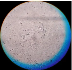
1) DIRECT EXAMINATION: a) 10% KOH MOUNT:
- Demonstrates yeast cells and hyphae
- Septate hyphae are easily seen in KOH
mount
b) GRAM STAIN:
It is more useful in identification of yeast
39
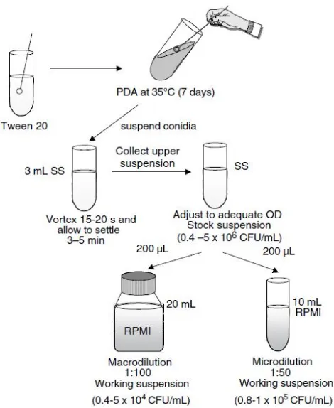
2) CULTURE:
Two sets of SDA with antibiotics are inoculated ,one at 25° c and
the other at 37°c. All cultures are examined everyday during the
first week and twice a week during next three weeks.
IDENTIFICATION:
The mycelial isolates are identified by the colony
characteristics and microscopic morphology by LPCB mount and
finally by slide cultures. The yeast are identified by using tests like
germ tube test, Chlamydospore formation on cornmeal agar, urease
test etc.
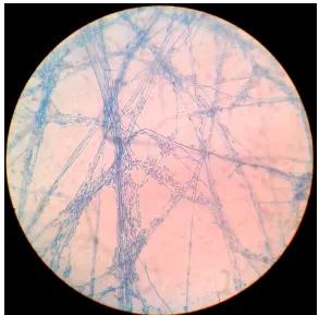
SABOURAUD S AGAR: 1) FUSARIUM
Macroscopic appearance:
Obverse: Colonies are pluffy to cottony owing to extensive
mycelium.
Reverse: Sometimes, diffusible pigment is seen.
Microscopic appearance:(LPCB Mount)
Conidiophores are single or grouped. Conidia are produced
40
septate. Microconidia are single and often found in chains.
Macroconidia are cylindrical but more often crescent shaped.
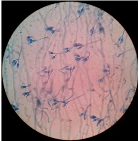
2) PENICILLIUM:
Miroscopic appearance (LPCB mount)
Conidiophores arise in various forms producing phialides
singly or in groups or from branched metulae giving brush like
appearance.
Conidia are unicellular and found in chains with the
youngest conidia at the base.
3) CANDIDA:
Macroscopic appearance: Cream coloured ,smooth and pasty
colonies.
Microscopic appearance: Presence of yeast cells and pseudohyphae
can be seen in Gram staining.
TREATMENT:
Topical antifungals are the main stay of treatment.
1) Filamentous Fungi:
First choice: 5% Natamycin ointment
Second choice: Amphotericin B 0.5%
eyedrops & Flucytosine
41
First choice: Amphotericin B 0.15% drops
Second choice: Fluconazole 0.5% drops42.
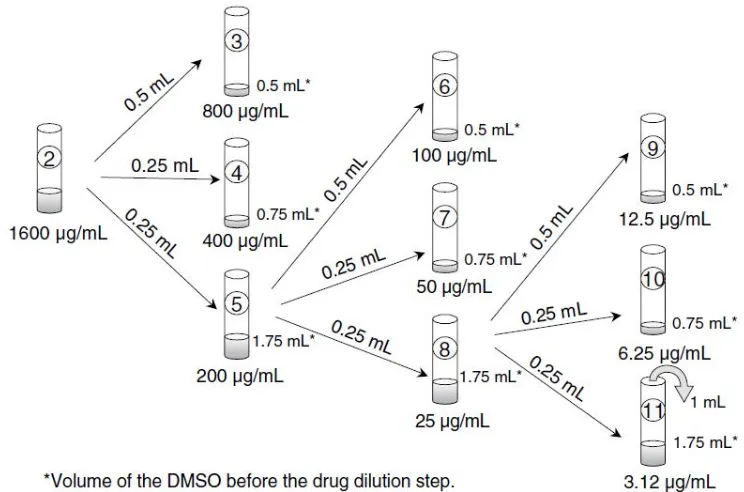
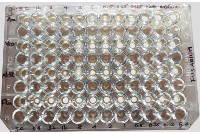
There is an increasing number of non responding
ocular fungal infection which needs necessity for anti
fungal susceptibility testing. There are a number of
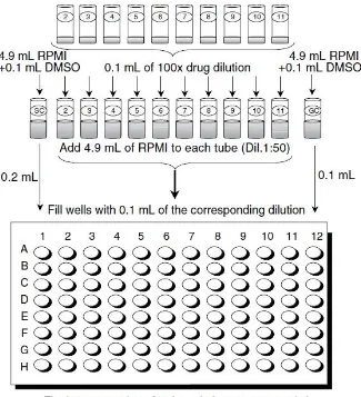
methods for anti fungal susceptibility testing that
include CLSI broth based methodology (M 27-A),CLSI
Methodology for moulds, E-test agar based testing
methods and flow cytometry.
For management of patients not responding to medical
treatment Penetrating keratoplasty or Lamellar keratectomy is done.
LACRIMAL SAC INFECTIONS
Dacryocystitis refers to the inflammation of the lacrimal sac
as a result of infection.
It has characteristic signs and symptoms which helps in the
diagnosis but the progression of the disease is slow and it has a
tendency to recur. Moreover it is associated with sequelae leading to
recurrent conjunctivitis, orbital cellulitis and endophthalmitis in
42
Under normal circumstances, mucosa of the lacrimal sac is
highly resistant to infections but however infections may develop
when trigerred by functional problems .
The main mechanism for the development of dacryocystitis is
the distal obstruction of the nasolacrimal duct leading to the
retention of the tears and development of infection.
It may be of two types acute or chronic
Acute Dacryocystitis
It is an acute inflammation of the lacrimal sac secondary to
nasolacrimal duct obstruction. The obstruction of the duct may
be due to idiopathic inflammatory stenosis or may be secondary
to trauma, infections, inflammation, neoplasm, or due to mechanical
obstruction43.
Obstruction of the nasolacrimal duct leads to the stagnation
of tears in the lacrimal system leading to dacryocystitis.
Aetiology:
It is more common in adult women.
It may occur as an exacerbation of chronic dacryocystitis or it
43
It is caused by pyogenic organisms like Staphylococcus spp.,
Streptococcus spp.,etc. Gram negative isolates accounted for 25% of
the isolates with E.coli being most frequently isolated44.
In a study on acute dacryocystitis in Universiti Sains
Malaysia, 23 patients with Dacryocystitis were studied and Females
(17) outnumbered males (6).Majority of the isolates were Gram positive
bacteria 10(43.4%) followed by Gram negative bacteria 2(12.9%).
Most common organisms were Streptococcus pneumoniae
(21.7%) followed by Staphylococcus epidermidis(13%)45.
Symptoms:
1) Excessive watering from the eyes
2) Redness and tenderness over the lacrimal sac region.
Signs:
1) Swelling and tenderness over the lacrimal sac area.
Treatment:
1) Oral antibiotics
2) I.v antibiotics
44
Chronic Dacryocystitis:
It is a chronic suppurative inflammation of the lacrimal sac. It
is a constant threat to cornea and orbital soft tissue46. It is more
common than acute dacryocystitis.
Types:
There are three types namely catarrhal, mucocele, suppurative
1) Catarrhal - There is intermittent epiphora with
mucoid discharge is seen.
2) Mucocele - There is swelling at the lacrimal sac
area and regurgitation of pus from it.
3) Suppurative - Due to pyogenic infection lacrimal
abscess results.There is reflux of purulent material
with pressure and the microorganisms can be
isolated.
Incidence :
It is more common in females over 40 yrs of age.
Aetiology:
Mixed bacterial isolates are common with preponderance
of Streptococcus pneumoniae and Staphylococcus spp. Staph.aureus
and Staph.epidermidis constitutes 45% and 24% of culture proven
45
Fungal infections are reported to present 4% to 7% ,the most
common isolated being Candida, although Aspergillus and Mucor
can also be found.
In a study by Prakash et al on Dacryocystitis, 80 cases were
studied over a period of one year,chronic dacryocystitis was most
common when compared to acute and congenital dacryocystitis.The
organisms isolated were Staph.aureus (26), Streptococcus
pneumoniae(22) ,Pseudomonas(14)47.
In a study, 44 patients with chronic dacryocystitis were
evaluated and the gram positive bacteria isolated was CoNS
(71%), and Staph.aureus ( 14%)48.
Treatment:
1) Dacryocystectomy in elderly individuals.
2) Dacryocystorhinostomy in young and adult patients.
ENDOPHTHALMITIS
Endophthalmitis is the inflammation of intraocular tissues or
cavities as a result of complication of any ocular surgery,
46
tissue, use of contaminated medications or penetrating ocular
trauma49 .
Depending on infectious agents, two categories are recognised
1) Bacterial endophthalmitis
2) Fungal endophthalmitis
The predominant organism depends on the normal
conjunctival flora and associated adnexal infection. In many cases
of endophthalmitis, an aetiological agent may not be detected on
laboratory cultures50.
POST OPERATIVE ENDOPHTHALMITIS:
It occurs as a complication following any intraocular
surgery. Blindness secondary to post-operative endophthalmitis has
been reported upto 18% of the patients50 .
Bacterial Endophthalmitis:
The gram positive organisms are responsible for 90% to 95%
of post surgical endophthalmitis.
The gram positive organisms causing endophthalmitis are
47
viridians, Streptococcus pyogenes and Corynebacterium. Of these the
predominant isolate is Staph.epidermidis in 20% to 50% the cases.
Although it is caused by Staph. epidermidis, poor visual
outcomes are associated with Staph.aureus, Streptococci, Enterococci
and gram negative organisms.
In a study on bacterial endophthalmitis, a total of 100
microorganisms were isolated. Among them 91% were gram positive
bacteria and 9% were gram negative bacteria. CoNS ( 48%) was
frequently isolated followed by Streptococcus viridians(18%) , and
Staph.aureus (13%)51.
The Gram negative isolates constitutes only 6%. Pseudomonas
aeuroginosa is the most common among them. Other organisms that
cause postoperative endophthalmitis are Proteus mirabilis, Klebsiella
pneumoniae, H.influenzae, E.coli and Enterococci.
In a study on postoperative endophthalmitis, among 170 cases
of culture proven postoperative endophthalmitis,71(41.7%) were
attributed to Gram negative bacteria, 64 (37.6%) to Gram positive
bacteria and 37 (21.8%) were due to Fungi52.
Fungal endophthalmitis:
The organisms causing fungal endophthalmitis are Aspergillus,
48
Fungal endophthalmitis is of two types
1) Exogenous endophthalmitis
2) Endogenous endophthalmitis
1) Exogenous endophthalmitis:
It occurs due to introduction of organisms into the eye
from outside. There is no underlying immunodeficiency. Although
Candida is the commonest cause , other agents include Aspergillus,
Fusarium,Paecilomyces, Curvularia etc. The first case of exogenous
Aspergillus endophthalmitis was reported in 1898 in Heidelberg.
Exogenous Aspergillus endophthalmitis usually follows ocular
surgery or trauma to the eye.
2) Endogenous Endophthalmitis:
It arises due to haematogenous spread from a focus of
infection elsewhere in the body. There is an underlying predisposing
condition and the patient is generally immunocompromised. Candida
spp. most commonly causes endogenous endophthalmitis in patients
with chronic diseases such as Diabetes mellitus and renal
insufficiency53.
Aspergillus is the commonest fungus causing endogenous
49
Endogenous endophthalmitis may arise due to i.v drug
abusers, immunosuppression associated with organ transplants.
Several species of Aspergillus particularly by Aspergillus
fumigatus and Aspergillus flavus are responsible.
Aspergillus spp. are less frequent cause of exogenous or
endogenous endophthalmitis than Candida spp.
Endophthalmitis is also classified into acute and chronic.
Acute endophthalmitis:
It develops between 5-7 days after post operative ocular
surgery .Most commonly it is caused by Staph.epidermidis or
Coagulase negative staphylococci and rarely by fungi.
Delayed endophthalmitis:
It develops one to several months after surgery and the
organisms involved are Staph. aureus, Propionibacterium acnes and
fungus.
Clinical features:
Bacterial Endophthalmitis:
1) There is sudden onset of severe pain and redness in the
affected eye.
2) Dimness of vision is seen.
50
4) 4) Hypopyon or fibrous exudate is seen in the anterior chamber.
5) There is associated vitritis and haze in the vitreous.
Fungal endophthalmitis:
1) It has an incubation period of several weeks.
2) Mild pain and redness is seen
3) Thick organised hypopyon is seen
4) The whole vitreous turns into a granulation mass.
Diagnosis:
Culture and sensitivity of the organism from the aqueous and
vitreous tap confirms the diagnosis of endophthalmitis.
Treatment of Bacterial endophthalmitis:
1) Intravitreal Antibiotics
- Amikacin,Vancomycin,Ceftazidime
- Ceftazidime is safe in cases of
exogenous endophthalmitis.
Generally ,a combination of Vancomycin and Ceftazidime is used
as an initial therapy.
2) Topical antibiotics
3) Subconjunctival antibiotics
51
TREATMENT OF FUNGAL ENDOPHTHALMITIS:
Candida:
- Vitrectomy and intravitreal Amphotericin
Amphotericin is very effective against ocular
Candidiasis.
Aspergillus and Fusarium:
- Vitrectomy , intravitreal Voriconazole ,Topical
Voriconazole and Systemic Voriconazole is
useful.
- Oral Voriconazole penetrates effectively into
the cornea.
PANOPHTHALMITIS:
Panophthalmitis is the purulent inflammation of all the layers
of the eyeball.
Aetiology : It is of two types namely
1) Exogenous
2) Endogenous
Exogenous:
It is usually due to an operative procedure in the eye or after
52
The organisms responsible are Staphylococcus spp.,
Streptococcus spp., E.coli, Pseudomonas pyocyanea, Clostridium
welchii etc.
Endogenous:
It is due to metastatis of the infected embolus in the retinal
artery and the choroid vessels.
Clinical features:
1) Severe pain and limited ocular movements of the eye is seen.
2) Corneal wound appears to be necrotic and hypopyon is
present.
Treatment:
Medical treatment: 1)Control of the infection by administration of broad spectrum antibiotics is helpful.
Surgical treatment:
1) Vitrectomy is done in early cases.
2) Evisceration of the eye is done in severe cases.
EYELID INFECTIONS:
Eyelid infections comprises of blepharitis, hordeolum externum,
hordeolum internum and chalazion
1) HORDEOLUM EXTERNUM: