EXPRESSION OF KI67, P53 AND MUC1 IN RENAL
CELL CARCINOMA IN CORRELATION WITH
NUCLEAR GRADE
Dissertation submitted in partial fulfilment of the requirements for the degree of
M.D. (PATHOLOGY)
BRANCH - III
INSTITUTE OF PATHOLOGY, MADRAS MEDICAL COLLEGE,
CHENNAI – 600 003.
THE TAMIL NADU
DR. M.G.R. MEDICAL UNIVERSITY CHENNAI
CERTIFICATE
This is to certify that this Dissertation entitled “ EXPRESSION OF KI67, P53 AND MUC1 IN RENAL CELL CARCINOMA IN
CORRELATION WITH NUCLEAR GRADE” is the bonafide original work
of Dr.G.SARUMATHY, in partial fulfillment of the requirement for M.D.,(Branch III) in Pathology examination of the Tamilnadu Dr.M.G.R Medical University to be held in April 2015.
Prof. Dr. RAJAVELU INDIRA, M.D., Prof.Dr.M.SARASWATHI, M.D.,
PROFESSOR OF PATHOLOGY DIRECTIOR & HOD,
Regional Institute of Ophthalmology& GOH Institute of pathology,
Madras Medical College, Madras Medical College,
Chennai – 600003 Chennai – 600003.
Prof. Dr. R. VIMALA, M.D.,
DEAN,
Madras Medical College and
Rajiv Gandhi Government General Hospital,
DECLARATION
I Dr.G.SARUMATHY, solemnly declare that the dissertation titled
“EXPRESSION OF KI67, P53 AND MUC1 IN RENAL CELL
CARCINOMA IN CORRELATION WITH NUCLEAR GRADE” is the
bonafide work done by me at Institute of Pathology, Madras Medical College under the expert guidance and supervision of Prof. Dr. RAJAVELU INDIRA,
M.D., Professor of Pathology, Regional Institute of Ophthalmology and GOH, Madras Medical College. The dissertation is submitted to the Tamilnadu Dr.M.G.R Medical University towards partial fulfillment of requirement for the award of M.D., Degree (Branch III) in Pathology.
Place : Chennai
ACKNOWLEDGEMENT
I express my sincere thanks to Prof. Dr. R. VIMALA, M.D., Dean, Madras Medical College and Rajiv Gandhi Government General Hospital, for
permitting me to utilize the facilities of the Institution.
I take this opportunity to express my heartfelt sincere gratitude to
Prof. Dr. M. SARASWATHI, M.D., Professor and Director of Institute of Pathology, Madras Medical College, Chennai, for her constant encouragement,
wholehearted support, valuable suggestions and expert guidance throughout
the study, without which this study would not have ever been possible.
I am extremely thankful to Prof. Dr. RAJAVELU INDIRA, M.D.,
Professor of Pathology, Regional Institute of Ophthalmology, Madras Medical
College, for her valuable suggestions, constant support, advice and
encouragements throughout the study
I am thankful to Prof. Dr. P.KARKUZHALI, M.D., Professor and former Director of Institute of Pathology, Madras Medical College for her
initial guidance and valuable suggestions during the study.
Prof. Dr. M. P. KANCHANA M.D., Prof. Dr. K. RAMA M.D., Prof. Dr. Prof. Dr. SUDHA VENKATESH M.D., and Prof. Dr. S. PAPPATHI M.D., D.C.H for their valuable suggestions and encouragement throughout the study.
I express my heartfelt sincere thanks to all my Assistant Professors
for their help and suggestions during the study.
I would like to thank the Institutional Ethics Committee for
approving my study. I am thankful to my colleagues, friends, technicians and
staff of the Institute of Pathology , Madras Medical College, Chennai for all
their help and support they extended for the successful completion of this
dissertation .
Above all I thank the Lord Almighty for His kindness and benevolence
ABBREVIATIONS
RCC - Renal Cell Carcinoma
PcNA - Proliferate cell nuclear antigen
VHL - Von Hippel-Lindau
CT Scan - Computed tomography
MRI - Magnetic Resonance Imaging
FNA - Fine Needle Aspiration
WHO - World Health Organisation
H & E - Hemotoxylin and Eosin
IHC - Immunohistochemistry
CK - Cytokeratin
EMA - Epithelial Membrane Antigen
VEGF - Vascular Endothelial Growth Factor
LI - Labeling Index
MIB – 1 - Monoclonal antibody directed against Ki-67 protein
CONTENTS
S.NO. TOPICS PAGE NO. 1. INTRODUCTION 1
2. AIM &OBJECTIVES 4 3. REVIEW OF LITERATURE 5
4. MATERIALS AND METHODS 46 5. OBSERVATION & RESULTS 54 6. DISCUSSION 88 7. SUMMARY 102 8. CONCLUSION 107 9. ANNEXURES
EXPRESSION OF KI67, P53 AND MUC1 IN RENAL CELL
CARCINOMA IN CORRELATION WITH NUCLEAR GRADE
ABSTRACT
AIMS AND OBJECTIVES:
Renal cell carcinoma comprises 2-3% of adult malignancies. It has
been very challenging to predict the prognosis of each of the patients with RCC;
when assessing cancer prognosis, classic prognostic factors, staging and grading
were also not always accurate in prediction. In different studies, Ki67, p53 and
MUC1 have been considered as a good predictive marker for RCC aggression,
prognosis and survival outcome of patients. In this study, an attempt has been
made to compare the expression of Ki67, p53 and MUC1 markers with nuclear
grade and other clinicopathological parameters.
MATERIALS AND METHODS:
The clinical and pathological findings of Renal cell carcinoma cases were
retrieved from the pathology records from august 2011 to august 2014 in Rajiv
Gandhi Government General Hospital, Chennai. Totally 52 cases of renal cell
carcinoma was studied and of this, 40 cases were randomly selected and
RESULTS:
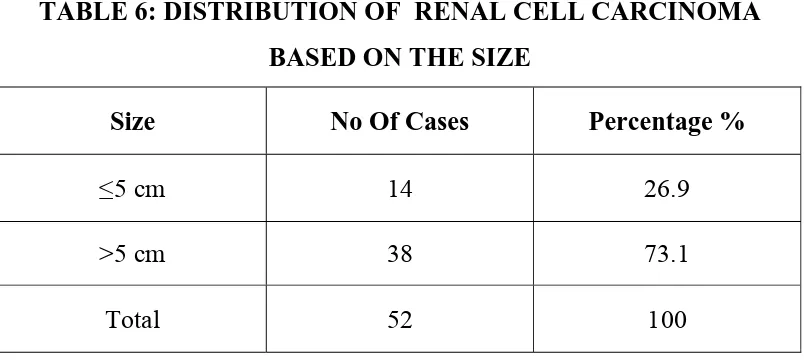
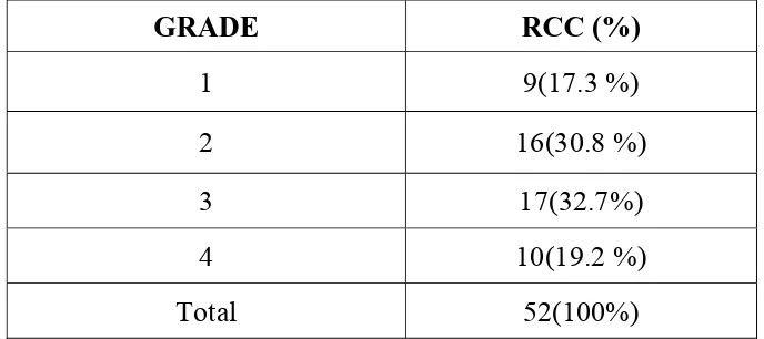
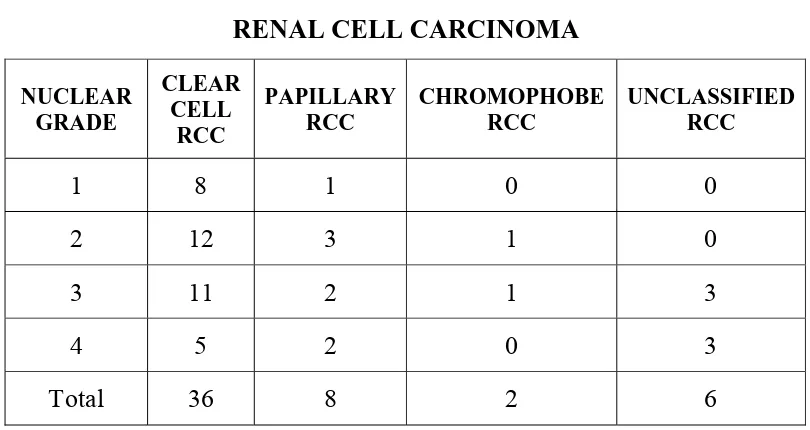
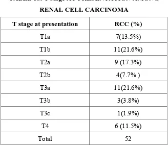
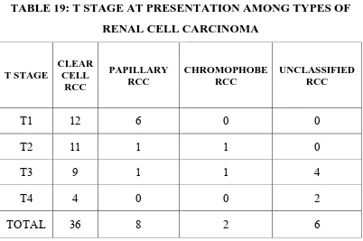
Among 52 cases studied, 36 were clear cell RCC, 8 were papillary RCC, 2
were Chromophobe RCC, 6 were unclassified RCC. Most common nuclear
grade was Furhman nuclear grade 3.
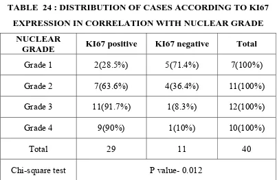
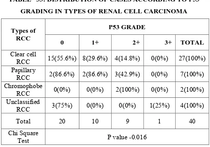
There was statistically significant association of Ki67 expression with nuclear
grade and stage at presentation. Association between p53 and histological type
was found to be significant. There is significant association of MUC1
expression with nuclear grade and stage.
CONCLUSION:
The combined detection of Ki67, p53 and MUC1 expressions, which are
superior to single marker along with nuclear grade and stage, could be used to
significantly improve the accuracy in predicting the prognosis of RCC patients.
KEY WORDS:
INTRODUCTION
Renal cell carcinoma comprises 2-3% of adult malignancies.(1) The
most lethal urological malignancy is Renal cell carcinoma and
annually100,000 deaths worldwide were caused by it.(2) Since 1970’s,
annualy there has been 2-4% rise in incidence of RCC. The use of
radiological imaging can find presymptomatic RCC lesion which has
been one of the reason for this recent rise in incidence and another reason
being the increased prevalence of smoking and obesity which are some of
the important predisposing risk factors. Among patients evaluated for
non-specific musculoskeletal and abdominal complaints, CT scan
incidentally picked up approximately approximately 30-60% of patients
having RCC.(3) RCC has been found in about 20–30% of patients after the
occurrence of metastasis.(4,5)
Prognosis of renal cell carcinoma is dependent on different factors
like early weight and dimensions, tumor stage and tumor cell
morphology. Different grading systems are used for RCC.(6) Nuclear
grading was found to correlate with patients survival.(7) Cellular
proliferation rate , apoptosis metastatic spread are another predictive
variable for biologic aggression of RCC and therefore affects
Cellular proliferation rate in RCC could be evaluated by studying
Ki67 antigen expression and PcNA (proliferate cell nuclear antigen).
Apoptosis degree in tumor can be measured by detecting mutant P53
antigen.(6) MUC1 have a role in cellular polarity, cell adhesion, and signal
transduction. In many epithelial cancers, there is a loss of polarized
cellular expression and there is diffuse circumferential distribution. In
carcinomatous cells ,these variations of expression are suspected to
participate in the metastatic dissemination. (8)
It has been very challenging to predict the prognosis of each of the
patients with RCC; when assessing cancer prognosis, classic prognostic
factors, staging and grading were also not always accurate in
prediction.(5,9) Treatment of metastatic RCC has dramatically changed in
the last decade and leads to revival of new hope to patients affected by
this malignancy and changed the traditional thinking of grave prognosis
in terms of survival among patients diagnosed in advanced stages. So
there has been a definite need for better tools in predicting the clinical
course of RCC in this era of evergrowing novel molecular targeted
therapies.
For proper counselling of the patient and for proper planning and
individualizing patient treatment, accurate prognostication is of utmost
considered as a good predictive marker for RCC aggression, prognosis
and survival outcome of patients.(6,8)
The purpose of this study was to access the expression of P53, Ki
67, MUC1 in different types of renal cell carcinoma. The expression of
these apoptotic, proliferative and metastatic marker was compared with
the nuclear grading. Increasing severity and reactivity rate to these
AIMS
AND
AIMS AND OBJECTIVES
To study the incidence and distribution of renal cell carcinoma in
patients who attended Rajiv Gandhi Government General Hospital
from august 2011 to august 2014.
To study the clinicopathological features of renal cell carcinoma
To determine the expression of ki67, P53 and MUC1 by
immunohistochemistry in renal cell carcinoma.
To study the correlation between ki67, P53 andMUC1 with nuclear
REVIEW OF LITERATURE
EPIDEMIOLOGY
Kidney tumor constitutes approximately 3% of all malignant
tumors in adults.(2) Renal cell cancer (RCC) comprises 90% of all
malignancies of the kidney that occur in adults in both sexes. Among
males it ranks 6th in industrialized areas and 16th in less developed area.
In women it ranks 12th in developed and 17th in developing countries
respectively.(10) RCC is a heterogeneous group of hereditary or sporadic
malignancies that arise from renal cells. Its frequency is next to prostate
and bladder cancer, but it is the most lethal of these malignancies.
The incidence of RCC has been reported to be relatively high in
North America, Scandinavia and Australia compared to other countries.(2)
In several Eastern and Western European countries and also in parts of
Italy, North America and Australia/New Zealand, incidence of RCC has
been generally the highest. The lowest incidence of RCC are found in
Africa and Asia.(11) The incidence of this malignancy has been increasing
steadily at the rate of 2-3%per year.(12,13) Around 20-30% of the RCC has
been estimated to present in the stage of metastasis.(14,15) It is also a well
known fact that advanced overall TNM stage tumours can have low
T-stage and they account for 25% of widely metastatic disease in few
AGE AND GENDER
Most commonly RCC occurs in the fourth to sixth decades of life,
but both sporadic and in particular hereditary tumors have been reported
in children.(17) Among both low and high risk countries, men were
affected two to three times more commonly than women.(18)
RISK FACTORS
Most common cause of renal malignancy is tobacco smoking and
in males, around 39% of all cases were caused by it.(19) Carcinogenic
arsenic compounds in industrial processes or drinking water increases the
risk by 30%.(20) Asbestos, cadmium, some organic solvents, pesticides
and fungal toxins are addressed as possible carcinogens for the kidney but
definitive evidence has not been established.(20,21)
Estrogens could be a risk factor for RCC in obese and overweight
individuals. Several epidemiological studies conducted in many different
populations have found out that the incidence of renal malignancy
increases steadily with increase in body mass index (BMI).(22) Cho et al
concluded in his prospective study that long term use of nonsteroidal
anti-inflammatory drugs may raise the incidence of renal cancer.(23) In
people suffering from chronic hypertension, the incidence of RCC is
obesity.(24,25,26) RCC has been established with exposure to analgesics
containing phenacetin.(20)
CLINICAL PRESENTATION
According to the mode of detection RCCs are classified in two
groups: symptomatic and incidental. The classic triad of presentation
with hematuria, abdominal pain and flank mass is encountered less
frequently than previously and is indicative of advanced disease. The
typical tumor presents with lack of warning signs in early stage and can
be clinically occult during majority of its time course. Majority of the
RCCs are now incidentally found during investigations for complaints
which are not expected in the renal cancer and due to the increasing use
of imaging investigations such as ultrasonography(USG), computed
tomography (CT)scan and magnetic resonance imaging (MRI) study.(27,28)
RCC remains a challenging malignancy due to its paraneoplastic
manifestations such as hypercalcaemia, erythrocytosis, increased
erythrocyte sedimentation rate, and non-metastatic hepatic dysfunction.
Most common presentations are
Abdominal pain (40%)
hematuria (40%)
loss of weight (33%)
fever (20%)
Systemic hypertension (20%)
Fatigue and varicocele, left side is usually affected, because of
testicular vein` obstruction by the tumour extension(2% of males). The
contribution of erythrocyte sedimentation rate (ESR) in prediction of
prognosis has been a matter of debate in several studies.(29,30) However, in
the recent studies of Kawai et al.(31) And Magera et al(32) preoperative
ESR has been identified as a significant independent prognostic factor in
patients with localized Clear Cell RCC. ESR is also found to be an
independent prognostic factor in patients with metastatic RCC treated
with or without cytoreductive radical nephrectomy.(33)
GENETICS
The Von Hippel-Lindau gene (VHL) is mutated or inactivated in
most sporadic clear cell carcinomas which is an early event in the
carcinogenesis of the tumor. The normal function of VHL includes
regulation of oxygen dependent expression of genes which will regulate
cellular response to hypoxia. These include genes associated with
erythropoiesis, angiogenesis and resistance to hypoxia. Von Hippel-
Lindau protein regulate ubiquitn –mediated destruction of hypoxia
which induces production of endothelin-1, erythropoietin, vascular
endothelial growth factor(VEGF), ceruloplasmin, transforming growth
factor, cyclin G2, and these leads to tumor progression.(34)
Some genetic syndromes are associated with RCC. Von Hippel-
Lindau disease is caused by mutation of Von Hippel-Lindau gene. This
gene encodes a tumor suppressor protein. In 75% of sporadic clear cell
carcinoma both gene copies are inactivated. Von Hippel-Lindau
syndrome, or VHL disease, is syndrome with an autosomal dominant
inheritance and it predisposes to a number of neoplasms, such as :
RCC having features of clear cell histology
Pancreatic islet cell tumors and cysts
Pheochromocytoma
Hemangioblastomas
Retinal angiomas
Tumors of Endolymphatic sac
Cystadenomas of epididymis.(35)
When compared to sporadic tumors these tumors occur in younger
Hereditary leiomyomatosis and renal carcinoma
This is a syndrome with autosomal dominant inheritance and they
occur due to germline mutation in fumarate hydratase gene.(37) This
inherited disease causes the affected individuals to have an increased
incidence to suffer from benign leiomyoma of skin and uterus and some
of them develop RCC with type 2 features.(38)
Hereditary papillary RCC
Hereditary papillary RCC is one of the genetic disorder with a
pattern of autosomal dominant inheritance; multifocal and bilateral
papillary RCC occur in the individuals with this syndrome. MET
protooncogene have been mutated in 85% of families(39)
TUBEROUS SCLEROSIS(TS)
This is a disorder of autosomal dominant pattern of inheritance . It
is due to mutation in TSC1 or TSC2 gene. TSC1 is located on
chromosome 9q34 .TSC2 is located on chromosome 6p13. Both of the
genes encode tumor suppressor proteins. This disorder is characterised by
multiple benign hamartoma in CNS, angiomyolipoma in kidney. There is
BIRT-HOGG-DUBE SYNDROME
This syndrome has an autosomal dominant pattern of inheritance.
These patients have a high predisposition to be affected by benign
neoplasms of the hair follicle, colonic polyps and pulmonary cysts.. There
is an increased incidence of renal tumors.(40)
HYPERPARATHYROIDISM (JAW TUMOUR SYNDROME)
Is a rare disorder with an autosomal dominant pattern of
inheritance. Characterised by fibromas of the jaw, parathyroid adenoma
and renal cell carcinoma.(41)
DIAGNOSTIC METHODS
The renal mass has a wide range of differential diagnosis and
includes pseudotumors, benign cysts, angiomyolipomas, vascular
malformations, Wilm’s tumor, sarcoma, lymphoma, and metastases.
However, percutaneous biopsy of a solid renal mass should not be
undertaken, as more than 80% of such masses are RCC,(42) benign lesion
and metastasis are rare .
As per the guidelines of National Comprehensive Cancer
Network(NCCN) for the RCC management, patients with known or
suspected renal cell carcinoma should be further evaluated with routine
partial thromboplastin time and prothrombin time,), abdomen and pelvis
computed tomography(CT) scan, X-ray of the chest, and chest CT scan if
the chest X-ray is abnormal or if there is extensive disease. Further
studies such as a magnetic resonance imaging (MRI) of the brain and a
bone scan should be undertaken only if clinically indicated.
CT or MRI
Renal mass can be usefully characterised by the MRI or CT scan of
the abdomen. In most patients, RCC be accurately diagnosed by these
imaging studies.
Information provided by abdominal CT are :
Morphology and function of the opposite kidney(43)
Anatomical extension of the primary tumour
Venous extension / invasion;
Regional lymph nodal involvement(enlargement) ;
Involvement of the adjacent adrenal glands and metastasis toliver
Contrast-enhanced biphasic abdominal CT angiography can be of
useful in surgical cases to obtain accurate information about the vascular
supply of the affected kidney for on table clamping of segmental renal
artery in cases planned for partial nephrectomy.(44,45) Biphasic MR
MR angiography is less accurate than CT angiography in accurate
depiction of accessory renal vessels .(46)
In patients with indeterminate CT results, MRI can give additional
valuable information like :
show any enhancing areas in renal masses (including enhancing
septations, wall and nodular components in complex cystic
masses).(47)
more accurately establish the anatomical extensions in case of
locally advanced malignancy.
more accurately establish venous extension of the tumour, if the
involvement of an inferior vena cava(IVC) tumour thrombus is
poorly depicted on CT scan.(48,49)
MRI is also indicated in pregnant patients without renal failure.(50,51)
Among imaging for chest staging of metastatic RCC, most accurate
investigation is the Chest CT. However, initially routine chest x-ray must
be done for evaluation of large lung metastasis, although this is less
accurate in finding small metastasis, when comparing CT chest. At the
time of diagnosis, most brain and bone metastases are symptomatic and it
is not generally advised to do routine bone or brain imaging in the further
brain or bone scan, may be carried out in patients presenting with related
clinical symptoms and signs.(54,55)
RENAL TUMOUR BIOPSY
Image guided percutaneous biopsies of renal tumour are of
increasing use :
1. For histological diagnosis in case of radiologically indeterminate
renal mass lesions;
2. For categorising patients into surveillance group, in case of small
renal mass lesions;
3. To get histological diagnosis before proceeding onto ablative
treatment procedures;
4. For deciding the most suitable means of targeted pharmacologic
therapy for the stage of metastatic disease .(56,57,58)
Image guided percutaneous sampling in case of a renal mass can be
done by means of trucut needle biopsy or fine needle aspiration. The
main aim is to determine the nature of malignancy, its exact histological
type, and its exact nuclear grade in view of its aggressiveness.
In view of the high accuracy in diagnosis of renal mass forming
before surgery is not always necessary, as in case of healthy individuals
having a long life expectancy.
Under local anesthesia, image guided percutaneous sampling in
case of a renal mass can be done with the guidance of either ultrasound or
CT.(59,60) Presently 18-gauge needles are considered ideal for trucut
biopsies of the renal mass, as we can obtain sufficient amount of tissue
and can be done with low morbidity for diagnostic purpose in the
majority of cases undergoing percutaneous biopsies. The complications
which we most frequently encounter in percutaneous biopsy of a renal
mass are spontaneously resolving hematoma (perinephric /subcapsular)
and hematuria; its unusual (0-1.4%) to encounter clinically significant
bleeding after biopsy and is usually self-limiting .(56,60)
On comparing to FNA, trucut needle biopsies are usually more
preferable in case of solid renal masses, in view of its higher diagnostic
yield and higher accuracy rate. For detailed histopatholigical analysis of
the malignancy, it is necessary to obtain at least two high quality biopsy
cores (> 10 mm in length and non-fragmented) and to avoid sampling
necrotic areas. In case of experienced biopsy centers, core needle biopsies
of renal solid masses have obtain 78-97% diagnostic yield for the
must be kept in mind that 2.5-22% of trucut needle biopsies are non
diagnostic.(61,62)
Tumour grade assessment on core biopsy specimen is very
challenging. The obtainable accuracy of Fuhrman grading on trucut
needle biopsies is poor (43-75%); For cystic renal masses, diagnostic
yield of needle core biopsies have been low and usually biopsy must not
be done on these lesions unless accesseble solid areas are present within
the lesion (Bosniak IV cysts).(59,60)
HISTOPATHOLOGICAL FEATURES
Renal cell carcinoma arises from the renal tubular epithelium. RCC
is characterised by having unique morphological features and distinct
genetic abnormalities.(63,64) The diagnosis of RCC is based on unique
histomorphological features. IHC and microRNA techniques are used if
histological findings are not concluvise in distinguishing the types of
RCC.(63) The Fuhrman grading system is used for nuclear grading of
RCC. This four-tiered system considers the nuclear features like size of
the nucleus and nucleolus, shape of nucleus and nuclear content for
WHO CLASSIFICATION OF RENAL CELL TUMOURS
Clear cell Renal cell carcinoma
Multilocular clear cell Renal cell carcinoma
Papillary Renal cell carcinoma
Chromophobe Renal cell carcinoma
Carcinoma of the collecting ducts of Bellini
Renal medullary carcinoma
Xp11 translocation carcinomas
Carcinoma associated with neuroblastoma
Mucinous tubular and spindle cell carcinoma
RCC, unclassified
Papillary adenoma
Oncocytoma
CLEAR CELL RCC
This constitutes 70–80% of RCCs. The genetic abnormalities most
frequently encountered in this type of RCC are von Hippel-Lindau (VHL)
gene mutations , the chromosome 5q duplication and chromosomal
Macroscopy
Clear cell renal cell carcinomas are randomly distributed cortical
tumours. They are usually solitary and occur with equal frequency in
either kidney. Less than 5 percent of cases are multicentric and
bilateral.(67) Hereditary cancer syndromes like Von Hippel-Lindau disease
are usually characterised by early age of onset, multicentricity and
bilaterality.
Clear cell RCCs are globular tumours .They protrude from the
renal cortex as a bosselated, rounded mass. The tumour and adjacent
kidney interface is usually well demarcated. The tumor is pseudocapsuled
with a "pushing margin" . The tumor average size is 7 cm in diameter. In
countries where radiologic imaging techniques are widely applied,
detection of small tumor lesions is increasing. Size itself is not a
determinant of malignancy though increasing size is associated with a
higher frequency of metastases. All tumours of the kidney with clear cell
type are considered as malignant tumours. Due to the rich lipid content of
cells, neutral lipids, cholesterol and phospholipids, the clear cell renal cell
carcinoma is typically golden yellow . Cysts, calcification, necrosis and
haemorrhage are commonly present. Radiologically10to 15percent of
Microscopy
On hemotoxylin and eosin (H&E) staining under light microscopy,
cytoplasm of Clear cell RCC appears more or less empty. This effect is
due to the intense glycogen and phospholipid accumulation in the
cytoplasm which in turn is attributed to the increase in
glucose-6-phosphate levels induced by decreased gluconeogenesis and increased
glycolysis.(70,71) In well differentiated tumours, the tumour cell nuclei are
more condensed. The tumour cell nuclei exhibit more polymorphism and
prominent nucleoli in less differentiated tumours.(72) Eosinophilic or
granular appearance of the cytoplasm is an another morphological variant
of Clear cell RCC, which in turn is caused by the mitochondrial
augmentation.
These tumors are characterised by variable architecture with acinar
or tubular patterns. The stroma is poorly defined inspite of rich
vasculature surrounding them. Occasionally, scattered bizarre nuclear
forms are seen in otherwise typical tumors, a phenomenon similar to that
more commonly seen in endocrine neoplasms and which should not be
equated to sarcomatoid or anaplastic transformation.(73) The stroma of
renal cell carcinoma is nondescript and, in general, not as abundant as in
collecting duct carcinoma or transitional cell carcinoma. A lymphocytic
Cases have also been described in which the red blood cells in the stroma
form clusters, resulting in a myospherulosis-like appearance.(74)
Immunoprofile
Clear cell RCCs frequently react with antibodies to brush border
antigens, low molecular weight cytokeratins(LMWCK), CK19, CK18,
AE1, CK8, vimentin and Cam 5.2. Detection of high molecular weight
cytokeratins(HMWCK) are rare. The most of clear cell RCCs react
positively for renal cell carcinoma marker epithelial membrane antigen
and CD10. MUCΙ and MUC3 are consistently expressed .(75,76)
MULTILOCULAR CYSTIC RCC
This tumor is characterised by numerous cysts in entirity. Within
the septa of the cyst lies the small clear cell groups which is similar to
clear cell carcinoma - grade Ι. There is male predominance. Mean age is
51 years.(77)
Multilocular cystic renal cell carcinoma are well-circumscribed
with serous or haemorrhagic fluid filled small and large cysts. A fibrous
capsule is seen separating this lesion from the normal kidney. More than
20% of tumors have calcification in the septa .Usually a single epithelial
cell layer lines the cysts or cyst may lack lining epithelium. The lining
Occasionally, the lining may be of cells of several layers or a few small
papillae are seen.(78) The nuclei are small and spherical with a dense
chromatin. Fibrous tissue forming the septa is often densely collagenous.
Within some of the septa, epithelial cell collections with a clear
cytoplasm are seen. These epithelial cells usually resemble cells those
lining the cysts and have small dark nuclei. These epithelial cells
resemble histiocytes, or lymphocytes surrounded by retraction artefacts.
The cells are strongly positive for cytokeratins(CK) and epithelial
membrane antigen(EMA) .
PAPILLARY RCC
Papillary RCC represents about 15% of all renal cell carcinomas.
They arise in patients on chronic hemodialysis.(79) Some of the papillary
renal cell carcinoma are hereditary, and these have been found to be
associated with the c-MET mutation.(80) It has a tendency towards
multicentricity and bilaterality. This tumour has a distinct papillary
growth pattern, with a solid pattern in undifferentiated areas. The
papillary structure are lined by a single layer of neoplastic cells with a
fibrovascular core containing foci of lipid-rich macrophages.(81,82)
This tumour can be divided into two types: type 1 papillary RCC,
in which the papillae are lined by a single layer of cells; The cells have a
lined by a pseudostratified epithelium. These cells are characterised by an
abundant acidophilic cytoplasm.(83,84) Type 1 tumors that are accompanied
by foamy macrophages and psammoma bodies and are immunoreactive
for keratin 7 and MUC1.(85,86) When compared to conventional renal cell
carcinoma, this tumour has a better prognosis.(87,81)
Papillary RCCs are characterised by the loss of Y chromosomes in
males and trisomy of chromosomes 8p, 3q, 7, 16, 12, 20 and17.(63)
Papillary renal cell carcinoma can undergo anaplastic or sarcomatoid
changes.(88) The presence of numerous foamy macrophages and
extensive tumour necrosis has been associated with a more favourable
prognosis.(81,89)
CHROMOPHOBE RCC
It comprises approximately 5% of renal epithelial tumours. It has a
lobulated surface with one or more solid tumour nodules. The cut surface
of this tumour appears homogeneously orange; after formalin fixation, it
turns beige or sandy.(90)
Microscopically, the characteristic feature is nesting (‘alveolar’)
arrangement of tumor cells. Microcystic and adenomatous patterns of
growth can also be seen sometimes.(91) The tumor cells have sharply
quality. There is often a clear perinuclear region.(92) Pale cytoplasm is
due to the presence of numerous cytoplasmic vesicles.(93) With Hale
colloidal iron technique, the microvesicles are stained blue.(94)
Calcification is seen in nearly half of cases. Immunohistochemically,
chromophobe renal cell carcinoma is positive for EMA, ck7, CD9, CD82,
paxillin, claudin-7 and -8, Ep-Cam (an epithelial adhesion molecule).(95)
COLLECTING DUCT CARCINOMA
It accounts for approximately less than 1% of RCCs.(66) These
tumors are more common in young males. They are centered in the
medulla and have a tubulopapillary architecture, and are surrounded by a
desmoplastic reaction.(96) The cells have a hobnail pattern with a
eosinophilic cytoplasm. The cells usually display (Fuhrman 3 and 4)
nuclear features. Both intraluminal and intracytoplasmic mucin may be
seen. Atypical changes in the adjacent ducts are common. Cases with
signet ring features are also reported.(97)
Vinculin is the immunohistochemical marker for this tumor type.
The characteristic feature is a positive reaction to Ulexeuropaeus and
coexpression of low molecular weight CKs and high molecular weight
CKs. Leu M1 and epithelial membrane antigen has a variable
expression.(90) This clinically aggressive tumour, often shows metastases
collecting duct carcinomas has poor prognosis . Two thirds of patients
die within two years of diagnosis.
RENAL MEDULLARY CARCINOMA
This is a very rare tumour. This tumour characteristically occurs in
young black patients suffering from sickle cell disease.(99) They are
centered in medulla and poorly circumscribed . Tumor` mean size is
approximately 7 cm. Most of these tumours have multiple areas of
haemorrhage and necrosis. Microscopically it exhibits a yolk sac-like ,
reticular or adenoid cystic appearance and poorly differentiated areas.
This tumour has desmoplastic stroma with neutrophils and marginated
by lymphocytes.(100) Immunohistochemically, they are consistently
positive for CEA. They are often reactive to CK20, CAM5.2, CK7
AE1/AE3, and vimentin. (101) It has a very aggressive behaviour and
usually present with metastasis.
RENAL CARCINOMAS ASSOCIATED WITH XP11.2 TRANSLOCATIONS / TFE3 GENE FUSION
These malignancies are characterised by different translocations in
chromosome Xp11.2. All of these translocations in turn can cause gene
fusions in the transcription factor binding to IGHM enhancer 3 (TFE3)
tumor.(102) These malignancies are usually characterised by an advanced
stage of presentation .
On gross examination, they resemble Clear cell RCC and most
commonly tan or yellow and often with necrosis and haemorrhage. The
most characteristic histopathologic feature is the papillary architecture
comprised of cells having a clear to granular eosinophilic type of
cytoplasm with distinct cell borders. These cells have vesicular nucleus
with prominent nucleoli. In all these tumours, there is constant presence
of psammoma bodies.(90)
TFE3 protein has a nuclear immunoreactivity and it is the most
chararcteristic immunohistochemical feature of these tumours. 50% of
tumors only express cytokeratin and EMA.(102) The tumours are also
positive for Renal Cell Carcinoma Marker antigen and CD10.
RENAL CELL CARCINOMA ASSOCIATED WITH NEUROBLASTOMA
It occurs in adolescents with history of childhood neuroblastoma.
Subsequent development of renal cell carcinoma in these patient is found
to be caused by Neuroblastoma treatment. Median age at the time of
diagnosis of Renal cell carcinoma was 13.5 years. Males and females
In these morphologically heterogeneous tumours, some are
characterized by solid and papillary architecture. The cells are with
abundant eosinophilic cytoplasm and some are with reticular cytoplasm,
exhibiting mild to moderate pleomorphism.(103) In other group, the
tumours are small, and clear cell RCC were detected incidentally. These
tumours are usually positive for vimentin, EMA, and keratins 8, 18, and
20 .They are negative for keratins 7, 14, and 19.
MUCINOUS TUBULAR AND SPINDLE CELL CARCINOMA
For the first time, this entity was included in the current WHO
classification . Mean age is 53 year at the time of diagnosis .There is a
female predominance. On ultrasound, it is usually found as an incidental
mass. They are well circumscribed, grey or light tan with uniform cut
surfaces. They are low-grade malignancies.
These tumors were composed of tubules which are tightly packed
with pale mucinous stroma separating these tubules. The tubular arrays
often have a spindle cell configuration. Distal nephron is likely to be the
site of origin. But some believe it to be of proximal tubule origin as a
RENAL CELL CARCINOMA, UNCLASSIFIED
This group accounts to 4-5% of RCC cases. Renal carcinoma that
could not be fit into one of the other categories should be classified into
this diagnostic category.(104) Since this variety is comprised of tumours
with varying appearances and genetic heterogenecity, it cannot be
described to have specific histological features. The features for defining
unclassified RCC include
a) Admixture of recognised types,
b) Mucin production,
c) Absence of recognisable epithelial elements with presence of
distinct sarcomatoid morphology,
d) presence of both epithelial and stromal elements rarely,
e) cell types with unrecognisable features.
At presentation, in comparison with clear cell RCC, unclassified
type was found to have larger size of tumours, increased incidence of
adrenal gland invasion, adjacent organs invasion, regional and
nonregional lymph nodal involvement and metastasis to bone. On
multivariate analysis, Unclassified histology itself was an independent
marker for poor outcome. Median survival of patients suffering from
NUCLEAR GRADING :
Skinner et al first proposed the nuclear grading system. Nuclear
morphology was the basis for this grading system. In RCC, for
demonstrating this system`prognostic value, Skinner et al carried out a
study comprised of 272 patients. This study demostrated a high
correlation of nuclear grade with patient survival rate in RCC.(105)
Skinner grading system(106)
G1 – Nuclei are small, indistinguishable from those seen in normal
tubular cells
G2 – Nuclei are slightly irregular and frequently pyknotic without
abnormal nucleoli
G3 – Nuclei are irregular, enlarged and pleomorphic with
prominent nucleoli
G4 – Nuclei are extremely giant and bizarre
In 1982, the nuclear grading system proposed by Skinner et al was
simplified by Furhman et al. This four-tier system used the features such
as size of nucleus and nucleoli, shape of nucleus and contents of nuclei.
In this system for nuclear grading, regardless of their percentage, highest
grade of any of its components is used to classify the entire neoplasm.
Worldwide, this grading system is currently used for nuclear grading of
Fuhrman grading system(107)
G1 – Nuclei are small, round and uniform (10 μm), with
inconspicuous or absent nucleoli.
G2 – Nuclei are slightly irregular (15 μm), with small nucleoli.
G3 – Nuclei are very irregular (20 μm), with large and prominent
nucleoli.
G4 – Nuclei exhibit large and pleomorphic often poly-lobed and
bizarre (> 20 μm).
This grading system is used to assess the RCC prognosis,
especially for conventional and papillary RCC. It is widely acceptable for
its simplicity. Its correlation with different pathologic variables has been
proven. Most of the controlled studies has confirmed its prognostic value
in RCC patients. Poor prognostic outcome has been associated with grade
3 or 4 of this system. Good prognostic outcome has been associated with
grade 1 or 2 of this system. Intraobserver variability and interobserver
variability has been the problem with this grading system and hence the
reproducibility problem among pathologists.(65)
TREATMENT OF LOCALIZED RCC
For localized RCC, radical nephrectomy has been the gold
(nephron-sparing surgery - NSS) is the standard treatment option
recommended for localised renal neoplasms measuring up to 7 cm in
diameter, and for larger neoplasms also, it can be the treatment option,
whenever surgically feasible.(109,110) In prospective randomized studies the
oncological efficacy of NSS is confirmed, and it has been proved that
with NSS, incidence of renal insufficiency and ill effects on day to day
health has been reduced, and cardiovascular ill effects also reduced on
comparison with radical nephrectomy.(111)
For some of the selected patients with RCC, laparoscopic resection
of renal masses has become the one of the standard treatment option.
When compared to open surgery, laparoscopic renal surgery is associated
with lower rate of morbidity,(110) for localised renal tumours that are not
suitable for NSS , laparoscopic nephrectomy is the standard procedure
and it also provides an equivalent prognostic outcomes in comparison
with open surgery .(112) With experienced hands and with careful selection
of patients, laparoscopic partial nephrectomy has been an effective
alternative method to partial nephrectomy by open laparotomy.
Partial nephrectomy by means of Robotic-assistance is under trial.
Role of lymphadenectomy in the management of RCC patients is
currently restricted only for the purpose of staging, principally at the
extended lymphadenectomy may improve the survival rates of RCC
patients. In case of patients with preoperative imaging showing normal
adrenal gland, routine adrenalectomy is to be done only for large upper
pole renal tumors or for renal tumours measuring more than 7 cm
diameter.(110)
Minimally-invasive procedures such as
cryoablation
percutaneous ablation by means of radiofrequency
microwave ablation
high-intensity focused ultrasound (HIFU), and
laser ablation(113)
are alternative procedure for surgical resection in some of the selective
RCC patients such as multiple tumours or poor overall health status.
Active follow up can be advised for small renal tumours and treatment
can be considered only if it shows significant progression.(110)
TREATMENT OF METASTASIZED RENAL CELL CARCINOMA
The typical feature of RCC is resistance against cytotoxic drugs,
radiotherapy, and hormones.(114) Immunotherapy with interleukin-2
(IL-2) or interferon alpha (IFN-α) can produce durable and complete
IL-2, response rate is 7-27%; this provides only a modest benefit in terms of
survival in patients with advanced stage of RCC. Currently, adjuvant use
with bevacizumab is the only role of immunotherapy in patients with
advanced stage of RCC.(110,114)
The Molecular targeted therapies are designed in order to block the
critical signalling pathways which underlies the pathogenesis of RCC.
Molecular targeted therapies are divided into three categories:
multikinase and tyrosine kinase inhibitors, VEGF antibodies and mTOR
inhibitors.(115) For metastasized RCC, these targeted therapies are applied
by a systemic route.(110) Clinical trials have proven the efficacy of
molecular targeted therapies where they have proved to improve both
progression-free and overall survival. Since these drugs do not eradicate
the disease. Durable remissions can occur.(114,115) Better efficacy,
tolerability, and oral administration are the advantages of molecular
targeted therapies over immunotherapy.(115)
Patient survival can be improved by means of cytoreductive
nephrectomy along with surgical resection of RCC metastases. They have
been advised for RCC patients with a good overall health status.(110) Role
of palliative surgery for symptomatic brain and bone metastases must be
re-evaluated by means of elaborative clinical trials, in view of the recent
is indicated if systemic treatment proves to be of no use. Severe pain due
to metastases can be relieved by means of embolization of paravertebral
and bone metastases.(110)
PROGNOSTIC AND SURVIVAL FACTORS IN RCC
RCC has a variable clinical course. Incidentally discovered and
small tumours have an indolent course even without treatment. Survival
rates are poor in metastasized RCC or recurrent disease . The overall
RCC prognosis has been greatly improved by means of diagnosis in early
stages of the tumor and by means of significant advancement in
anatomical imaging, surgical staging and different modes of
treatment(both medical and surgical means).(117,5) Stage and grade are
currently the most important RCC prognostic factors.(9)
Clinical prognostic factors
Patients who are presenting with clinical symptoms are found to
have decreased survival, whereas patients with incidentally found tumors
are likely to have a more favourable prognosis, which can be explained
by the incidence of smaller size of the renal mass and lower tumour stage
at the time of diagnosis.(118,119,120) More than 10% body weight loss in 6
months is found to have significantly lowered survival rate. For
predicting poor prognosis, cachexia have been an independent
scales or Eastern Cooperative Oncology Group (ECOG), can estimate the
impact of RCC on the overall wellbeing of the patient, and have been
accepted as the significant and established prognostic factors for RCC.(64)
Younger age of diagnosis has been established as an independent
indicator of a more favourable outcome.(121) Gender does not have any
prognostic potential.(119)
Patient survival rate can be correlated with many laboratory
indices. Excessive interleukin-6 (IL-6) production by the advanced RCC
leads to a relatively high CRP level compared to early stage RCC. This
IL-6 is a cytokine with multifunctional growth factor activities and in turn
can be a predictor for a more poor prognosis. Poor outcome of the RCC
patient can also be predicted by the elevated erythocyte sedimentation
rate and thrombocytosis.(64) In addition, haemoglobin, serum calcium,
lactate dehydrogenase, albumin, neurone-specific enolase (NSE, γ
-enolase) and alkaline phosphatase are also found to be of prognostic
value to some extent in RCC.(122)
Prognostic anatomical factors: stage at diagnosis
The tumor staging is currently the most reliable indicator in RCC
prognosis. For localized stage of disease, 5-year survival rates following
RCC, it is 65–80%; for tumours extending into inferior vena cava, it is
40–60%; for RCC with lymph nodal extension, it is 10–20%; and for
RCC with metastasis, it is 0–5%.(64) Staging takes into account features
like size of the tumour, venous extension , invasion into the renal
capsule, involvement of adrenal gland, lymph nodal involvement and
metastasis to distant organs, and all of which are established
independent prognostic markers and when they are assessed in
combination by means of staging, provide the most dependable
prognostic information in RCC patients.(9,123) The TNM classification for
staging of RCC was revised recently in 2009.(124) In comparison to the
previous 2002 version of staging(125), T2 tumour class is now sub
classified into T2a(tumours more than 7 cm but less than 10 cm in
diameter) and T2b( tumours more than 10 cm in diameter), but both not
extending beyond the limits of the kidney. In addition, RCC with a
tumour thrombus extending only into the corresponding renal vein is now
staged as T3a and invasion into adjacent adrenal gland is now staged as
T4.(110)
Histological prognostic factors
Despite strong criticism regarding the predictive value and validity
of the Fuhrman grading system, untill now it is considered the most
of the independent prognostic marker for clear cell variety of RCCs.(121)
The RCC-specific 5-year survival rate is 89% in case of grade 1 tumours,
65% in case of grade 2 tumours and 46% in case of grades III-IV
tumours.(16) In general, the overall prognosis for papillary type of RCC
and chromophobe type of RCC is much better than overall prognosis of
clear cell type of RCC. The overall survival rate of collecting duct type of
RCC is poor.(9,110) Among papillary type of RCCs, type 1 tumours
generally have a better prognosis than the prognosis of type 2, which
itself is one of the independent indicators of a poor prognosis.(84) Among
the different histological features of RCC, microscopic invasion of
veins(MVI), sarcomatoid differentiation, collection system invasion and
areas of tumoural necrosis are correlated with decreased survival rates.
Cystic component of RCC is accepted as an independent marker to
indicate more benign clinical outcome.(110,121)
Molecular prognostic factors
Numerous molecular markers are being investigated which
includes carbonic anhydrase IX (CaIX), vascular endothelial growth
factor (VEGF), hypoxia-inducible factor (HIF), Ki67 (proliferation), p53,
E-cadherin, C-reactive protein (CRP), CD44 (cell adhesion) and
osteopontin. Gene expression profiling seems a promising method, to
Prognostic Factors In Metastasized Renal Cell Carcinoma
In advanced RCC, classic anatomical and histological features of
the primary tumour had limited predictive value.(9) The prognostic
factors identified in metastasized disease are performance status, time of
appearance of metastases, number and locations of metastatic sites, prior
nephrectomy and surgical resection of metastases.(120) Metastases to bone
have been regarded as a marker of shorter survival. Number of metastatic
sites is considered as a more important prognostic marker than location
of metastasis.(64)
IMMUNOHISTOCHEMISTRY:
Albert Coons et al in 1941 first labelled antibodies directly with
fluorescent isocyanate. Nakane and Pierce et al in 1966, introduced the
indirect labeling technique in which the unlabelled antibody is followed
by second antibody or substrate. Various stages of development of
Immunohistochemistry include peroxidase – antiperoxidase method
(1970), alkaline phosphatase labeling (1971), avidin biotin method
(1977) and two layer dextrin polymer technique (1993).(127)
ANTIGEN RETRIEVAL:
Antigen retrieval can be done by the following different techniques
1. Proteolytic enzyme digestion
2. Microwave antigen retrieval
3. Pressure cooker antigen retrieval
4. Microwave and trypsin antigen retrieval
PROTEOLYTIC ENZYME DIGESTION:
Huank et al in 1976 introduced this technique to breakdown
formalin cross linkages and to unmask the antigen determinants. The
most commonly used enzymes include trypsin and proteinase.(128) The
disadvantages include over digestion, under digestion and antigen
destruction.
MICROWAVE ANTIGEN RETRIEVAL:
This is a new technique most commonly used in current practice.
Microwave oven heating involves boiling formalin fixed paraffin sections
in various buffers for rapid and uniform heating.(127)
PRESSURE COOKER ANTIGEN RETRIEVAL:
Miller et al in 1995 compared and proved that pressure cooking
method has fewer inconsistencies, less time consuming and can be used
PITFALLS OF HEAT PRETREATMENT:
Drying of sections at any stage after heat pretreatment destroys
antigenicity. Nuclear details are damaged in poorly fixed tissues. Fibers
and fatty tissues tend to detach from slides while heating. Not all antigens
are retrieved by heat pretreatment and also some antigens like PGP 9.5
show altered staining pattern.
DETECTION SYSTEMS:
After addition of specific antibodies to the antigens, next step is to
visualize the antigen antibody reaction complex. The methods employed
are direct and indirect methods.
In the direct method, primary antibody is directly conjugated with
the label. Most commonly used labels are flouro-chrome, horse radish
peroxidase and alkaline phosphatase. Indirect method is a two-step
method in which labelled secondary antibody reacts with primary
antibody bound to specific antigen. The use of peroxidase enzyme
complex oravidin biotin complex further increases the sensitivity of
immunohistochemical stains.(127)
In 1993, Pluzek et al introduced enhanced polymer one step
enzymes are attached to dextran polymer back bone. This is the rapid and
sensitive method.(130)
Dextran polymer conjugate two step visualization system is based
on dextran technology in Epos system. This method has greater
sensitivity and is less time consuming.
Ki-67
Ki-67 also recognized as MKI67 is a protein encoded by the
MKI67 gene (131) which was discovered by Gerdes. Originally this protein
was defined by the prototype monoclonal antibody Ki-67 and it was
generated by immunizing mice with nuclei of the Hodgkin lymphoma cell
line L428. It was named based on the city of origin (Kiel, Germany) and
the number of the original clone in the 96-well plate.
Ki-67 is a nuclear protein that is necessary for cellular proliferation
and ribosomal RNA transcription. It is present during all active phases of
the cell cycle (G1, S, G2, and M), but is absent from resting cells (G0).
The protein is predominantly localized in the peri-nucleolar region in the
G 1 phase, in the later phases it is also detected throughout the nuclear
interior, being predominantly localized in the nuclear matrix having a half
life of is 60-90 minutes. In mitosis, it is present on all chromosomes.(131)
fraction of a specified cell population and is widely as a proliferation
marker in many of the human tumours. The fraction of Ki-67-positive
tumor cells is often associated with the clinical course of various
neoplasms. The monoclonal antibody generally used to detect the Ki-67
antigen is MIB-1. One of its major merits over the original Ki-67
antibody is that it can be applied on formalin-fixed paraffin-embedded
sections, after heat-mediated antigen retrieval. Ki-67 labeling index is
calculated by the percentage of tumours cells showing distinct brown
staining of the nucleus with strong intratumoural heterogeneity.
Studies on RCC, gastric cancer , bladder cancer , lymphomas,
colorectal cancer and breast cancer have shown that overexpression of
Ki67 is correlated with biological behaviour and prognosis of these
malignancies.(132)
The other methods of detection of Ki-67 are by Western blot
analysis and immunofluorescence. The various other markers of
proliferation include AgNOR staining, PCNA and Topoisomerase II. The
novel markers being evaluated for identifying cell proliferation include
Fen-1, MCM proteins (mini-chromosome maintenance), mitosin, polo –
P53
p53 was first identified in 1979 by David P. Lane, Lionel, Lloyd
Old, and Crawford Arnold Levine. in 1985, human TP53 gene was
cloned. The role of P53 as a tumor suppressor gene was discovered by
Bert Vogelstein in 1989. It is considered as the “Guardian of the
genome”. This tumour suppressor gene is located on chromosome
17p13.1. It encodes a nuclear phosphoprotein of 53kDa.(133) p53 plays a
central role in cell – cycle regulation, in cell apoptosis and in DNA
repair. When there is cellular insult or DNA damage there is increased
p53 production; then it induces cell cycle arrest at the G1/S junction.
Therefore, for control of tumor growth, apoptosis and maintaining
genome stability, p53 is essential. Normal p53 protein, is rapidly
removed from the nucleus but mutant forms have prolonged
half-life.This favours intranuclear accumulation and so it can be detectable
immuno-histochemically. P53 appears mutated in a wide variety of
human carcinomas, such oral and oropharyngealcarcinoma, colorectal
carcinoma, breast carcinoma, esophageal carcinoma, gall bladder
carcinoma and gastric carcinoma. In numerous studies there was
correlation between over expression of p53 gene and the poor prognosis
in patients with these tumors. The p53 is also involved in regulating the
metallo-proteinase-2 (MMP-2), MMP-13 and the tissue inhibitor of
metalloproteinase-3 (TIMP3).
P53 appears mutated in about 50% of many malignancies, but in
RCC, incidence of p53 mutations is low. P53 mutations has been
detectedin 3-33% of patients with RCC. Although in the majority of
RCCs p53 remains wild type, this does not mean, that it is functional.
P53 function can also be repressed by mechanisms, which involve loss
of positive regulators, such as Arf or overexpression of natural negative
regulators,MDM2 or MDMX or by viral proteins, such as E6 of the
human papilloma virus.
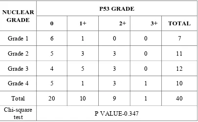
On comparing the association of p53 expression and nuclear grade,
there are number of controversial studies. Some investigators have found
no association but some of them demonstrated a strong relationship.
However, p53 is considered as a potential marker in determining
prognosis of patients with RCC.(133) It is now known that like melanoma,
RCC also belongs to the type of tumors with a low incidence of p53
mutations when compared to prostate and bladder.(134)
The most commonly used methods for detection of p53 mutations
includes immunohistochemistry, polymerase chain reaction-single-strand
sequencing. Although sequencing is the most unambiguous method, it is
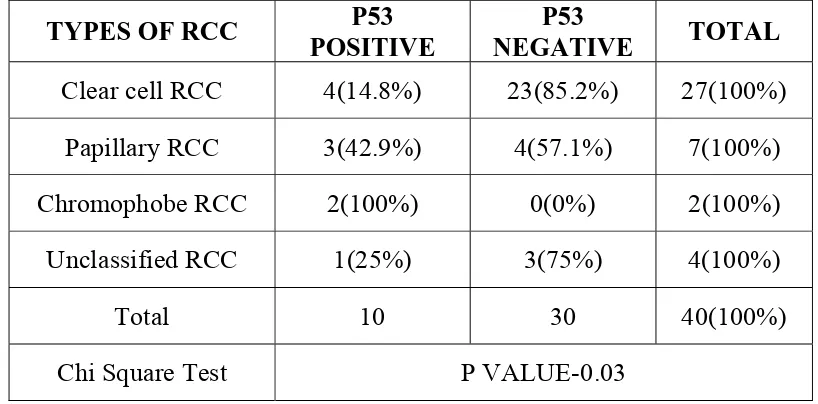
technically cumbersome. Therefore, both immune-detection and PCR
have been used as alternative methods.
MUC1
Mucins are high-molecular-weight glycoproteins with 4200 kDa
with oligosaccharides are attached to an apomucin protein by
O-glycosidic linkage.(135)
The MUC1 gene is located on chromosome 1q21-24 .It is a
member of mucin family which encodes a transmembrane glycoprotein.
MUC1 is membrane-associated and membrane-secreted. MUC1 also is
known as polymorphic urinary mucin, or PUM, and epithelial membrane
antigen (EMA). MUC1 has a apical membranous distribution of many
glandular epithelia like epithelium of the colon, breast, lung, pancreas and
kidney. MUC1 is supposed to play a role in cellular polarity, cell
adhesion and signal transduction. In the kidney, MUC1 is expressed in
normal distal convoluted tubules, collecting ducts .(8)
Sialylated MUC1 mucin expressed on tumor cells suppresses
cellmatrix adhesion and homotypic cellular aggregation and promotes
lymphocyte–target cell interactions. They also induce apoptosis of
lymphocytes.
Thus, with these findings cancer cells with a high level of
sialylated MUC1 expression are able to detach easily from the primary
lesion and they survive in circulation and in distant organs of metastasis
by escaping immune surveillance.(136)
In many epithelial cancers, there is loss of polarized cellular
expression and there is diffuse circumferential distribution. These
variations of expression of MUC1 in malignant cells are suspected to be
responsible for metastatic dissemination by destabilization of cell-cell and
cell–extracellular matrix interactions. In various studies, MUC1 is
MATERIALS
AND
MATERIALS AND METHODS
This study is a combined retrospective and prospective study of
renal cell carcinoma, conducted in the Institute of Pathology, and Rajiv
Gandhi Government General Hospital, Chennai for a period of 3 years
between august 2011 to august 2014 .
Total of 52 cases of resected specimens of renal cell carcinoma
were received for histopathological examination in Madras Medical
College during the period between august 2011 to august 2014.
INCLUSION CRITERIA:
All resected specimens of renal cell carcinoma, irrespective of the
age and stage were included for the study.
EXCLUSION CRITERIA:
1. Renal biopsy specimen
2. Renal malignancies other than renal cell carcinoma
3. Nephrectomy done for Benign and non-neoplastic lesion of kidney
METHOD OF DATA COLLECTION:
Detailed history of the cases regarding age, sex, clinical
presentation, investigations done along with the findings, type of
received during the period of study. Haematoxylin and Eosin stained 4
micron thick sections of the paraffin tissue blocks of the cases were
prepared from nephrectomy specimens and cases reported as renal cell
carcinoma were selected. 40 patients were randomly selected for
Immunohistochemical analysis using ki67, p53 and MUC1
Variables studied:
The following clinical and pathological parameters were evaluated:
Age, gender, size, laterality (right or left side), histological types
(clear cell RCC, papillary RCC, chromophobe RCC, unclassified RCC)
Nuclear grading according to FURHMAN grading system.(107)
G1 – Nuclei are small, round and uniform (10 μm), with
inconspicuous or absent nucleoli.
G2 – Nuclei are slightly irregular (15 μm), with small nucleoli.
G3 – Nuclei are very irregular (20 μm), with large and prominent
nucleoli.
G4 – Nuclei exhibit large and pleomorphic often poly-lobed and
bizarre (> 20 μm).
Presence of capsular infiltration, renal vessel invasion, ureter
invasion and lymph node involvement, distant metastasis and TNM
fixed, paraffin embedded tissue samples were subjected to
immunohistochemical analysis with a panel of 3 markers i.e., ki67, p53
and MUC1
Antigen Vendor Species
(clone) Dilution control
KI67 PATHINSITU MOUSE Ready to use
Malignant phyllodes
P53 DAKO MOUSE Ready to use malignancy Colonic
MUC1 PATHINSITU RABBIT Ready to use
Distal Convoluted
Tubule
Immunohistochemistry procedure:
Slide Preparation:
1. Sections with a thickness of 4 μ were cut from formalin fixed
paraffin embedded tissue samples and transferred to
gelatin-chrome alum coated slides.
2. The slides were incubated for overnight at 58ºC.
3. The sections were deparaffinised in xylene for 15 minutes x 2
4. The sections were dehydrated with absolute alcohol for 5 minutes
for 2 changes.
5. The sections were then washed in tap water for 10 minutes.
6. The slides were then immersed in distilled water for 5 minutes.
Antigen Retrieval:
1. Heat induced antigen retrieval was done with microwave oven in
appropriate temperature with appropriate buffer for 20 minutes.
This step unmasks the antigenic determinants of fixed tissue
sections.
2. The slides were then cooled to room temperature for 20 minutes
and washed in running tap water for 5 minutes.
3. The slides were then rinsed in distilled water for 5 minutes.
4. They were washed with appropriate wash buffer (phosphate buffer)
for 5 minutes x 2 changes.
5. Peroxidase block was applied over the sections for 10 minutes.
6. The slides were washed in phosphate buffer for 5 minutes x 2
changes.
Antibody application:
1. The sections were drained (without washing) and appropriate
primary antibody was applied over the sections and incubated for
30 minutes.
2. The slides were washed in phosphate buffer for 5 minutes x 2
changes.
3. The slides were covered with Primary antibody amplifier for 10
minutes.
4. The slides were washed in phosphate buffer for 5 minutes x 2
changes.
5. The slides were covered with HRP micropolymerQuanto for 10
minutes.
6. The slides were washed in phosphate buffer for 5 minutes x 2
changes.
Chromogen application:
1. DAB substrate was prepared by diluting 1 drop of DAB
Quantochromogen to 1 ml of DAB Quanto buffer.
2. DAB substrate solution was applied on the sections for 5 minutes.
3. The slides were washed in distilled water for 2 minutes.
4. The sections were counterstained with Hematoxylin for 2 seconds.
6. The slides were air dried, cleared with xylene and mounted with
DPX.
INTERPRETATION AND SCORING SYSTEM Ki67
The immunohistochemically stained slides were analyzed for the
presence of reaction and percentage of cells stained. Immunoreactivity
was identified by nuclear brown color. The percentage of nuclei with
immunoreactive ki67 was counted for each tumor slide.
Immunoreactivity was classified as continuous data from undetectable
levels (0%) to homogeneous (100%). The reaction is considered positive
when 10% or more of the tumor cells showed staining, according to
previous study.(134)
P53
Immunoreactivity was identified by nuclear brown color. The
percentage of nuclei with immunoreactive p53 was counted for each
tumor slide. Immunoreactivity was classified as continuous data from
undetectable levels (0%) to homogeneous (100%).
Expression of p53 was evaluated separately using the following scale:(133)
3+ = high level (91-100% of positive cells)
1+ = low level (up to 10% of positive cells)
0= negative cells (0% of positive cells).
For purpose of statistical analysis a sample is said to be positive if
5% of cells are positive for p53.(137)
MUC1
A cell was estimated as positive when the cytoplasm, cell
membrane, or both were stained. The percentage of positively stained
cells (positive rate) was determined for each tumor. Immunoreactivity
was categorized as follows:
0 - no reactivity
1 - less than 10% of cancer cells positive
2 - 10–25% positive
3 - 25–50% positive
4 - 50–75% positive
5 - 75–90% positive
6 - more than 90% of cancer cells positive.
For statistical analysis, in accordance with previous studies, sample
with more than 10% of tumor cells positive immunostaining were
STATISTICAL ANALYSIS:
The statistical analysis was performed using statistical package for
social science software version 15.5 which consisted computing the
frequency counts and percentages for qualitative variables and mean for
the quantitative variables. The expression of KI67, P53, MUC1 was
correlated with clinico-pathological factors like age, gender, tumor size,
histological types, nuclear grade, stage using pearson’s chi-square test.
OBSERVATION
AND
OBSERVATION AND RESULTS
From august 2011 to august 2013, a total of 31,237 cases were
received for histopathological examination in the institute of pathology,
Madras Medical College. Among this, 52 were nephrectomy specimens
done for RCC. Of this, 36 were clear cell RCC, 8 were papillary RCC, 2
were Chromophobe RCC, 6 were unclassified RCC. Clear cell RCC was
the most common ty