THE STUDY OF HISTOLOGICAL GRADING BY MODIFIED BLOOM RICHARDSON GRADING SYSTEM AND ER, PR,
HER-2/neu STATUS IN INVASIVE BREAST CARCINOMA BY
DIAGNOSTIC IMMUNOHISTOCHEMISTRY USING TISSUE MICROARRAY
DISSERTATION
SUBMITTED TO THE TAMILNADU DR.M.G.R. MEDICAL UNIVERSITY CHENNAI
In partial fulfillment of the requirements for the degree of
M.D. (PATHOLOGY) BRANCH – III
DEPARTMENT OF PATHOLOGY
TIRUNELVELI MEDICAL COLLEGE HOSPITAL TIRUNELVELI - 627011
CERTIFICATE
I hereby certify that this dissertation entitled “THE STUDY OF HISTOLOGICAL GRADING BY MODIFIED BLOOM RICHARDSON GRADING SYSTEM AND ER, PR, HER-2/neu STATUS IN INVASIVE BREAST CARCINOMA BY DIAGNOSTIC IMMUNOHISTOCHEMISTRY
USING TISSUE MICROARRAY” is a record of work done by Dr. MANORANJANI. R, in the Department of Pathology, Tirunelveli Medical
College, Tirunelveli, during her postgraduate degree course period from 2013- 2016. This work has not formed the basis for previous award of any degree.
The DEAN
CERTIFICATE
This is to certify that this Dissertation entitled “THE STUDY OF HISTOLOGICAL GRADING BY MODIFIED BLOOM RICHARDSON GRADING SYSTEM AND ER, PR, HER-2/neu STATUS IN INVASIVE BREAST CARCINOMA BY DIAGNOSTIC IMMUNOHISTOCHEMISTRY
USING TISSUE MICROARRAY” is the bonafide original work of Dr. MANORANJANI. R, during the period of her Post graduate study from 2013
– 2016, under my guidance and supervision, in the Department of Pathology Tirunelveli Medical College & Hospital, Tirunelveli, in partial fulfillment of the requirement for M.D., (Branch III) in Pathology examination of the Tamilnadu Dr. M.G.R Medical University will be held in APRIL 2016.
Dr. ARASI RAJESH, M.D Dr. K. SHANTARAMAN, M.D
Professor, Professor and HOD of Pathology,
Department of Pathology, Department of Pathology, Tirunelveli Medical College, Tirunelveli Medical College,
DECLARATION
I solemnly declare that this dissertation titled “THE STUDY OF HISTOLOGICAL GRADING BY MODIFIED BLOOM RICHARDSON GRADING SYSTEM AND ER, PR, HER-2/neu STATUS IN INVASIVE BREAST CARCINOMA BY DIAGNOSTIC IMMUNOHISTOCHEMISTRY USING TISSUE MICROARRAY” submitted by me for the degree of M.D, is the record work carried out by me during the period of 2013-2016 under the guidance of Prof. Dr. ARASI RAJESH, M.D, Professor of Pathology, Department of Pathology, Tirunelveli Medical College, Tirunelveli. The dissertation is submitted to The Tamilnadu Dr. M.G.R. Medical University, Chennai, towards the partial fulfilment of requirements for the award of M.D. Degree (Branch III) Pathology examination to be held in April 2016.
Place: Tirunelveli Dr. MANORANJANI. R,
Date: Department of Pathology,
Tirunelveli Medical College,
ACKNOWLEDGEMENT
Though only my name appears on the cover of this dissertation, a great many people have been behind this task and I take this opportunity with immense pleasure to place on record my heartfelt gratitude and respect to all my distinguished resources.
I thank the DEAN Dr. SITHY ATHIYA MUNAVARAH, M.D for permitting me to conduct this study and to avail the resources of the hospital.
I am greatly indebted to my esteemed Professor and Head, Department of Pathology DR. SHANTARAMAN. K M.D, who amidst his tight schedule has always provided me the necessary help. His valuable suggestions, unsparing support and concern bring the successful completion of this project
I express my heartfelt gratitude to my revered mentor and guide DR. ARASI RAJESH M.D, Professor, Department of Pathology, but for whose
expert guidance, ever available help and constant encouragement, this dissertation would have been impossible.
I am extremely thankful to the respected Professors of my Department,
DR. VALLIMANALAN. S M.D, DR. SWAMINATHAN.K M.D, DR. SURESH DURAI. J M.D, , Associate Professor; DR. VASUKI
I also thank all the lab technicians and my fellow postgraduates for their cooperation which enormously helped me in the study. Without their humble cooperation, this study would not have been possible
I express my sincere gratitude to my husband DR. VIJAYARAGHAVAN. He encouraged and supported me from time to time throughout and especially during the tough times and helped me in the completion of this dissertation.
ABBREVIATIONS 1. DCIS Ductal Carcinoma In Situ 2. IDC Invasive Ductal Carcinoma
3. IDC, NOS Invasive Ductal carcinoma, Not otherwise specified 4. ICC Invasive Cribriform carcinoma
5. MBR Modified Bloom and Richardson 6. ASCO American Society Of Oncology 7. CAP College of American pathologists
8. ER Estrogen Receptor
9. PR Progesterone Receptor
10. HER2/neu Human Epidermal Growth Factor/neuroblastoma
11. CK Cytokeratin
12. GCDFP Gross cystic disease fluid protein
13. BRCA Breast cancer
14. EGFR Epidermal growth factor receptor
15. IHC Immunohistochemistry
16. HRP Horse Radish Polymer
17. TRIS – EDTA Trizma base ( Tris – hydroxyl methyl aminomethane) – Ethylene diamine tetra acetic acid
18. TMA Tissue Microarray
CONTENTS
S.NO TITLE PAGE.NO
1. 2. 3. 4. 5. 6. 7. 8.
INTRODUCTION
AIM AND OBJECTIVES REVIEW OF LITERATURE MATERIALS AND METHODS OBSERVATION AND ANALYSIS DISCUSSION
SUMMARY CONCLUSION BIBILIOGRAPHY ANNEXURES MASTER CHART
ABSTRACT
BACKGROUND: Breast carcinoma, the most common malignant tumour among women contributes to a significant proportion of all cancers in women worldwide. It was graded based on levels of nuclear pleomorphism, tubular formation and mitotic index. The commonly used predictive immunohistochemical markers are estrogen receptors, progesterone receptors and HER2/neu status. In our study we use microarray technique where small representative tissue samples from many cases were assembled on a single histology slide and subjected to Immunohistochemical analysis. AIMS AND OBJECTIVES: To study histological grading by Modified Bloom Richardson grading system and ER, PR & HER 2/ neu status in invasive breast carcinoma by diagnostic immunohistochemistry using Manual Tissue Microarray and assess the cost-effectiveness of IHC done on TMA slides.
MATERIALS AND METHODS: 50 cases of invasive breast carcinoma were included in the study. Histopathological examination of the haematoxylin and eosin stained slides were done and the tumour was graded according to Modified Bloom Richardson grading system. A standard method of microarray preparation was done. First the design for TMA construction was laid out. Paraffin embedded tissue blocks were collected and the areas of invasive carcinoma were cored from donor blocks and transferred to the recipient blocks using bone marrow needle. Thus tissue microarray was constructed manually. Immunohistochemical analysis using ER, PR and Her2/neu were done for all these cases. Evaluation was done with Allred scoring system for ER and PR status, and ASCO guidelines for HER2/neu status.
RESULTS: Of the 50 patients analysed, majority were invasive ductal carcinoma (84%). Majority of the invasive breast carcinoma were of MBR grade II (50%) followed by grade III tumors (42%) and grade I tumors (8%). Among 50 cases, ER and PR were positive in 24 cases (48%) and 31 cases (62%) respectively. HER-2/neu expression was seen in 25 cases (50%). A statistically significant correlation was noted between histologic grading and ER, PR and HER2/neu status. The tissue microarray uses only one seventh of the reagent consumed by conventional immunohistochemistry.
CONCLUSION: The process of immunohistochemistry using tissue microarray obviates the need for control and standardisation. This allows the study of different cases on a single slide, thus reducing the amount of reagent, duration and labour of the procedure and making it cost effective. Tissue loss due to technical problems can be overcome by following standard protocols or by obtaining more number of tissue cores.
1
INTRODUCTION
Breast carcinoma is the most common malignant tumor among women. It contributes to a significant proportion of all cancers in women worldwide (25%). Annually about one million women are diagnosed with breast cancer worldwide(1). Breast cancer accounts for maximum number of deaths in the age group of 15-54 years(2).
There is an increased trend in the detection of breast carcinoma, which can be attributed to increased mammographic screening and changes in lifestyle(3).But the mortality has decreased due to early screening, which detects the tumor at an early curable stage and also by means of better effective treatment modalities. Nowadays the incidence of breast carcinoma has increased in less developed countries owing to gradual changes in lifestyle of women.
Breast cancer is a heterogeneous disease with distinct biological subtypes. Major types includes invasive ductal carcinoma and invasive lobular carcinoma. Of these, invasive ductal carcinoma is the most common subtype accounting for 70-80%, it is further sub classified as well differentiated (grade1), moderately differentiated (grade2) and poorly differentiated (grade3) based on levels of nuclear pleomorphism, tubular formation and mitotic index(4).
2
In addition to this there are certain predictive factors like(6), 1. Estrogen and progesterone receptor(ER, PR).
2. HER2/neu amplification.
3. Proliferative markers like Ki-67
The outcome of the tumor varies in each individual and is believed to be due to the heterogeneous nature of the tumor.
3
AIM:
To study histologic grading by Modified Bloom Richardson grading system and ER, PR & HER 2/ neu status in invasive breast carcinoma by diagnostic Immunohistochemistry using Manual Tissue Microarray.
OBJECTIVES:
¾ To grade Invasive Breast carcinoma by Modified Bloom Richardson grading system.
¾ To apply a panel of IHC markers on Invasive breast carcinoma.
4
REVIEW OF LITERATURE
HISTORY:
The first description of breast cancer dates back more than 3500 years. The Egyptians were the first to describe breast cancer as bulging tumor in breast .The descriptions of Edwin Smith and George Ebers Papyrus about breast tumors match with the present day scientific descriptions of breast cancer. The etiology of breast cancer was a matter of great speculation. Hippocrates was the first to describe breast cancer as a humoral disease and also named cancer as karkinos which is a Greek word for crab(8).
EMBRYOLOGY
The breast is a modified apocrine sweat gland and forms an important accessory organ of female reproductive system.
The mammary glands develop at the fifth week of fetal development from the ectodermal mammary ridges which are present on the ventral surface of the fetus bilaterally from the axillary to the inguinal region. During the normal course of development majority of the mammary ridge disappears at the seventh week of gestation.
A small portion of the mammary ridge persists as primary mammary buds
which are present in fourth or fifth intercostal spaces. Development proceeds by
5
primary mammary buds develop into secondary buds at around 3rd month of
gestation and the primary bud later contributes to mammary lobules.
At 20 weeks of gestation, the developing breast is penetrated by multiple
radial ingrowths of ectoderm. This is followed by the canalization of the buds
which are the precursors of the lactiferous ducts and their branches. The
mammary pit which is formed during gestation by the convergence of the
lactiferous ducts, then transforms into nipple during infancy.
In males, there is no significant postnatal development of the breast but in
females during puberty under the influence of sex hormones the parenchyma and
the ductal system proliferates rapidly(9).
ANATOMY
The breast is covered by skin and subcutaneous tissue and it lies on the
pectoral muscle which is separated by a fascia. It extends from the 2nd to 6th rib
vertically and from the lateral border of sternum to the mid axillary line
horizontally. A small extension called axillary tail of spence extends laterally
towards the axilla.
The nipple lies at the level of 4th intercostal space and is pierced by
15-20 lactiferous ducts. Surrounding the nipple is the areola which is a circular
pigmented area and is rich in modified sebaceous glands. Fibrous strands extend
6
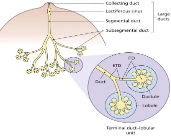
Fig 1(A)-Diagramatic representation of Terminal Ducto-lobular unit
The breast parenchyma is composed of glandular tissue which is arranged
topographically into lobes. The lobes are made up of terminal duct lobular unit
[TDLU] and the large duct system. The TDLU is the secretory portion of breast
and consists of lobule and terminal ductule. Each lobule in turn is a cluster of
acini. The TDLU connects with the lactiferous (collecting) duct by means of sub
segmental and segmental duct. The lactiferous duct opens in to the nipple. A
fusiform dilatation called the lactiferous sinus is present between the lactiferous
and segmental duct [Fig 1(A)] (10).
[image:17.595.139.483.80.353.2]7
cells admixed with scattered lymphocytes. The interlobular stroma is made of dense fibrous connective tissue admixed with adipose tissue(10).
BLOOD SUPPLY
Arterial supply of breast is by,
i. Internal thoracic artery.
ii. Branches of the lateral thoracic, superior thoracic and
acromiothoracic arteries.
iii. Lateral branches of posterior intercostal arteries.
Venous drainage of the breast follows the course of arteries forming an
anastomotic circle in the subcutaneous tissue beneath the nipple-areola complex.
From this the veins run as,
1. Superficial veins draining into internal thoracic vein.
2. Deep vein draining into internal thoracic, axillary and posterior intercostal
veins(10).
NERVE SUPPLY:
Nerve supply is by anterior and lateral cutaneous branches of 4th and 6th
8 LYMPHATIC DRAINAGE
1. Axillary lymph nodes: Lymphatic drainage is mainly into the anterior
group of axillary nodes. Posterior, lateral, central and apical groups of
nodes also receive lymphatic drainage either directly or indirectly.
2. The internal mammary nodes which lies along internal thoracic vessels.
3. Supraclavicular node, cephalic node, posterior intercostal, Sub
diaphragmatic and sub peritoneal lymph plexus(10).
4. The superficial lymphatics drains overlying skin of breast except
nipple and areola .They pass radially to the surrounding lymph nodes
(axillary, internal mammary, supraclavicular and cephalic node ).
5. The deep lymphatics drain the parenchyma, nipple and areola of
breast. About 75% of lymph drains into axillary nodes, 20% into internal
mammary nodes and 5 % into posterior intercostal nodes(10).
HISTOLOGY
The nipple and the skin are lined by the keratinizing stratified squamous epithelium. The entire ductal-lobular unit lies on a continuous basement membrane. It is lined by two cell layers: luminal epithelial cells and basally located myoepithelial cells [Fig 1(B)]. Luminal cells are either columnar or
cuboidal depending on their function. Other cell types present in the breast
9
Fig. 1(B) - Photomicrograph of Terminal Ducto-lobular unit in a normal adult female (H & E).
The nipple is formed by the lactiferous duct along with the sebaceous
unit. The main difference of epidermis of nipple and areola is the increased
melanin content in basal layer compared to the normal skin. The basal layer also
contains occasional Toker cells(10).
Lactatory function of the breast is a co-ordinated effort of the lobular and myoepithelial cells. The secretory function is by the luminal cells in the lobules. The ejection of milk is by the contractile myoepithelial cells which also renders a structural support to lobules(10).
When analyzed immunohistochemically the luminal epithelial cells stain
[image:20.595.160.471.71.324.2]10
positive for S-100, Smooth Musle Actin, calponin, caldesmon (duct portion) and
also shows nuclear reactivity for p63(10).
CARCINOMA BREAST
Breast cancer is the most common malignant cancer among women(1).
The incidence has increased nowadays due to increasing awareness of the
people and use of diagnostic modalities like mammography, fine needle
aspiration and core biopsy(3).
EPIDEMIOLOGY
Breast cancer is one of the most prevalent diseases affecting women. Observation is that the incidence of breast cancer has increased globally over the last several decades(11). And the more worrisome fact is that the greatest increase has been in Asian countries(12). Worldwide, breast cancer is the most frequent cancer in women and represents the second leading cause of cancer death among women (after lung cancer)(13,14). The number of new cases occurring each year is estimated to be around 1,00,000(15).
11
cancer is the commonest cancer in women in cities whereas in rural areas, it is second to cervical cancer.
CLINICAL PRESENTATION
Breast cancer can present in many different patterns. The most common
symptoms are breast lump (60-70%) followed by pain (14-18%).Nipple
discharge (7-9%) is the least common presenting symptom. With the
introduction of mammographic screening there is an increased detection of
asymptomatic cases. Anatomically upper outer quadrant contributes to majority
of cases (40-50%) followed by central, upper inner, lower outer to lower inner
quadrant(17).
Breast mass should be evaluated by triple assessment which includes
clinical examination, imaging studies (mammography, ultrasound) and tissue
sampling either by fine needle aspiration cytology or core needle biopsy.(17)
MICROSCOPIC TYPES 1. Carcinoma In Situ 2. Invasive Carcinoma
INVASIVE DUCTAL CARCINOMA
12
Two major categories of invasive carcinoma are - ductal and lobular type. Invasive ductal carcinoma comprises 75-85% of mammary carcinoma. Invasive ductal carcinoma, not otherwise specified comprises majority of duct carcinoma. Other relatively infrequent forms of infiltrating ductal carcinoma include tubular, medullary, metaplastic, colloid carcinoma etc(18).
CYTOARCHITECTURAL TYPES
1. INVASIVE DUCTAL CARCINOMA, NOS TYPE
The major chunk of all breast carcinomas is comprised by IDC, NOS type (75 %). and is considered as the prototype of all breast carcinomas(19).
GROSS:
The tumor is usually an ill circumscribed firm tumor. Cut section reveals a yellowish gray cut surface with multiple trabeculae radiating through the surrounding parenchyma in to the adjacent fat with a crab like or stellate configuration. In case of larger tumors, areas of necrosis, hemorrhage and cystic degeneration may be present. Scirrhous carcinoma was the term used synonymously with these tumors as they are hard in consistency due to large amounts of stroma.
MICROSCOPY:
13
cells are usually large and pleomorphic. The tumor shows standard features of malignancy like prominent nuclei and nucleoli and increased mitotic figures. About 60% of the cases show areas of necrosis and calcification. The amount of stroma varies from scant to abundant desmoplastic stroma. The interphase between the tumor and stroma shows mononuclear cell inflammatory infiltrates(10).
Fisher et al. noted lymphatic, blood vessel and perineural invasion in 33%, 5% and 28% of the cases respectively(20).
IHC:
The tumor cells are positive for ER, PR, HER2/neu, low molecular weight keratin (8, 18 and 19) and EMA. Other sensitive breast related markers are mammoglobin and GCDFP 15.The basement membrane components collagen 4 and laminin shows a discontinuous linear pattern or it may be totally absent(10). 2. INVASIVE CRIBRIFORM CARCINOMA
Invasive cribriform carcinoma is a rare form of breast malignancy which has an excellent prognosis.
MICROSCOPY:
14
3. TUBULAR CARCINOMA
Pure tubular carcinoma contributes only a small portion of invasive breast cancer. But in mammographic screening 9-19 % of cases are noted as speculate nature and cellular stroma which is characteristic of this tumor can be easily noted.
GROSS:
In gross section the tumor appears poorly circumscribed and is hard in consistency and the size of the tumor is usually small with a mean diameter of 1 cm.
MICROSCOPY:
The characteristic feature of tubular carcinoma is the irregular and angulated contour of the glands which shows no organoid configuration. The lining cells show apocrine type snouts in the apical cytoplasm. This tumor typically lacks myoepithelial cells and basement membrane. The lumina of the glands are open and filled with basophilic secretion. The tumor shows cellular desmoplastic reaction with fat invasion in the periphery. It can confused with benign disorders because of the well differentiated nature of the glands, scant pleomorphism and absence of necrosis(10).DCIS can be seen in majority of the cases. The in situ component is usually of low grade showing cribriform or papillary pattern.
15
carcinoma there can be a diagnostic dilemma. In such instances, the term tubular NOS and tubular mixed can be employed. The term tubular carcinoma can be best employed for tumors in which tubular pattern is present for at least 90 % of the tumor. These tumors are associated with favorable prognosis
4. MUCINOUS CARCINOMA
Mucinous carcinoma was classified under mucin producing carcinoma. The age groups usually affected by this tumor are postmenopausal women. It is
also called as mucoid, gelatinous or colloid carcinoma(17). GROSS:
The tumor is well circumscribed .Cut surface of the tumor shows a characteristic glistening and gelatinous appearance.
MICROSCOPY:
The tumor cells are arranged usually in small clusters floating in a mucinous pool which are surrounded by bands of fibrous septa. The malignant cells are characterized by little pleomorphism and a low mitotic rate. The mucin is usually extracellular.
16
Two different types of mucinous carcinoma are seen based on the endocrine differentiation. Type A tumors shows trabeculae of malignant cells with minimal intracytoplasmic mucin. The cells are conspicuous by their absence of argyrophilia. Type B tumor shows sheets of tumor cells with abundant intracytoplasmic mucin with argyrophilia.
Nodal metastasis is very low which accounts for 2-4% of all cases. IHC:
They are positive for estrogen and progesterone receptors. They usually do not show HER2/neu overexpression or p53 accumulation.
4. MEDULLARY CARCINOMA
The tumor is most common in patients under 50 years of age and is common among carriers of BRCA1 mutation.
GROSS:
The tumor is well circumscribed, solid and homogenous. MICROSCOPY:
17
IHC:
They are positive for CK, p53 and negative for hormone receptors (ER, PR), Her2/neu and come under triple negative phenotype. The tumor expresses HLA-DR antigen which could be the possible reason for the prominent lymphoplasmacytic infiltrate. Though axillary lymph node involvement is common, only low axillary group of lymph nodes will be usually involved. The prognosis will be better than IDC, NOS type(10, 18).
5. ATYPICAL MEDULLARY CARCINOMA: The tumor shows following characteristic features,
a. Syncytial growth comprising > 75% of the tumor b. Atypical features
c. Focal tumor infiltration at the margins d. Uniform nuclei and rare mitosis
e. Mild to absent lymphoplasmacytic infiltration at the margins. f. Focal tubule formation(23).
6. INVASIVE PAPILLARY CARCINOMA
18
papillary pattern is seen, metastatic papillary carcinoma from other sites should also be excluded(10, 18).
The tumor may have axillary lymph node metastasis particularly in solid variant of papillary carcinoma. Prognosis of the tumor is better compared to that of invasive ductal carcinoma, NOS type(24).
7. INVASIVE MICROPAPILLARY CARCINOMA
Invasive micro papillary carcinoma is a distinct rare variant of invasive ductal carcinoma. When the micro papillary pattern is found throughout the tumor it is referred as pure invasive micro papillary carcinoma. When it is present as a part of conventional IDC it is called as mixed invasive micro papillary carcinoma. But the criteria to distinguish these two are not clear cut. Some authors suggest at least 50 % of the tumor should be micro papillary to call it as pure invasive micro papillary carcinoma.
MICROSCOPY
The tumor is composed of clusters of cells arranged in micro papillary or tubular pattern and tumor cells are found floating in clear spaces. Fibro vascular core will be absent in the micro papillary clusters. The clusters exhibit a “inside out” arrangement in which the apical cells are polarized outside and this can be evidenced by MUC 1 staining.
19
Lymphatic invasion is reported in more than 50 % of the cases. The tumor has a bad prognosis.
IHC:
In a study done by Cruz et al, estrogen receptor was positive in 72-75 % of cases, 45 % cases were positive for progesterone receptor and 36 % of the cases show Her 2-neu overexpression(25).
8. APOCRINE CARCINOMA
Apocrine carcinoma is very rare comprising 0.5% of all breast carcinomas. The tumor is composed entirely or predominantly of apocrine type cells with tumor cells being large with abundant eosinophilic cytoplasm with vesicular nucleus and prominent nucleolus. Glandular differentiation can be seen with apocrine snouts. Diagnosis of apocrine carcinoma should only be made when the architectural features are those of a malignant tumor. Immunohistochemically they are positive for GCDFP-15.Estrogen and progesterone receptors will be negative(26).
9. SECRETORY CARCINOMA
Secretory carcinoma is a rare tumor and usually seen in children. It can also occur in adults and has an excellent Prognosis.
GROSS:
20
MICROSCOPY:
The tumor is composed of tubuloalveolar and papillary structures. The lumina contain eosinophilic PAS positive, diastase resistant material. The malignant cells have a pale staining vacuolated cytoplasm. Nucleoli may be prominent and mitosis is scanty.
IHC:
There is a strong immunoreactivity for S-100 and a-lactalbumin. 10. METAPLASTIC CARCINOMA
Metaplastic carcinoma represents tumor predominantly with cell type other than epithelial and glandular component. It includes many categories which may overlap with each other. Metaplastic carcinoma is more aggressive than invasive ductal carcinoma. Metastasis is usually hematogenous rather than lymph node metastasis.
GROSS:
21
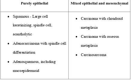
[image:32.595.94.531.147.414.2]CLASSIFICATION OF METAPLASTIC CARCINOMA
TABLE 1: CLASSIFICATION OF METAPLASTIC CARCINOMA
Purely epithelial Mixed epithelial and mesenchymal
• Squamous - Large cell keratinizing, spindle cell, acantholytic
• Adenocarcinoma with spindle cell differentiation
• Adenosquamous, including mucoepidermoid
• Carcinoma with chondroid metaplasia
• Carcinoma with osseous metaplasia
• Carcinosarcoma
A. SQUAMOUS CELL CARCINOMA GROSS
The tumors are large showing cystic spaces filled with keratin. MICROSCOPY:
In pure squamous cell carcinoma the central cystic cavity is lined by malignant squamous cells. Most cases represent squamous metaplasia.
22
carcinoma is said to have a favourable prognosis whereas acantholytic squamous cell carcinoma has an aggressive behavior(27).
B. CARCINOSARCOMA
When the transition between sarcomatous and carcinomatous component is gradual and sharp, it is termed carcinosarcoma. Microscopically the sarcoma like component can be malignant fibrous histiocytoma, osteosarcoma, chondrosarcoma, angiosarcoma or a combination of various components.
When the transition to cartilaginous or osseous elements is direct without an intervening spindle cell component or osteoclastic giant cells, it is called matrix producing carcinoma.
IHC:
The sarcoma like elements acquires vimentin and other mesenchymal features which is referred to as the phenotypic switch. The cells are occasionally positive for epithelial markers.
SPREAD RELATED VARIANTS 1. INFLAMMATORY CARCINOMA
23
Histopathological examination of some of the cases shows an undifferentiated carcinoma.
The prognosis is usually bad. Studies done by Charafe-Jauffret et al. found that most of the inflammatory carcinomas are negative for estrogen and positive for MIB1, E-Cadherin and HER2/ neu(28).
INVASIVE LOBULAR CARCINOMA: 1. CLASSIC TYPE
It is the most typical form of Invasive Lobular Carcinoma which is characterized by the presence of small, uniform tumor cells which grows singly, in Indian file and in a concentric fashion around lobules. The stroma is usually abundant, of dense fibrous type(10, 18).
2. PLEOMORPHIC LOBULAR CARCINOMA
This form of invasive breast tumor has the pattern of growth of a classic breast carcinoma but exhibits a marked degree of nuclear pleomorphism and abundant cytoplasm. It also frequently shows apocrine differentiation, focal signet ring morphology.
IHC:
24
3. HISTIOCYTOID CARCINOMA
Histiocytoid carcinoma is characterized by a diffuse growth of tumor cells which displays abundant granular, foamy cytoplasm. It may simulate the appearance of a granular cell tumor (myoblastoma). It is currently viewed as a variant of invasive lobular carcinoma exhibiting apocrine differentiation.
IHC:
Immunohistochemical reactivity for GCDFP-15 and the demonstration of mRNA for the related prolactin-inducible protein (PIP) by in situ hybridization. In most cases E-cadherin is absent. The mucins expressed by this tumor include some ‘non-mammary’ types, such as MUC2 and MUC5AC(10).
4. SIGNET RING CARCINOMA
Signet ring carcinoma is a type of breast carcinoma in which the tumor cells show intracytoplasmic mucin accumulation, resulting in the typical signet ring appearance. It is important to separate this tumor from mucinous carcinoma (in which the mucin is extracellular).
IHC:
Signet ring carcinoma is positive for CK7 and MUC1, and usually negative for E-cadherin.
5. TUBULOLOBULAR CARCINOMA
25
cords of tumor cells growing in a lobular configuration similar to that of invasive lobular carcinoma. The in situ component, if present, may be of lobular, ductal or mixed type. It is associated with a higher incidence of multifocality and positive axillary nodes than pure tubular carcinoma
IHC:
Immunohistochemical profile is intermediate between those of ductal and lobular carcinoma, in that it shows positivity for both E-cadherin and HMW keratin(10, 20).
HISTOLOGICAL GRADING OF DUCTAL CARCINOMA
Grading of breast cancer was first attempted by Greenhough in 1925(29). It was an extensive system with 18 features and it is not used now. In 1993 Haagensen evaluated 15 histological features which mainly include growth pattern, cell morphology and the stromal reaction.
The most popular grading system till date was proposed by Bloom in 1950(30).The original classification proposed by him was based on three main features which include degree of tubule formation, nuclear features and mitotic activity. He classified breast carcinomas into 2 group low grade and high grade tumors.
26
TABLE 2: BLOOM AND RICHARDSON GRADING SYSTEM 1957
Elston further modified this classification by applying this only to invasive ductal carcinoma and excluding special types like mucinous, medullary carcinoma. Elston and Ellis modified Bloom and Richardson grading system by quantifying the mitotic activity(32). This is also referred as the Nottingham modification of Bloom and Richardson system.
TABLE 3: NOTTINGHAM MODIFICATION OF BLOOM RICHARDSON HISTOLOGICAL GRADING SYSTEM Score 3-5 Grade 1 Well differentiated tumors
Score 6-7 Grade 2 Moderately differentiated tumors
Score 8-9 Grade 3 Poorly differentiated tumors
CRITERIA SCORE
Tubule and gland formation Majority of tumor (>75%) Moderate degree (10-75%) Little or none (< 10% )
27
Mitotic count
Mitotic count is also graded as 1-3. But it depends on the field diameter used. Mitotic figures are to be counted from the most mitotically active area. 10 high power fields should be counted from the same area. Poorly preserved area should be ignored.
TABLE 4: SCORING OF MITOTIC COUNT
Field diameter 0.59mm Field diameter 0.44mm Score
0-9 0-5 1
10-19 6-10 2
>20 >11 3 Nuclear pleomorphism
Small, regular, uniform
Moderate variation in size, shape Marked variation in size, shape
28
TABLE 5: FINAL GRADE OF NOTTINGHAM MODIFICATION OF BLOOM RICHARDSON HISTOLOGICAL GRADING SYSTEM
GRADE SUM OF POINTS
I 3–5
II 6–7
III 8–9
PROGNOSTIC FACTORS 1. Age
Prognosis is better in patients <50 years. Older patients have a higher rate of recurrence and distant metastasis.
2. Pregnancy
Carcinoma breast manifesting during pregnancy is generally aggressive with over expression of HER2/neu and low expression of hormone receptor. 3. BRCA-1 status
29
4. Skin and nipple invasion
T4a lesions are associated with decreased survival rate. Nipple involvement is associated with high incidence of axillary node metastasis(33).
5. Presence or absence of invasion
The single most important prognostic determinator of breast carcinoma is the presence of invasive component. The invasive component of a tumor correlates with the nodal metastasis. The in situ component is proportionate to the incidence of multicentricity and indirectly with occult metastasis(10).
6. Size of the tumor
Tumor diameter should be measured in three planes to the nearest millimeter. The greatest diameter is taken as the size of the tumor. For lesions less than 1 cm stage micrometer is used in histological sections for tumor size estimation. The invasive component is the better predictor of the total tumor size than the DCIS component.
It has been proved beyond doubt that tumor size correlates with the prognosis. Multivariate analysis by Nottingham/Tenovas Primary Breast cancer study showed tumor size is an independent prognostic variable(34).
7. Histological type
30
carcinoma. Signet ring cell carcinoma in particular is associated with a poor prognosis.
8. Histological grade
The poorer the grade of the tumor by Nottingham modification of the Bloom–Richardson system, the worse is the outcome, particularly IDC, NOS type(35).
9. Tumor necrosis
Spontaneous tumor necrosis is associated with tumors showing high histological grade and increased incidence of lymph node metastases(36) and hence a poorer prognosis.
10. Lymphovascular emboli
Presence of tumor emboli within endothelial lined spaces in the peritumoral area under H & E is taken as positive lymphovascular invasion. Lymphatic emboli within the breast correlate with local recurrence and vascular invasion correlates with distant spread(37). Tumors with vascular emboli have a poor prognosis.
11. Lymph node status
31
Ten year survival rate for node negative patients will be 75 % while compared to that only 25-30% in node positive patients. Prognosis is also dependent on the number and the level of regional lymph nodes. The prognosis will be poor if greater number of nodes is involved(39).
NSABR categorises patients under two divisions for therapeutic purpose. They are categorized as patients with 1-3 positive nodes and cases with 4 or more positive nodes.
12. Hormone receptors
Estrogen receptor positive tumors have a longer disease free survival. 13. HER2 /neu expression
HER2 / neu over expression correlate with the tumor grade. It has a poor prognosis especially when associated with lymph node metastasis. It is an excellent predictor of response to the drug trastuzumab but a weak predictor for chemotherapy(40).
14. Cell proliferation
32
15. Microvessel density.
Invasive breast carcinoma with prominent vascular component in the surrounding stroma behaves aggressively than other tumors(42).
HORMONE RECEPTORS
The presence of hormone receptors (estrogen, progesterone) correlates with the response of the tumor to endocrine therapy and chemotherapy. Though estrogen and progesterone are codependent variables, estrogen receptor status is the powerful predictive factor in breast cancer management(43).
ER and PR are expressed in 80 % and 60 % of the breast cancer, respectively. There is not much correlation between receptor positivity and cyto architectural type of breast. But however some studies suggest that mucinous, tubular, lobular carcinoma show high estrogen positivity. Medullary, apocrine, metaplastic carcinomas are estrogen negative(44).
There are two parameters evaluated in immunohistochemical assessment of hormone receptors which include,
• Number of tumor cell nuclei stained-expressed as percentage of entire tumor nuclei population.
• Intensity of the reaction
33
(0 to 3) and a score for the proportion of nuclear staining (0 to 5). The final score is obtained by the sum of proportion score and intensity score which ranges from 0 to 8. Invasive tumor with an Allred score of more than 2 was considered to be positive for hormonal receptors(45).
Estrogen and Progesterone receptors
The ovarian hormones, primarily estrogen are believed to play a role in breast cancer etiology. These steroid hormones influence their effect on breast cell proliferation by the estrogen receptor (ER) and progesterone receptor (PR). The estrogen receptor is expressed in two different types, usually referred to as α and β. They are encoded by gene ESR1 and ESR2 on the sixth and fourteenth chromosome (6q25.1 and 14q), respectively(46). The main function of the estrogen receptor is as a DNA binding transcription factor, which regulates gene expression(47).
The binding of estrogen to ER stimulates proliferation of mammary cells, resulting in increase in cell division and DNA replication leading to mutations. Secondly, estrogen metabolism produces genotoxic waste. Both processes lead to disruption of cell cycle, apoptosis and DNA repair, progressing to tumor formation. This is the proposed mechanism of effects of estrogen on breast cancer. ER-α is certainly associated with more differentiated tumors, while evidence of ER-β involvement is controversial(48).
34
also triggers this cell proliferation process. Hunter et al. have studied this effect of oral contraceptives as sources of exogenous estrogen for the increased risk of breast cancer for women using these products(49).Estrogen receptor status is an important diagnostic parameter when a patient presents with breast cancer. Patients are classified as either estrogen receptor positive (ER+) or estrogen receptor negative (ER-) based on immunohistochemistry of the biopsy sample for ER-α.
The introduction of adjuvant hormonal therapy, in particular tamoxifen, has been one of the major breakthroughs in the fight against breast cancer mortality.
For women with estrogen receptor (ER)-positive breast cancer, 5 years of tamoxifen in an adjuvant setting decreases the risk of death. The same effect cannot be said for ER-negative patients as risk of death from breast cancer was significantly increased in those treated with tamoxifen(50).
35
The prognostic significance of elevated PR levels is that these tumors are less aggressive tumors. The other good prognostic feature is that they are associated with a longer overall survival time in metastatic disease, whereas PR negative tumors are more aggressive(52). The American Society of Clinical Oncology (ASCO) recommended that ERα and PR should be measured on every primary invasive breast cancer.
Human Epidermal growth factor Receptor 2 (HER-2/neu)
HER-2 (Human Epidermal growth factor Receptor 2) also known as proto-oncogene Neu .The oncogene neu is so-named because it was derived from a rodent glioblastoma cell line, which is a type of neural tumor. HER-2 protein is known to form clusters in cell membranes that might play a role in tumorigenesis(53). HER cellular signalling occurs through transmembrane receptor tyrosine kinases and can induce cell proliferation, motility, and invasion. Dysregulated expression and activity of HER family members is prevalent in human neoplasia. Overexpression of this protein leads to constitutively activated tyrosine kinase, resulting in mitogenic transduction and poorer prognosis(54).
36
It also occurs in other cancers such as ovarian cancer, stomach cancer, and uterine cancer(56). The American Society of Clinical Oncology (ASCO) recommended that should be determined Her-2 as a part of diagnostic routine on every primary invasive breast cancer.
Studies show that HER2/neu is an independent prognostic indicator for overall survival of patients with breast carcinoma.
IMMUNOHISTOCHEMISTRY
Immunohistochemistry is one of the powerful ancillary methods used in pathology today which has revolutionized the study of disease and its prognosis. The most useful aspect of IHC is that it is a powerful and cost effective tool applicable in light microscopy. The morphologic observations made by pathologists are validated by the use of IHC. Immunohistochemistry (IHC), or immunocytochemistry, is a method for localizing specific antigens in tissues or cells based on antigen-antibody recognition(57).
37
USES OF IHC:
¾ Classifying undifferentiated tumors, lymphomas, neuroendocrine and soft tissue tumors.
¾ Detection and accurate assay of tumor biologic factors of prognostic and predictive values such as hormone receptors (ER, PR) and HER-2/neu in breast cancer.
¾ Detection of metastatic cells in bone marrow, lymph nodes& serous fluids when the cell groups are too less or confusing.
38
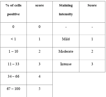
TABLE 6: ER, PR SCORING BY IMMUNOHISTOCHEMISTRY (ALLRED SCORING SYSTEM)
% of cells positive
score Staining intensity
Score
0 0 - -
< 1 1 Mild 1
1 – 10 2 Moderate 2
11 – 33 3 Intense 3
34 – 66 4
67 – 100 5
TOTAL SCORE =PROPORTION SCORE + INTENSITY SCORE ( 0 TO 8)
INTERPRETATION: 0, 2 - Negative
39
TABLE 7: GRADING OF THE IMMUNOHISTOCHEMICAL STAINING FOR HER 2 / NEU OVEREXPRESSION
Score Staining Pattern HER 2 / neu protein
overexpression assessment
0 No staining at all or very slight partial membrane staining in less than 10% of tumor cells.
Negative
1+ Faint barely perceptible membrane staining in more than 10% of tumor cells. Cells stained in only part of the membrane.
Negative
2+ Weak to moderate complete membrane staining observed in more than 10% of tumor cells.
Weakly Positive
3+ Strong complete membrane staining in more than 30% of tumor cells
40
MOLECULAR SUBTYPE OF BREAST CARCINOMA
In the year 2000 Perou and colleagues tried to segregate breast carcinoma based on their gene expression profiles into distinct subgroups. They used microarray to demonstrate gene expression. It was accepted with a hope that this will provide new approach to the biology of breast cancer and was also believed that it may impact on the therapeutic approach of the patient(60).
The subtypes recognized by the gene expression are luminal A, luminal B, HER2/neu type, basal like and normal breast like. It was suggested that normal breast like subtype is most propably an artifact rather than genuine subtype. It is due to lack or sparsity of tumor in the tissue samples used for microarray analysis.
As the different subtypes show specific characteristics, they may benefit from different therapeutic approach. Among the subtypes, basal like type shows the worst prognosis(60).
LUMINAL A
41
LUMINAL B
It comprises 20% of invasive breast carcinoma. It corresponds to invasive ductal carcinoma NOS type and micropapillary carcinoma. It expresses cytokeratins with moderate to weak expression of hormone receptors. Proliferation rate is higher compared to that of luminal A type. It shows variable expression for chemo and endocrine therapy.
HER 2/NEU
It comprises about 15% of invasive breast cancer. The tumors are usually high grade with lymph node metastasis. They are negative for hormone receptors. HER2/neu amplification and high proliferation is seen. Patients respond to trastuzumab and anthracycline based chemotherapy and usually have a poor prognosis.
BASAL LIKE
It comprises about 15% of invasive breast carcinoma. High grade IDC NOS, metaplastic and medullary carcinoma come under this category. They are negative for hormone receptors and HER2/neu but show a high proliferation rate.
42
[image:53.595.81.544.176.441.2]Based on the reactivity they are classified as follows,
TABLE 8: MOLECULAR TYPING OF BREAST CARCINOMA ACCORDING TO EXPRESSION OF IHC MARKERS
Immuno profile Luminal A Luminal B HER2/neu Basal-like
ER, PR ER and/or PR+
ER and/or PR+
ER–, PR– ER–, PR–
HER2 and others
HER2 – Low Ki-67 (<14%)
HER2+ or HER2 – Ki-67 =14%
HER2+
HER2– CK5/6 and/or EGFR+
43
TISSUE MICROARRAYS
Dr. Hector Battifora introduced a new technique in 1986 in which a number of tissues from various organs are thrown into the same block and tissue distribution of a particular antigen was processed. This was called sausage block technique(62).Wan et al in a modification of the original technique produced a library of paraffin embedded cores and used it to determine various staining patterns of many number of monoclonal antibodies(63).Later this was modified by Kononen et al who introduced the term ‘Tissue microarray’ in 1998 which is widely used nowadays, which includes the usage of 4mm skin biopsy punch needle(64).
The major focus of TMAs at the present time is in the fields of cancer and non-cancer research; and quality control in modern pathology(65).It has greatly facilitated the in situ analysis of molecular targets at the DNA, mRNA, and protein levels under standardized conditions in a large number of archived pathology specimens(66).
44
of available arrayers. The specialized commercial automated and semi- automated equipments as well as the inexpensive simple manual tissue array methods have been used to construct the TMA blocks(69). The cost of available arrayers and technical difficulties of routine array construction have limited the development of tissue microarray technique in developing countries like India(70).
The disadvantages associated with this technique are that the small size of TMA cores may not be representative of the whole heterogeneous cancers and may not provide enough data about the entire tissue profile(71). The technical issues commonly noted with the application of microarray technique are sectioning difficulties, irregular cutting plane due to variable thickness of donor paraffin blocks, core losses and instability of the TMA blocks in routine TMA.
This technology thus offers a way to fill the gap between the discovery of new molecular markers, derived from high throughput genomic analysis, and their application in the clinical setting.
Materials for TMA
45
constructed using frozen sections in which case they are called as Cryoarrays or and can be constructed using paraffin embedded cell lines and by using cell blocks(74).
TMA categorization
Tissue microarray is categorised based upon the usage of instruments and purpose of tissue microarray. Based upon the usage of the instruments and microarrayer used it has been classified into manual, semi-automated and automated tissue microarray. There are many instruments which are available nowadays which includes, the manual and automated tissue arrayer from Beecher instruments, the semi-automated tissue arrayer from Veridiam, the quick ray manual and the automated tissue arrayer from Unitma and the manual array mold tissue arrayer. Apart from this many number of home- made tissue arraying methods have been published(75, 76).
The common feature between all these devices is that they use hollow needles or punches and they adopt the technique of skin biopsy to extract tissue cores from a donor block and make a new paraffin recipient block. After cutting all the tissue cores appear as circular samples arranged in grid like fashion.
46
varies widely from tissue to tissue. If task is to compare the expression pattern of a marker from tumor centre and periphery, the cores from the particular location may be compatible. However the method of tissue sampling is entirely different when the task is to characterize the overall expression of the protein in a tumor. Targeted sampling technique is followed in the cases of comparing the expression patterns of tumor centre and periphery whereas Random sampling technique is best suited to study the overall expression patterns(77).
. Fig. 2(A) shows taking out a tissue core from the donor block with skin
biopsy needle.
[image:57.595.176.453.286.523.2]47
Although no standard and universally agreed sampling methods are in the records, it is intuitive when more samples are taken it becomes the representative area for donor tissue. The concordance of Tissue microarray technique with full section eventually depends upon the number of cores obtained. Most of the studies seem to indicate that the results from triplicate Tissue microarray cores have upto 98% concordance with the results from full section. However Goethal through his study suggests that atleast four cores are needed to achieve greater accuracy of more than 95% which is obtained using two core tissue sampling method(79).
It should be also noted that there are also technical reasons which increases number of cores taken from each tissue block(80).The reasons being, tissue folding and complete loss of tissues during processing and section cutting. Total number of lost cases accounts for as high as 23% in the Tissue microarray construction study by using tissue cores from the cases of renal cell carcinoma(81).There are controversial data regarding the size of the cores that should be used in Tissue microarray technique and their influence over the technique.
48
introduced by Hoose et al, which uses a row of tissue cores which will not be analysed and can be any tissue that is available in plenty in the laboratory(82). Any confusion in identifying the origin of the cores after Tissue microarray construction makes the staining and analysis very difficult and hence orientation of the tissue cores needs to be perfect. Many use orientation cores in the specific position, usually outside the geometric margin of the array. Using the intentionally left empty core position, it is possible to identify and orient the position of the cores macroscopically as well as to orient cut section microscopically. In addition insertion of control tissue array may be of more value in orientation of the tissue cores. Thus the control core serves as an orientation control and also as both internal positive and negative control(83).
49
problem of bubble formation which is commonly encountered when paraffin recipient blocks are used(83).
Tissue Arrayers
Both automated and manual tissue arrayers are available for the construction of Tissue microarray. Automated tissue arrayers are easy to use. The instrument usually marks, edits and saves punch co-ordinates by using an on screen display and software tools. Automated tissue arraying instruments are commonly used in laboratories with high volume of Tissue microarray and can punch upto 180 cores/hour.
Automated HT-1 Tissue Microarray
50
instruments. The key steps in automated tissue microarray are punching array pores in recipient blocks and embedding multi-tissue cylinders into the recipient blocks(84).
Recipient block formation
This method uses three types of recipient block maker which are called as recipient block-molding machine that can accommodate 24, 42, 56 tissue cylinders. The spacing between the cores is usually fixed. Recipient paraffin blocks can be made within several seconds by using block-molding machine. The instrument is composed of an array pores forming metal tamp series of metal embedding boxes and punch needles with corresponding inner cores. The array pores-metal consist of metal plates and a bracket. The lower plate is fixed with hollow cores while upper plate is fixed with inner cores. The inner diameter is designed from 0.5 to 2.5mm.The instrument for making array recipient blocks is fixed in the manipulator with the help of the bracket. The lower plate with the hollow array can move up and down by controlling the handle. The residual paraffin in array hollow punch is automatically removed by the piston(84).
Negative-pressure embedding
51
embedding box consists of metal mesh, which can effectively adjust the negative pressure so that the air bubbles which are formed can be drained out of recipient paraffin blocks. Screen mesh can also be used to adjust the temperature for the embedding media(84).
TMA without prefabricated recipient block
Array construction done with the help of automated tissue arrayers proved to be very costly and hence not suitable in developing laboratories. Hence efficient microarray system which is cost effective was designed using manual tissue arrayer technique. Most of the manual methods used pre constructed paraffin recipient block into which holes are punched followed by insertion of tissue cores. The use of paraffin blocks proved difficult because of block breakage during punching, non alignment of holes, and mismatch between the size of the recipient hole and the tissue core. With the desire to overcome these difficulties, TMA construction was done without the use of prefabricated paraffin blocks(85).
52
is cut according to the size of embedding mould and the top surface is exposed to receive the tissue cores. The site for the attachment is marked with the ruler and the felt pen. The area to be cored is marked on the block by superimposing the marked area over the slides. After obtaining the core from the donor block, the core is transferred to adhesive tape using the forceps. Then it is transferred to stainless steel mould and after which melted paraffin wax is poured. Later after uniform setting of the block the adhesive tape is peeled off to expose the cutting surface. This method can give rise upto 20 sections from each core. This is reliable, readily reproducible and does not need any specialisation(85).
Simple manual Tissue Microarray
This is the modification of conventional manual tissue microarray which used skin punch biopsy needle. This technique used bone marrow aspiration needle for the construction of manual tissue microarray. Both 14 gauge and 16 gauge needles are used. After the selection of donor block, the area of interest is marked and empty recipient paraffin block is made using the mould. By using 16gauge needle pores are made in the recipient block(85).
53
tissue cylinders are levelled with the blocks using a glass slide. Then the array is incubated at 600c for 15 minutes after which the array is chilled on ice [fig 2(B)].
Fig. 2(B) – shows the final recipient paraffin block
Types of TMA based on application
[image:64.595.182.447.136.415.2]54
as they involve the collation of tissues from the patients of same disease, those who were exposed to similar pattern of treatment and have been followed up for significant period. Progression arrays are used in analysis of role of proteins in the progression of cancer. Tumor characteristic based array is constructed based on the given characteristics such as patient age and tumor grade(83).
55
[image:66.595.188.438.69.357.2]56
MATERIALS AND METHODS
The study was conducted after obtaining approval from Institutional Ethical Committee of Tirunelveli Medical College, Tirunelveli. The study was carried out in the Department of Pathology, Tirunelveli Medical College and Hospital, Tirunelveli from January 2014 to September 2015.
SOURCE
Formalin fixed, paraffin embedded tissue blocks from 50 surgically resected breast tissues which were diagnosed as invasive breast carcinoma by histopathological examination were retrieved along with their haematoxylin and eosin stained slides and they were examined and the tumor was graded according to Modified Bloom and Richardson grading system.
SAMPLE SIZE
A total of 50 cases were included in this study. Clinical data like patient age, sex and other relevant history were noted from the Pathology records.
INCLUSION CRITERIA
• Invasive ductal carcinoma - NOS type
• Papillary carcinoma
• Medullary carcinoma
• Mucinous carcinoma
57
• Metaplastic carcinoma
• Invasive lobular carcinoma
EXCLUSION CRITERIA
• Benign tumors of breast.
• Ductal carcinoma in situ.
• Lobular carcinoma in situ
MATERIALS REQUIRED
1. Lay out for constructing Tissue microarray.
2. Metal moulds and molten wax for preparing empty recipient paraffin block.
3. Donor blocks which contain formalin fixed paraffin embedded tissue obtained from all the cases of Invasive breast carcinoma.
4. Hematoxylin and eosin stained tissue sections made from the donor blocks.
5. Black glass marking pen for marking area of interest.
6. 16 gauge bone marrow aspiration needle for making punches in the recipient block and 14 gauge needle for obtaining core from the donor block.
7. Microtome and incubator for obtaining tissue sections and to dewax the sections
8. Positively charged slides for holding tissue sections for IHC.
58
10.Pressure cooker for antigen retrieval.
11.Kit for performing immunohistochemistry which includes primary antibody (ER, PR & HER 2 neu) and universal kit. Microscope, used for interpretation and grading of IHC.
METHODOLOGY
The method of performing immunohistochemistry over the paraffin tissue microarray includes the following steps.
1. Designing the layout for TMA construction. 2. Collection of the donor blocks.
3. Preparation of the recipient paraffin blocks. 4. Immunohistochemistry and analysis.
DESIGNING THE LAY OUT
59
COLLECTION OF THE DONOR BLOCKS
The hematoxylin and eosin stained sections which were prepared from formalin fixed paraffin embedded blocks of all the cases of invasive breast carcinoma in the Department of pathology during the study period were retrieved. The corresponding formalin fixed paraffin embedded tissues were also obtained which constituted the donor block. Then the hematoxylin and eosin stained slides which contained full sections were examined and the area of interest was marked by using black glass marking pen. The area of interest is the area of tumor containing well preserved and well stained malignant cells. Then these marked areas on the slides were matched with the donor blocks and the corresponding areas over the donor blocks were also marked with the help of black glass marking pen. This area was used as the site for obtaining cores for the recipient block.
PREPARATION OF THE RECIPIENT PARAFFIN BLOCKS
The empty paraffin recipient blocks with minimum size of 25mm x 25mm were first prepared by freshly poured molten wax in the metal moulds. Then it was allowed to cool. Later using 16 gauge needle, paraffin wax cylinders of 2mm diameter were punched from the recipient blocks. Each block contained 3x3 cylinder matrix at a distance less than 2mm. In our study seven such blocks containing 50 cases were prepared.
60
donor blocks, after which it was injected into the recipient blocks into the corresponding empty cylinders with the help of predesigned layouts so that six recipient blocks contained seven cases and one recipient block contains eight cases. After the recipient block was embedded with the tissue cores, the block was incubated at 400c for 15 minutes and then it was allowed to cool for few minutes at room temperature and then the array was chilled on the ice for few minutes.
IMMUNOHISTOCHEMISTRY SECTION CUTTING
Sections were taken at 5 microns thickness after tissue microarray construction on the surface of the Poly-L-Lysine coated slides. This was followed by incubation of slides at 58-600c for one hour.
ANTIGEN RETRIEVAL SOLUTION
We used two antigen retrieval solution and a wash buffer as prescribed by the manufacturer (PATH IN SITU).
1. Citrate buffer at a pH of 6.2 for HER 2/ neu. 2. Tris EDTA at a pHof 9 for ER, PR.
61
ANTIGEN RETRIEVAL
In our institution we followed antigen retrieval by using pressure cooker as it produces even heating with lesser disadvantages compared to other methods.
PROCEDURE FOR IMMUNOHISTOCHEMISTRY AS GIVEN BY MANUFACTURER
1. Section cutting and incubation is followed by Xylene wash (2 changes) for 10 minutes each.
2. Rehydrated in graded alchohol containing 100%, 80%, 70% for ten minutes each.
3. Rinsed in distilled water for 2 minutes. 4. Antigen retrieval.
5. Cooling for 15 minutes.
6. Washed in TRIS wash buffer- 2 changes 5 minutes each. 7. Treated with peroxide block for 5 minutes.
8. Washed in TRIS wash buffer- 2 changes 10 minutes each. 9. Kept in protein block for 10 minutes.
10.Application of primary antibody (ER, PR, HER 2 neu) – 30 minutes. 11.Washed in TRIS wash buffer- 2 changes 10 minutes each.
12.Amplifier application for 15 minutes.
13.Washed in TRIS wash buffer- 2 changes 10 minutes each.
62
15.Washed in TRIS wash buffer- 2 changes 10 minutes each.
16.Application of Diamino-benzidine tetrachloride (DAB) chromogen 2 - 4 minutes.
17.Washed in distilled water – 2 changes.
18.Counterstaining is done with Hematoxylin for 30 seconds to impart background staining.
19.Wash in running tap water.
20.This is followed by dehydration, clearing and mounting. IMMUNOHISTOCHEMICAL EVALUATION
63
TABLE 8: BIOMARKERS USED IN IHC
Antigen Species(clone) Dilution
HER2/neu Rabbit monoclonal Ready to use
64
OBSERVATION AND ANALYSIS
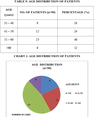
[image:75.595.112.512.230.726.2]Of the 50 patients included in the study majority of the patients were between 50 and 60 years. Who constituted 46% of the group. The youngest patient was 33years old and the oldest patient is 75 years old.
TABLE 9: AGE DISTRIBUTION OF PATIENTS AGE
(years) NO. OF PATIENTS (n=50) PERCENTAGE (%)
31 – 40 9 18
41 – 50 12 24
51 – 60 23 46
>60 6 12
CHART 1: AGE DISTRIBUTION OF PATIENTS
9
12
23 6
AGE DISTRIBUTION (n=50)
<40 41-50
51-60 >60
AGE GROUP
65
DISTRIBUTION OF SAMPLES BASED ON HISTOLOGICAL SUBTYPES
[image:76.595.102.525.321.612.2]Of the 50 patients analysed 42 (84%) were IDC, NOS. Invasive lobular carcinoma constituted 6% of the group followed by papillary and mucinous carcinoma each constitutes 4%. Metaplastic carcinoma was the least common type constituting 2% of the cases.
TABLE 10: DISTRIBUTION OF SAMPLES BASED ON HISTOLOGICAL SUBTYPES
HISTOLOGICAL SUBTYPES
NO. OF PATIENTS(n=50)
PERCENTAGE (%)
Invasive Ductal carcinoma, NOS 42 84
Invasive Lobular carcinoma 3 6
Papillary carcinoma 2 4
Mucinous carcinoma 2 4
66
CHART 2:DISTRIBUTION OF SAMPLES BASED ON HISTOLOGICAL SUBTYPES
DISTRIBUTION OF SAMPLE BASED ON MODIFIED BLOOM RICHARDSON GRADE
Modified Bloom Richardson scoring system was applied to all these cases. Among these cases, majority of the patients 25 (50%) were of MBR grade II followed 21 cases were grade III (42%) and 4 cases (8%) were under grade I.
67
TABLE 11: DISTRIBUTION OF SAMPLES BASED ON MODIFIED BLOOM RICHARDSON GRADE
MBR GRADE NO. OF PATIENTS
(n=50) PERCENTAGE (%)
I 4 8 II 25 50 III 21 42
CHART 3: DISTRIBUTION OF SAMPLES ACCORDING TO MODIFIED BLOOM RICHARDSON GRADE
4
25 21
MODIFIED BLOOM RICHARDSON GRADE
DISTRIBUTION
(n=50)
I II III
MBR GRADE
68
DISTRIBUTION OF ER, PR, HER 2 / neu RECEPTORS:
[image:79.595.103.516.312.743.2]There was an almost equal distribution of estrogen receptor positive (48%) and negative cases (50%), 2% of sample was lost during tissue processing. While analyzing the progesterone receptor status, majority of tumors were receptor positive (62%). Only 32% of cases did not express progesterone receptor and remaining 6% of samples were lost during tissue processing. 50% of the cases demonstrated HER-2/neu positivity and 40% of cases were negative. 10% of samples were lost during tissue processing.
TABLE 12: DISTRIBUTION OF ER STATUS ER STATUS NO. OF PATIENTS
(n=50)
Percentage (%)
Positive 24 48
Negative 25 50
Tissue loss 1 2
CHART 4: DISTRIBUTION OF ER STATUS
24 25
1 0
5 10 15 20 25 30
Positive Negative Tissue loss
NUMBER OF CASES
69
TABLE 13: DISTRIBUTION OF PR STATUS
PR STATUS NO. OF PATIENTS (n=50)
Percentage (%)
Positive 31 62
Negative 16 32
Tissue loss 3 6
CHART 5: DISTRIBUTION OF PR STATUS
31
16
3
0 5 10 15 20 25 30 35
Positive Negative Tissue loss
NUMBER OF CASES