OF ETHANOLIC EXTRACT OF LANTANA CAMARA LEAVES
”
Dissertation Submitted to
THE TAMILNADU Dr.M.G.R.MEDICAL UNIVERSITY
Chennai - 32
In Partial fulfillment for the award of the degree of
MASTER OF PHARMACY
Submitted by
REGISTER NUMBER 261425225
Under the guidance of
Mr.S.VENKATESH, M.Pharm.,
This is to certify that the work embodied in this dissertation entitled “ ANTI-HYPERGLYCEMIC AND ANTI-OXIDANT ACTIVITIES OF ETHANOLIC EXTRACT OF LANTANA CAMARA LEAVES” submitted to “The Tamil Nadu Dr. M.G.R. Medical University”, Chennai, in partial fulfillment and requirement of university rules and regulation for the award of Degree of Master of Pharmacy in Pharmacology, is a bonafide work carried out by Reg.No. 261425225 during the academic year 2015-2016, under the guidance and supervision of Mr. S.VENKATESH, M.Pharm., Assistant Professor, Department of Pharmacology, J.K.K. Nattraja College of Pharmacy, Kumarapalayam.
CERTIFICATE
This is to certify that the dissertation “ANTI-HYPERGLYCEMIC AND ANTI-OXIDANT ACTIVITIES OF ETHANOLIC EXTRACT OF LANTANA CAMARA LEAVES” is a bonafide work done by Reg. No. 261425225, J.K.K.Nattraja College of Pharmacy, in partial fulfillment of the University rules and regulations for award of Master of Pharmacy
inPharmacologyunder my guidance and supervision during the academic year 2015-2016.
Dr. R. SAMBATHKUMAR, M.Pharm, Ph.D., Professor & Principal
Dr. R. SHANMUGA SUNDARAM, M.Pharm, Ph.D., Vice Principal & Professor
This is to certify that the work embodied in this dissertation entitled “ ANTI-HYPERGLYCEMIC AND ANTI-OXIDANT ACTIVITIES OF ETHANOLIC EXTRACT OF LANTANA CAMARA LEAVES” submitted to “The Tamil Nadu Dr. M.G.R. Medical University”, Chennai, in partial fulfillment and requirement of university rules and regulation for the award of Degree of Master of Pharmacy in Pharmacology, is a bonafide work carried out by Reg.No. 261425225 during the academic year 2015-2016, under my guidance and direct supervision in the department of Pharmacology, J.K.K. Nattraja College of Pharmacy, Kumarapalayam.
Place: Kumarapalayam. Date:
Mr. S. Venkatesh, M. Pharm., Assistant Professor,
Department of Pharmacology,
J.K.K. Nattraja College of Pharmacy. Kumarapalayam.
This is to certify that the work embodied in this dissertation entitled entitled
“ANTI-HYPERGLYCEMIC AND ANTI-OXIDANT ACTIVITIES OF
ETHANOLIC EXTRACT OF LANTANA CAMARA LEAVES” submitted to “The Tamil Nadu Dr. M.G.R. Medical University”, Chennai, in partial fulfillment and requirement of university rules and regulation for the award of Degree of Master of Pharmacy in Pharmacology, is a bonafide work carried out by LATHA R [Reg.No. 261425225] during the academic year 2015-2016 under the guidance and supervision of Mr. S.VENKATESH, M. Pharm., Assistant Professor, Department of Pharmacology, J.K.K. Nattraja College of Pharmacy, Kumarapalayam.
Place: Kumarapalayam. Date:
Dr. R. Sambathkumar, M.Pharm.,Ph.D. Principal & Head,
Department of Pharmaceutics,
This is to certify that the work embodied in this dissertation entitled entitled
“ANTI-HYPERGLYCEMIC AND ANTI-OXIDANT ACTIVITIES OF
ETHANOLIC EXTRACT OF LANTANA CAMARA LEAVES” submitted to “The Tamil Nadu Dr. M.G.R. Medical University”, Chennai, in partial fulfillment and requirement of university rules and regulation for the award of Degree of Master of Pharmacy in Pharmacology, is a bonafide work carried out by LATHA R [Reg.No. 261425225] during the academic year 2015-2016 under the guidance and supervision of Mr. S.VENKATESH, M.Pharm., Assistant Professor, Department of Pharmacology, J.K.K. Nattraja College of Pharmacy, Kumarapalayam.
Place: Kumarapalayam.
Date:
Dr. R.Shanmugasundaram, M.Pharm.,Ph.D. Professor & Head,
Department of Pharmacology,
J.K.K. Nattraja College of Pharmacy. Kumarapalayam.
I hereby declare that the dissertation entitled “ANTI-HYPERGLYCEMIC AND ANTI-OXIDANT ACTIVITIES OF ETHANOLIC EXTRACT OF LANTANA CAMARA LEAVES” has been carried out under the guidance and supervision Mr. S.VENKATESH, M. Pharm., Assistant Professor, Department of Pharmacology, J.K.K. Nattraja College of Pharmacy, Kumarapalayam, in partial fulfillment of the requirements for the award of degree of Master of Pharmacy in Pharmacology during the academic year 2015-2016.
I further declare that, this work is original and this dissertation has not been submitted previously for the award of any other degree, diploma associateship and
fellowship or any other similar title.
Place: Kumarapalayam. LATHA R
I am proud to dedicate my deep sense of gratitude to the founder, (Late) Thiru J.K.K. Nattaraja Chettiar,providing us the historical institution to study.
My sincere thanks and respectful regards to our reverent Chairperson
Smt. N. Sendamaraai, B.Com., Managing Director Mr. S. Omm Sharravana, B.Com., LLB.,
Kumarapalayam for their blessings, encouragement and support at all times.
It is most pleasant duty to thank our beloved Principal Dr. R.SambathKumar, M.Pharm., Ph.D., J.K.K.Nattraja College of Pharmacy, Komarapalayam for ensuring all the facilities were made available to me for the smooth running of this project.
I express my whole hearted thanks to my guide Mr.S.VENKATESH, M.Pharm., Assistant Professor, Department of Pharmacology, for suggesting solution to problems faced by me and
providing indispensable guidance, tremendous encouragement at each and every step of this
dissertation work. Without his critical advice and deep-rooted knowledge, this work would not have
been a reality.
My sincere thanks toDr. R. Shanmuga Sundaram, M.Pharm., Ph.D.,Vice Principal and Head, Department of Pharmacology.
Dedicated to
Almighty,
S.NO
CHAPTER
PAGE NO
1
INTRODUCTION
1
2
REVIEW OF LITERATURE
13
3
PLANT REVIEW
38
4
AIM AND OBJECTIVES
49
5
PLAN OF WORK
50
6
MATERIALS AND METHODS
51
7
RESULTS
71
8
DISCUSSION
80
9
CONCLUSION
83
1. INTRODUCTION Traditional Medicine
Traditional medicine (TM) is a very valuable resource because of the long history of its use and thus being evidence based. It is employed extensively in developing countries for primary health care, but of late has aroused increasing interest in developed countries too; 1) as an alternative to high cost medicines for promotive and preventive health care; 2) for disease conditions with inadequate modern drugs and 3) for non-life threatening diseases of lower incidence side effects reported than with modern drugs1. WHO defined TM as the sum of the total knowledge, skills and practices based on the theories, beliefs and experiences indigenous to different cultures, whether explicable or not used in the maintenance of the health and in the prevention, diagnosis, improvement or treatment of physical and mental illness. The major resource base of the TM is medicinal plants with the introduction of modern medicine.
Medicinal plants continued to play a very significant role in the healthcare of human kind. The advancement in modern medicine caused a rapid decline of traditional medicine particularly in developed nations. But medicinal plants continued to meet the health needs of 80% of population in developing countries. Towards the end of the 20th century there began a revival of interest in traditional medicine not only in developing countries, but also in the developed countries. The resurgence of plant based medicine is mainly due to the increasing evidence/ realization of the health hazards associated with harmful side effects of many synthetic medicines and also the hazards associated with the indiscriminate use of modern medicine such as antibiotics, steroids and other synthetic drugs. The increasing popularity in plant based drugs is now felt all over the world leading to a fast growing market for plant based drugs, pharmaceuticals, nutraceuticals, functional foods and even cosmaceuticals2.
1.1 Indian traditional systems of medicine
The carriers of these traditions are millions of housewives, thousands of traditional birth attendants, bone setters, practitioners skilled in acupressure, eye treatment or treatment of snakebites, the traditional village level herbal physicians (the “Vaidyas”) and tribal physicians in the tribal areas. These local health
traditions thus represent an autonomous community supported system of health delivery at the village level which runs parallel to the state supported system. A second level of traditional health care system is the academic or classical system. This consists of codified and organized medical wisdom with sophisticated theoretical foundations and philosophical explanations, expressed in thousands of regional manuscripts covering treatises on all branches of medicine and systems like Ayurveda, Siddha, Unani, Yoga, Naturopathy and Amchi, which are expressions of this stream3.
1.1.1 Ayurvedic system of medicine
Ayurveda is perhaps the oldest among the organized traditional medicine. It spread with Vedic and Hindu culture as far in east as Indonesia and to the west it influenced the Greek, who developed a similar form of medicine. The Buddhists added many new insights to it and they took it along with their religion to many different countries. In this way, Ayurveda became the basis of the healing tradition of Tibet, Sri Lanka, Burma and other Buddhist lands and exchanged/ influenced Chinese and Greek medicine. Ayurveda is thus a rich tradition, adaptable to many different times, cultures and climates4.
welfare of human beings. Thus Ayurveda becomes one of the oldest systems of health care dealing with both the preventive and curative aspects of life in a most comprehensive way and presents a close similarity to the WHO’s concept of
health propounded in the modern era. In India Ayurveda, Siddha and Unani systems are the formal and most organized ones amongst the traditional systems of medicine. The Tibetan system of medicine is considered as an off-shoot of Ayurveda5.
Ayurveda remains one of the most ancient medical systems widely practiced in the Indian subcontinent and has a sound philosophical, experiential and experimental basis. Charak Samhita and Sushrut Samhita (100–500B.C.) are main Ayurvedic classics, which describe over 700 plants along with their classification, pharmacological and therapeutic properties. Knowledge of Ayurvedic medicine has unfortunately been confined to India and the west is largely ignorant of it. Even in India, this traditional medical practice has lost a lot of its importance in the urban situations. One of the main reasons for this is that much of the early and core medical literature on Ayurveda is in Sanskrit, the ancient language which ceased to be a day-to-day language. Even today a considerable bulk of Ayurvedic knowledge is in the form of ancient palm leaf manuscripts hidden in remote libraries and private collections, and as treasured personal knowledge of a few individuals. The net result is that Ayurveda has been away from limelight and does not enjoy the importance and popularity it deserves. 1.2 Diabetes
Diabetes is defined as a state in which homeostasis of carbohydrate and lipid metabolism is improperly regulated by insulin. This results primarily in elevated fasting and postprandial blood glucose levels. If this imbalanced homeostasis does not return to normalcy and continues for a protracted period of time, it leads to hyperglycemia that in due course turns into a syndrome called diabetes mellitus6. Diabetes as a disorder was known and recognized by man since
most obvious sign of diabetes being excessive urination. "Mellitus" comes from a Latin word that means "sweet".
Diabetics tend to believe that there is no cure yet for diabetes in any system of medicine anywhere in the world, which is partly because diabetes is not a single disease but is a complex disorder with multiple syndromes making it difficult to cure the cause of the disease. Diabetes is a cluster of symptoms like in an aging process this manifests as graying of hair or wrinkling of skin. As with aging symptoms, diabetes may occur at a very early age in a few, and at some stage of the life in most. Diabetes manifests in different persons in many different ways, and depending on the age, severity of the symptoms and involvement of other organs in the body, medical treatment greatly varies. For example a diabetic, either in young age, during pregnancy (gestational diabetes), if suffering from tuberculosis or a foot ulcer or, with recent heart attack or paralysis, should be given insulin injections only, and not other anti diabetic drugs of any systems of medicine6.
Diabetes leads to the development of numerous complications due to hyperglycemia. Likelihood of developing complications, whether acute or chronic, is ultimately a reflection of the level of blood sugar control. Diabetics are susceptible top three major complications, hypoglycemia, diabetic ketoacidosis (primarily affects type I diabetics), non ketogenic hyperosmolar syndrome (primarily affects type II diabetics). On a long term basis, the diabetic’s health
cells. Type 1 diabetes always requires insulin replacement. Type 2 diabetes usually occurs in mature adults and has slow and progressive onset. Type 2 diabetes is accompanied both by insulin resistance and by impaired insulin secretion, each of which are important in its pathogenesis. Treatment is initially dietary although oral hypoglycemic drugs usually become necessary, and about one third of patients ultimately require insulin8.
1.2.1. A rising global burden
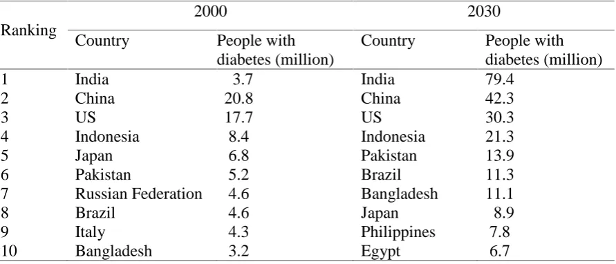
Diabetes mellitus (predominantly type 2 diabetes) is a major and growing health problem in almost all the countries. Globally, the prevalence of diabetes in adults aged over 20 years was estimated to be 4% in 1995 and is projected to rise to 5.5% by 2025. The number of people with diabetes will be more than double over the next 25 years, to reach a total of 366 million by 2030. These projections of the number of people with diabetes in 2030 take into account the fact that there will be more people in the world (population growth) and that there will be more elderly people (population ageing). They also take into account trends in urbanization - the fact that people are moving from rural areas to cities, particularly in developing countries. This affects the number of people who are likely to have diabetes, because people living in cities in developing countries tend to be less physically active and have higher levels of overweight and obesity than people in rural areas. In fact, current trends in obesity suggest that these projections are conservative and that the increase in the prevalence of diabetes may be even greater9. In 2005, an estimated 1.1 million people died from diabetes. Almost 80% of diabetes deaths occur in low and middle-income countries. Almost half of diabetes deaths occur in people under the age of 70 years; 55% of diabetes deaths are in women10. WHO projects those diabetes deaths will increase by more than 50% in the next 10 years without urgent action. Most notably, diabetes deaths are projected to increase by over 80% in upper-middle income countries between 2006 and 2015.
12.1%). It has remained an urban phenomenon so far and all the previous epidemiological studies have illustrated a 4-fold difference in the prevalence of diabetes between the urban and rural population. National urban diabetes survey in 2000 by a group of doctors found that Hyderabad topping the list (16.6% of its population) followed by Chennai (13.5%), Bangalore (12.4%), Kolkatta (11.7%), Delhi (11.6%) and Mumbai (9.3%). The incidence of diabetes in most metros and cities in India presently is 10-15%.11
Table.1. List of countries with the highest number of estimated cases of diabetes for year 2000 and 203013
Ranking
2000 2030
Country People with
diabetes (million)
Country People with diabetes (million)
1 India 3.7 India 79.4
2 China 20.8 China 42.3
3 US 17.7 US 30.3
4 Indonesia 8.4 Indonesia 21.3
5 Japan 6.8 Pakistan 13.9
6 Pakistan 5.2 Brazil 11.3
7 Russian Federation 4.6 Bangladesh 11.1
8 Brazil 4.6 Japan 8.9
9 Italy 4.3 Philippines 7.8
1.3 Free radicals, diabetes and antioxidant
Much of the morbidity and mortality associated with diabetes is primarily attributed to micro vascular and macro vascular changes, such as atherosclerosis, retinopathy, nephropathy, coronary artery disease, cerebral vascular disease, and peripheral artery disease15. One of the reasons for injury related to hyperglycemia is the formation of glycated proteins, glucose oxidation, and increased free fatty acids16.
Moreover, some recent studies suggest that reactive oxygen species/free radicals may also be involved in the initiation and development of vascular complications in diabetics17.Oxidative stress combined with mitochondrial dysfunction leads to the activation of inflammatory signaling pathways, which may damage insulin-producing cells and further aggravate the complications of diabetes18. Free radicals meet many of the criteria required for a role in the pathogenesis of diabetic vascular disease. They have a direct toxic effect on tissues; and under certain conditions, glucose molecules can also induce free radical production19. Free radicals may also modulate oxidative stress in diabetes by nonenzymatic glycosylation of proteins, monosaccharide auto-oxidation, polyol pathway, and indirect production of free radicals through cell damage from other causes20.
potential to impart therapeutic effect holistically in complicated disorders like diabetes and its complications.
1.4 Present status and future prospects
Diabetes is becoming something of a pandemic and despite the recent surge in new drugs to treat and prevent the condition, its prevalence continues to soar. Although several drugs targeted for carbohydrate hydrolysing enzymes (pseudosaccharides), release of insulin from pancreatic b-cells (sulphonyl urea), glucose utilization (biguanides), insulin sensitizers, PPARg agonists (glitazones) are in clinical practice, the growing diabetes market observes a number of changes. The glitazones are meant to target the problem of insulin resistance and enhance insulin action at the cellular level; however, some of these drugs are linked to liver toxicity (troglitazone), including a number of deaths from hepatic failure28,29,30 and raising the symptoms and risk factors of heart disease leading to heart failure (rosiglitazone)29. Therefore, as the long term of risk and effect on the complications of diabetes related with these drugs are not yet clear, UK Drug and Therapeutic Bulletin warrants that patients taking glitazones be monitored for signs of heart failure31.
theories of polyherbal formulation have the synergistic, potentiative, agonistic/antagonistic pharmacological agents within themselves due to incorporation of plant medicines with diverse pharmacological actions. These pharmacological principles work together in a dynamic way to produce maximum therapeutic efficacy with minimum side effects. Traditional medicinal preparations therefore, should not be considered just as a collection of therapeutic recipes. They are formulated and prepared keeping in mind the conditions of sickness and the healing properties of individual ingredients. It is important therefore, that herbal medicines and preparations should be taken with the consideration of their holistic therapeutic approach. The multiple activities of plant-based medicinal preparations meant for diabetes offer enormous scope for combating the threat of the diabetic epidemic.
To achieve a blockbuster status, clear evidence of the advantage over the existing therapy is the most important requirement of the day. The ability of modern medicine and health care systems to adequately manage symptoms of chronic and terminal disease is a central theme. The systematic reviews and meta analysis of clinical trials are the foundation of their success. Unfortunately, despite the apparent supremacy in terms of multiple therapeutic approaches of herbal medicines, well-organized, rigorous clinical trail evidences are not adequately available in order to advocate their scientific merit and supremacy over the existing drugs. Though the markets for herbal medicines are booming34 and evidence for effectiveness is growing, it is also being simultaneously counterbalanced by inadequate regulation35. Therefore, the product standardization, efficacy, safety and therapeutic risk/benefit associated with the use of herbal medicines need proper evaluation. A sound basic and rigorous clinical investigation to confirm and advocate the excellence over the existing therapies of traditional medicinal plants, preparation(s) mechanism(s) of action and therapeutic effects is absolutely required.
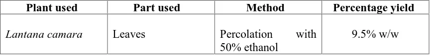
In the present study, attempt was made to prove the anti-hyperglycemic effect of Lantana camara.
Plants have always been an exemplary source of drugs and many of the currently available drugs have been derived directly or indirectly from them. The ethnobotanical information reports about 800 plants that may possess anti-diabetic potential36. Several such herbs have shown anti-diabetic activity when assessed using presently available experimental techniques 37, 38,39,40,41. A wide array of plant derived active principles representing numerous chemical compounds have demonstrated activity consistent with their possible use in the treatment of NIDDM 42, 43, 44. Among these are alkaloids, glycosides, polysaccharides, peptidoglycans, hypoglycans, guani-dine, steroids, carbohydrates, glycopeptides, terpenoids, amino acids and inorganic ions. Thus, plants are a potential source of anti-diabetic drugs (and others too) but this fact has not gained enough momentum in the scientific community. The reasons may be many including lack of belief among the practitioners of conventional medicine over alternative medicine, Although, oral hypoglycemic agents/insulin are the mainstay of treatment of diabetes and are effective in controlling hyperglycemia, they have prominent side effects and fail to significantly alter the course of diabetic complications45. As the knowledge of heterogeneity of this disorder increases, there is need to look for more efficacious agents with lesser side effects. Though development of modern medicine resulted in the advent of modern pharmacotherapeutics including insulin, biguanides, sulfonylureas and thiazolidinediones46
2. REVIEW OF LITERATURE
DIABETES MELLITUS–A REVIEW
Diabetes
Diabetes Mellitus is a group of syndromes characterized by hyperglycemia, altered metabolism of lipids, carbohydrates and proteins and a risk of complications from vascular disease. It can be classify clinically as either type I diabetes mellitus (type I DM, formally known as insulin dependent diabetes or IDDM) or type 2 disease (type 2 DM, formally known as non-insulin dependent diabetes or NIDDM)52. Some facts related to diabetes are given below:
1. Glucose in the blood is produced by the liver from the foods you eat.
2. In a healthy person, the blood glucose level is regulated by several hormones, one of which is insulin. Insulin is produced by the pancreas, a small organ near the stomach that also secretes important enzymes that help in the digestion of food.
3. Insulin allows glucose to move from the blood into liver, muscle, and fat cells, where it is used for fuel.
4. People with diabetes either do not produce enough insulin (type 1 diabetes) or can not use insulin properly (type 2 diabetes), or both.
5. In diabetes, glucose in the blood cannot move into cells, and it stays in the blood. This not only harms the cells that need the glucose for fuel, but also harms certain organs and tissues exposed to the high glucose levels.
2.1 Pancreas
pancreas is digestive in function, and the endocrine functions are performed by the
islets of Langerhans. They are small, highly vascularized masses of cells scattered
throughout the pancreas, forming only 1 to 3 percent of the entire organ. Pancreatic islets are scattered throughout the pancreas. Like all endocrine glands, they secrete their hormones into the bloodstream and not into tubes or ducts like the digestive pancreas. Because of this need to secrete their hormones into the blood stream, pancreatic islets are surrounded by small blood vessels.
The islets of Langerhans contain four type of secretory cells: 1) Alpha (A) cell, secretes glucagons 2) Beta (B) cells, secretes insulin 3) Delta (D) cells, secretes somatostatin 4) PP (F) cells, secretes pancreatic polypeptide53.
2.2 Insulin
the liver re-enters the bloodstream as muscle cells lack the necessary export mechanism54
2.2.1 Synthesis and secretion
Insulin is synthesized as a precursor (preproinsulin) in the rough endoplasmic reticulum. Preproinsulin is transported to the Golgi apparatus where it undergoes proteolytic cleavage first to proinsulin and then to insulin plus a fragment of uncertain function called C-peptide. Insulin and C-peptide are stored in granules in B-cells. The main factor controlling the synthesis and secretion of insulin in the blood glucose concentration. B-cells respond both the absolute glucose concentration and also to the rate of change of blood glucose. There is a steady basal release of insulin and also a response to a change in blood glucose. The response to an increase blood glucose has two phases– an initial rapid phase reflecting release of stored hormone and a slower, delayed phase reflecting both continued release of stored hormone and new synthesis.
ATP-sensitive potassium channels determine the resting membrane potential in B-cells. Glucose enters B-cells via a membrane transporter called Glut-2, and its subsequent metabolism via glucokinase (the rate limiting enzyme that act as the “glucose sensor” linking insulin secretion to extracellular glucose) and glycolysis increases intracellular ATP. This blocks KATP causing membrane depolarization and opening of voltage-dependent calcium channels, leading to Ca2+ influx. This Ca2+ signal induces insulin secretion, but only in the presence of amplifying messengers including diacylglycerol (DOC), non-esterified arachidonic acid. Phaspholipases are commonly activated by Ca2+, but free arachidonic acid is liberated in B-cells by an ATP-sensitive Ca2+-insensitive (ASCI) phaspholipase A2. Consequently, in B-cells,
Ca2+ entry and arachidonic acid production are both driven by ATP, linking cellular energy status to insulin secretion
2.2.2 Actions of insulin
2.2.3 Effect of insulin on carbohydrate metabolism
Insulin influences glucose metabolism in most tissues, especially the liver where it inhibits glycogenolysis (glycogen breakdown) and gluconeogenesis (synthesis of glucose from non-carbohydrate sources) while stimulating glycogen synthesis. It also increase glucose utilization (glycolysis), but overall effect is to increase hepatic glycogen stores. In muscle, unlike liver, uptake of glucose is slow and is the rate-limiting step in carbohydrate metabolism. The main effect of insulin is to increase facilitated transport of glucose via a transport called Glut-4, and to stimulate glycogen synthesis and glycolysis. Insulin increase glucose uptake by Glut-4 in adipose tissue as well as muscle, enhancing glucose metabolism.
2.2.4 Effect of insulin on fat metabolism
Insulin increase the fatty acid as well as triglyceride synthesis in adipose tissue and liver. It inhibit lipolysis, partly by dephosphorylation of lipase. It also inhibits the lipolytic actions of adrenaline, growth hormone and glucagons by opposing their action on adenylate cyclase.
2.2.5 Effect of insulin on protein metabolism
Insulin stimulates uptake of amino acids into muscle and increase protein catabolism and inhibit oxidation of amino acid in the liver.
2.2.6 Other metabolic effects of insulin
Degradation of insulin occurs primarily in liver, kidney, and muscle. About 50% of the insulin that reaches the liver by the portal vein is destroyed and never reaches general circulation. Insulin is filtered by renal glomeruli and is reabsorbed by the tubules, which also degrade it. Several enzyme involved in degradation of insulin, the primary insulin degradation enzyme is thiol metalloproteinase. It is primarily localized in hepatocytes56.
2.3 Glucose metabolism
Because insulin is the principal hormone that regulates uptake of glucose into most cells from the blood (primarily muscle and fat cells, but not central nervous system cells), deficiency of insulin or the insensitivity of its receptors plays a central role in all forms of diabetes mellitus.
Much of the carbohydrate in food is converted within a few hours to the monosaccharide glucose, the principal carbohydrate found in blood. Some carbohydrates are not converted. Notable examples include fruit sugar (fructose) that is usable as cellular fuel, but it is not converted to glucose and does not participate in the insulin / glucose metabolic regulatory mechanism; additionally, the carbohydrate cellulose (though it is actually many glucose molecules in long chains) is not converted to glucose, as humans and many animals have no digestive pathway capable of handling cellulose. Insulin is released into the blood by beta cells (β-cells) in the pancreas in response to rising levels of blood glucose (e.g., after a meal) (Fig 1). Insulin enables most body cells (about 2/3 is the usual estimate, including muscle cells and adipose tissue) to absorb glucose from the blood for use as fuel, for conversion to other needed molecules, or for storage. Insulin is also the principal control signal for conversion of glucose (the basic sugar used for fuel) to glycogen for internal storage in liver and muscle cells. Reduced glucose levels result both in the reduced release of insulin from the beta cells and in the reverse conversion of glycogen to glucose when glucose levels fall, although only glucose thus recovered by the liver re-enters the bloodstream as muscle cells lack the necessary export mechanism.
signal in converting many of the bidirectional processes of metabolism from a catabolic to an anabolic direction, and vice versa. In particular, it is the trigger for entering or leaving ketosis (ie, the fat burning metabolic phase).
If the amount of insulin available is insufficient, if cells respond poorly to the effects of insulin (insulin insensitivity or resistance), or if the insulin itself is defective, glucose will not be handled properly by body cells (about ⅔ require it) or stored appropriately in the liver and muscles. The net effect is persistent high levels of blood glucose, poor protein synthesis, and other metabolic derangements, such as acidosis.
Fig. No.2. Mechanism of insulin release in normal pancreatic beta cells. Insulin production is more or less constant within the beta cells, irrespective of blood glucose levels. It is stored within vacuoles pending release, via exocytosis, which is triggered by increased blood glucose levels.
[image:43.595.141.522.274.437.2]Europe, though this varies by geographical location. This type of diabetes can affect children or adults but was traditionally termed "juvenile diabetes" because it represents a majority of cases of diabetes affecting children.
The main cause of beta cell loss leading to type 1 diabetes is a T-cell mediated autoimmune attack57. The principal treatment of type 1 diabetes, even from the earliest stages, is replacement of insulin. Without insulin, ketosis and diabetic ketoacidosis can develop and coma or death will result.
Currently, type 1 diabetes can be treated only with insulin, with careful monitoring of blood glucose levels using blood testing monitors. Emphasis is also placed on lifestyle adjustments (diet and exercise). Apart from the common subcutaneous injections, it is also possible to deliver insulin by a pump, which allows continuous infusion of insulin 24 hours a day at preset levels and the ability to program doses (a bolus) of insulin as needed at meal times. An inhaled form of insulin, Exubera, was approved by the FDA in January 200658.
Type 1 treatment must be continued indefinitely. Treatment does not impair normal activities, if sufficient awareness, appropriate care, and discipline in testing and medication is taken. The average glucose level for the type 1 patient should be as close to normal (80–120 mg/dl, 4–6 mmol/l) as possible. Some physicians suggest up to 140–150 mg/dl (7-7.5 mmol/l) for those having trouble with lower values, such as frequent hypoglycemic events. Values above 200 mg/dl (10 mmol/l) are often accompanied by discomfort and frequent urination leading to dehydration. Values above 300 mg/dl (15 mmol/l) usually require immediate treatment and may lead to ketoacidosis. Low levels of blood glucose, called hypoglycemia, may lead to seizures or episodes of unconsciousness.
2.4.2 Type 2 diabetes mellitus
improve insulin sensitivity or reduce glucose production by the liver, but as the disease progresses the impairment of insulin secretion worsens and therapeutic replacement of insulin often becomes necessary. There are numerous theories as to the exact cause and mechanism for this resistance, but central obesity (fat concentrated around the waist in relation to abdominal organs, and not subcutaneous fat, it seems) is known to predispose individuals for insulin resistance, possibly due to its secretion of adipokines (a group of hormones) that impair glucose tolerance. Abdominal fat is especially active hormonally. Obesity is found in approximately 55% of patients diagnosed with type 2 diabetes (UKPDS, 1998). Other factors include aging (about 20% of elderly patients are diabetic in North America) and family history (type 2 is much more common in those with close relatives who have had it), although in the last decade it has increasingly begun to affect children and adolescents, likely in connection with the greatly increased childhood obesity seen in recent decades in some places.
Type 2 diabetes may go unnoticed for years in a patient before diagnosis, as visible symptoms are typically mild or non-existent, usually without ketoacidotic episodes, and can be sporadic as well. However, severe long-term complications can result from unnoticed type 2 diabetes, including renal failure due to diabetic nephropathy, vascular disease (including coronary artery disease), vision damage due to diabetic retinopathy, loss of sensation or pain due to diabetes neuropathy, liver damage from non-alcoholic steatohepatitis, etc.
attenuate insulin resistance (e.g., thiazolidinediones). According to one study, overweight patients treated with metformin compared with diet alone, had relative risk reductions of 32% for any diabetes endpoint, 42% for diabetes related death and 36% for all cause mortality and stroke (Armenian Medical Network, 2006). When oral medications fail (cessation of beta cell insulin secretion is not uncommon amongst Type 2s), insulin therapy will be necessary to maintain normal or near normal glucose levels. A disciplined regimen of blood glucose checks is recommended, most particularly and necessarily when taking medications.
2.4.3 Type 3 diabetes mellitus (Gestational diabetes)
Gestational diabetes also involves a combination of inadequate insulin secretion and responsiveness, resembling type 2 diabetes in several respects. It develops during pregnancy and may improve or disappear after delivery. Even though it may be transient, gestational diabetes may damage the health of the fetus or mother, and about 20%–50% of women with gestational diabetes develop type 2 diabetes later in life.
Gestational diabetes mellitus (GDM) occurs in about 2%–5% of all pregnancies. It is temporary and fully treatable but, if untreated, may cause problems with the pregnancy, including macrosomia (high birth weight), fetal malformation and congenital heart disease. It requires careful medical supervision during the pregnancy.
Fetal/neonatal risks associated with GDM include congenital anomalies such as cardiac, central nervous system, and skeletal muscle malformations. Increased fetal insulin may inhibit fetal surfactant production and cause respiratory distress syndrome. Hyperbilirubinemia may result from red blood cell destruction. In severe cases, perinatal death may occur, most commonly as a result of poor placental profusion due to vascular impairment. Induction may be indicated with decreased placental function. Cesarean section may be performed if there is marked fetal distress or an increased risk of injury associated with macrosomia, such as shoulder dystocia.
2.4.4 Other types of diabetes
1. Genetic defects in beta cells (autosomal or mitochondrial): Both type 1 and type 2 diabetes are at least partly inherited. Type 1 diabetes appears to be triggered by some (mainly viral) infections, or in a less common group, by stress or environmental exposure (such as exposure to certain chemicals or drugs). There is a genetic element in individual susceptibility to some of these triggers which has been traced to particular HLA genotypes (i.e., the genetic "self" identifiers relied upon by the immune system). However, even in those who have inherited the susceptibility, type 1 diabetes mellitus seems to require an environmental trigger. A small proportion of people with type 1 diabetes carry a mutated gene that causes maturity onset diabetes of the young (MODY).
Wolfram's syndrome - Wolfram's syndrome is an autosomal recessive neurodegenerative disorder that first becomes evident in childhood. It consists of diabetes insipidus, diabetes mellitus, optic atrophy, and deafness, hence the acronym DIDMOAD (Armenian Medical Network, 2006).
2. There is a stronger inheritance pattern for type 2 diabetes. Those with first-degree relatives with type 2 have a much higher risk of developing type 2, increasing with the number of those relatives. Concordance among monozygotic twins is close to 100%, and about 25% of those with the disease have a family history of diabetes. Candidate genes include KCNJ11 (potassium inwardly rectifying channel, subfamily J, member 11), which encodes the islet ATP-sensitive potassium channel Kir6.2, and TCF7L2 (transcription factor 7–like 2), which regulates proglucagon gene expression and thus the production of glucagon-like peptide-1(Rother, 2007).
Another risk factor is obesity, particularly central obesity (i.e., that in and around abdominal organs), which is found in approximately 85% of North American patients diagnosed with this type, so some experts believe that inheriting a tendency toward obesity also contributes.
2.5 Diabetes Causes
Type 1 diabetes is believed to be an autoimmune disease. The body's immune system attacks the cells in the pancreas that produce insulin.
A predisposition to develop type 1 diabetes may run in families but much less so than for type 2.
Environmental factors, such as certain types of viral infections, may also contribute.
Type 1 diabetes is most common in people of non-Hispanic white persons of Northern European descent, followed by African Americans and Hispanic Americans. It is relatively rare in those of Asian descent.
Type 1 diabetes is slightly more common in men than in women.
Type 2 diabetes: Type 2 diabetes is believed to have a strong genetic link, meaning that it tends to run in families. Several genes are being studied that may be related to the cause of type 2 diabetes. Risk factors for developing type 2 diabetes include the following:
High blood pressure
High blood triglyceride (fat) levels
Gestational diabetes or giving birth to a baby weighing more than 9 pounds
High-fat diet
High alcohol intake
Obesity or being overweight
Ethnicity: Certain groups, such as African Americans, Native Americans, Hispanic Americans, and Japanese Americans, have a greater risk of developing type 2 diabetes than non-Hispanic whites.
Aging: Increasing age is a significant risk factor for type 2 diabetes. Risk begins to rise significantly at about age 45 years, and rises considerably after age 65 years.
2.6 Symptoms of Diabetes
Unusual thirst,
frequent and profuse urination,
loss of weight despite increased appetite and food intake,
weakness and drowsiness,
Itching of the skin and boils.
2.7 Complications of diabetes
Both forms of diabetes ultimately lead to high blood sugar levels, a condition called hyperglycemia. Over a long period of time, hyperglycemia damages the retina of the eye, the kidneys, the nerves, and the blood vessels.
Damage to the retina from diabetes (diabetic retinopathy) is a leading cause of blindness.
Diabetes accelerates atherosclerosis, or the formation of fatty plaques inside the arteries, which can lead to blockages or a clot (thrombus), which can then lead to heart attack, stroke, and decreased circulation in the arms and legs (peripheral vascular disease).
Diabetes predisposes people to high blood pressure and high cholesterol and triglyceride levels. These independently and together with hyperglycemia increase the risk of heart disease, kidney disease, and other blood vessel complications.
In the short run, diabetes can contribute to a number of acute (short-lived) medical problems.
Many infections are associated with diabetes, and infections are frequently more dangerous in someone with diabetes because the body's normal ability to fight infections is impaired. To compound the problem, infections may worsen glucose control, which further delays recovery from infection.
Hypoglycemia, or low blood sugar, occurs from time to time in most people with diabetes. It results from taking too much diabetes medication or insulin (sometimes called insulin reaction), missing a meal, doing more exercise than usual, drinking too much alcohol, or taking certain medications for other conditions. It is very important to recognize hypoglycemia and be prepared to treat it at all times. Headache, feeling dizzy, poor concentration, tremors of hands, and sweating are common symptoms of hypoglycemia. You can faint or have a seizure if blood sugar level gets too low.
Hyperosmolar hyperglycemic nonketotic syndrome is a serious condition in which the blood sugar level gets very high. The body tries to get rid of the excess blood sugar by eliminating it in the urine. This increases the amount of urine significantly and often leads to dehydration so severe that it can cause seizures, coma, even death. This syndrome typically occurs in people with type 2 diabetes who are not controlling their blood sugar levels or have become dehydrated or have stress, injury, stroke, or medications like steroids.
2.8 Medications
Many different types of medications are available to help lower blood sugar levels in type 2 diabetes. Each type works in a different way. It is very common to combine 2 or more types to get the best effect with fewest side effects.
Sulfonylureas: These drugs stimulate your pancreas to make more insulin.
Biguanides: These agents decrease the amount of glucose produced by your liver.
Alpha-glucosidase inhibitors: These agents slow absorption of the starches you eat. This slows down glucose production.
Thiazolidinediones: These agents increase your sensitivity to insulin.
Meglitinides: These agents stimulated the pancreas to make more insulin.
Incretin mimetics: Incretin mimetics promote insulin secretion by the pancreas and mimic other blood sugar level lowering actions that naturally occur in the body. Exdenatie (Byetta) is the first incretin mimetic agent approved in the United States. It is indicated for diabetes mellitus type 2 in addition to metformin or a sulfonylurea when these agents have not attained blood sugar level control alone.
Insulins: Human insulin is the only type of insulin available in the United States; it is less likely to cause allergic reactions than animal-derived varieties of insulin. The type of insulin chosen to customize treatment for an individual is based on the goal of providing optimal blood sugar control. Different types of insulin are available and categorized according to their times of action onset and duration. Commercially prepared mixtures of some insulins may also be used to provide constant (basal) control and immediate control60.
o Rapid-acting insulins
Regular insulin (Humulin R, Novolin R)
Insulin lispro (Humalog)
Insulin aspart (Novolog)
Insulin glulisine (Apidra)
Prompt insulin zinc (Semilente, slightly slower acting)
Inhaled insulin (Exubera)
o Intermediate-acting insulins
Isophane insulin, neutral protamine Hagedorn (NPH) (Humulin N, Novolin N)
Insulin zinc (Lente)
o Long-acting insulins
Extended insulin zinc insulin (Ultralente)
Insulin glargine (Lantus)
2.9 Free radicals
2.9.1 Chemistry of Free radicals
In every atom, electrons inhabit shells or orbits around the nucleus. Each orbit has sub - orbits known as orbitals. Each orbital can accommodate two electrons and it is stable if it has two electrons. If one electrons in the pair is lost or missing, then the atom or molecule has an intense thirst to retrieve the electron from other molecules to attain stability. Such an unstable and reactive atom (or molecule) is called a free radical. These free radicals are capable of independent existence and have an unpaired electron in an orbital (12, 14). Free radicals are natural by-products of our own metabolism. These are electrically charged molecules that attack our cells, tearing through cellular membranes to react and create havoc with the nucleic acids, proteins, and enzymes present in the body. These attacks by free radicals, collectively known as oxidative stress, are capable of causing cells to lose their structure, function and can eventually destroy them. Free radicals may be designated as molecular sharks that damage molecules in cell membranes, in mitochondria (the cells energy plants), and in the DNA (the cell’s intelligence) and are very unstable, tend to rob electrons from the molecules in the immediate surrounding in order to replace their own losses.
The energy required to dissociate the covalent bond can be provided by heat, electromagnetic radiation etc. The presence of an unpaired electron increases reactivity as the solitary electron seeks a partner for stability. As partner can be obtained by abstracting an electron from a co-reactant. This reaction results in quenching by reduction (election addition) of the radical and formation of a new radical by oxidation (electron loss) of the co-reactant.
The toxicity of oxygen to humans has been of interest in relation to diving underwater, swimming and in the use of hyperboric oxygen in the treatment of cancer, infections, multiple sclerosis and lung diseases. High pressure oxygen frequently causes acute central nervous system (CNS) toxicity in animals producing convulsions. ROS is a collective term, which includes not only the oxygen radicals (O2.-,
and OH.) but also some non radical derivatives of oxygen. These include H2O2,
HOC1 and ozone (O3). ROS.
Radicals Non radicals
Superoxide radical (O2.-) H2O2
Hydroxyl radical (OH.) O3
Peroxyl radical (RO.) Singlet oxygen Alkoxyl radical (RO.) Peroxynitrile
Hydroperoxyl radical (ONOO)
(HO2.)
Various sources of ROS have been identified in living organism. The superoxide anion radical appears to play the central role and other reactive intermediates are formed from it. ROS are also produced in the organism as a part of the primary immune defense. Phagocytic cells such as neutrophils or marophages defend against foreign organisms by generating O2. -and nitric oxide as a part of the
killing mechanism. These ROS can be studied under the two headings biologically important radicals and biologically important non-radicals.
1. High impact energy sources (thermal, microwave etc) 2. Metals (Cadmium, copper, iron, mercury, Zinc etc)
3. Enzymes (metallic or otherwise) dispersed or grouped in cytoplasmic organelles
4. Mechanic action
7. Sources and characteristics of ROS
Any free radical involving oxygen is then referred to as reactive exygen species (ROS). The most commonly formed ROS are superoxide anion radcial (O2.-)
and hydroxyl radical (
.
OH). O2.- is formed when one electron is added to an oxygenmolecule, and is considered the least reactive type of ROS. Once O2.- is produced, it
triggers a rapid cascade of events that creates other free radicals, eventually terminating in the formation of H2O. In humans, O2.-is the most commonly produced
free radical. Phagocytic cells such as macrophage and neutrophils are prominent sourced of O2.-. In an inflammatory response, these cells generate free radicals that
attack invading pathogens such as bacteria. Production of O2. - by activated
radical-plays an important role in atherosclerosis. The process of formation of reactive oxygen species (ROS) shown below.
2.9.2Biologically important radicals
Nitric oxide, sulphur radicals, peroxyl and alkoxyl radicals, superoxide radical, Hydroxyl radical, Transition metals.
2.9.3Free radicals and diseases
Despite the existence of endogenous defense mechanisms against ROS, it has been observed that whenever either the level of the cellular antioxidant system goes down or when the ROS reach abnormally high levels, oxidative damage to the cells occurs leading to several pathological conditions. Over about 100 disorders like rheumatoid arthritis (RA), hemorrhagic shock, CVS disorders, cystic fibrosis, metabolic disorders, neurodegenerative diseases, gastrointestinal ulcerogenesis and AIDS have been reported as ROS mediators. Some specific examples of ROS mediated diseases include Alzheimer’s disease, Parkinson’s disease, Atherosclerosis, Cancer, Down’s syndrome and ischemic reperfusion injury in different tissues
Fig no: 3 ROS in diseases (Source: Comparative Biochemistry and Physiology (11) In principle, disease associated oxidative stress could result from either or both of the following.
1. Diminished antioxidants due to the mutations affecting antioxidant enzymes like SOD, GPX or disease that cause depletion of such defenses. Depletion of dietary antioxidants and other essential dietary constituents can also lead to oxidative stress.
LPO. In most diseases however, increased oxidant formation is a consequence of the disease activity. For example, infiltration of a large number of neutrophils into a localized site, followed by activation of these cells to generate (O2.-) and H2O2 can
produce an intense localized oxidative stress which appears to happen in rheumatoid arthritis and also in some forms of the adult respiratory distress syndrome.
Atherosclerosis, Hypertension, Diabetes are perfusion injury, Inflammation, Autoimmune diseases, Rheumatoid arthritis, Asthma, Cystic fibrosis, cancer, Parkinson’s disease (PD), Free radicals in aging biology.
2.9.4 Antioxidant and diabetes
The elevated levels of blood glucose in diabetes produce oxygen free radicals which cause membrane damage due to peroxidation of membrane lipids and protein glycation62 .Glucose auto-oxidase in the presence of transition metal ions generates oxygen free radicals which make the membrane vulnerable to oxidative damage. The lipid oxidation of the cell membrane has been associated with a number of pathological phenomenon such as increase the membrane rigidity, decrease cellular deformability and lipid fluidity in erythrocytes. The action of diabetes induction agents produced reactive free radicals, which have shown to be cytotoxic to the β -cells of the pancreas63. As the diabetogenic action can be prevented by the SOD, CAT, and other hydroxyl radical scavengers such as ethanol, dimethyl urea, there is evidence to suggest that the incidence of diabetes involves superoxide anions and hydroxyl radicals. The harmful effects of superoxide and hydroxyl radicals can be counteracted by antioxidant enzymes such as SOD, CAT, and glutathione peoxidase (GSH-px). In addition to these enzymes, GSH-R and glutathione-S-transferase (GST) provide GSH and help to neutralize toxic electrophiles respectively. There is clear cut evidence to show the role of free radicals in diabetes and studies indicate that tissue injury in diabetes may be due to free radicals64.
agreement that vitamin C levels are lower than normal in plasma, despite the fact that elevated blood glucose has been reported to inhibit the uptake of ascorbate and of dehydro ascorbate in to calls.
Possible sources of oxidative stress in diabetes include shifts in redox balance resulting from altered carbohydrate and lipid metabolism, increase generation of ROS and decrease levels of antioxidant defenses such as GSH. In physiologic concentrations, endogenous reactive oxygen species (ROS) help to maintain homeostasis. However, when ROS accumulate in excess for prolonged periods of time, they cause chronic oxidative stress and adverse effects. This is particularly relevant and dangerous for islet, which is among those tissues that have the lowest levels of intrinsic antioxidant defenses.
2.9.5 Antioxidant defence
It has been postulated that the etiology of the complications of diabetes involves oxidative stress perhaps as a result of hyperglycemia65. The elevated levels of blood glucose in diabetes produce oxygen-free radicals (OFR), which cause membrane damage due to peroxidation of membrane lipids and protein glycation66. Baynes67 reported that plasma thiobarbituric acid reactive substance (TBARS) levels increased in diabetic patients due to vascular lesions induced by hyperglycemia. Diabetic patients thus have an increased incidence of vascular diseases and it has been suggested that free radical activity increased in diabetes68. It has also been shown that glucose under physiological conditions produces oxidants that possess reactivity similar to the hydroxyl-free radicals.
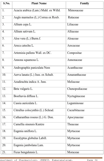
Table.2. LIST OF PLANTS REPORTED FOR ANTI-DIABETIC ACTIVITY
S.No. Plant Name Family
1. Acacia arabica (Lam.) Muhl. ex Willd. Mimosaceae
2. Aegle marmelos (L.) Correa ex Roxb. Rutaceae
3. Allium cepa L. Liliaceae
4. Allium sativum L. Alliaceae
5. Aloe vera (L.) Burm.f. Aloaceae
6. Areca catechu L. Arecaceae
7. Artemisia pallens Wall. ex DC. Compositae
8. Annona squamosa L. Annonaceae
9. Andrographis paniculata Nees Acanthaceae
10. Aerva lanata (L.) Juss. ex Schult. Amaranthaceae
11. Azadirachta indica A. Juss. Meliaceae
12. Beta vulgaris L. Chenopodiaceae
13. Boerhavia diffusa L. Nyctaginaceae
14. Cassia auriculata L. Leguminosae
22. Gymnema montanum Hook.f. Asclepiadaceae
23. Gymnema sylvestre R. Br. Asclepiadaceae
24. Hibiscus rosa sinensis L. Malvaceae
25. Ipomoea batatas (L.) Lam. Convolvulaceae
26. Momordica cymbalaria Fenzl ex Naudin Cucurbitaceae
27. Momordica charantia L. Cucurbitaceae
28. Murraya koeingii (L.) Spreng. Rutaceae
29. Ocimum sanctum L. Lamiaceae
30. Picrorrhiza kurroa Royle ex Benth. Scrophulariaceae
31. Phyllanthus amarus Schumach. & Thonn. Euphorbiaceae
32. Pterocarpus santalinus L. f. Leguminosae
33. Salacia reticulata Wight. Celastaceae
34. Salacia oblonga Wall. Celastaceae
35. Swertia chirayita (Roxb. ex Fleming) H. Karst.
Gentianaceae
36. Terminalia catappa L. Combretaceae
37. Terminalia pallida Brandis Combretaceae
38. Tinospora cordifolia (Willd.) Hook.f. & Thomson
Menispermaceae
39. Zingiber officinale Roscoe Zingiberaceae
3. PLANT REVIEW
LANTANA CAMARA - A REVIEW
3.1 Plant monograph74 Lantana camara. (Family: Verbenaceae)
3.1.1 Scientific classification
Kingdom : Plantae
Order : Lamiales
Family : Verbenacea
Genus : Lantana
Species : camara
3.1.2 Common Names:
Tamil : Unnichedi,Arasimala
Hindi : Raimuniya
Kannada : Kakke
Telegu : Pulikampa
Malayalam : Kongini, Konkini, Aripoo
3.1.3 Description:
Lantana camara is a thorny shrub upright, half climbing or sometimes
3.1.4 Distribution75:
Lantana camara is a tropical origin plant and native to Central and
Northern South America and Caribbean. Lantana camara is now spreaded to nearly 60 countries viz, New Zealand, Mexico, Florida, Trinidad, Jamaica and Brazil. It is reported in many African countries including Kenya, Uganda, Tanzania and South Africa.
3.1.5 Traditional uses74:
Lantana camara has been used in many parts of the world to treat a wide
3.2 PHARMACOLOGICAL REVIEW
ALLELOPATHIC IMPACT OF LANTANA CAMARA ON VEGETATIVE GROWTH AND YIELD COMPONENTS OF GREEN GRAM
(PHASEOLUS RADIATUS)
K.C. Lenka et.al., (2014) was carried out by An experiment was conducted to understand the allelopathic effects of different concentrations derived from leaflitter dust of Lantana camara on the vegetative growth parameters such as -development of total number of leaves per plant, height of the plant, total leaf area, leaf area index and components of yield such as - production of number of heads per plant, production of seeds per head, weight of seeds, seed yield per plant of green gram (Phaseolus radiatus).Results showed different concentrations of leaf-litter dust caused significant inhibitory effect on vegetative growth and yield of the test crop. The study indicates that the allelochemicals released from the leaf-litter dust into the soil suppressed the above parameters of the of the green gram plant.
ANALGESIC AND ANTI-INFLAMMATORY ACTIVITY OF TOPICAL PREPARATION OF LANTANA CAMARA LEAVES
K.Lakshman et.al., was carried out by The present study was undertaken to evaluate the analgesic and anti-inflammatory activity of topical preparation of
Lantana camara leaves, in literature it is found that leaves of Lantana camara
having analgesic and anti-inflammatory activity. Due to the toxicity of Lantana
camara on oral administration, topical formulations containing 1%, 2%, 4%, 6%
phenolic compounds and the pharmacological studies have revealed that preparations containing 4%, 6% and 8% AELC showed significant analgesic and anti-inflammatory activity. In conclusion the formulations containing 4%, 6% and 8% AELC was found to possess good analgesic and anti-inflammatory activity.
ANTIBACTERIAL ACTIVITY OF LANTANA CAMARA LINN AND LANTANA MONTEVIDENSIS BRIG EXTRACTS FROM CARIRI-CEARÁ, BRAZIL
A review of work was done by F.S. Baretto et.al., 2010 Jan-Mar; 2(1): 42–44. The use of medicinal plants with therapeutics properties represents a secular tradition in different cultures, mainly in underdeveloped countries. Lantana
camara Linn and Lantana montevidensis Briq (Verbenaceae) found in tropical and
subtropical areas around the world are popularly known as “camará” or
cytoprotective compounds. Morphological examinations indicated apoptosis induction as the mechanism of activity on Jurkat cells. In conclusion, L. camara's root and leaf extracts might be subjects for further fractionation and identification to find new anticancer agents.
ANTIDIABETIC ACTIVITY OF LANTANA CAMARA LINN FRUITS IN NORMAL AND STREPTOZOTOCIN-INDUCED DIABETIC RATS
Venkatachalam, T et.al., Journal of Pharmacy Research;May2011, Vol. 4 Issue 5, p1550 To evaluate the hypoglycemic activity of methanolic extract of
Lantana camara linn fruits in normal and streptozotocin induced diabetic rats.
Material and methods: Methanolic extract of Lantana camara linn fruits were orally tested at the dose of 100 and 200 mg/kg for hypoglycemic activity for normal and streptozotocin induced diabetic rats. In addition changing in body weight, HbA1c, assessed in the methanol-treated diabetic rats, were compared with diabetic control and normal animals. Histopathological observations during 21 days treatment were also evaluated. Statistical analysis: Analysis of Variance (ANOVA) followed by Multiple comparison student two-tail’t’test was used for statistical analysis of collected data. Differences were considered significant at p<0.05. All the values were indicated in the tables and figures as Mean ± SEM. Results and discussion: Methanolic extract of Lantana camara linn fruit 200 mg/kg produced a significant reduction in fasting blood glucose level in the normal and streptozotocin induced diabetic rats. Significant differences were observed change of body, HbA1c by methanol extract treated- diabetic rats, when compared with the diabetic control and normal animals. Concurrent histopathological studies of the liver these animals showed comparable regeneration by extract which were earlier encored by Streptozotocin.
ANTINOCICEPTIVE AND ANTI-INFLAMMATORY EFFECTS OF LANTANA CAMARA L. EXTRACT IN MICE
of plants within the same family show the existence of anti-inflammatory activity in paw edema induced by carrageenan, serotonin and histamine and analgesic activity in the acetic acid writhing and tail-flick tests. The present study investigated whether the L. camara extract (ACE) also exerts these effects. The ACE toxicity was studied in male mice, and the percentage of mortality recorded 7 days after treatment was assessed. The ACE was evaluated as an antinociceptive agent in the hot plate, tail-flick and acetic acid writhing tests at a nontoxic dose of 1.0 g/Kg. The results showed that 1.5 g/Kg of ACE was not able to cause death, and doses of 3.0 and 4.0 g/Kg caused 50% and 60% death, respectively, in male mice. In all of the antinociceptive tests, 1 g/Kg of ACE markedly reduced responses to pain. Our findings suggest that ACE may have active anti-inflammatory and antinociceptive properties in much smaller doses than toxic.
ANTIULCEROGENIC ACTIVITY OF LANTANA CAMARA LEAVES ON GASTRIC AND DUODENAL ULCERS IN EXPERIMENTAL RATS
Bhushan Vyawaharea, et.al., Journal of Ethnopharmacology Volume 134, Issue 1, 8 March 2011, Pages 195–197 was carried out by A Review was carried out by Lantana camara L. (Verbenaceae), a widely growing shrub has been used in the traditional medicine for treating many ailments. The objective of the present study was to evaluate the effects of methanolic extract of Lantana camara leaves on gastric and duodenal ulcers. The antiulcerogenic effect of methanolic extract of
Lantana camara was evaluated in aspirin induced gastric ulcerogenesis in pyloric
methanolic extract of Lantana camara leaves shown healing of gastric ulcers and also prevents development of duodenal ulcers in rats.
ANTIULCEROGENIC ACTIVITY OF LANTANA CAMARA LEAVES ON GASTRIC AND DUODENAL ULCERS IN EXPERIMENTAL RATS
R. Sathish et.al., Journal of ethnopharmacology 134(1):195-7, 2010 Lantana
camara L. (Verbenaceae), a widely growing shrub has been used in the traditional
medicine for treating many ailments. The objective of the present study was to evaluate the The antiulcerogenic effect of methanolic extract of Lantana camara was evaluated in aspirin induced gastric ulcerogenesis in pyloric ligated rats, ethanol induced gastric ulcer, and cysteamine induced duodenal ulcer models. The extract was administered orally at two different doses of 250 mg/kg and 500 mg/kg. The lipid peroxidation, reduced glutathione levels of ethanol induced gastric ulcer model and inhibition zone in diameter against Helicobacter pylori also determined.
EVALUATION OF ANTIMOTILITY EFFECT OF LANTANA
CAMARA L. VAR. ACUELATA CONSTITUENTS ON NEOSTIGMINE INDUCED GASTROINTESTINAL TRANSIT IN MICE