LANGUAGE DYSFUNCTIONS IN
STROKE
Dissertation submitted in partial fulfillment of the requirements
for
the degree of
BRANCH – 1 D.M. (NEUROLOGY)
OF
THE TAMILNADU DR.M.G.R. MEDICAL UNIVERSITY
CHENNAI, TAMILNADU
DEPARTMENT OF NEUROLOGY
TIRUNELVELI MEDICAL COLLEGE,
TIRUNELVELI
CERTIFICATE
This is to certify that this dissertation entitled
“Language
dysfunctions in stroke”
submitted by
Dr. E Bobby
appearing for
D.M., Neurology
Degree examination in August 2014 is a bonafide
record of work done by her under my direct guidance and supervision
in partial fulfillment of regulations of the Tamil Nadu Dr. M.G.R.
Medical University, Chennai. I forward this to the Tamil Nadu
Dr.M.G.R. Medical University, Chennai, Tamil Nadu, India.
Dr. S.Saravanan, M.D., D.M.,
Guide
Tirunelveli Medical College
Tirunelveli – 627011
Dr. S.Saravanan, M.D., D.M.,
Professor and HOD
Department of Neurology
Tirunelveli Medical College
Tirunelveli – 627011
DEAN
Tirunelveli Medical College
DECLARATION
I, Dr.E.Bobby, solemnly declare that this dissertation “LANGUAGE DYSFUNCTIONS IN STROKE, TIRUNELVELI MEDICAL COLLEGE
HOSPITAL” was done by me at the Department of Neurology, Tirunelveli Medical College, Tirunelveli under the guidance and supervision of the Professor of Neurology, Tirunelveli Medical College, Tirunelveli between April 2012 and November 2013.
This dissertation is submitted to the Tamil Nadu Dr.M.G.R. Medical University, Chennai – 32 in partial fulfilment of the University requirements for the award of the degree of D.M., Neurology.
Place : Tirunelveli Date : 12.03.2014
ACKNOWLEDGEMENT
I gratefully acknowledge and sincerely thank the DEAN, Tirunelveli medical college ,Tirunelveli for allowing me to do this Dissertation and utilize the Institutional facilities.
I would like to express my sincere gratitude to my Respected Professor and Head of the department, Department of Neurology, Prof. S Saravanan, MD., D.M., for his motivation, guidance and encouragement in fulfilling this dissertation work.
I profoundly thank my Associate Professor Dr.P K Murugan MD., D.M., for his advice, guidance and valuable criticism which enabled me to do this work effectively.
This is one another fine moment to express my gratitude and indebtedness to my Asst. Professors DR.V Sriramakrishnan M.D., D.M., DR.M. Radha M.D., D.M., and DR.C Rachel Packiaseeli MD., D.M., for their supervision and guidance in making this work successfully.
I am thankful to my postgraduate colleagues for their constant support and sharp constructive criticism.
I am thankful to the Neurochemistry laboratory, Radiology Department and and all technicians who helped me to conduct this study.
CONTENTS
Sl. No.
Title
Page
No.
1.
INTRODUCTION
1
2.
AIM OF THE STUDY
3
3.
MATERIALS AND METHODS
4
4.
REVIEW OF LITERATURE
13
5.
RESULTS
33
6.
DISCUSSION
66
7.
CONCLUSION
73
8.
APPENDIX
Bibliography
Proforma
Western aphasia battery
Consent form
NIH Stroke scale
Handedness questionarre
Master chart I
1
INTRODUCTION
Worldwide stroke is one of the leading causes of death, with stroke mortality being very high in Asia and Eastern Europe according to WHO (2004). The annual stroke death in developing countries by 2020 is estimated to be around 19 out of 25 millions (1). Stroke not only causes death, but also worse disabilities in adults.
Among the stroke survivors, approximately 30% require assistance for daily living, 20% require assistance for ambulation and 16% require institutional services.
Among the disabilities of stroke, post stroke language disorders are frequent which include aphasia, alexia, agraphia, acalculia and apraxias (2). Aphasia, being a common consequence of left hemispheric lesion is one of the most common neuro psychological consequences of stroke, prevalent in nearly one third of all stroke patients. Recovery pattern of aphasias vary in various aphasias.
2
cognitive and social disability (4,5) and higher mortality (5,6). Aphasia outcome remains poor – 32% to 50% of aphasics still suffer from aphasia six months after stroke. Therefore it is important to evaluate and treat post stroke aphasias.
The incidence of stroke follows a rising trend in India. Post stroke language disorders are frequent and disable the quality of life. There is necessity to differentiate and classify aphasias for prognosis and outcome assessment.By more precise and detailed description of the language functions of the brain damaged subjects, it is possible to plan novel reeducative strageties and to monitor their recovery in performance. Early rehabilitation for language dysfunction and other associated neurological deficits restores the quality of life in affected subjects. Aphasia treatment have so far have been given less importance by physicians and speech therapists. Studies regarding language dysfunction are less in our place.
3
AIM OF THE STUDY
The aims of the study are
1. To find out the different types of aphasias prevalent among adult stroke patients attending Medicine and Neuromedicine departments of Tirunelveli Medical College hospital.
2. To study the recovery patterns among the different types of aphasias. 3. To correlate the types of aphasias with the imaging CT/MRI.
4
MATERIALS AND METHODS
Study Design:
This study is a single centered hospital based,cross-sectional,descriptive study.
Study Population:
Study was carried out in patients attending medicine and Neuromedicine departments of Tirunelveli medical college hospital with stroke and language dysfunctions.
Period of study:
The study was conducted during the period of April 2012-November-2013.
Ethical Approval:
Study was approved by the Institutional Ethical Committee.
Consent:
5
Sample Size:
A total of 100 stroke patients with language dysfunction.
Inclusion Criteria:
Adult stroke patients of age 18 to 80 years with language dysfunction, attending medicine and Neuromedicine departments (both outpatients and inpatients) , with confirmation by CT/MRI and willing to participate in the study were included.
Exclusion Criteria:
Patients with
1. Psychiatric disorders 2. Developmental dyslexia
3. Head injury, space occupying lesions etc 4. Stuttering speech
5. Disorders of phonation 6. Demented patients 7. Mental retardation
6
METHODS OF STUDY
In all patients epidemiological data including age, sex, literacy status and presence of risk factors for stroke like hypertension, diabetes, heart disease, smoking, alcoholism, family history were collected. Patients handedness was assessed using handedness questionnaire either from the patient or care givers(Appendix 8).
General physical examination and neurological examination was done in all patients. Routine biochemical investigations were done. Neurological status was assessed using National Institutes of Health Stroke Scale. It contains 11 items with the minimum score 0 and the maximum score 42. The reliability (7) and validity (8) of the NIHSS is well documented. (Appendix (8)
NIHSS-0 – Normal examination
NIHSS 1-7 – Mild neurological deficit
NIHSS 8-14 – Moderate neurological deficit
NIHSS > 15 – Severe neurological deficit
ASSESSMENT OF LANGUAGE FUNCTION
Bedside assessment of language was done in all patients as follows
7
a. Informal interview b. Structured task
c. Automatic sequences 2. Auditory comprehension 3. Naming
4. Repetition
The first step is to listen to spontaneous speech. The patient is asked simple questions like describing the weather outside or the reason for consulting the doctor. The following observations are done from the patient‘s speech like initiation difficulty, fluency, articulation, phonation, speech rate, prosody, length of the phrase, word finding difficulties, presence of circumlocution, paraphasias and neologisms.
Fluency was assessed using animal naming test in one minute. Normal individuals can name 18-22 animals in one minute. A score less than 13 suggest impairment in verbal fluency.
8
Comprehension was tested by asking patients specific questions that can be answered with a ―yes‖ or ―no‖ response or by asking patients to point out various objects in the room.
Repetition of spoken language was tested by presenting material in ascending order of difficulty beginning with single monosyllabic words and proceeding to complex sentences. Normal people can usually repeat sentences of 19 syllables or 6 words accurately.
In patients who were found to be aphasic by clinical language examination, detailed language function was done based on western aphasia battery (kertesz, 1979, 1982 (9))(Appendix 8). The battery provides insight into patients speech and language function and groups patient into various aphasia syndromes. The WAB has four subtests – spontaneous speech (fluency and information content), comprehension, repetition and naming. The spontaneous speech subtest carries a maximum score of 20 while other three subtests carries a maximum of 10 each.
9
table. These discrete cut off scores in the WAB criteria for classification are based on Kertesz‘s review of 150 patients.
The questionnaire was translated in tamil, which is the native language in our place. The battery was administered thrice, during first admission in hospital, 12 weeks and 24 weeks.
Classification of Aphasias
10
Table 1
Criteria for classification of aphasia
Types of Aphasia Fluency Comprehension Repetition Naming
1. Global 0-4 0-3.9 0-4.9 0-6
2. Broca 0-4 4-10 0-7.9 0-8
3. Wernicke 5-10 0-6.9 0-7.9 0-7
4. Conduction 5-10 7-10 0-6.9 0-9
5. Anomic 5-10 7-10 7-10 0-9
6. Transcortical Motor 0-4 4-10 8-10 0-8 7. Transcortical Sensory 5-10 0-6.9 8-10 0-9
Identification of cortical language areas in CT/MRI scan
11
1. Broca’s cortical area
Situated in the frontal lobe, lateral to the inferior portion of anterior horn of the left lateral ventricle. Cortical representation of the area is present lateral to the anterior horn of the left lateral ventricle.
2. Wernicke’s cortical area
This area (area 22) is present, lateral to the third ventricle and the quadrigeminal cistern in the temporal lobe. The area is lateral and just anterior to the atrium of the left lateral ventricle. The density of the calcified choroid plexus often present in the atrium is a useful anatomic landmark relative to the Wernicke‘s area on CT scan.
3. Supra marginal and angular gyrus areas
The supra marginal and angular gyrus cortical areas (areas 40 and 39) are usually observed, lateral to the posterior portions of the body of the left lateral ventricle.
12
a- maximum transverse diameter b- maximum vertical diameter c- no of slices of CT with bleed
Statistical Tools
The information collected regarding all the selected cases were recorded in a Master Chart. Data analysis was done with the help of computer using SPSS – version19 statistical package.
13
REVIEW OF LITERATURE
Speech and language functions are of fundamental human, significant both in social interaction and in private intellectual life. In general ―language‖ should be separated from ―speech‖ which refers to the neuromechanical process of articulation.
Language is a complex system of communication symbols and rules for their use. Speech is the articulation and phonation of language sounds.
Aphasia is an acquired language disorder causing deficits of production and comprehension of verbal messages in individuals with a normal language acquisition history (10).Aphasia can involve the entire linguistic system, but can also impair single components or modalities: phonology, lexicon, morpho-syntax and semantics. Input and output, oral and written language
Aphasia does not include
1. Developmental disorders of language, often called dysphasia;
2. Purely motor speech disorders, limited to articulation of speech, referred to as stuttering, dysarthria, and apraxia of speech; or
14
Encompassed under the term aphasia are selective, acquired disorders of reading (alexia) or writing (agraphia).
Elementary review of linguistic components
1. Phonemes - the smallest meaning carrying sounds (11);
2. Morphemes - the smallest meaningful units of a word (11),
3. Morphology - use of appropriate word endings and connector words for tenses, possessives, and singular versus plural;
4. Semantics/Graphemes refers to word meanings.
5. Lexicon is the internal dictionary.
6. Syntax is the grammatical construction of phrases and sentences.
7. Discourse refers to the use of these elements to create organized and logical expression of thoughts.
8. Pragmatics refers to the proper use of speech and language in a conversational setting, including pausing while others are speaking, taking turns properly, and responding to questions.
15
of temporal lobe is larger on left side in about 2/3 of population. The left hemisphere controls speech in 98% of dextral and also in about 50% of the non right handed persons explaining low incidents (2% - 3%) of crossed aphasia (12).
HISTORICAL BACKGROUND
Modern study of aphasia began with the publication by Paul Broca (1861) of a postmortem study on a patient with speech difficulty (13). Broca attributed this aphasia to the lesion of the posterior portion of inferior frontal gyrus, now called Broca‘s area. Later Broca brought to the world the fact that left hemisphere was dominant for language.
16
Anatomy of language
17
and visual language centers just anterior to the visual association cortex, Exner‘s writing area in the posterior part of the second frontal convolution.
The Wernicke‘s area is concerned with the perception of spoken language along with the areas 41 and 42 (Heschl‘s gyri). Perception of written language carried out by the angular gyrus, anterior to the visual receptive areas. Supramarginal gyrus integrates the visual and auditory language functions. Broca‘s area mediates the motor aspects of speech. Exner‘s writing area concerned with the writing act.
These sensory and motor areas are connected by rich network of nerve fibres, arculate fasciculus and subcortical white matter of the insula. Many additional corticocortical connections leading to the perisylvian zones and project from them to other parts of the brain. The visual receptive and somatosensory zones are integrated in the parietal lobe, and the auditory receptive zones in the temporal lobe. Short association fibres join the Broca‘s area with lower rolandic cortex, innervating the muscles of lips, tongue, larynx and pharynx. Similarly the Exner writing area is integrated with the motor apparatus for the muscles of the hand.
18
through corpus callosum and anterior commissure.Damage to input or output modalities of areas of central language process results in aphasias (1).
CLASSIFICATION OF APHASIA
Aphasia can be broadly divided as fluent and non fluent (Benson 1967) or as anterior and posterior (Goodglass and Kaplan, 1972), or as a motor and sensory (originally proposed by Wernicke). The Boston Aphasia Classification System, classifies the aphasia syndromes into eight types (15) .
1. Broca‘s aphasia 2. Wernicke‘s aphasia 3. Conduction aphasia 4. Global aphasia
5. Transcortical motor aphasia 6. Transcortical sensory aphasia 7. Isolation aphasia
8. Anomic aphasia
Broca’s Aphasia
19
small grammatical words. This is termed as telegraphic speech or agrammatism. Wide range of variation in the motor speech deficit occur. The mildest poverty of speech and so-called cortical dysarthria with entirely intact comprehension and ability to write (Broca's area aphasia;"mini-Broca"). The complete loss of all means of lingual, phonetic, and gestural communication.
Paraphasia is a speech error in which the patient substitutes a wrong sound or word for the intended sound or word. Paraphasic errors may be phonemic(literal) where there is addition ,deletion or substitution of phoneme or semantic, where the patient substitutes a wrong word. Phonemic paraphasias are characteristics of anterior lesions and semantic paraphasias in posterior perisylvian lesions .
Paraphasic errors in naming more frequently are of literal type; they make many phonemic errors . Naming difficulty is present. Patients may repeat a few stereotyped utterances over and over again, as if compelled to do so—a disorder referred to as monophasia (Critchley), recurring utterance (Hughlings Jackson), verbal stereotypy, or verbal automatism.
20
Writing is also difficult. Letters are malformed and words misspelled. While writing to dictation is impossible, letters and words can still be copied. The patient may be able to sing inspite of dysarthria.
The patient recognizes his mistakes. Repeated failures in speech cause exasperation or despair. They very frequently develop depression (16).
"Pure" Word Mutism (Aphemia, Pure Motor Aphasia Of Déjerine)
It is a variant of Broca's aphasia. The patient initially is mute and starts to speak with substitution of phonemes and pauses, with other language functions being normal. This is due to small lesions in Broca's area and its adjacent subcortical white matter or of the inferior precentral gyrus.
Wernicke's Aphasia
The expression of speech is fluent with impaired comprehension. Speech contains large numbers of function words (e.g., prepositions, conjunctions) but few substantive nouns or verbs that refer to specific actions. The output is therefore voluminous (logorrhea) but uninformative.
21
unintelligible speech, termed Jargon aphasia or word salad.Gestures and pantomime do not improve communication.
Patient has difficulty in naming with impaired repetition, reading and writing.
The deficit associated with Wernicke‘s aphasia may be a partial or complete right homonymous hemianopia. As the patient is unaware of his problem, the chances of depression are less.
Pure Word Deafness
Rare but striking syndrome of isolated loss of auditory comprehension and repetition, without any abnormality of speech, naming, reading, or writing. The lesion is usually in the bilateral or left superior temporal gyrus which affects the flow of information from the unimodal to Wernicke's area.
It is a disconnection syndrome due to the loss of white matter connections.. In time, patients with pure word deafness teach themselves lip reading and may appear to have improved.
Global Aphasia
22
reading, and writing. Associated signs in the patients are right hemiparesis, hemisensory impairment, and hemianopia.
The patients have large lesions with the involvement of inferior frontal, superior temporal regions, and parietal lobe. Patients with the sparing of superior temporal gyrus have tendency to recover their auditory comprehension and they evolve into Broca‘s aphasia. The recovery is prolonged in global aphasia.
Conduction Aphasia (Central aphasia/ Deep aphasia)
Speech output is fluent, comprehension of spoken language is intact, and repetition is severely impaired .Patient may have literal paraphasias. There may be naming difficulty with intact auditory comprehension. Repetition is worst affected.
23
Anomic (Amnesic, Nominal) Aphasia
Naming difficulty is the principal deficit. There are typical pauses in speech, groping for words, circumlocution, and substitution of another word or phrase that is intended to convey the meaning. Articulation, comprehension, and repetition are intact, but confrontation naming, word finding, and spelling are impaired. Inability to produce nouns is characteristic of temporal lobe lesions whereas, inability to produce verbs occur in frontal lesions (19).
Anomic aphasia is the single most common language disturbance seen in head trauma, metabolic encephalopathy, and Alzheimer's disease. No specific areas could be localized for this aphasia.
Transcortical Aphasia
24
This syndrome occurs with lesions in the frontal lobe, in the deep frontal white matter, or in the medial frontal region, in the vicinity of the SMA. All of these lesion sites are within the territory of the anterior cerebral artery, separating this syndrome from the aphasia syndromes of the middle cerebral artery (Broca‘s, Wernicke‘s, global, and conduction aphasia). An analog of Wernicke‘s aphasia in which fluent, paraphasic speech, paraphasic naming, impaired comprehension, and abnormal writing coexist with normal repetition. This syndrome is relatively uncommon, occurring with strokes of the left temporo-occipital area and in dementias.
Mixed transcortical aphasia (Isolation Syndrome)
An analog of global aphasia in which the patient repeats, but has no propositional speech or comprehension. The patient may parrot fragments of heard conversations (echolalia). The lesions present as large, watershed infarcts of left hemisphere. Lesions are patchy and can be associated with anoxia, carbon monoxide poisoning.
Subcortical Aphasias
Two types of subcortical aphasias are described.
Thalamic lesions
25
Thalamic Aphasia
Left thalamic lesions present with the Wernicke‘s -like fluent aphasia, but the comprehension is better when compared with the cortical Wernicke‘s aphasia. Reading and writing may or may not be affected.
Characteristically, the patient's ability to repeat dictated words and phrases preserved.
The patient fluctuates between an alert state and drowsy state. The language is near normal in an alert state with paraphasias and poor comprehension in a drowsy. can occur even with a in a Left-handed patients with right thalamic lesion may present with thalamic aphasia indicating the hemispheric language dominance of thalamus.
Aphasia in Basal Ganglia
26
Wernicke‘s aphasia. Finally, larger subcortical lesions involving both the anterior and posterior lesion sites produce global aphasia.
Alexia(Word blindness, text blindness, visual aphasia)
Acquired sensory aphasia, inability to read in a previously literate person.
It may present as
1. Alexia with agraphia 2. Alexia without agraphia
3. Alexia with aphasia( Frontal alexia)
Neurolinguistic model of the reading process. Evidence from studies of the alexias points to three separate routes to reading: 1, the phonological (or grapheme-phoneme conversion) route; 2, the semantic (or lexical-semantic-phonological) route; and 3, the nonlexical phonological route.In deep dyslexia, only route 2 can operate; In phonological dyslexia, 3 is the principal pathway; In surface dyslexia, only 1 is functional.
Aphasic Alexia
27
Letter-by-letter dyslexia is equivalent to pure alexia without agraphia. In Deep dyslexia patients is able to recognize and read aloud familiar words with semantic or visual errors in reading and not able to read nonsense syllables or nonwords. Word reading is not affected by word length , for example, one could read ―ambulance‖ but not ―am.‖
In Phonological dyslexia, similar to deep dyslexia, patient is not able to read nonwords, but able to read single nouns and verbs. Patients appear to read words without understanding. Surface dyslexia involves spared ability to read laboriously by grapheme-phoneme conversion but inability to recognize words at a glance. These patients can read nonsense syllables but not words of irregular spelling, such as ―colonel‖ or ―yacht.‖
"Pure" Word-Blindness (Alexia without Agraphia, Visual Verbal Agnosia)
28
Associated deficits include right hemianopia and frequently deficit of short-term memory. Lesion involves the left visual cortex and geniculocalcarine tract, as well as the callosal connections of the right visual cortex (Disconnection Syndrome).
Alexia with Agraphia
It is the inability to read or write in a previously literate person. Usually speech, naming, auditory comprehension and repetition are intact. Most of the patient have a fluent speech with paraphasias and naming difficulty.
Associated deficits include right hemianopia and elements of Gerstmann‘s syndrome: agraphia, acalculia, right-left disorientation, and finger agnosia. The lesions typically involve the inferior parietal lobule, especially the angular gyrus. Etiologic disorders include strokes in the territory of the angular branch of the left MCA and mass lesions in the same region.
Agraphia
29
Phonological agraphia involves the inability to convert phonemes into graphemes, or to write pronounceable nonsense syllables, in the presence of ability to write familiar words. Deep dysgraphia is similar to phonological agraphia, but the patient can write nouns and verbs better than articles, prepositions, adjectives, and adverbs. Lexical or surface dysgraphia, patients can write regularly spelled words and pronounceable nonsense words but not irregularly spelled words.
Language In Right Hemisphere Disorders
Left-handed patients may have right hemisphere language dominance and may acquire aphasic syndromes. Right-handed patients occasionally become aphasic after right hemisphere strokes, a phenomenon called crossed aphasia rare, occurring in only 1 percent of cases (Bakar et al., 1996). These patients presumably have crossed or mixed dominance. Even right-handed persons with typical left hemisphere dominance have altered language function . Affective aspects of language are impaired, such that the speech sounds flat and unemotional; the normal prosody, or emotional intonation, of speech is lost (Aprosodias). Motor aprosodia involves loss of expressive emotion with preservation of emotional comprehension;
30
pragmatics of communication. In other words, right hemisphere–damaged patients understand what is said, but not how it is said. The related issue of the accent of speech, which carries such a strong regional identity, probably also has an anatomic meaning.
Recovery And Rehabilitation Of The Patient With Aphasia
Patients with aphasia from acute disorders, such as stroke, generally show spontaneous improvement over days, weeks, and months. The greatest recovery occurs during the first 3 months, but improvement may continue over a prolonged period. Aphasia type changes during recovery: Global aphasia evolves into Broca‘s aphasia, and Wernicke‘s aphasia into conduction or anomic aphasia.
31
MECHANISMS OF RECOVERY IN POST STROKE APHASIA
The greatest recovery in patients with aphasia due to stroke occurs during the first 3 months, but may be prolonged in global aphasics (1). Mechansisms of language recovery have been a subject of several studies PET and SPECT scans studies of language activation are advancements in understanding neuroanatomy of language recovery. Recovery in the first few days are due to the resolution of acute cellular derangements like oedema and restoration of blood flow to the ischaemic penumbra. The recovery of aphasias due to ischaemic stroke are better than the haemorrhagic stroke.
The finding of an excessive activation of homologous right sided brain regions in aphasics compared with normal subjects has suggested the role of non dominant hemispheric activation in the recovery process. Some studies have suggested that the preserved left hemispheric perilesional area plays an important role in the recovery of language function (20).
INFLUENCING FACTORS IN POST STROKE APHASIA
32
language areas organization and influence on vascular pathology and infarct locations contributing to the poor outcome in older patients.
The next debated question is the sex difference in the functional organization of the brain for language. The functions of language are hypothesized to be highly
33
RESULTS
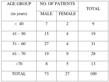
Total number of patients included in the study was 100. Out of this the maximum number of patients with stroke and aphasia were between 51 to 60 years age group in the males and 61 to 70 age group in the females. The mean age was
[image:40.612.127.483.292.560.2]56.34years(Table 1).
Table 1:
AGE GROUP (in years)
NO. OF PATIENTS
TOTAL MALE FEMALE
< 40 7 2 9
41 – 50 15 4 19
51 – 60 27 4 31
61 – 70 19 9 28
>70 8 5 13
TOTAL 73 27 100
The majority were males – 73% and females contributing to 27%
34
Table 2:
LITERACY STATUS
MALE PERCENTAGE FEMALE PERCENTAGE
LITERATE 50 68.49% 14 51.85%
ILLITERATE 23 31.50% 13 49.15%
Out of the 100 patients, 98 were right handed and 2 were left handed(Table 3).
Table 3:
HANDEDNESS NO. OF PATIENTS
Right 98
Left 2
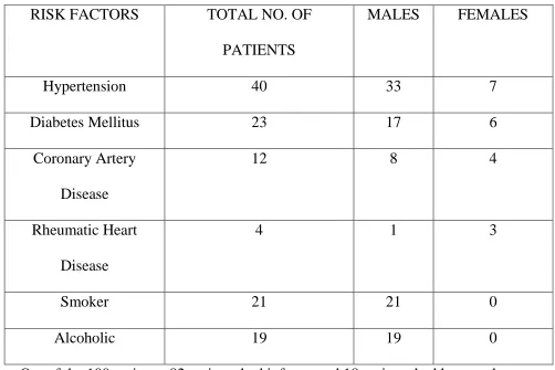
Among these, 40 patients had hypertension(33 – males , 7- females) while 23 were diabetics(17 – males , 6- females), 12 with Coronary Artery Disease(8 – males , 4- females), 4 patients had Rheumatic Heart Disease(1 – male , 3- females), 21
35
Table 4:
RISK FACTORS TOTAL NO. OF PATIENTS
MALES FEMALES
Hypertension 40 33 7
Diabetes Mellitus 23 17 6
Coronary Artery Disease
12 8 4
Rheumatic Heart Disease
4 1 3
Smoker 21 21 0
Alcoholic 19 19 0
[image:42.612.54.556.110.445.2]Out of the 100 patients, 82 patients had infarcts and 18 patients had haemorrhage. Out of the 82 infarcts, 5 patients had haemorrhagic transformation(Table 5).
Table 5:
PATHOLOGY NO. OF PATIENTS PERCENTAGE
Infarct 82 82%
Haemorrhage 18 18%
36
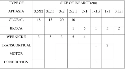
Table 6:
TYPE OF APHASIA
SIZE OF INFARCT(cm)
3.5X2 3x2.5 3x2 2x2.5 2x1 1x1.5 1x1 0.5x1
GLOBAL 18 13 20 10
BROCA 1 6 1 5 2
WERNICKE 3 3 3 5 4
TRANSCORTICAL MOTOR
1 2
CONDUCTION 1
[image:43.612.68.547.112.378.2]The volume of bleed calculated in Aphasia patients with Haemorrhagic stroke are given below
Table 7:
TYPE OF APHASIA VOLUME OF BLEED(ml)
15 – 20 10 - 15 < 10
GLOBAL 7 3
BROCA 1 1
WERNICKE 3 3
37
Table 8:
NEUROLOGICAL DEFICIT NIHS SCORE
NO. OF PATIENTS
Mild 1 – 7 8
Moderate 8 – 14 22
Severe >15 70
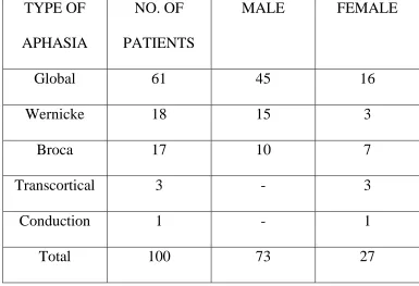
Among the 100 patients majority had Global Aphasia - 61 patientss, Wernicke‘s Aphasia – 18 patients, Broca‘s Aphasia in 17 patients, Transcortical Aphasia in 3 patients, Conduction Aphasia in 1 patient(Table 9).
Table 9:
TYPE OF APHASIA
NO. OF PATIENTS
MALE FEMALE
Global 61 45 16
Wernicke 18 15 3
Broca 17 10 7
Transcortical 3 - 3
Conduction 1 - 1
38
[image:45.612.89.517.269.581.2]Fig 1:
Fig 2:
Distribution in patients with different aphasia syndromes. The mean age of Global Aphasia patients was 57.13 and 59.22 in Wernicke‘s Aphasia patients. But the
45 15
10 3
0
Types of Aphasia in Males
Global Wernicke Broca Transcortical Conduction
16 3
7
0 1
Types of Aphasia in Females
39
[image:46.612.155.459.509.717.2]mean age was younger in Broca‘s Aphasia patients 47.1 years. The mean age in Transcortical motor Aphasia was 50.3 years(Table 10).
Table 10:
Type of Aphasia No. of patients Mean Age(in years)
Global 61 57.13
Wernicke‘s 18 59.22
Broca‘s 17 47.1
Transcortical Motor 3 50.3
The relationship between age and initial aphasia type was analysed and found to be statistically insignificant p>0.05 (Table 11)
Table 11:
Relationship between age and initial aphasia type
Initial aphasia
Age of patients
(years)
Mean SD
Global 57.1 10.4
40
Broca 50.6 16.9
Transcortical 54.0 11.0
Conduction 61 0
‗p‘ 0.2273
Not significant
41
[image:48.612.74.516.363.638.2]Figure 3 Risk Factors of Stroke and Types of Aphasia in male patients
Fig 4 Fig 5:
0 5 10 15 20 25 Global Wernicke's Broca's Transcortical Conduction 4 2 2 1
Risk Factors and Global
Aphasia with
Haemorrhage in Males
Hypertension Diabetics Coronary Artery Disease Smoker 20 8 4 14 14Risk Factors and
Global Aphasia
42
[image:49.612.73.525.272.617.2]Among the risk factors in females, Diabetics leads other risk factors, with Global Aphasia in 6 patients, Wernicke‘s Aphasia in 1 patients and Broca‘s Aphasia in 1 patients. Hypertension with Global Aphasia were 4 patients, Wernicke‘s Aphasia in 3 patients, Broca‘s in 2 patient and Conduction Aphasia in 1 patients. Coronary Artery Disease with Global Aphasia were found in 2 patients. Rheumatic Heart Disease with Global Aphasia in 1 patient and Broca‘s Aphasia in 2 patients.
Fig 6: Risk Factors of Stroke and Types of Aphasia in female patients
0 1 2 3 4 5 6
Hypertension Diabetes Coronary Artery Disease
Rheumatic Heart Disease
43
Among the risk factors for Global Aphasia in females, diabetics were high – 6 patients followed by hypertensives – 4 patients and Coronary Artery Disease in 2 patients. All patients had Ischemic strokes.
Fig 7
Among the risk factors for Wernicke‘s Aphasia in males, Diabetics and Hypertensives are equal – 4 patients each followed by smoker and alcoholics – 3 patients each, followed by Coronary Artery Disease in 2 patients. In females, Hypertension with Wernicke‘s Aphasia was found in 3 patients followed by Diabetes with Wernicke‘s Aphasia in 1 patient.
In males, hemorrhage with hypertension was one patient and other 3 infarcts .Infarcts and hemorrhage were equal in diabetics two each. In females all the 4 were infarcts.
4
6 2
1
Risk Factors and Global Aphasia with
Infarct in Females
Hypertension Diabetics
[image:50.612.98.516.178.409.2]44
Fig 8 Fig 9
Figure 10: Risk Factors for Wernicke's Aphasia and tye of Lesion in Males
Among the female patients with Wernicke‘s Aphasia all the 4 patients had infarcts, 3 with Hypertension and 1 with Diabetes.0
3 1
Risk Factors and
Wernicke's Aphasia in
Females
Hypertension Diabetes 0 0.5 1 1.5 2 2.5 3 3.5Hypertension Diabetes Coronary Artery Disease Smoker Alcoholics Infarct Haemorrhage 4 4 2 3 3
Risk Factors and
Wernicke's Aphasia in
45
Figure 11: Risk Factors for Wernicke's Aphasia in Females
Among the risk factors for Broca‘s Aphasia in males, Diabetes in 1 patient. Smoker – 3 patients and alcoholics – 2 patients.In females, Hypertension with Broca‘s Aphasia was found in 2 patients followed by Diabetes in 1 patient, Rheumatic Heart Disease with Broca‘s Aphasia in 2 patients.
0 0.5 1 1.5 2 2.5 3 3.5
Hypertension Diabetes
46
Fig 12 Fig 13:
Among the male patients with Broca‘s Aphasia, all the 6 patients had Infarcts with 1 patient being Diabetic, 3 Smokers and 2 Alcoholics. Among the female patients with Broca‘s Aphasia, 2 were hypertensives, 1 had infarct and 1 had haemorrhage and other patients with diabetes and rheumatic heart disease had infarcts.
Figure 14: Risk Factors for Broca's Aphasia with Infarct in Males
2
1 2
Risk Factors for
Broca's Aphasia in
Females
Hypertension Diabetes Rheumatic Heart Disease 0 0.5 1 1.5 2 2.5 3 3.5Diabetes Smoker Alcoholics
Infarct 2
1 3
Risk Factors for
Broca's Aphasia in
Males
47
Figure 15: Risk Factors for Broca's Aphasia and type of Lesion in Females
Transcortical Motor Aphasia was found in 3 male patients, out of them 2 had Hypertension and Coronary Artery Disease and they all had Infarcts. Conduction Aphasia was found in 1 female patient with Hypertension and Infarct.
FOLLOW UP
Among the 100 patients enrolled, 40 were lost to follow up because of
premature deaths/ absence of follow up.
Among the 60 patients followed up, 51 were male patients and 9 were female patients. Out of the 9 female patients, 8 had Infarcts with 4 Global Aphasia, 2 Broca‘s and 2 Wernicke‘s Aphasia and 1 had Haemorrhage with Broca‘s Aphasia. Among the 51 male patients, 36 patients had Global Aphasia with 27 Infarcts and 9
0 0.5 1 1.5 2 2.5
Hypertension Diabetes Rheumatic Heart Disease
48
[image:55.612.89.525.408.618.2]Haemorrhage, Wernicke‘s Aphasia in 8 patients, all being Infarcts and Broca‘s Aphasia in 7 patients with all 7 Infarcts.
Figure 16: Aphasia and Type of Lesions in followed up males
Figure 17: Aphasia and Type of Lesions in followed up females
0 5 10 15 20 25 30
Global Aphasia Broca's Aphasia Wernicke's Aphasia
Infarct Haemorrhage
0 0.5 1 1.5 2 2.5 3 3.5 4 4.5
Global Aphasia Broca's Aphasia Wernicke's Aphasia
49
EVOLUTION OF APHASIA
Out of the 9 female patients followed up, 2 Wernicke‘s remained as Wernicke‘s, 1 Broca‘s Aphasia evolved to Transcortical Motor, 2 Broca‘s remained as Broca‘s, 3 Global Aphasia evolved to Broca‘s, 1 Global remained as Global.
[image:56.612.62.552.458.718.2]Out of the 51 male patients, Global Aphasia constituted 36 patients out of which 12 Global Aphasia evolved to Broca‘s Aphasia with Infarct in 9 patients and Haemorrhage in 3 patients, 24 remained as Global Aphasia with 6 Haemorrhage and 18 Infarcts. 8 Wernicke‘s Aphasia remained as Wernicke‘s Aphasia. Out of 7 Broca‘s Aphasia patients 3 evolved to Transcortical Motors and rest remained as Broca‘s(Table 12).
Table 12:
APHASIA EVOLUTION
MALES FEMALES
ME AN AGE NO. OF PATI ENT S PERCENTAGE NO. OF PATIEN TS PERCENTAGE WITHOUT
EVOLUTION 36 70.8% 5 55.5%
57.1 2 EVOLUTION TO
OTHER TYPE 15 29.41% 4 44.44%
50
[image:57.612.150.459.186.421.2]The relation between age and evolution of aphasia was found to be statistically significant p<0.001(Table 13)
Table 13: Age and evolution of Aphasia
Type of case
Age of patients
(years)
Mean SD
Cases with evolution 61.1 10.2 Cases without evolution 53.2 12.2
‗p‘ 0.0011
Significant
Out of the 51 male patients, 15(29.41%) patients evolved to other type of Aphasia. In the females, out of the 9 patients, 4(44.44%)patients evolved to other type, with the percentage being higher in females. The mean age of patients with evolution was 46.57 and in the patients without evolution the mean age was 57.12 years. The mean age of male patients with evolution was 46.3 and in females the mean age was 47.5 years.
51
Table 14:
Age and evolution
Type of case
Age of patients
(years)
Mean SD
Cases with evolution to other type 46.2 11.7
Cases without evolution 56.5 11.0
‗p‘ 0.0016
Significant
52
Table 15:.
Sex and evolution
Type of case
Sex of patients
Male Female
No. % No. %
Cases with evolution to other type(19)
15 78.9 4 21.1
Cases without evolution (41) 37 90.2 4 9.8
‗p‘ 0.2114
Not Significant
GLOBAL APHASIA AND TEST SCORES
53
Table 16:
CASE NO.
T1(1ST WEEK)
T2(12 WEEKS)
T3(24 WEEKS)
INITIAL TYPE OF APHASIA
FINAL
EVOLUTION
3 2.8 2.8 11.6 Global Global
4 2.8 11.2 24 Global Global
7 14.4 39.6 66 Global Broca‘s
8 24 42.2 62.4 Global Broca‘s
9 2.8 2.8 4.2 Global Global
12 2.8 2.8 4.2 Global Global
14 7.2 30.4 51.6 Global Broca‘s
15 2.8 3.6 3.6 Global Global
17 12 35.6 56.4 Global Broca‘s
19 12 37.4 57.6 Global Broca‘s
27 7.2 36 55.6 Global Broca‘s
29 2.8 4.2 16.2 Global Global
32 13.2 35 57.6 Global Broca‘s
33 2.8 3.6 4.2 Global Global
54
43 8.2 36 52 Global Broca‘s
44 2.8 5.2 17.2 Global Global
45 13.2 48.8 60 Global Broca‘s
46 7.4 30.8 52 Global Broca‘s
47 3.6 4.2 4.2 Global Global
48 2.0 12.2 22.2 Global Global
53 2.8 8.2 24.2 Global Global
55 13.2 39 62 Global Broca‘s
60 2.8 12 16.2 Global Global
61 12 42 65.6 Global Broca‘s
63 2.8 11.6 15 Global Global
67 13.4 28.4 54.4 Global Broca‘s
68 2.8 7.2 9.2 Global Global
72 2.8 9.2 20.2 Global Global
75 12 41.6 64 Global Broca‘s
76 7.4 33.6 54 Global Broca‘s
77 2.8 9.6 20.2 Global Global
79 2.8 7.2 20.2 Global Global
80 2.8 3.6 5.6 Global Global
55
93 2.8 2.8 6.8 Global Global
94 3.6 2.8 25 Global Global
98 2.8 9.6 20.2 Global Global
99 2.8 7.6 11.6 Global Global
100 2.8 2.8 18.6 Global Global
BROCA‘S APHASIA AND TEST SCORES
4 patients evolved to Transcortical Motor Aphasia and 6 patients remained as Broca‘s Aphasia. The mean AQ score in evolved patients was 36.05 and in the unevolved 23.06(Table 18).
Table 18:
CASE NO. T1(1ST WEEK)
T2(12 WEEKS)
T3(24 WEEKS)
INITIAL TYPE OF APHASIA
FINAL EVOLUTION
13 27.94 50.8 75.2 Broca‘s Transcortical Motor 22 60.4 65.2 89.6 Broca‘s Transcortical
56
28 25 34 43.6 Broca‘s Broca‘s
39 58 65.2 89.6 Broca‘s Transcortical
Motor 49 29.04 54.8 65.2 Broca‘s Transcortical
Motor
51 22 36.4 50.2 Broca‘s Broca‘s
69 23 32.4 44.8 Broca‘s Broca‘s
73 21.6 25 34 Broca‘s Broca‘s
83 22 36.4 51.2 Broca‘s Broca‘s
90 21 33 49.2 Broca‘s Broca‘s
WERNICKE‘S APHASIA AND TEST SCORES
57
Table 19:
CASE NO. T1(1ST WEEK)
T2(12 WEEKS)
T3(24 WEEKS)
INITIAL TYPE OF APHASIA
FINAL EVOLUTION
16 22.8 33 42.4 Wernicke‘s Wernicke‘s 35 18.4 21.6 30 Wernicke‘s Wernicke‘s 36 20.4 24 28.4 Wernicke‘s Wernicke‘s
57 22 28 30 Wernicke‘s Wernicke‘s
59 21.8 30 37.2 Wernicke‘s Wernicke‘s 70 25 30.8 32.4 Wernicke‘s Wernicke‘s 84 21.6 28.4 38.8 Wernicke‘s Wernicke‘s 87 20.8 26 34.8 Wernicke‘s Wernicke‘s 95 20.2 29 42.8 Wernicke‘s Wernicke‘s 97 18.4 30.4 32.8 Wernicke‘s Wernicke‘s
58
Table 20:
TYPE OF APHASIA
NO. OF PATIENTS
MEAN AQ EVOLVED
MEAN AQ NOT EVOLVED
MEAN AQ (FINAL SCORE
– INITIAL SCORE) Global 40 47.1 11.46 24.83 Broca‘s 10 36.05 23.06 28.26 Wernicke‘s 10 - 13.82 13.82
The relation between age group and aphasia quotient was not significant (p>0.5) initially ,but at the end of six months it was highly significant (p<.001)(Table 21
Table 21: Age group and Aphasia quotients
Age group
Aphasia quotient
Initial At the end of 6 months
Mean SD Mean SD
Upto 40 years 27.9 19.2 66.0 18.0
59
[image:66.612.117.498.441.711.2]The relation between sex and aphasia quotient at the end of six months was statistically significant (p<0.001)(Table 22)
Table 22: Sex and aphasia quotients
Sex
Aphasia quotient
Initial At the end of 6 months
Mean SD Mean SD
Male 14.3 14.3 33.4 22.1
Female 17.8 20.1 52.5 19.4
‗p‘
0.341 Not significant
0.0249
Significant
51-60 years 15.8 14.5 31.1 20.0
61-70 years 12.7 18.2 32.2 22.0
>70 years 15.9 15.2 26.8 18.4
‗p‘
0.1099
Not significant
0.0009
60
[image:67.612.109.500.186.483.2]The relation between NIHSS score and aphasia quotient was significant initially and at the end of six months (p<0.001)(Table 23)
Table 23: NIHS score and aphasia quotients
NIHS score
Aphasia quotient
Initial
At the end of 6
months
Mean SD Mean SD
Mild 33.4 17.8 59.9 25.9 Moderate 15.9 7.7 49.0 10.1 Severe 12.7 16.1 26.1 20.5
61
[image:68.612.116.497.188.637.2]The relation between aphasia type and aphasia quotient was statistically significant(p<0.001)(Table 24)
Table 24: Aphasia type and aphasia quotients
Aphasia type Aphasia quotient
Initial At the end of 6
months
Mean SD Mean SD
Global 5.5 4.4 14.0 7.6
Wermicke 21.0 1.9 35.0 5.2
Broca 32.6 15.7 54.2 7.9
Transcortical 51.1 0.6 79.9 11.9
Conduction 81.7 0 - -
‗p‘ <0.0001
Significant
<0.0001
Significant
62
initial aphasia quotient was negatively correlated. The correlation efficient between hemorrhage volume and final aphasia quotient was found to be negatively
[image:69.612.70.531.220.469.2]correlated(Table 25).
Table 25: CORRELATION
Correlation coefficient
between
Initial Alphasia
quotient
Final Alphasia quotient
Age -0.1699 -0.4575
NIHSS score -0.3709 -0.6641 Correlated
Infarct area -0.6651 Correlated -0.4709
Haemorrhage volume -0.4634 -0.5457 Correlated
CORRELATION OF IMAGING WITH APHASIA
63
Table 26:
SIZE OF THE INFARCT(cm) NO. OF PATIENTS
3 x 2.5 6
2 x 2.5 3
3 x 2 3
[image:70.612.68.547.114.246.2]The size of the infarcts in patients who remained as Global are given as follows
Table 27:
SIZE OF THE INFARCT(cm) NO. OF PATIENTS
3.5 x 2 8
3 x 2 10
2 x 2.5 4
64
Table 28:
SIZE OF THE INFARCT(cm) NO. OF PATIENTS
0.5 x 1 2
1 x 1.5
[image:71.612.69.548.297.396.2]In patients who remained as Broca‘s , the size of the infarcts are given as follows
Table 29:
SIZE OF THE INFARCT(cm) NO. OF PATIENTS
1 x 1 3
2 x 0.5 1
Patients with Wernicke‘s Aphasia had infarct in left temparo parietal region
including Wernicke‘s, supra marginal gyrus, cortical and sub cortical regions. The size of the infarcts in patients with Wernicke‘s Aphasia are given as follows
Table 30:
SIZE OF THE INFARCT(cm) NO. OF PATIENTS
3.5 x 2 3
3 x 2 2
2 x 2.5 4
65
The patients who had small size infarcts had better recovery than the patients with large size infarcts. The mean size of infarcts was 5.12 cm2
The volume of bleed in Hemorrhagic stroke calculated by the formula abc/2 in the different types of aphasia are given below
Table 31:
VOLUME OF BLEED NO. OF PATIENTS APHASIA TYPES INITIAL FINAL
15 – 20 ml 3 Global Global
10 – 15ml 2
1
Broca‘s Global
Broca‘s Broca‘s
< 10ml 2 Global Broca‘s
Patients who had bleed less than 10ml had better recovery than the patients with large volume of bleed.
66
DISCUSSION
The mean age group of patients in this study was 56.34 years with the maximum number of patients in the 5th to 6th decade. Majority of the patients(73%) were males and right handed. In Indian studies done by Paithankar et al (22) the mean age was 71.3 and maximum number of male patients. Study conducted by O.Godefroy et al (23) France on characteristics of aphasia in acute stroke reported the mean age of patients 62 with male predominance and right handed. This data is consistent with other studies which have reported higher prevelance of stroke in elderly age group and in males.
67
There was no significant age difference among the patients with different types of aphasia in this study, though the patients with Broca‘s aphasia were slightly younger (mean age – 47.1years) than patients with Wernicke‘s(59.2years) and Global Aphasia(57.13years). This is consistent with Kertesz et al‘s (26) study which reported Broca‘s aphasia in younger age group than others and related the slight age difference to pathophysiological factors favouring embolic strokes in the anterior division of middle cerebral artery territory. Godefroy et al reported that age did not significantly differ across various aphasias except Conduction aphasias in younger patients and Subcortical aphasias in elder patients. Studies done by Pashek and Holland (27) reported better recovery in younger patients. Studies done by Sarno (28) and Levita using a subjective and functional assessment of language failed to show significant changes in correlation with age, education and initial performance.
There was no significant sex difference among various aphasias in this study. This is consistent with studies by Kertesz ae al and Godefroy et al. he spontaneous recovery of language in patients with aphasia studied by Lendren W et al revealed that age, sex and type of aphasia were not related to the amount of improvement (29).
68
the 40 patients with global aphasia 15 evolved to Broca‘s aphasia and the rest remained as global. Among the 10 Broca‘s aphasia patients, 4 evolved into transcortical motor and the rest remained as Broca‘s. all the wernicke‘s aphasias remained as Wernicke‘s. This is consistent with the data in the Copenhagen aphasia study which concluded that non fluent aphasia could evolve into a fluent aphasia whereas a fluent aphasia never evolve into a non fluent one. Andrew Kertesz arid, McCabe noted good recovery in Broca‘s aphasia and Vignolo (30) noted poor outcome in his study. In our study Broca‘s aphasia had a good outcome.
Kertesz and McCabe noted a bimodal pattern of recovery in Wernicke‘s aphasia ie patients with initially low scores had poor outcome and those with high scores had good outcome. All the Wernicke‘s patients in our study had low initial scores and showed fair recovery.
The relation between age and evolution of aphasia was statistically significant. More females (44.44%) had evolution of aphasia compared to males(29.41%) suggesting that language outcome is better in females.
69
brain compared to the male brain. Gender differences in aphasia studied by Heir et al showed no gender differences in aphasia due to hemorrhagic stroke, but infarct more frequent in women than men (32).
In our study global aphasia was higher in male patients(65%) than female patients(48%). Broca‘s aphasia was higher in female patients(25.92%) than male patients(13.69%). Wernicke‘s aphasia was also higher in male patients(20.5%) than female patients(11.11%).
The change in aphasia quotient(final AQ – initial AQ) was significantly higher in patients whose aphasia evolved than the patients whose aphasia not evolved in this study. McDermott et al reported a significant changes in aphasia scores in patients whose aphasia type evolved into another and the study also concluded that initial severity was one of the factors predicting aphasia recovery. Sarno, Silverman and Sands (33) noted good outcome in patients who had high initial scores. Similar results was noted by Kertesz and McCabe et al. The relation between aphasia quotient and NIHSS, aphasia quotient and aphasia type, sex and aphasia quotient were statistically significant in our study.
70
especially in auditory word recognition for numbers, bodyparts and objects, in patients with global and Wernicke‘s and low aphasia scores (34).
Among the risk factors, hypertension leads followed by diabetes mellitus, coronary artery disease and rheumatic heart disease. In males smoking and alcoholism constitute major risk factor.
In this study, increase in aphasia quotient in the first 3 months following the stroke was higher than the subsequent months, indicating that maximum recovery of language functions occurred in the first 3 months. This is consistent with the studies by Kertesz et al and McDermott et al in which they attributed the improvement to the resolution of acute ischemic changes like perilesional edema and restoration of blood flow to the ischemic penumbra.
Correlating the change in AQ to the initial severity of stroke as measured by the NIHS scale, there was asignificant negative correlation.
71
where hypertension is the major risk factor followed by diabetes mellitus, coronary artery disease and smoking and alcoholism in males.
The size of the infarct had significant correlation with outcome of aphasia. Patients with small sized infarcts had a better outcome. The volume of bleed also had similar significant correlation with outcome of aphasia.The correlation efficient between infarct and initial aphasia quotient and correlation efficient between hemorrhage volume and final aphasia quotient were found to be negatively correlative.
There was good correlation with the anatomical location of the lesion and CT scan. Separate lesion sites for Broca, Wernicke‘s, Conduction and transcortical motor aphasia were demonstrated on CT scan. The lesion sites were consistent with Geschwind‘s concept of aphasia (36).
72
Studies by Margaret Naeser and Robert Hayward (39) with CT brain showed that the lesions in Broca‘s aphasia was large involving the Broca‘s cortical and subcortical areas. In Wernicke‘s aphasia the lesion extended into the subcortical areas along with the Wernicke‘s cortical area and also the supra marginal angular agyrus. Patients with conduction aphasia had lesions deeper to the Wernicke‘s area compatible with the involvement of arcuate fasciculus. The above findings are compatible with the Kertesz radionuclide brain scan study. Dominant frontal lobe lesions associated with Transcortical motor aphasia and did not involve Broca‘s area. Large lesions involving the perisylvian areas were associated with Global aphasia. Large lesions involving cortical and subcortical areas of frontal, parietal and temporal lobes were noticed by Kertesz and Yarnell and also noted poor recovery in these patients.
73
CONCLUSION
Global aphasia is the most common aphasic syndrome in acute stroke patients followed by Wernicke‘s aphasia, Broca‘s aphasia, Transcortical motor aphasia and Conduction aphasia. Age and sex did not differ significantly in various types of aphasia.
Recovery in patients with aphasia due to stroke is a dynamic process with evolution of aphasia from one type to another. There was some improvement in language function as reflected by the aphasia quotient in most of the patients followed up. The type of aphasia change to a less severe form in 30.1%.
Patients with Global aphasia evolved into Broca‘s aphasia in 15 patients out of 40 patients. Out of the 10 Broca‘s aphasia patients, 4 evolved into Transcortical motor aphasia. Significant improvement was noted after 12th week of stroke.
Hypertension and Diabetes were the major risk factors.
BIBLIOGRAPHY
1. Jose Biller et al- vascular diseases of nervous system- Bradley’s Neurology in clinical practice-6th
Edition
2. Osman Sinanovic et al Tusla University Post stroke language disorders- Acta Clin Croat 2011
Mar;50(1):79-94
3. Engelter ST, Gostynski M,Papa S, Frei M, Bornc et al- Epidemiology of aphasia attributable to first
ischemic stroke: incidence, severity, fluency, etiology and thrombolysis. Stroke 2006:37:1379-84 4. Tatemichi TK, Desmond DW, Stern Y, Paik M, Sano M,Bagiella E- Cognitive impairment after
stroke : frequency,patterns and relationship to functional abilities. J Neurol Neurosurg Psychiatry. 1994:57:202-07
5. Wade DT ,Hewer RL, David RM, Enderby PM. Aphasia after stroke: Natural history and assosciated deficits. J Neurol Neurosurg Psychiatry 1986;49:11-16
6. Pederson PM, Jorgensen HS, Nakayama H, Raaschou HO, Olsen TS-Aphasia in acute stroke:
incidence ,determinants and recovery. Ann Neurol 1995;38:659-666
7. Goldstein LB, Samsa GP: Reliability of the national institute of health stroke scale Extension to
non-neurologist in the context of clinical trial-Stroke 28:307,1997
8. Goldstein LB, Berter s C, Davis JN interreliablility of the NIH stroke scale - Archives of Neurology
46(6) 660-2,1989 June
9. Kertesz A: Western Aphasia Battery; Grune Strulton New York 1982
10. Benson DF, Geschwind N-Aphasias and related disturbances Clinical Neurology Vol 1 edited by Baker AR, Bake LH,Newyork ,Harper-Row,1971 Chap 8
11. Damasio AR, Aphasia-Review article- New England Journal of Medicine Vol 326,NO 8 pp 531-539 12. Geshwind M - Disconnection syndrome in animal and man- Brain 1965:88:585-644
13. Broca P-Loss of speech,Chronic softening and partial destruction of the anterior lobe of brain – In Wilkins RH Neurosurgical classics New York, Johnson Reprint corporation 1965,pp 61-68
14. Wernicke C: The symptom complex of aphasia in church A modern clinical medicine:Diseases of the
nervous system, New York, Appleton , 1908 pp 265-324
15. Goodglass H, Kaplan E, The assessment of aphasia and related disorders Lee and Febiger 1972 16. Robinson RG- Neuro Pyschiatric consequences of stroke, Annual review of medicine 1997
17. Alexander MB, Benson DF: The aphasias and related disturbances clinical neurology Vol 1
Lippincott ,1991 chap 10
18. Hickok G , Poeplel D: Towards a functional neuro anatomy of speech perception. Trends Cogn Sci
2000 Apr 4
19. Damasio AR, Damasio H: Brain and Language, Sci Am 1992 sep ,267(3)
21. Shaywitz BA, Shawitz SE, Pugh KR, Constable RT-Sex differences in the functional organization of the brain for language.Nature 1995 Feb16 ;373(6515):607-9
22. Paithankar MM,Dabhi RD- Functional Recovery in Ischemic stroke -Neurol India 2003;51:414-416 23. O Godefroy, C Dubois,B Debachy et al- Vascular aphasias –Main characteristic of patients
hospitalized in acute stroke units for the Lillie stroke program,Stroke 2002,33:702
24. Frances Bouyer McDermott, Jennifer Horner, Elizabeth Delong- Evolution of acute aphasia as measured by western aphasia battery.Clinical Aphasiology Vol24 1996
25. Pederson PM, Vinter K, Olsen TS-Aphasia after stroke: Type ,severity and prognosis: The Copenhagen aphasia study, cerebrovascular diseases 2004; 17:35-43.
26. Kertesz A, McCabe P- Recovery pattern and prognosis in aphasia: Brain (1977) 100 1-18
27. Pashek GV, Holland AL: Evolution of aphasia in the first year post stroke Cortex 24, pg 411-423
28. Sarno M, Levita E-Recovery in treated aphasia in the first year post stroke Stroke Vol 10 No 6,1979 29. Landren W, Lincoln NB-Spontaneous recovery of language in patients with aphasia between 4 and 34
weeks after stroke J. Neurol Neurosurg 1985 Aug 48:743-8
30. .Vignolo L-Evolution of aphasia and language rehabilitation a retrospective study;Cortex
1:344-367,1964
31. Pizzamigliol L, Mammukari A:Evidence for sex differences in brain organization in recovery of
aphasia ,Brain Lang 1985 Jul:25(2)213-23
32. Heir DB, Yoon WB, Mohr JP: Gender and aphasia in the stroke data bank. Brain Lang 1994 Jul ;47(1);165-67
33. Sarno MT, Silverman M, Sands E;Speech therapy and language recovery in severe aphasia J Speech Hear Res 13:607-623,1970
34. Vijayaraghavan V, Nadarajan V, Sablha Sultana M, Viruthagirinathan BS, Arjun Das G; recovery of language functions in strokes with dysphasia-Our experience with 16 cases XIV world congress of
Neuro sciences 1989
35. Kulshrestha M,Vidhyananth-Analysis of risk factors of stroke in North India-J Clin .Diagn Res 2013
Jan 7(1) 127-31
36. Geschwind N; Aphasia N Engi Jmed 1971,284:654-6
37. Kertesz A, Ghent C, Poole E: Localisation of Lesions in Aphasia –Neurology India: Supp 3 :463-465,1973
38. Yarnell P,Monroe MA , Sobel L;Aphasia outcome in stroke: Clinical and Neuroradiological correlation, Stroke 7:516-522,1976
39. Margarat A Naeser, Robert W. Hayward: Lesion localization in aphasia with cranial computed tomography and the Boston Diagnostic aphasia exam. Neurology 28:545-551,1978
40. Mohr JP: Broca’s Area and Broca’s Aphasia :Studies in Neuro Linguistics: New York; Academic
PROFORMA
1)
Name of the patient
:
2)
Age
:
3)
Sex
:
4)
Literacy Status
: Literate/Illiterate
5)
Handedness
:
6)
InPatient Number
:
7)
Occupation
:
8)
Date of Admission
:
9)
Duration of Illness
:
10)
Past similar episodes
:
11)
Comorbid Illness
: DM/HT/CAD/RHD/ S
MOKER/ALCOHOLIC
12)
Details of treatment
:
13)
Family history
: Yes/No
14)
Biochemical profile
:
16)
Follow up after 12
thweek :
24
thweek :
Static
Improving
17)
Associated Deficits
:
18)
NIHS Score
:
19)
LOBAR Functions
:
Frontal
:
Temporal
:
Parietal
:
Occipital
:
20)
WAB Score
:
During Admission
:
12 weeks
:
24 weeks
:
21)
Type of Aphasia
:
NIH STROKE SCALE
1.a. Level of Consciousness: 0 Alert
1 Not alert, but arousable with minimal stimulation
2 Not alert, requires repeated stimulation to attend
3 Coma
1.b. Ask patient the month and their 0 Answers both correctly age:
1 Answers one correctly
2 Both incorrect
1.c. Ask patient to open and close eyes and
0 Obeys both correctly
1 Obeys one correctly
2 Both incorrect
0 Normal
2. Best gaze (only horizontal eye movement):
1 Partial gaze palsy
2 Forced deviation
3. Visual Field testing: 0 No visual field loss
1 Partial hemianopia
2 Complete hemianopia
3 Bilateral hemianopia (blind including cortical blindness)
4. Facial Paresis (Ask pat