“A CLINICAL COMPARATIVE STUDY BETWEEN
DEXMEDETOMIDINE AND CLONIDINE AS AN ADJUVANT TO BUPIVACAINE IN BRACHIAL PLEXUS BLOCK BY
SUPRACLAVICULAR APPROACH”
Dissertation submitted in partial fulfilment of the
Requirement for the award of the Degree of
DOCTOR OF MEDICINE - BRANCH X ANAESTHESIOLOGY
APRIL 2015
TIRUNELVELI MEDICAL COLLEGE HOSPITAL
THE TAMIL NADU DR.M.G.R. MEDICAL UNIVERSITY CHENNAI ,
CERTIFICATE
This is to certify that the Dissertation “A CLINICAL
COMPARATIVE STUDY BETWEEN DEXMEDETOMIDINE
AND CLONIDINE AS AN ADJUVANT TO BUPIVACAINE IN
BRACHIAL PLEXUS BLOCK BY SUPRACLAVICULAR
APPROACH” presented herein by Dr. B.SUNDARI is an original work
done in the Department of Anaesthesiology, Tirunelveli Medical College
Hospital, Tirunelveli for the award of Degree of M.D. (Branch X)
Anesthesiology under my guidance and supervision during the academic
period of 2012 - 2015.
THE DEAN,
Tirunelveli Medical College,
CERTIFICATE
This is to certify that this dissertation “A CLINICAL
COMPARATIVE STUDY BETWEEN DEXMEDETOMIDINE
AND CLONIDINE AS AN ADJUVANT TO BUPIVACAINE IN
BRACHIAL PLEXUS BLOCK BY SUPRACLAVICULAR
APPROACH” entitled submitted by DR. B.SUNDARI to the faculty of
ANAESTHESIOLOGY, The Tamil Nadu Dr. M.G.R. Medical
University, Chennai, in partial fulfillment of the requirement in the award
of degree of M.D. Degree, Branch -X (ANAESTHESIOLOGY), for the
March 2014 examination is a bonafide research work carried out by her
under our direct supervision and guidance.
Guide DR.S.SHENBAGARAJAN M.D
Assistant professor
Tirunelveli medical college
Tirunelveli
DR.A.THAVAMANI M.D.,D.A
Prof. and Head of the Department,
Department of Anaesthesiology,
Tirunelveli medical college,
DECLARATION
I, Dr.B.SUNDARI, declare that the dissertation titled “A
CLINICAL COMPARATIVE STUDY BETWEEN
DEXMEDETOMIDINE AND CLONIDINE AS AN ADJUVANT TO
BUPIVACAINE IN BRACHIAL PLEXUS BLOCK BY
SUPRACLAVICULAR APPROACH” has been prepared by me. This
is submitted to The Tamil Nadu Dr. M.G.R. Medical University, Chennai,
in partial fulfilment of the requirement for the award of M.D. Degree,
Branch X (ANAESTHESIOLOGY) degree Examination to be held in
April 2015.
Place : TIRUNELVELI
Date :
Dr. B.SUNDARI. MBBS.,
POST GRADUATE,
M.D. (ANAESTHESIOLOGY),
TIRUNELVELI MEDICAL COLLEGE,
ACKNOWLEDGEMENT
I am extremely thankful to Dr.L.D.THULASIRAM M.S
(ORTHO), Dean, Tirunelveli Medical College, for his permission to
carry out this study.
I am immensely grateful to Prof.Dr.A.THAVAMANI M.D, D.A,
Professor and Head of the Department, Department of Anaesthesiology and
Critical Care, for encouraging me and rendering timely suggestions and
guiding me throughout the course of this study. I will be forever indebted
to him for his constant support.
I am very grateful to Dr.A.Balakrishnan M.D.,D.A
(Anaesthesiology), Dr.Amutha Rani M.D (Anaesthesiology) and
Dr.R.Selvaraj M.D (Anaesthesiology) Professors, department of
Anaesthesiology and Critical Care, for their constant motivation and
valuable suggestions.
I am greatly indebted to my guide Dr.S.Shenbagarajan M.D
(Anaesthesiology) for his inspiration, guidance, and comments on all
stages of this study.
I am thankful to all Assistant Professors and senior residents for
I wish to express my gratitude to my parents, my sister’s and my
husband for their support throughout my study.
I am thankful to all my colleagues for the help rendered in carrying
out this dissertation.
I thank Mr.Selvaprakash and Miss. M. Uma selvi statisticians for
their useful inputs.
Last, but not least, I thank all the patients for willingly submitting
CONTENTS
S.NO
PAGE NO
1. Introduction 1
2. Aim of the study 5
3. Review of literature 6
4. Anatomy of brachial plexus 11
5. Techniques of supraclavicular block 20
6. Physiology of nerve conduction 29
7. Pharmacology of Bupivacaine 35
8. Pharmacology of Dexmedetomidine 43
9. Pharmacology of clonidine 53
10. Materials and Methods 66
11. Observation and Results 72
12. Discussion 95
13. Summary 98
14. Conclusion 99
15. Consent 100
16. References 104
17. Proforma
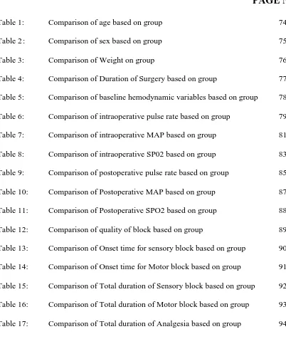
LIST OF TABLES
PAGE NO
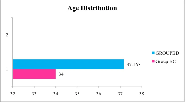
Table 1: Comparison of age based on group 74
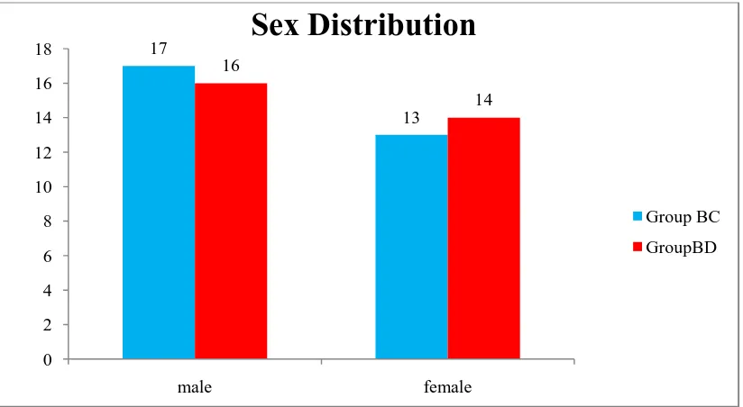
Table 2 : Comparison of sex based on group 75
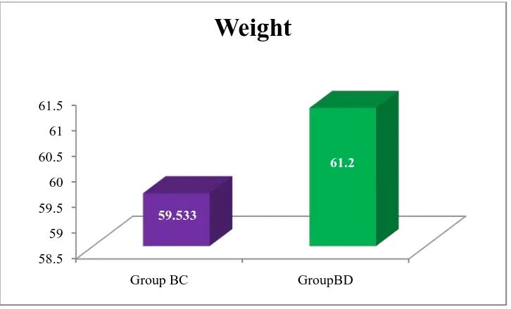
Table 3: Comparison of Weight on group 76
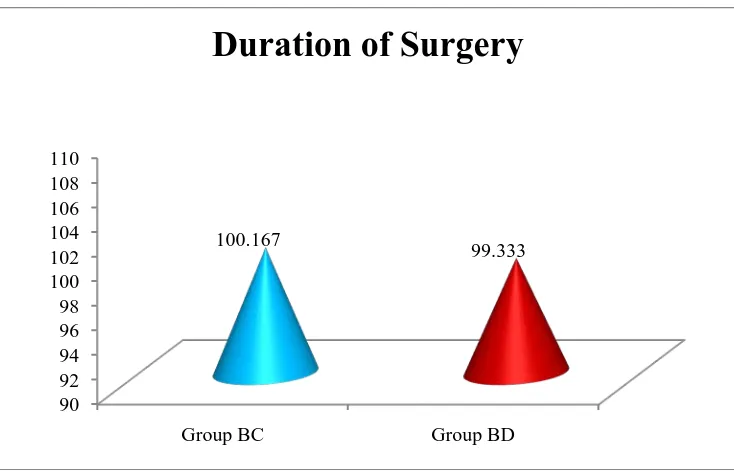
Table 4: Comparison of Duration of Surgery based on group 77
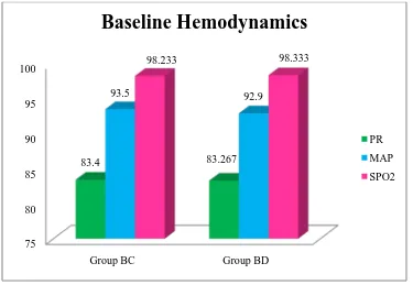
Table 5: Comparison of baseline hemodynamic variables based on group 78
Table 6: Comparison of intraoperative pulse rate based on group 79
Table 7: Comparison of intraoperative MAP based on group 81
Table 8: Comparison of intraoperative SP02 based on group 83
Table 9: Comparison of postoperative pulse rate based on group 85
Table 10: Comparison of Postoperative MAP based on group 87
Table 11: Comparison of Postoperative SPO2 based on group 88
Table 12: Comparison of quality of block based on group 89
Table 13: Comparison of Onset time for sensory block based on group 90
Table 14: Comparison of Onset time for Motor block based on group 91
Table 15: Comparison of Total duration of Sensory block based on group 92
[image:10.595.112.514.112.588.2]Table 16: Comparison of Total duration of Motor block based on group 93
LIST OF FIGURES
S.NO PAGE NO
Figure 1: Course of the brachial plexus 13
Figure 2: Cutaneous distribution of cervical roots 16
and peripheral nerves
Figure 3: Supraclavicular block 22
Figure 4: Plumb bob technique 25
Figure 5: Structure of sodium channel 31
Figure 6: preganglionic and postganglionic alpha 44
receptors of sympathetic nervous system
Figure 7: Comparison of age based on group 74
Figure 8: Comparison of sex based on group 75
Figure 9: Comparison of Weight on group 76
Figure 10: Comparison of Duration of Surgery based on group 77
Figure 11: Comparison of baseline hemodynamic variables 78
on group
Figure 12: Comparison of intraoperative pulse rate based on group 80
Figure 13: Comparison of intraoperative MAP based on group 82
Figure 14: Comparison of intraoperative SP02 based on group 84
Figure 15: Comparison of postoperative pulse rate based on group 86
Figure 16: Comparison of Postoperative MAP based on group 87
Figure 18: Comparison of quality of block based on group 89
Figure 19: Comparison of Onset time for sensory block based 90
on group
Figure 20: Comparison of Onset time for Motor block based 91
on group
Figure 21: Comparison of Total duration of Sensory block based 92
on group
Figure 22: Comparison of Total duration of Motor block based 93
on group
A CLINICAL COMPARATIVE STUDY BETWEEN
DEXMEDETOMIDINE AND CLONIDINE AS AN ADJUVANT TO
BUPIVACAINE IN BRACHIAL PLEXUS BLOCK BY
SUPRACLAVICULAR APPROACH
AIM OF THE STUDY
Recently alpha 2 agonists are playing a vital role as an adjuvant in
neuraxial block and peripheral nerve block. The purpose of this study is to
compare the efficacy of Dexmedetomidine and Clonidine with Bupivacine
in brachial plexus block by supraclavicular approach.
MATERIAL AND METHODS :
It is a prospective randomised single blinded study, conducted in
unilateral upper limb surgeries under brachial plexus block. Patients were
divided into two groups as Group B&C, GroupB&D, Group B&C (N=30) –
35 ml of 0.357% Bupivacaine with Clonidine 2microgm/kg. Group B&D
(N=30) - 35 ml of 0.357% Bupivacaine with Dexmedetomidine
2microgm/kg.
INCLUSION CRITERIA
ASA I, II
Age 20 to 50
Unilateral upper limb orthopaedic surgeries
EXCLUSION CRITERIA
Patient Refusal
Patient on adrenoreceptor agonist or antagonist therapy.
Suspected coagulopathy
Infection at the site of block
History of respiratory, cardiac, hepatic or renal failure.
Patients with medical complications like severe anemia, severe
hypovolemia, shock, septicemia.
Allergy to local anaesthetics and study drug.
Pregnant women.
Objectives:
∗ Sensory block- onset time
∗ Motor block-onset time
∗ Complete duration of sensory and motor block
∗ Total duration of analgesia
∗ Adverse effects
RESULTS:
Comparison of quality of block
Quality Mean SD p value t value
Group BC 3.13 0.82
< 0.001 4.52 Significant
Bupivacaine dexmedetomidine group has better quality than
bupivacaine clonidine group.
Comparison of onset time of sensory block(minutes)
Mean SD p value t value
Group BC 8.47 1.04
< 0.001 17.19 Significant
Group BD 4.7 0.59
The mean time for onset of sensory block in Group BD was 4.7
minutes which was lower than Group BC -8.47 minutes. This was
statistically significant(p<0.05)
Comparison of onset time of motor block between two groups
OTMB Mean SD p' value t value
Group BC 13.1 1.42
< 0.001 11.32 Significant
Group BD 9.63 0.89
The mean time for onset of motor block in Group BD was 9.63
minutes which was lower than Group BC -13.1minutes.This was statistically
significant(p<0.05).
Comparison of total duration of sensory block between two groups (mt)
TDSB Mean SD p' value t value
Group BC 319.1 32.74
< 0.001 25.89
The mean time for total duration of sensory block in Group BD was
537.8minutes. This was higher than the Group BC -319.1minutes.It was
statistically significant(p<0.05).
Comparison of total duration of motor block between two groups
TDMB Mean SD p' value Tvalue
Group BC 222.23 17.84
< 0.001
40.27 Significant Group BD 466.87 28.08
The mean time for total duration of motor block in Group BD was
466.87minutes. This was higher than in Group BC 222.23 minutes. It was
statistically significant(p<0.05).
Comparison of total duration of analgesia between two groups
DOA Mean SD p value t value
Group BC 375.23 32.6
< 0.001 32.55
Significant
Group BD 666.27 36.54
The total duration of Analgesia in Group BD was 666.27 minutes.
This was higher than in Group BC – 375.23 minutes. It was statistically
significant. (p<0.05).
SUMMARY
In adult patients undergoing orthopaedic forearm and hand surgeries
under brachial plexus block, the addition of 2μg/kg of dexmedetomidine to
motor blockade. It also prolongs the duration of sensory and motor blockade.
Postoperatively the duration of analgesia is prolonged with minimal
reduction in pulse rate,blood pressure.
CONCLUSION
The addition of Dexmedetomidine (2μg/kg) to bupivacaine
(0.357%) in brachial plexus block by supraclavicular approach results
in a shorter onset time for sensory and motor blockade, prolongs the
duration of sensory and motor blockade and also the duration of
analgesia.
KEYWORDS : Supraclavicular block, Bupivacaine, Dexmedetomidine,
INTRODUCTION
The surgeries in the upper limb can be done by general or regional
anaesthesia or both. Nowadays regional anesthesia have wide
application in providing surgical anaesthesia, complete muscle
relaxation, better hemodynamic stability and post operative analgesia
as well as in treating chronic pain syndromes. The sympathetic
block produced by regional anesthesia reduces vasospasm, edema.
Nowadays most of the anaesthesiologists practicing combined general
anesthesia and regional anesthesia in paediatric patients. It reduces the
anesthetic requirements and provides smooth extubation.
Regional anaesthesia has several advantages in the postoperative
period compared with general anaesthesia including decreased sedation,
decreased nausea and vomiting, early discharge from the recovery room
and a smooth transition to pain control as the block effects gradually
dissipate.
Brachial plexus provide sensory innervations of the upper limb.
William halsted first demonstrated the brachial plexus block by axillary
approach.1 There are various available approaches and techniques in
brachial plexus blockade.
These include
a) Interscalene approach
b) Supraclavicular approach
c) Parascalene approach
d) Axillary approach
e) Infraclavicular approach
LOCAL ANAESTHETICS
These are the drugs that block the conduction of impulses in
the electrically excitable tissues. Local anaesthetics provide anaesthesia
and analgesia by blocking the transmission of pain sensation along
the nerve fibres. They are classified into
1. Aminoamide group (Lignocaine, bupivacaine, levobupivacaine etc)
2. Amino ester group (cocaine, Chloroprocaine, procaine,
tetracaine)
Bupivacaine is the commonly used local anaesthetic agent. It is a
racemic mixture with two enantiomers, levobupivacaine, S (-) isomer and
dextrobupivacaine, R(+)isomer.
ADJUVANTS
Adjuvants are added to local anaesthetic agents to
- Prolong the duration of anaesthesia and analgesia
- Reduces the dose requirement
- Reduces the incidence of toxic effects(2)
Various adjuvants like morphine, fentanyl, sufentanil,
dexamethasone, midazolam, ketamine, neostigmine, sodabicarbonate
are added to local anesthetic agents. Alpha 2 receptor agonists clonidine
and dexmedetomidine are of new interest in regional anaesthesia because
of their better haemodynamic stability, sedation and longer duration of
postoperative analgesia.
Adjuvants are administered by various routes like epidural,
intrathecal and intravenous.
DEXMEDETOMIDINE
Alpha 2 adrenergic receptor agonist dexmedetomidine gain the
focus of interest for its sedative, analgesic, perioperative sympatholytic
and hemodynamic stabilizing properties. Dexmedetomidine is a new
highly selective alpha 2 adrenergic receptor agonist(3). FDA approved
dexmedetomidine as an ICU sedation for mechanical ventilation(4).
Nowadays new researches are going on about dexmedetomidine as an
excellent adjuvant in neuraxial blocks, peripheral nerve blocks and
intravenous regional anaesthesia. Dexmedetomidine improves the quality
of anaesthesia by means of fast onset, prolonged duration with sedative
effect. It provides excellent post operative analgesia, when compared to
other adjuvants.
AIM OF THE STUDY
Recently alpha 2 agonists are playing a vital role as an adjuvant in
neuraxial block and peripheral nerve block. The purpose of this study is
to compare the efficacy of Dexmedetomidine and Clonidine with
Bupivacine in brachial plexus block by supraclavicular approach.
MATERIAL AND METHODS :
It is a prospective randomised single blinded study, conducted in
unilateral upper limb surgeries under brachial plexus block. Patients were
divided into two groups as Group B&C, GroupB&D, Group B&C (N=30)
– 35 ml of 0.357% Bupivacaine with Clonidine 2microgm/kg. Group
B&D (N=30) - 35 ml of 0.357% Bupivacaine with Dexmedetomidine
2microgm/kg.
Objectives:
a) Sensory block- onset time
b) Motor block-onset time
c) Complete duration of sensory and motor block
d) Total duration of analgesia
e) Side effects
REVIEW OF LITERATURE
Swami SS et al(5) studied the efficacy of dexmedetomidine
and clonidine with bupivacaine in brachial plexus block by
supraclavicular approach. They found that dexmedetomidine increases
the duration of motor and sensory block with better quality and
better post operative analgesia when compared to clonidine
Rachana G et al(6) studied about dexmedetomidine with
bupivacaine in brachial plexus block by supraclavicular approach.
They concluded that dexmedetomidine provided longer duration of
motor and sensory block, increased duration of post operative
analgesia and better hemodynamic stability when added with
bupivacaine
Sandhya agarwal, et al(7) compared dexmedetomidine with
bupivacaine in brachial plexus block by supraclavicular approach. They
concluded that dexmedetomidine hastens the onset time, increases the
sensory and motor block duration and post operative analgesia.
Ammar AS et al (8) studied the effects of dexmedetomidine with
bupivacaine in brachial plexus block by infraclavicular approach. The
result was dexmedetomidine enhances the sensory and motor block onset
time, increases the duration of analgesia, increases the sensory and motor
blockade duration, produce less VRS (verbal response scale) pain scores
and reduces supplemental opioid requirements when added with
bupivacaine
Jang ho song et al (9) studied the effect of dexmedetomidine and
epinephrine when added to 1%mepivacaine in infraclavicular brachial
plexus block. They concluded that dexmedetomidine is a better
alternative than epinephrine.
JE kim et al (1p0) studied the effects of dexmedetomidine with
bupivacaine intrathecally in TURP surgery. They concluded that
dexmedetomidine produces fast onset, increases the sensory block
duration and post operative analgesia, but recovery of motor block could
be delayed.
Deepika shukla et al(11) studied the effects of dexmedetomidine
and magnesium sulphate intrathecally with bupivacaine. They concluded
that dexmedetomidine has rapid onset and prolonged duration than
magnesium sulphate.
Rancourt et al (12) evaluated the effect of dexmedetomidine with
ropivacaine in posterior tibial nerve block. They conclude that sensory
blockade is increased by dexmedetomidine.
Cengiz Kaya et al (13) compared the effect of dexmedetomidine
premedication intra muscularly on hemodynamics and stress response.
They conclude that dexmedetomidine premedication reduces the dose of
opioid requirement in induction and better post operative pain relief. It
also reduces the stress response.
Kaygusuz K et al(14) studied the efficacy of adding dexmedetomidine (1μg/kg) to levobupivacaine (0.5%) in axillary block.
They concluded that dexmedetomidine shortens the onset time for
sensory block , increases the duration of motor and sensory block and
extends the post operative analgesia
Esmaoglu et al(15) evaluated the effects of dexmedetomidine
(100μg) to levobupivacaine in axillary block. They found that
dexmedetomidine decreases the onset time for motor and sensory block,
extends the sensory and motor blockade duration and extends the duration
of analgesia.
Feroz Ahmad Dar et al(16) did a study about dexmedetomidine
added to ropivacaine in brachial plexus block by axillary approach. They
concluded that dexmedetomidine shortens the sensory and motor
blockade onset time. It also prolongs the duration of sensory and motor
blockade and increases the duration of analgesia.
Marhofer et al(17) did a study on the adjuvant action of systemic or
perineural dexmedetomidine with ropivacaine in peripheral nerve block.
They found that systemic and perineural dexmedetomidine increases the
motor block duration by 10% and 60%.
Obayah et al (18) evaluated the effect of adding dexmedetomidine
to bupivacaine for post operative analgesia in children who were operated
for cleft palate repair. He concluded that adding dexmedetomidine with
bupivacaine for peripheral nerve block extends the postoperative
analgesia with clinically no relevant side effects.
Bajwa et al (19) studied the efficacy of fentanyl and
dexmedetomidine to epidural ropivacaine for lower limb orthopaedic
surgeries. They found that dexmedetomidine provides stable
hemodynamics, early onset of sensory block, prolonged post operative
analgesia, less consumption of local anaesthetics postoperatively and
better sedation scores.
Al-mustafa et al (20) did a study of adding dexmedetomidine in
lower dose to bupivacaine in spinal anaesthesia for urological procedures.
They found that dexmedetomidine produces a dose dependent action in
the onset and duration of sensory and motor blockade.
Memis D et al (21) evaluated the efficacy of adding
dexmedetomidine to lignocaine in intravenous regional anaesthesia. They
found that dexmedetomidine added to lidocaine produces the better
quality of anaesthesia and perioperative analgesia without causing side
effects.
M.A.Abosedira et al (22) compared the effects of clonidine and
dexmedetomidine added to lignocaine in bier’ s block. They concluded
that dexmedetomidine lignocaine mixture enhances the quality of
anaesthesia and improves tourniquet tolerance. It also enhances the
intraoperative and postoperative analgesia when compared to clonidine
Solanki S et al(23) compared the effects of dexmedetomidine and
clonidine with bupivacaine in trauma patients posted for lower limb
surgeries. They observed that dexmedetomidine (5 μg) added to
bupivacaine (15 mg) intrathecally provides longer duration of
postoperative analgesia than clonidine(50 mcg).
Esmaoglu et al al (24) studied the effects of adding
dexmedetomidine to lignocaine in bier’s block. They found that
dexmedetomidine added to lignocaine causes a better quality of
anaesthesia and perioperative analgesia without any side effects.
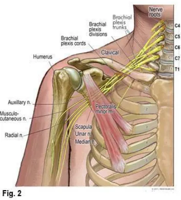
ANATOMY OF BRACHIAL PLEXUS
The brachial plexus provides innervation of the upper limb.
The plexus consists of the roots, trunks, divisons, cords, terminal
nerves.
ROOTS :
Roots are formed from the anterior primary rami of C5, C6,
C7, C8 and T1. In addition there may be contributions from C4 and
T2. If the plexus is formed from C4-C8, it is called as prefixed
plexus. If the plexus is formed from T2, then it is called as post
fixed plexus. The roots are joined together to form the trunks.
TRUNKS:
• C5and C6 roots join to form the upper trunk
• C7 root forms the middle trunk
• C8 and T1 roots join to form the lower trunk.
DIVISIONS:
Trunks are divided into ventral & dorsal divisions which
supply the anterior and posterior aspects of the limb.
CORDS :
• Ventral divisions of the upper and middle trunk unites to form
the lateral cord.
• Ventral division of the lower trunk forms the medial cord.
• Dorsal divisions of all the trunks unite to form the posterior
cord.
BRANCHES :
Branches from the Root:
1) Long thoracic nerve (C5,C6,C7)
Motor supply- serratus anterior
2) Dorsal scapular nerve
Motor supply-Rhomboids (C5)
Levator scapulae
3) Nerve to subclavius
Branches from the Trunk:
1) Suprascapular Nerve (C5,C6)
Motor supply- supra spinatus & infra spinatus
2) Nerve to Subclavius (C5,C6)
FIG.1 ANATOMY OF BRACHIAL PLEXUS
Branches from the Cord:
Lateral cord:
1) Lateral pectoral Nerve (C5,C6,C7)
Motor supply- pectoralis major and minor
2) Musculocutananeous Nerve(C5,C6,C7)
Motor supply- Coracobrachialis, biceps, brachialis
Sensory supply-Lateral cutaneous nerve of arm
3) Median nerve- Lateral root ( C5, C6,C7)
Medial cord:
1) Medial pectoral Nerve (C8,T1)
2) Medial cutaneous nerve of arm (C8,T1)& forearm(C8,T1)
3) Ulnar nerve (C7,C8,T1) .
a. Motor supply-Flexor digitorum profundus, Palmaris brevis,
Flexor carpi ulnaris.
b. Sensory supply-Dorsal and palmar cutaneous branches.
c. Deep terminal branch of ulnar nerve
Motor supply-Flexor digiti minimi, abductor digiti
minimi, opponens digiti minimi, four palmar
interossei, four dorsal interossei, two lumbricals,
adductor pollicis.
4) Medial root of median nerve(C8,T1)
a. Motor supply- Pronator teres, flexor carpi radialis, flexor
digitorum superficialis, Palmaris longus, lateral two
lumbricals.
b. Anterior interosseus branch: Flexor digitorum profundus,
pronator quadratus, abductor pollcis brevis, flexor pollicis
longus, flexor pollicis brevis, opponens pollicis.
Posterior cord:
1) Upper subscapular nerve(C5,C6)
Motor supply- subscapularis
2) Thoraco dorsal Nerve
Motor supply- Lattismus dorsi(C6,C7,C8)
3) Lower subscapular nerveC5, C6)
Motor supply-subscapularis
4) Axillary nerve (C5,C6)
a. Motor supply-Teres minor, deltoid
b. Sensory supply- Upper lateral cutaneous nerve of arm
5) Radial nerve(C5,C6,C7,C8,T1)
a. Motor supply- Triceps, brachioradialis, extensor carpi
radialis longus.
b. Sensory supply- Posterior cutaneous nerve of arm and
forearm, lower lateral cutaneous nerve of arm.
c. Posterior interosseous branch of radial nerve
Motor supply- Supinator, extensors of thumb
d. Superficial branch of radial nerve
Sensory supply- Dorsum of hand
Fig 2. Cutaneous distribution of cervical plexus and peripheral
nerves
ANATOMICAL LOCATION:
The plexus which is formed by the C5-C8, T1 nerve roots are
coming from the corresponding intervertebral foramen and passes
behind the foramen transversorium. Then it lies between the anterior and
posterior tubercles of the corresponding transverse process. The five roots
are situated between the anterior and medial scalene muscles. C5&C6
roots unite to form the upper trunk, C7 continues as middle trunk, C8
&T1 unite to form the lower trunk.
The trunks emerge between the two scalene muscles and passes
downwards and laterally across the base of posterior triangle and then it
passes across the 1st rib. At the lateral border of the 1st rib each trunk
further divides into anterior and posterior division behind the clavicle.
The anterior and posterior divisions unite to form the three cords.
a) Lateral cord- it is formed by the anterior divisions of the upper and
middle trunks.
b) Medial cord- it is a continuation of the anterior division of the
lower trunk.
c) Posterior cord - formed by all the three posterior divisions.
Sympathetic contributions of this plexus are derived from middle
cervical ganglion and stellate ganglion.
Relations
ROOTS
This part of the plexus lies above the second part of the subclavian
artery and between the scalene muscles.
TRUNKS
In the posterior triangle, the trunks are covered by prevertebral
fascia. It is superficially placed, covered by skin, platysma and deep
fascia.
Structures crossing the trunk:
Omohyoid-Inferior belly
External jugular vein
Transverse cervical artery
Supraclavicular nerves
The upper and middle trunks are situated above the subclavian
artery as they pass across the first rib. The lower trunk lies behind the
artery and may groove the rib immediately posterior to the subclavian
groove.
DIVISIONS
At the level of lateral border of first rib and behind the clavicle,
subclavius muscle, suprascapular vessels (which lies immediately
posterior to the clavicle), the trunks are bifurcate into divisions and then it
descends into the axilla.
CORDS:
Cords are formed at the apex of the axilla. These cords are named
in relation to the axillary artery. Lateral cord- lateral to the axillary
artery.Posterior cord- At first it lies lateral to the artery, when it comes
behind the pectoralis minor it lies posterior to the artery. Medial cord- At
first it lies behind the artery, but when it comes behind the pectoralis
minor it lies medial to the artery.
History:
• In 1880 von anrep had injected cocaine under the skin of his arm
and he realised the insensitivity of that area.
• 1879&1880 –william halsted and Alfred hall injected 4%cocaine in
the forearm. They found that it produces analgesia below the level
of injection and not above.
Then hall injected 2ml into the ulnar nerve at the level of
elbow. It produced loss of sensation along the ulnar
distribution.
• After 1893, George crile, surgically exposed the neck and injected
each nerve directly.
• G.Hirschel first discovered the axillary approach.
• D.Kulenkampff first discovered the supraclavicular approach. He
injected 10 ml of procaine in midclavicular area lateral to
subclavian pulse.
• L.Bazy and V.Pauchet 1917-introduced infraclavicular approach
• Intravenous regional anesthesia was first introduced by August bier
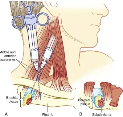
SUPRACLAVICULAR BLOCK:
Indications:
Surgery on distal upper extremity
To palliate acute pain emergencies, herpes zoster, neuritis,
upper extremity trauma, cancer
Alternative to stellate ganglion block
Blockade level occurs at distal trunk- proximal division level.
ADVANTAGES:
• compactly arranged nerve fibres
• intensive blockade
• Small volume of drug
• Rapid onset of reliable blockade
• Can be performed with patients arm in position.
DISADVANTAGES:
• Less suitable for shoulder problems, requires cervical plexus
block for supplementation
• Demonstrable paraesthesias required which is unpleasant
for the patient
• 0.5 - 6% of pneumothorax incidence seen
• 40-60% phrenic nerve blockade seen
• 70-90% stellate ganglion blockade recorded
• Possibility of neuritis also seen.
Various Technique:
Classic approach : Kulenkampef
Subclavian perivascular approach : Winnes and Collins
Modified lateral paravascular approach of Moorthy
Technique
Several anatomic points are important in performance of the
supraclavicular approach. The trunks are situated vertically at the level of
the first rib, with the relation to the subclavian artery cephaloposteriorly,
which can often be palpated in a slender, relaxed patient. At the level of
the midpoint of the clavicle, the neurovascular bundle lies inferiorly. The
1st rib acts as a medial barrier to the needle reaching the pleural dome and
is short, broad, and flat, with an anteroposterior orientation at the site of
the plexus.
Position : supine,
Head turned to the opposite side
Arm in adducted position, hand should be extended towards the
same side of the knee.
Classical technique :- Identify and mark the midpoint of the clavicle.
The posterior border of the sternocleidomastoid can be palpated easily
when the patient raises the head slightly. Then palpate the belly of
scalenus anterior muscle, in the interscalene groove which may be
situated at the level of midpoint of the clavicle around 1.5 – 2 cm
[image:39.595.111.510.299.680.2]posteriorly. We can palpate the subclavian pulse at this level.
FIGURE 3 SUPRACLAVICULAR BLOCK
After appropriate preparation, a skin wheal should be created at the
landmark. A 22-gauge, 4-cm needle is inserted lateral and posterior to
the subclavian pulse in a caudal, medial and posterior direction until the
paresthesia or motor response is elicited or the first rib is encountered. If
a syringe is attached, this causes the needle and syringe to lie parallel to a
line joining the skin entry site and the patient's ear. If the first rib is
encountered without paresthesia, the needle should be systematically
walked anteriorly and posteriorly along the first rib until the plexus or the
subclavian artery is located, which results in a paresthesia or motor
response. After confirming negative aspiration for blood, the local
anesthetics are injected incrementally.
The rib is usually contacted at a needle depth of 3 to 4 cm.
However, in an obese patient or in the presence of tissue distortion from
hematoma or injection of solution, the depth may exceed the length of the
needle. Nonetheless, before the needle is advanced farther, gentle probing
in the anterior and posterior directions should be done at the 2- to 3-cm
depth if paresthesias are not obtained. Multiple injections may improve
the quality or may shorten the onset of blockade.
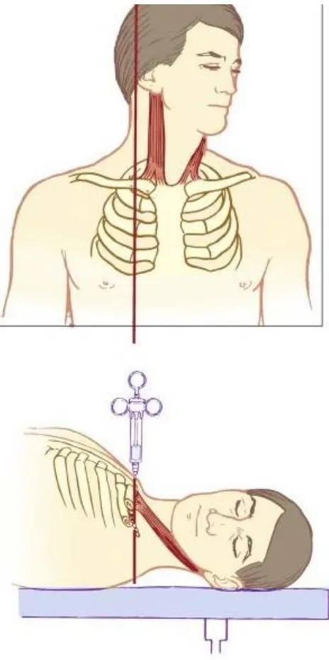
The modified plumb-bob approach uses similar patient positioning,
although the needle entry site is at the point where the lateral border of
the sternocleidomastoid muscle inserts into the clavicle. After preparation
and raising of a skin wheal, a 22-gauge, 4-cm needle is inserted while
mimicking a plumb-bob suspended over the needle entry site. Frequently,
a paresthesia or motor response is elicited before contacting the first rib
or artery. If no paresthesia or motor response is elicited, the needle is
reinserted while angling the tip of the needle cephalad and then caudad in
small steps until the first rib is contacted. The modified plumb bob
technique reduces the unwanted complications of classic approach(26).
FIGURE 4 PLUMB BOB TECHNIQUE
NERVE IMAGING STUDY WITH ULTRASOUND: [25-30]
The Fascicles of peripheral nerves can be detected with a
high-resolution ultrasound imaging. The fascicular echotexture is often the
most distinguishing feature of nerves namely “honeycomb” architecture.
More central nerves, such as the cervical ventral rami, with fewer
fascicles, therefore can appear as monofascicular on ultrasound scans.
One of the most powerful techniques to clinch the nerve fascicles is
to slide a broad linear transducer on the area of peripheral target
nerve.
Nerves can appear round, oval, or triangular. Although nerve
shape can be changing on course, cross-sectional area is same and
constant in the absence of major branching. The Peripheral nerves are
pathologically enlarged also by entrapment or in certain other
neuromuscular disorders such as Charcot-Marie-Tooth disease of type IA.
There is also some evidence to suggest that the patients with diabetic
neuropathy are also having enlarged peripheral nerves.
It is true that direct nerve imaging has led to a phenomenal good
increase in ultrasound-guided regional anesthesia, but still the
identification of other nearby structures like the fascia and other
connective tissue is critical in this endeavor.
These significant structures permit favorable distribution of local
anaesthetic that the nerve contact with the block needle is not mandated.
Successful drug injections must always clarify the borders of the nerve.
ULTRASOUND AND ITS ARTIFACTS IN REGIONAL
ANAESTHESIA: (31-41)
There are several common assumptions in the ultrasound imaging.
First of all, the velocity of sound is assumed to be around 1540 msec.
This estimate was achieved from measurements on soft tissue at
physiological body temperature.
When the local heterogeneities exist, then artifactual bending of
the block needle can be seen with sonography, the so-called bayonet
artifact. The Speed of sound artifacts relate to the time-of-flight
considerations and to the refraction at the interface of tissues with the
different speeds of sound. [33,34,35]
Comet tail artifact is a type of reverberation artifact. At the low
receiver gain, the comet tail is seen as a typical tapering series of discrete
and clear echo bands just deep to a strongly reflecting structure.
Then spacing between the bands represents the distance seen
between the anterior and posterior side walls of the object. Internal
clear reverberations which arising from within the object cause the artifact
of comet tail, that is most intensely observed while the object is
perpendicular to the beam.
Moreover the pleura is a strong reflector that causes the comet tail
artifact. Reverberation echoes are usually seen while strong specular
reflections are being received.
During supraclavicular block, the mirror-image artifacts can be
observed from the reverberation. While the pleura is adjacent to the
subclavian artery, the mirror-image artifacts can occur with gray-scale
type sonographic imaging.
Third to say, all reflectors are assumed to be on one central ray of
the transducer beam. When this is not occurring true, out-of-plane
artifacts are also observed that are slice thickness artifacts.
Definitive proof of the out-of-plane artifacts requires multiple
views that are recommended when ambiguities arise.
Not like the adjacent tissue, biologic fluids are not significantly
attenuating the sound beam and therefore will cause acoustic
enhancement. The Acoustic enhancement artifacts deep to vessels may
be erroneously interpreted as the nerves. For example, acoustic
enhancement lying deep to the axillary artery that is in the axilla can
mislead.
PHYSIOLOGY OF NERVE CONDUCTION
Autonomic postganglionic efferent and nociceptive afferent C
fibres are nonmyelinated. These axons have only single Schwann cell
sheath. Large motor and sensory fibres are myelinated. The myelin
sheath enhances the nerve conduction and causes the action potential
impulse to flow through the axoplasm to node of Ranvier. Active
impulses are regenerated in nodes of Ranvier. Sodium channels are rich
in nodes of Ranvier in myelinated nerve fibres. These channels are
essential for impulse generation and propagation(42). In unmyelinated
nerve fibers, these sodium channels are present throughout the length.
Physiology of nerve conduction:
The Nerve cells maintain a negative resting potential difference of
-60 to -90mv. During rest, it is impermeable to sodium ions and
permeable to potassium ions. This gradient was maintained by
Na+K+ATPase (transport of three sodium ions out of the cell for two
potassium ions into the cells). Permeation of these ions occur via an ion
channel, a specialised protein.
Action potential:
During the stimulus, the nerve cell membrane permeable to sodium
ions and changing the membrane potential to positive(43) . The threshold
for sodium ion channel opening is -55mv. During depolarisation, both
sodium and potassium ion channels are in open configuration (Na>K). So
excessive positive ions enter intracellularly and reversal of membrane
potential to +35mv. The membrane depolarization extends to nearby the
area and cause more opening of sodium channels and increasing the
inward current. This event continues until some of the sodium channels
became inactivated and also k+ channels are still opened and result in a net
outward current and produces repolarization. Now the threshold above
the resting state, so it is refractory to next stimulus. Over time, the
sodium channel inactivation decays and the potassium channel changes to
closed state. Thus the resting threshold is restored.
Sodium channel has one large alpha subunit and one or two small
beta subunits. Alpha subunit has four domains which is homologous
DI-DIV and each has six helical regions (S1-S6) to span the membrane. It
was in three states, open, inactivated and resting state.(44-45)
FIGURE 5 STRUCTURAL FEATURES OF SODIUM CHANNEL
LOCAL ANAESTHETICS
Local anesthetics bind with alpha subunit and block from inside of
the cell (Tetrodotoxin which binds and block from outside of the cell).
Local anesthetics does not alter the resting membrane potential. When
increasing the concentration of local anesthetics, it slows the impulse
conduction and decrease the rate of rise and magnitude of the action
potential and threshold for excitation is raised progressively. Thus the
propagation of impulse is abolished.
Local anesthetics have greater affinity to activated and inactivated
state than the resting state. Its action is both voltage and time dependent.
Frequency dependent blockade: (46,47)
Local anesthetic action is effective, when the nerve fibres are
activated rapidly. Also called as use dependent blockade or phasic block.
More depolarization causes more affinity with local anesthetics.
a) Guarded receptor model: This theory states that binding sites
are more available during firing of nerve.
b) Modulated receptor model: Local anesthetics dissociates
from inactivated channels slowly than from resting
channels.(48)
Order of sensitivity to local anesthetics (49)
a) Small myelinated axons A γ ,Aδ
b) Large myelinated Aα, Aβ
c) Small non myelinated C fibres.
Other channels blocked by the local anaesthetics: Calcium, Potassium,
NMDA
Structure of local anaesthetics:
It has three groups
1) Lipophilic – benzene ring
2) Hydrophilic end – tertiary amine
3) Intermediate chain by ester /amide
These local anesthetics are weak bases. Lipid solubility determines
the potency. Local anaesthetics are poorly soluble in water, but soluble in
hydrophobic organic solvents. So these drugs are prepared as water
soluble hydrochloride salts with a PH of 6-7. Potency is determined
mainly by lipid solubility(50).
Epinephrine is unstable in alkaline solution. So commercial
preparations of local anaesthetics with adrenalin solutions are available as
acidic solution(PH4-5).
Aminoester : Procaine, Chloroprocaine, Tetracaine, Cocaine
Aminoamides : Lignocaine, Bupivacaine, Ropivacaine,
Mepivacaine, Prilocaine, Editocaine.
Cm- minimum concentration of local anesthetic that will block the
nerve impulse conduction. Factors determining the cm are fibre size,
type, myelination, PH(acidic PH antagonize the block), frequency of
nerve stimulation, electrolytes level. (hypokalemia, hypercalcemia
antagonize block)(51).
Onset of action depends on lipid solubility and concentration of
non ionized form. Local anesthetics with pka closest to physiological PH
will have a higher concentration of non ionized base and have the fast
onset property. Increasing the dose or concentration of local anesthetics
cause prolonged duration of action and reduce the onset time(52,53). But it
will cause local anesthetic toxicity. Adjuvants are added with local
anesthetics to avoid the side effects of local anesthetics and improve the
efficacy and onset.
PHARMACOLOGY OF BUPIVACAINE
Bupivacaine is an amide type of local anaesthetic drug. It is a
hydrochloride salt of 1-butyl-N-(2, 6-dimethylphenyl) piperidine-2-
carboxamide.
It was synthesized in Sweden by Ekenstam and his colleagues in
1957. First used clinically by L.J. Telivuo in 1963. Pka is 8.2
Molecular weight - 288
Protein binding - 95%
Lipid solubility - 28
Elimination half life - 210mts
Toxic plasma concentration - >1.5µg/ml
Approximate duration of action - 175mts
The drug is very stable to acids, alkalis and repeated autoclaving.
Bupivacaine 0.5% is the preferred strength. Higher concentration result
in greater variability of spread. Bupivacaine is 4 times potent than
lidocaine, hence 0.5 % solution is equivalent to 2 % lidocine. It is more
cardiotoxic than lidocaine. Cardiotoxicity is aggravated by hypoxia,
hypercapnia and pregnancy. It causes less motor block compared to
sensory block. It is not recommended for intravenous regional analgesia.
Duration of action is between 5 to 16 hours and is the longest acting local
anaesthetics, which is related to binding to nerve tissue. Small percentage
of a given dose of drug is excreted unchanged in the urine and the
remainder is metabolized in the liver.
Uses:
Spinal anaesthesia
Epidural anaesthesia
Caudal anaesthesia
Continuous epidural anaesthesia
Peripheral nerve block
Infiltration anaesthesia
Onset time and duration of action
Site of action Onset (minutes) Duration (minutes)
Intrathecal 5 90-120
Epidural 15-20 165-225
Brachial plexus 10-20 600
Pharmacokinetics:
Once injected intrathecally, it gets absorbed by the nerve rootlets
and it is rapidly absorbed from the site of injection, but the rate of
absorption depends on the vascularity and the presence of
vasoconstrictors. Because of high lipid solubility it easily penetrates
nerve and vascular tissue. 80-95% of absorbed bupivacaine binds to the
plasma proteins.
Distribution:
Rapid distribution phase: (α)
Slow disappearance phase: (β)
Biotransformation:
Possible pathways of metabolism of bupivacaine include aromatic
hydroxylation and conjugation. Only the dealkylated metabolite,
N-desbutyl bupivacaine has been measured in blood or urine after
epidural/spinal anaesthesia. Alpha1 acid glycoprotein is the most
important plasma protein binding site of bupivacaine and its
concentration is increased by many clinical situations including post
operative trauma.
Excretion:
4-10% of the drug is excreted in urine as unchanged form.
Mode of action
Site of action:
The spinal nerve rootlet fine nerve filaments having a large
surface area are exposed to the local anaesthetics.
Posterior and lateral aspects of the spinal cord.
Sodium Channel blockade:
They impede the sodium ion access to the axon interior by
occluding the transmembrane sodium channels, thus delaying the process
of depolarization and the axon remains polarized. It is a
non-depolarisation blockade. Thus the resting membrane potential is
maintained and depolarization in response to stimulation is inhibited.
The mechanism by which local anaesthetics block sodium channel
conductance is as follows,
a) Local anesthetics in the cationic form act on the receptors within
the sodium channels, on the cell membrane and block it. The local
anaesthetic can reach the sodium channel either via the lipophilic
pathway directly across the lipid membrane or via the axoplasmic
opening. This mechanism accounts for 90% of the nerve blocking
effects of amide local anaesthetics.
b) The second mechanism of action is by membrane expansion. This
is a non specific action in contrast to the more specific drug
receptor interaction.
Pharmacodynamics:
It has got a longer duration of action but a slower onset.
Cardiovascular system:
Bupivacaine reduces the cardiac output by reducing the
sympathetic tone, by slowing the heart rate and by reducing the venous
return. It produces a fall in arterial blood pressure, but it is relatively
slow and is seldom very profound. Central venous pressure is reduced. It
causes an increase in lower limb blood flow, thereby it reduces the
incidence of deep vein thrombosis.
Respiratory System:
It relaxes the bronchial smooth muscle. It causes apnea due to
phrenic and intercostal nerve paralysis or depression of the medullary
respiratory center following direct exposure to drug.
Gastro intestinal tract:
There is an increase in gastro intestinal motility and emptying of
the gastric contents.
Toxicity:
Toxicity is related to the plasma level of unbound drug and more
likely due to an inadvertant intravenous injection. Systemic toxicity
reactions primarily involve central nervous system and cardio vascular
system. The blood level required to produce central nervous system
toxicity is less than that required to produce circulatory collapse.
Central Nervous System Toxicity:
The patient may have circumoral numbness, dizziness and tongue
paresthesia immediately. Tinnitus and blurred vision may follow.
Excitatory signs such as restlessness, agitation, nervousness, paranoia will
precede central nervous system depression (slurred speech, drowsiness,
unconsciousness). Muscle twitching followed by tonic clonic seizures.
Respiratory arrest often follows. Selective blockade of inhibitory
pathways causes these excitatory reaction.
Cardiovascular System Toxicity(54-56)
The rate of depolarization in fast conducting tissue of purkinje
fibres and ventricular muscle is decreased. The rate of recovery of
bupivacaine induced block is slower than that of lignocaine. Extremely
high concentration of the drug causes sinus bradycardia, hypotension,
atrioventricular heart block, idioventricular rhythms and life threatening
arrhythmias such as ventricular tachycardia, ventricular fibrillation and
cardiac arrest. Levobupivacaine S(-) isomer is devoid of some of the
CNS and CVS adverse effects. It will prevent the toxic effects
following inadvertent intravascular injection of bupivacaine.
The dosage of bupivacaine depends on,
Area to be anaesthetized
The vascularity of the tissue to be blocked
The number of neuronal segments to be blocked
Individual tolerance
Technique of local anaesthesia.
Available concentrations:
0.25%,0.5%
0.25%,0.5% soluble in isotonic saline
0.5% 0.75% solution in 8% dextrose hyperbaric
These doses can be repeated in 3-4 hours but maximum dose is
400mg in 24 hours.
Dosage and concentration of bupivacaine in various blocks
Type of block Concentration Dosage in ml Dosage in mg
Local infiltration 0.25-0.5% 5-20ml Upto 75 mg
Brachial plexus
block
0.25-0.5% 20-40ml 75-225 mg
Intercostal block 0.25-0.5% 3-5ml
15-20mg per each
nerve
Epidural block 0.25-0.5% 15-20ml 50-200mg
Caudal block 0.25-0.5% 15-30ml 75-150mg
Subarachnoid
block
0.5% 2-4 ml 10-20mg
PHARMACOLOGY OF DEXMEDETOMIDINE
Dexmedetomidine is an ∝2- agonist that received FDA approval in
1999. It is used as a short-term sedative analgesic especially in the ICU
and usually not used for more than 24 hours(57). Dexmedetomidine is a
selective ∝2 – adrenoceptor agonist. It is used in high doses for sedation
and analgesia. It has a reversal drug Atipamezole for its sedative effect.
It is used in perioperative period as sedative and analgesic, as
premedication, as an anesthesic adjunct for general as well as regional
anesthesia and also for post operative sedative and analgesic.
Physiology of ∝2 -adrenoceptors.
Alpha 2 – adrenoceptors are found in peripheral and central
nervous systems, also in effector organs like liver, kidney, pancreas,
eye, vascular smooth muscles and platelets.
They are divided into 3 subtypes.
∝2 A- predominant subtypes in CNS, this is responsible for the
sedative, analgesic and sympatholytic effect. Dexmedetomidine is
8 to 10 times more selective towards ∝2 AR than Clonidine.
∝2 B –found mainly in the peripheral vasculature, and is
responsible for the short term hypertensive response.
∝2 C-found in the CNS, which is responsible for the
anxiolytic effect(58) ,startle response.
Startle response is the response of mind and body to a sudden
unexpected stimulus, such as flash of light, loud noise. In human beings,
the reaction includes physical movement away from the stimulus, the
contraction of the muscles of arms and legs,blinking , respiratory and
[image:61.595.124.476.525.693.2]blood pressure changes.
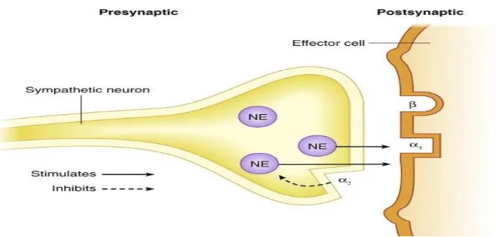
Fig 6 preganglionic and postganglionic alpha receptors of
sympathetic nervous system
All these subtypes produce cellular action by signalling through a
G-Protein which couples to effector mechanisms, and the coupling differs
depending on receptor sub-type and location. The ∝2 A-Subtype appears
to couple in an inhibitory fashion to the calcium channel in the locus
ceruleus of the brain stem and in the vasculature. The ∝2 B subtype
couple in an excitatory manner to the same effector mechanism.
Mechanism of action of dexmedetomidine:
Dexmedetomidine possess unique properties and it differs from
other sedative drugs. ∝2 – adrenoceptors are found in many sites
throughout the CNS, but the highest densities are found in the locus
ceruleus, the predominant noradrenergic nuclei of the brainstem which is
an important modulator of vigilance(59). Presynaptic activation of ∝2
adrenoceptor in the locus ceruleus inhibits nor epinephrine (NE) release
and results in sedative and hypnotic effects. Locus ceruleus is the site of
origin for descending medullospinal noradrenergic pathway which is an
important modulator of nociceptive neuro transmission. Stimulation of
the ∝2 –adrenoceptors in this area terminates mainly the propagation of
pain signals leading to analgesia. Post synaptic activation of ∝2 –
adrenoceptors in the CNS causes decrease in sympathetic activity which
leads to hypotension and bradycardia. Also cardiac vagal activity is
augmented and all the effects together produce analgesia, sedation and
anxiolysis.
Stimulation of ∝2 –receptors at the substantia gelatinosa causes
inhibition of the nociceptive neurons firing and inhibition of substance
P release.∝2adrenoceptors also have analgesic mechanisms by inhibiting
norepinephrine release at the nerve endings whereas the reason for
analgesic effect is by the spinal mechanism.
∝2 - Receptors located on blood vessels which mediates
vasoconstriction whereas those located on sympathetic terminals inhibit
norepinephrine release. In other areas these ∝2 adrenoceptors cause
contraction of vascular and other smooth muscles, decreased salivation,
secretion and bowel motility in the gastrointestinal tract. It also inhibit
the release of renin , increased glomerular filtration rate, decreased
insulin release from pancreas, decreased intraocular pressure, decreased
platelet aggregation and decreased shivering threshold by 2oC.(60)
Pharmacokinetics:
Absorption and distribution:
Dexmedetomidine with the dose of 0.2 to 0.7 µg/kg /hr exhibits
linear pharmacokinetics and it is administered as intravenous infusion
upto 24 hours. Also it has the rapid distribution phase, its distribution half
life is around 6 minutes (61), and elimination half life is 2 hours.
The volume of distribution is 118L. Average protein binding is
94%. Context- sensitive half life ranges from 4 minutes after a 10-minute
infusion to 250 minutes after an 8-hour infusion. Its oral bioavailability is
poor, which is because of extensive first-pass metabolism. The
bioavailability of sublingual route is high (84%) and it offers a potential
role in pediatric sedation and premedication.
Metabolism and excretion
Dexmedetomidine undergoes biotransformation through direct N-
glucuronidation and cytochrome P-450 (CYP 2A6) mediated aliphatic
hydroxylation to its inactive metabolites. Metabolites are excreted in the
urine(95%) and in the feces (4%). Dose has to be reduced in patients with
hepatic failure.
Pharmacodynamics
∝- adrenoceptor agonists have different ∝2 / ∝1 selectivity.∝2 / ∝1
selectivity of dexmedetomidine is 1620:1 whereas it is low for clonidine
and hence dexmedetomidine is 8 times more powerful ∝2 – adrenoceptor
than clonidine.
Cardiovascular system
Dexmedetomidine has no effects on the heart directly. It causes a
dose dependent increase in coronary vascular resistance and oxygen
extraction and the supply / demand ratio is unaltered. It evokes a biphasic
blood pressure response. A short hypertensive phase and subsequent
hypotension and the 2 phases are mediated by 2 different ∝2 –AR
Subtypes: the ∝-2B AR is responsible for the initial hypertensive phase.
Hypotension is mediated by the ∝2A –AR.(62) Younger patients with
high level of vagal tone develop bradycardia and sinus arrest which were
effectively treated with anticholinergic agent.
Respiratory system
Dexmedetomidine does not produce respiratory depression even at
high doses (63). It can be safely used in spontaneously breathing patients
after surgery in ICU. It maintains sedation without cardiovascular
instability or respiratory drive depression. Hence it is used during
weaning and extubation in trauma / surgical ICU Patients in whom
previous attempts at weaning have failed because of agitation associated
with hyperdynamic cardio pulmonary response (64).
Central nervous system
Dexmedetomidine reduces cerebral blood flow and cerebral
metabolic requirement of oxygen. Dexmedetomidine enhances
cumulative performance and also possess sedative, analgesic and
anxiolytic action through ∝2 –AR(65) .It reduces levels of circulating and
brain cetecholamines, thus balancing the ratio between cerebral oxygen
supply and demand, reduces excitotoxicity and improves the perfusion in
the ischemic penumbra, hence it possess excellent neuroprotective action.
In subarachnoid haemorrhage it reduces the levels of glutamate which is
responsible for cellular brain injury.
Endocrine and renal effects
Dexmedetomidine activates peripheral presynaptic ∝2–AR, thus
catecholamine release is reduced and hence sympathetic response to
surgery is also reduced. It is an imidazole agent but does not inhibit
steroidogenesis when used as an infusion for short term sedation(66).
Adverse Effects:
Side effects reported are hypotension, hypertension, nausea,
vomiting, dry mouth, bradycardia, atrial fibrillation, pyrexia, chills,
pleural effusion, atelectasis, pulmonary edema, hyperglycemia,
hypocalcaemia, acidosis, etc., Transient hypertension is produced when
dexmedetomidine infusion is rapidly administered (Loading dose of
1µg/Kg / hr if given less than 10 minutes) and this is mediated by
peripheral ∝2B –AR vasoconstriction(67).
The occurrence of Hypotension and bradycardia is mediated by
central ∝2A-AR, causing decrease in noradrenaline release from the
sympathetic nervous system. Supersensitization and up regulation of
receptors occur during long term use, hence abrupt discontinuation not
advised. Withdrawal symptoms of nervousness, agitation, headache and
hypertensive crisis occur during abrupt discontinuation.
Clinical applications of dexmedetomidine premedication
Dexmedetomidine is used as an adjuvant for premedication since
this drug possess sedative, anxiolytic, analgesic, sympatholytic, and
stable homodynamic profile. Premedication dose is 0.33 to 0.67 mg /kg
IV given 15 minutes before surgery. Oxygen consumption is decreased in
intraoperative period and in post operative period (68).
Intra operative use:
Dexmedetomidine attenuates the homodynamic stress response
which occurs during intubation and extubation by sympatholysis(69).
Dexmedetomidine potentiates anesthesic effect of all the anesthesic
agents, thus reducing their requirement (70).
Regional anaesthesia
Highly lipophilic nature of dexmedetomidine facilitates rapid
absorption into the cerebrospinal fluid. It binds to ∝2 – AR of spinal cord
for its analgesic action(79). Sensory and motor block produced by local
anesthetics is prolonged. It is also used in intravenous regional
anaesthesia (IVRA), brachial plexus block and intraarticularly. It is also
given through intraarticular route in arthroscopic knee surgeries to
improve the duration of postoperative analgesia(71).
Sedation in ICU
Dexmedetomidine produces cooperative sedation. It does not
interfere with the respiratory drive hence it facilitates early weaning from
ventilator, thus reducing ICU stay and costs (72). Many studies have
recommended their use for longer than 24 hrs. Their other beneficial
effects are minimal respiratory depression, analgesic sparing effects,
reduced delirium and agitation, and desirable cardio vascular effects.
Procedural sedation
Dexmedetomidine is used for short term procedural sedation like
transesophageal echocardiography(73), colonoscopy(74), awake carotid
endarterectomy(75), shockwave lithotripsy(76), elective awake fiberoptic
intubation (77),pediatric MRI(78). The dose is 1 µg/kg with a maintainance
dose of 0.2µg/kg/h.
Controlled hypotension(80-81)
Spinal fusion surgery for idiopathic scoliosis, septoplasty and
tympanoplastyoperations and maxillofacial surgery have been done with
dexmedetomidine induced hypotension.
Analgesia
Dexmedetomidine activates ∝2 –AR in the spinal cord, thus the
transmission of nociceptive signals is reduced. It possesses significant
opioid sparing effect.
Cardiac surgery(82)
Dexmedetomidine reduces the extent of myocardial ischemia
during cardiac surgery.
Neurosurgery(83)
Dexmedetomidine possess neuro protective effect. It also
attenuates delirium and agitation, so that postoperative neurological
evaluation will be easier. It has a role in functional neurosurgery like
awake craniotomy surgeries and implantation of deep brain stimulators
for Parkinson’s disease.
Obesity
In morbidly obese patients this drug does not cause respiratory
depression in the dose of 0.7µg /kg intra operatively.
Obstetrics(84)
Dexmedetomidine is also used in obstetrics due to its maternal
hemodynamic stabilizing property. It also produces anxiolysis and
stimulation of uterine contractions. Since it is highly lipophilic it does not
cross placenta and hence it causes less chance of fetal bradycardia.
Pediatrics
Recently it is used in pediatric patients for sedation during
non-invasive procedures in radiology like CT scan and MRI(85) .
Other uses
a) Used as an anti-shivering agent
b) Used as an alternative to clonidine unresponsive patient
c) Used in the treatment of withdrawal from benzodiazepines,
opioids and alcohol.