A STUDY ON
“CLINICAL AND ENDOSCOPIC PREDICTORS OF THE
OUTCOME OF CORROSIVE INGESTION”
Submitted in partial fulfilment
of Requirements for
M.D. DEGREE BRANCH I GENERAL MEDICINE OF
THE TAMILNADU DR.M.G.R. MEDICAL UNIVERSITY,
CHENNAI
INSTITUTE OF INTERNAL MEDICINE MADRAS MEDICAL COLLEGE
CHENNAI – 600 003
CERTIFICATE
This is to certify that the dissertation entitled “CLINICAL AND ENDOSCOPIC PREDICTORS OF THE OUTCOME OF CORROSIVE INGESTION” is a bonafide original work of Dr. KARTIKAYAN. R. K., in partial fulfilment of
the requirements for M.D. Branch– I (General Medicine) Examination of the
Tamil Nadu Dr. M.G.R. Medical University to be held in APRIL 2014 under
my guidance and supervision during the period of June 2013 - December
2013.
PROF. DR. E. DHANDAPANI, M.D.,
Professor of Internal Medicine, Guide and supervisor
Institute of Internal Medicine Madras Medical College Rajiv Gandhi Government
General hospital, Chennai – 600003.
Dr. V.KANAGASABAI, M.D., M.B.A.,
Dean
Madras Medical College & Rajiv Gandhi Government
General Hospital, Chennai-3
PROF. DR. K. SIVASUBRAMANIAM, M.D.,
Director and Professor
Institute of Internal Medicine Madras Medical College
Rajiv Gandhi Government General Hospital,
DECLARATION
I hereby solemnly declare that this dissertation entitled “CLINICAL AND ENDOSCOPIC PREDICTORS OF THE OUTCOME OF CORROSIVE INGESTION” was done by me at Madras Medical College & Rajiv Gandhi Government General Hospital, Chennai-3 during June 2013- December 2013
under the guidance and supervision of Prof. Dr. E. DHANDAPANI, M.D. This
dissertation is submitted to the Tamil Nadu Dr. M.G.R. Medical University
towards the partial fulfilment of requirement for the award of M.D. Degree
Branch I (General Medicine).
Place: SIGNATURE OF THE CANDIDATE
Date:
DR. KARTIKAYAN. R.K.
MD GENERAL MEDICINE,
Post Graduate Student,
Institute of Internal Medicine,
Madras Medical College,
ACKNOWLEDGEMENT
I express my heartful gratitude to the Dean, Dr.V.KANAGASABAI.,M.D. M.B.A., Madras Medical College & Rajiv Gandhi Government General Hospital, Chennai-3 for permitting me to do this study.
I am deeply indebted to Prof. Dr. K. SIVASUBRAMANIAN, M.D., Director & Professor, Institute of Internal Medicine, Madras Medical College & Rajiv
Gandhi Government General Hospital for his support and guidance.
I am very grateful to Prof. Dr. E. DHANDAPANI, M.D., Professor of Medicine, Institute of Internal Medicine, Madras Medical College & Rajiv Gandhi
Government General Hospital who guided and trimmed my work
throughout the period of my study.
I am very grateful to Dr. MOHAMMED ALI, M.D.,D.M., Professor and HOD, Dept of Medical Gastroenterology, Madras Medical College & Rajiv Gandhi
Government General Hospital for guiding and helping me throughout my
study.
I am thankful to Prof. S. RAGHUNANTHANAN M.D., Additional Professor, Toxicology unit, Madras Medical College & Rajiv Gandhi Government
I am very much thankful for the help rendered by my Assistant Professor
Dr. V. RAJENDRAN, M.D. and Dr. K. VIDHYA, M.D., for their constant help and encouragement.
I am extremely thankful to all the Members of the INSTITUTIONAL ETHICAL COMMITTEE for giving approval for my study.
I also thank all the patients who were part of the study and my Professional
CONTENTS Page No. 1. INTRODUCTION 1 2. AIMS AND OBJECTIVES 3 3. REVIEW OF LITERATURE 4 4. MATERIALS & METHODS 45 5. OBSERVATIONS & RESULTS 50 6. DISCUSSION 73 7. LIMITATIONS OF THE STUDY 80 8. SUMMARY & CONCLUSION 81 9. REFERENCES 84 10. ANNEXURE
a. Proforma
b. Information sheet c. Patient consent form
d. Ethical committee approval order e. Master chart
f. Plagiarism report
ABBREVIATIONS
ABG - Arterial Blood Gas
aPTT - activated Partial Thromboplastin Time
ATN - Acute Tubular Necrosis
BAPN - β -Amino Proprio Nitrile
CAPE - Caffeic Acid Phenethyl Ester
CNS - Central Nervous System
CT - Computed Tomography
DIC - Disseminated Intravascular Coagulation
ECG - Electrocardiogram
EGD - Esophago Gastro Duodeno scopy
GERD - Gastro Esophageal Reflux Disease
GI - Gastro Intestinal
IV - Intra Venous
LDH - Lactate Dehydrogenase
NAC - N – Acetyl Cysteine
NPO - Nil Per Oral
pH - negative logarithm of Hydrogen ion concentration
pKa - dissociation constant of acid/alkali
PT - Prothrombin Time
TAR - Titrable Acid/Alkali Reserve
Tc99m - Technitium 99 metastable
TPN - Total Parenteral Nutrition
Clinical and endoscopic predictors of the outcome of corrosive ingestion
Abstract
Corrosives are common substances to be ingested either with a suicidal intent or accidentally. Following ingestion they cause a wide spectrum of injury to the GI tract both acute and delayed. The consequences are associated with significant mortality and morbidity.
The aim of this study was to identify the factors in a given patient with history of corrosive ingestion the predictors of significant corrosive injury and also to predict the long term outcome.
Key words: “corrosives”, “esophageal and gastric burns”, “strictures”, “Upper GI endoscopy”
Study
50 patients with history of corrosive ingestion who presented within 24 hours of consumption and in whom the initial upper GI endoscopy was done within 24 hours of consumption were taken as the study after excluding complications such as respiratory distress and perforation. Detailed history regarding the consumption and symptoms was taken and thorough physical examination was done. They all underwent initial upper GI endoscopy and their injuries were categorized and managed accordingly.
Corrosive ingestion was more common in males and in the age group of 20 – 30 years. Acids were more commonly consumed than alkalis in our study. Suicidal intention was the most common circumstance of poisoning and it was associated with significant injury along with accidental consumption under the influence of alcohol where again the incidence of injury was higher. Consumption of more than 50 ml was associated with significant injury. Symptoms and signs (oropharyngeal burns) were not reliable in predicting the injury.
The extent of the lesions on initial endoscopy had the highest correlation in predicting the occurrence of strictures with higher grade lesions having the most significant association. Placement of a NG tube for providing nutrition did not serve the purpose of reducing the occurrence of strictures in patients with significant injury.
Conclusion:
1
INTRODUCTION
“All substances are poisons; there is none which is not a poison. The right
dose differentiates a poison from a remedy.”
Paracelsus
Toxicology is the study of adverse effects of xenobiotics on human beings.
Modern toxicology goes beyond the study of adverse effects of
exogeneous agents to the study of molecular biology, using toxicants as
tools.
Corrosive substances are common household substances that can be
ingested either accidentally or intentionally with suicidal intent. Ingestion
of corrosive chemicals causes a wide spectrum of injury to the Upper
gastrointestinal tract that may be moderate or fatal and may lead to
lifelong handicap1. Hence patients who present with the history of having
consumed corrosive substances should be evaluated in an emergency basis
not only to identify early complications such as perforation and
haemorrhage but also to take care of the nutrition of the patient and to
2
Corrosives substances may be broadly classified into acids and alkalis. Acids
and alkalis produce damage to the mucosa of the gastrointestinal tract by
means of coagulative necrosis and liquefactive necrosis respectively.
The initial investigative modality of choice for evaluating the extent of
injury in patients with history of consumption of corrosives is Upper GI
endoscopy. The ideal time for an Upper GI endoscopy in patients who have
consumed corrosives is usually in the initial 12 – 24 hrs. An initial Upper GI
endoscopy gives us useful data on the location, severity and extent of the
post-corrosive injuries and the endoscopic classification is of substantial
importance for establishing the diagnosis1.
The most important predictor of the occurrence of complications in a
patient who has consumed corrosives is the extent of tissue injury that has
occurred which can be assessed by an Upper GI endoscopy. The extent of
injury in turn is influenced by several factors such as the type of substance
consumed, amount and concentration of the substance, duration since
3
AIMS AND OBJECTIVES
1. To evaluate 50 cases of corrosive injury of GI tract
2. To analyse the clinical profile of 50 cases of corrosive injury of GI
tract.
4
REVIEW OF LITERATURE
Definitions
Corrosive substances
A corrosive is a substance that causes damage on contact with tissue
surfaces both histologically and functionally. Corrosive substances can be
classified typically into two types based on their pH and proton
donating/accepting nature.
Acids are substances which act as proton donator and usually have a
pH below 3.
Alkalis are substances which act as proton acceptors and usually
have a pH above 11.
When corrosive substances come in contact with tissues, there is release of
thermal energy for neutralising the corrosive. This thermal energy is
responsible for the damage caused to the tissues
There are several factors which influence the extent of injury caused by the
corrosive to the GI tract like the volume of the substance consumed, pH of
5
substance to penetrate tissues and a property of the corrosive known as
titrable acid/alkali reserve (TAR). TAR is a quantification of the amount of
neutralizing xenobiotic needed to bring the pH of a corrosive to that of
physiologic tissues. The ability of the substance to cause tissue damage is
usually directly proportional to the TAR of the substance.
Corrosives are present in many household items that are used in
day-to-day practice. They are available in both solid and liquid forms, with
variations in viscosity, concentration of solution, and pH. The circumstance
of consumption of corrosive agent is usually with suicidal intent in adults.
Accidental unintentional exposure can occur in children or in adults who
6
Table 1 - List of alkalis and their common applications.
Chemical Applications used as
Sodium and potassium
hydroxide (lye)
Detergents and washing powders, paint
removers, drain cleaners, oven cleaners,
denture cleanser
Sodium hypochlorite Bleaches, cleansers
Sodium borates,
carbonates, phosphates
and silicates.
Detergents, electric dishwashing preparations,
water softeners, purex bleach
Ammonia (ammonium
hydroxide)
Toilet bowl cleaners, metal cleaners and
polishers, hair dyes, jewellery cleaners.
Potassium permanganate Illegitimate abortifacient
Phosphorus Matches, rodenticides, insecticides, fireworks
Benzalkonium chloride Detergents, floor and toilet bowl cleaners.
7
Table 2 – List of acids and their common applications
Chemical Applications used as
Hydrochloric acid Metal cleaners, toilet bowl cleaners (5 –
25%), swimming pool cleaners
Sulfuric acid Battery acid (25 – 30%), drain cleaners (95 –
99%), toilet bowl cleaners (8 – 10%)
Sodium bisulfite Toilet bowl cleaners
Oxalic acid Disinfectant, furniture polish, rust and stain
remover
Hydrofluoric acid Anti rust products
Formaldehyde (formic acid) Deodorizing tablets, plastic members,
fumigants, embalming agents (60%)
Carbolic acid (phenol) Antiseptic, preservatives
Acetic acid Permanent wave neutralizers, photographic
stop bath (6 – 40%)
Boric acid Roach powders, water softeners, germicide
8
Pathophysiology of injury following corrosive ingestion
Alkali ingestion
The primary pathology that occurs in the tissues following alkali exposure is
liquefaction necrosis. The basic mechanism is the formation of hydroxide
ions from the alkali once they come in contact with the tissues. The entire
process includes protein dissolution, collagen destruction, fat
saponification, cell membrane emulsification, transmural thrombosis, and
cell death. Vascular thrombosis occurs following the necrosis.
In case of alkali ingestion, the site most commonly affected is the
esophagus. The stomach is relatively spared of the damage of
neutralization by acid; with few patients having damage in the small
intestine as well3. Acutely, injury can range from erythema and edema,
through erosion and ulceration, to necrosis and perforation. The initial
alkali injury hence can be transmural and if associated with perforation can
lead to mediastinitis, and peritonitis2. External sloughing and ulceration
occur a few days after ingestion. In animal models of alkali injury, the
tensile strength of the esophagus is lowest from day 3 to day 14, a
vulnerable period for esophageal perforation. Finally, extensive granulation
tissue, fibroblastic activity, and collagen deposition occur over weeks,
9
epithelial repair may continue for months, severely affected segments have
permanent shortening, dysmotility or atony, and stricture. Finally, the
incidence of squamous cell carcinoma at the sites of strictures in the
esophagus increases 20 to 40 times after alkali injury, with a latency period
of decades.
Alkali solutions have the hazard of being consumed in larger quantities,
even when consumed accidentally, by virtue of being odorless and
tasteless. Moreover unlike with acids, there is little or no immediate pain
to deter an accidental ingestion and hence it is well recognized that even
an accidental ingestion of a small amount of concentrated alkali can result
in significant injury.
The factors that influence the severity of alkali injury include
concentration, volume, pKa, pH, TAR, formulation (solid vs. liquid), and
viscosity of the alkali, as well as transit time, pre-existing contents (food,
secretions), and premorbid condition of the gastrointestinal tract. Solid or
granular alkali tends to cause localized injury, especially at sites of
anatomic narrowing, whereas liquid alkali causes more diffuse,
circumferential injuries. Intentional ingestions generally involve larger
10
Acid ingestion
Acids induce tissue injury by means of tissue protein desiccation to
produce coagulation necrosis in contrast to liquefactive necrosis caused by
alkali ingestion. This occurs by a process in which the dissociated protons
(H+) from the ingested acid, after hydration with H2O obtained from the
cells form hydronium ions (H3O+), results in cellular protein desiccation,
denaturation, and precipitation. This process of protein precipitation
results in eschar formation and hence the acid-induced damage is usually
limited to the more superficial layers of mucosal tissue as penetration into
the deeper layers is impeded by the presence of the eschar. Even though
this eschar is postulated to be protective, acid ingestion may induce
full-thickness burns secondary to tissue sloughing, with resultant esophageal or
gastric perforation and even potentially fatal complications.
Caustic-induced injury to the tissues can be generally characterized by
three phases. First is the inflammatory phase (which lasts for about 4 – 7
days) in which there are thrombotic events in the vasculature with cell
necrosis eventually leading to the destruction of the columnar epithelium
of the mucosa and the submucosa. Generally, at 72 to 96 hours after
ingestion an ulcer develops after superficial mucosal necrosis and
11
begins around 3 days and lasts up to 2 weeks after ingestion. Attempts to
repair the necrotic areas initially by filling them with granulation tissue and
subsequently with collagen occur at this time. Lastly, if the gastrointestinal
mucosa has sustained a severe caustic-induced injury, an excessive amount
of fibrous tissue may form, resulting in stricture formation 2 or more weeks
after ingestion.
The pattern of injury of the gastrointestinal tract following is also different
from that following alkali ingestion, the most common pattern being
concomitant involvement of the esophagus and stomach5. In rare
occasions, the esophagus is spared of damage with severe damage noted
in the stomach and is probably related to the rapid transit time of liquid
acids through the upper gastrointestinal tract6. In stomach the injuries are
common in the antrum. The reason for the predilection to affect the
antrum is due to the “magenstrasse” flow of liquid acids along the lesser
curvature of the stomach with resultant pooling in the pylorus secondary
to acid-induced pylorospasm. Skip lesions from acid ingestions may be a
function of viscosity and contact time. The relative sparing of the
duodenum may be due to the pylorospasm and the alkaline pH of the
12
Another important feature is that acids when ingested, the amount
ingested is likely to be low when compared to alkali due to the offensive
smell and immediate pain following ingestion. Thus in certain cases acid
ingestion causes less overall damage when compared to alkalis.
Clinical presentation
Clinical presentation in a patient who has consumed corrosive can range
from being entirely asymptomatic to being extremely moribund. The
common symptoms include
Pain which can be at multiple sites such as oropharyngeal pain, chest
pain, epigastric or abdominal pain
Burns in the oral cavity and oropharynx - Examination of this region
may be unremarkable or reveal burns ranging from erythematous
mucosa to mucosal erosions with pseudomembrane formation to
actual necrosis of the buccal mucosa and uvula. The absence of
burns in the oropharyngeal region after the ingestion of corrosive
does not preclude the presence of esophageal or gastric injury8.
Nausea, vomiting, dysphagia, refusal to swallow and drooling of
secretions
About 40% patients are asymptomatic with normal physical
13
The important symptoms that should arouse the suspicion of complications
include
Haematemesis or melena indicates upper gastrointestinal bleeding
which could be due to the corrosive injury induced burns per se, due
to perforation or due to great vessel erosion.
Respiratory distress if present may be due to
o aspiration of contents (especially when the patient has history
of vomiting),
o esophageal perforation (when present with persistent
retrosternal pain, fever, subcutaneous emphysema over the
neck and chest, Hamman’s mediastinal crunch) or
o due to corrosive induced laryngeal edema (which manifests as
hoarseness, stridor and aphonia)
Perforation of subdiaphragmatic viscera is rare and may present with
fever, abdominal guarding and rigidity with ileus.
Acid can readily be absorbed across the gastric mucosa leading to a
more severe systemic acidemia. Depending on the acid ingested, an
anion gap (e.g., sulfuric acid) or non–anion gap (hydrochloric acid)
14
Rarely in patients who present late may show signs of end stage
complications like shock, metabolic acidosis, DIC, signs of vital organ
hypoperfusion
Certain corrosives have specific systemic toxicities when absorbed into the
[image:27.595.89.531.309.615.2]circulation9.
Table 3 – systemic effects of specific corrosive agents35
Corrosive Agent Systemic Symptoms
Formaldehyde Metabolic acidosis; formate poisoning Hydrofluoric acid Hypocalcemia; hyperkalemia
Methylene chloride
CNS depression; cardiac arrhythmias; converted to carbon monoxide
Oxalic acid Hypocalcemia; renal failure
Paraquat Pulmonary fibrosis
Permanganate Methemoglobinemia
Phenol Seizures; coma; hepatic and renal damage (ATN – olive green urine)
Respiratory depression, pulmonary edema Phosphorus Hepatic and renal injury (see Phosphorus) Picric acid Renal injury
Silver nitrate Methemoglobinemia Tannic acid Hepatic injury
Delayed complications:
Those patients surviving a few weeks after a grade II or III injury may
15
formation. Strictures may also present with esophageal motility disorders
caused by impaired smooth muscle reactivity23.
Other complications reported include motility abnormalities of the pharynx
and esophagus, formation of aorto- and tracheoesophageal fistulas,
delayed massive hemorrhage from erosion into a great vessel, and
pulmonary thrombosis33.
Another dreaded long term complication is the association of malignant
potential in patients with strictures following alkali ingestion34. Long-term
survivors of moderate and severe injury of the esophagus have a risk of
esophageal carcinoma (squamous cell type) that is 1000 times higher than
that of the general population and appears to present with a latency of up
to 40 years.
Approach to a patient with history of Corrosive ingestion
History
Name, concentration, and amount of acid/alkali ingested
Time of ingestion
Accidental (small volumes) vs. suicidal (large volumes)
Vomiting after ingestion (aspiration risk)
16
Signs and symptoms of organ damage
Pain – oropharyngeal, retrosternal, epigastric
Respiratory distress, stridor, hoarsensess of voice
Oral and/or oropharyngeal burns
Dysphagia, drooling, vomiting
Haematemesis or melena
Fever
Shock, DIC, Metabolic acidosis
Figure 1 – Picture showing burns in the lips and tongue of a patient with
[image:29.595.160.458.444.695.2]17
Table 4 - Differential diagnosis of patients with history of
corrosive ingestion
Airway signs and symptoms
Infections (epiglottitis, croup, deep neck space infections) Foreign body Anaphylaxis/Angioneurotic edema Thermal burns Asthma/Bronchiolitis/Croup Esophageal obstruction Foreign body Malignancy Dysmotility Achalasia
Upper GI bleeding
Trauma
Peptic ulcer disease Esophageal varices Malignancy
Gastritis
Mallory-Weiss tear
Mediastinitis
Trauma (Boerhaave syndrome, iatrogenic, penetrating trauma) Malignancy
Shock – septic, cardiogenic (especially in elderly)
Investigations
All patients with history of corrosive ingestion should be subjected to
routine blood investigation such as Complete blood counts, blood
grouping, renal and liver function test, coagulation parameters, ECG and
ABG. Elevated Prothrombin time (PT) and activated Partial Thromboplastin
time (aPTT) and an arterial pH below 7.2210 are suggestive of DIC and
18
the levels of Uric acid (increase), phosphate and alkaline phosphatase
(both decrease) were found to be useful in predicting esophageal injuries
in children in one prospective study 11.
Radiography
X rays of the chest and abdominal in the initial stages may not be useful in
grading the severity of injury directly. But they are nevertheless indicated
for detecting gross signs of esophageal or gastric perforation which include
pneumomediastinum, pneumoperitoneum, and pleural effusion (any of
which would be an indication for emergency surgical management) and
also to rule out aspiration. The major disadvantage of, these studies
however is their limited sensitivity, and hence an absence of findings
cannot be taken as an evidence of the absence of perforation33.
An X-ray Chest PA view is best to visualize intraperitoneal although
occasionally a pneumoperitoneum may be seen only in a lateral chest X
ray. Free intraperitoneal air adjacent to the liver demonstrated in lateral
abdominal radiographs are also suggestive of perforation and may be used
in patients who are too ill for an upright chest X ray. In patients with
significant corrosive ingestion where there is high risk of perforation, CT is
19
Contrast studies – A contrast esophagram is useful for defining the extent
of esophageal injury in both in the acute as well as in the delayed phases.
An enteric contrast study (esophagram and upper GI series) can be
obtained 24 hours after the ingestion especially in patients for whom there
is a high suspicion for esophageal perforation in whom adequate
visualization of the upper gastrointestinal tract by endoscopy is not
possible due to the extensive nature of injury (as in grade IIb
circumferential burns or grade III burns). The presence of perforation is
indicated by extravasation of contrast outside the GI tract33. Late after the
ingestion contrast studies are useful for detecting stricture formation and
also for assessing esophageal motility.
The choice of contrast agent to be used is also to be considered before
taking up the contrast study as both Barium contrast and water soluble
contrast have their pros and cons33,34. Water-soluble contrast would be
ideal when perforation is suspected as it is has less chance of causing
irritation to mediastinal and peritoneal tissues even if extravasated.
However, the resulting study with water soluble contrast is suboptimal
because of the fact that barium contrast agents are more radiopaque than
water-soluble agents and have the advantage of offering greater
20
barium being administered only after a negative study using water-soluble
contrast medium. In addition, if there is risk of aspiration, barium is
preferred because water-soluble contrast material can cause a severe
chemical pneumonitis. The findings on contrast studies that should arouse
a suspicion of perforation include esophageal dilation, displacement of the
pleural reflection, and widening of the pleuroesophageal –line33.
Figure 2 – Barium swallow in a patient with alkali ingestion showing
atonicity and poor coating of the esophagus, suggesting edema and
[image:33.595.189.431.398.732.2]21
Figure 3 – Repeat Barium swallow in the same patient showing
mid-esophageal stricture with sparse passage of barium beyond the stricture
and thinning of the distal portion of the esophagus.
Upper GI endoscopy:
As the signs and symptoms do not match the degree of injury, an upper
endoscopy examination should be performed in the first 24 to 48 hours in
all patients with history of corrosive ingestion who do not have any clinical
or radiographic evidence of perforation14. The usefulness of a promptly
[image:34.595.209.410.183.520.2]22
grading the extent of injury but also in assessing the long term prognosis
and in guiding therapy. Other well recognized advantages of an early upper
GI endoscopy include shortening of time that the patient has to forego
nutritional support, permitting more precise therapeutic regimens and also
in early discharge of patients with normal findings or minimal evidence of
GI tract injury15. However endoscopic examination may be normal with no
evidence of injury in as many as 40 – 80% of the patients with history of
corrosive ingestion.
Indications
All symptomatic patients
All Young Children even if asymptomatic.
All Patients who have intentionally ingested
Endoscopy may be omitted in patients with history of accidental ingestion
of small quantity with no symptoms or signs and symptoms described
above. An upper GI endoscopy if performed in such cases would be to
document absence of injury in the GI tract
Contra indications
23
Esophageal or gastric necrosis with pleural irritation
Mediastinal irritation
Peritonitis
Free air in the abdomen
The ideal time for performing an endoscopy in a patient who has
consumed corrosive would be in the 1st 24 hours following ingestion. It
may be done up to 48 – 72 hours following ingestion but should not be
done between 5 days and 2 weeks post-ingestion as it is at this time that
wound strength is least and the risk of perforation is greatest. A delay of 4
to 6 hours before initial endoscopy is recommended to avoid
underestimating the severity of injury16.
If upper airway edema due to burns is known or suspected, an
endotracheal tube should be placed prior to esophagoscopy. However,
esophagoscopy should not be deferred until resolution of upper airway
swelling. Previous recommendations to abandon the endoscopic procedure
at the level of the first full-thickness or circumferential burn originated in
the era of rigid endoscopy. The comfort and experience of the endoscopist
mainly decides the choice between a rigid and a flexible endoscope33. The
24
diameter but however requires gentle insufflation of air for achieving
better visualization. A prospective evaluation of the role of fiberoptic
endoscopy in the management of corrosive ingestions recommended the
following guidelines:
a. direct visualization of the esophagus prior to advancing the
instrument,
b. minimal insufflation of air,
c. passage into the stomach unless there is a severe (particularly
circumferential) esophageal burn, and (a rule if violated can
incresase the risk of endoscopy associated perforation)
d. Avoidance of retroversion or retroflexion of the instrument within
the esophagus.
Hence in a patient who is hemodynamically stable with no evidence of
perforation, an earnest attempt should be made to visualize the
esophagus, stomach and duodenum after a corrosive ingestion regardless
of the signs and symptoms.
The major pitfall in endoscopy is the limited evaluation of gastrointestinal
injury, as only the mucosa is visualized and evaluated sparing the serosal
side. Ulcers in the stomach are particularly susceptible to this pitfall as they
25
through the layers of the stomach, or due to the effect of stomach acid on
the blood exposed from a shallow lesion. To overcome these issues the use
of endosonography has shown to improve the assessment of depth of
injury in recent studies17. Often definitive evaluation is possible only with
laparoscopy or laparotomy which allows the serosal as well as the mucosal
surfaces to be visualized directly.
The risk of perforation following endoscopy is usually associated with
advancement of the endoscope beyond severe circumferential lesions or
with the use of rigid endoscope in a child or in an un-cooperative patient34.
Endoscopic grading of Corrosive injury to the GI tract
Several systems are available for endoscopically grading the extent of
injury to the GI tract following corrosive ingestion. The most commonly
26
Figure 4 – corrosive injury to the esophagus showing sloughing of
squamous epithelium in a linear pattern along with edema of the mucosa
Figure 5 – corrosive injury to the stomach showing edematous and
[image:39.595.198.421.147.375.2] [image:39.595.196.422.477.702.2]27
Table 5 – Zargar grading system for Corrosive injury to the GI tract
Grade Visible appearance Clinical significance
0 History positive, no symptoms
and visible damage
Able to take fluids immediately
I Edema, hyperemia, loss of
normal mucosal pattern, no
transmucosal injury
Temporary dysphagia, able to
swallow liquids in 0 – 2 days, no
long term sequelae
II a Transmural injury, friability,
blistering, exudates,
haemorrhage, scattered
superficial ulceration
Scarring, no stenosis, no long
term sequelae
II b 2a plus deep discrete ulceration
and/or circumferential
ulceration
Small risk of perforation,
scarring may result in later
stenosis (75%)
III a Scattered deep ulceration with
necrosis of tissue
Risk of perforation,
High risk of later stenosis (70 –
100%)
III b Extensive necrotic tissue High risk of perforation and
death(65%), High risk of stenosis
28
Other grading systems which can also be used are Kikendall grading system
and Hollinger Fridman grading system. Sometimes a grade IV is used to
describe a perforation.
Other diagnostic modalities that can be used:
Endoscopic ultrasonography – may be superior to conventional endoscopy
in its ability to assess the depth of injury beneath the gastrointestinal
mucosa induced by the corrosive which is not reliably assessed by the
latter17.
Computed tomography scanning – there is a role for CT in detecting
extraluminal air in the body cavities as a sign of perforation for which it has
higher sensitivity when compared to plain or contrast radiographs33. CT
also has an additional role in visualizing the esophagus and stomach distal
to severe corrosive burns for which endoscopy or contrast radiographs may
not be safe. Chest CT may also be useful to non-invasively determine the
response of strictures to dilation procedures18.
Sucralfate swallow labeled with Tc99m has been suggested to have a role
29
Transabdominal USG was used to evaluate corrosive gastritis in a
10-year-old with acid ingestion. It may supplement other diagnostic modalities in
the early management of severely ill patients20.
More studies are needed to better define the role, if any, of these
techniques in assessing caustic injuries.
Predictors of injury
Several studies and analyses have attempted to identify a constellation of
signs and symptoms whose presence or absence at presentation correctly
predicts the presence of gastrointestinal tract injury and thus the need for
endoscopy. Most of these studies have been conducted in the pediatric age
group or in young adults and were done in patients with history of alkali
ingestion. Studies of the presence or absence of oropharyngeal burns
identified on examination as a predictor of distal esophagogastric injury
have repeatedly found this finding to be poorly predictive.
A retrospective study of 378 children admitted for a caustic injury found
that signs or symptoms could not be used to predict significant esophageal
injury. However, one prospective study of 79 children evaluated for
vomiting, drooling, and stridor found that a combination of two or more of
30
visualized on endoscopy, with a 95% confidence interval for the sensitivity
of these criteria ranging from 100% down to only 59% 21. In another
retrospective study conducted with 115 children younger than 15 yrs of
age revealed the fact that all 20 patients who had complications such as
strictures had atleast one sign or symptom in the Crain score
supplememented by oral burns and dyspnoea(95% confidence limits for
sensitivity ranging from 100 – 85%). A prospective study of alkali ingestions
by both adults and children found 100% specificity for stridor in predicting
significant injuries in the esophagus, but was based on only three patients
with this sign22.
The abdominal examination is likewise an unreliable indicator of the
severity of injury. The presence of abdominal pain suggests tissue injury,
but the absence of pain or findings on abdominal examination do not
preclude life-threatening gastrointestinal damage. For the prediction of
long term complications, especially stricture formation, one study
suggested that involvement of the esophagus in its entirety, the presence
of hematemesis and increased serum LDH, are useful indicators for the
31
Management
Acute management:
Basic decontamination and observation of universal precautions by the
examiner essentially form the first step in patients with history of corrosive
ingestion. Airway inspection and protection should be followed by basic
resuscitation principles.
Immediate assessment and stabilization of the airway are essential in
patients with stridor, dyspnea, or evidence of significant oropharyngeal
swelling. Health care providers should be protected from secondary
exposure by personal protection equipment such as gowns, gloves, and
goggles. In patients with signs of life-threatening upper airway
involvement, direct visual evaluation of the pharynx and larynx with a
direct or a fibreoptic laryngoscopy, with sedation as necessary, should be
undertaken immediately as blind nasotracheal intubation attempts always
carry the risk of perforation of edematous tissues of the pharynx and
larynx. An endotracheal tube (generally 1 to 2 mm smaller than usual)
should be placed if significant laryngeal edema is identified.
Both tracheostomy and cricothyrotomy can interfere with the surgical field
non-32
surgical airway management is usually recommended whenever possible24.
However in case of patients with severe airway obstruction requiring
surgical management, the decision depends upon the clinical status of the
patient, the ability to successfully secure an airway via a laryngoscope and
the comfort of the physician performing the procedure. In patients with
signs of corrosive-induced airway edema steroids in the dose of
dexamethasone 10 mg IV in adults and 0.6 mg/kg up to a total dose of 10
mg in children is widely used though studies are not available. A proactive,
anticipatory approach is warranted, an approach paralleling airway
management in cases of infectious epiglottitis or thermal airway burn.
Similarly, aggressive volume resuscitation and circulatory support may be
needed as both acid and alkali ingestions can cause third spacing of
intravascular fluid into the interstitial space resulting in hypotension.
Individual fluid requirements should be based upon the clinical assessment
of neck vein distension and central venous pressure measurement.
Patients should be kept NPO and given analgesia as needed32.
Decontamination, dilution and neutralization:
Removal of any residual corrosive agent by copious and careful irrigation of
the patient's skin and eyes forms the essential step in decontamination of
33
emesis (as it carries the risk of reintroducing the corrosive to the upper
gastrointestinal tract and airway) or activated charcoal (as most corrosives
are not adsorbed to activated charcoal and it also interferes with
endoscopic tissue evaluation and precludes a subsequent treatment plan)
is contraindicated.
Gastric emptying via a narrow, carefully placed nasogastric tube may be
attempted with gentle suction in case of patients with history of acid
consumption who present within 30 minutes. Although this technique has
carries the risk of perforation, it can be tried for removing residual acid
from the stomach in patients with intentional ingestions of large amount of
acid as these patients have a grave outcome with limited treatment
options. Gastric emptying in these cases prevents absorption of some
portion of the ingested acid and hence has potential benefit in reducing
systemic toxicity and hence has a favorable risk-to-benefit analysis in case
of presumed lethal acid ingestions.
In contrast, in case of alkaline and unknown caustic ingestions there is a
definite risk of perforation that clearly outweighs the benefit for a
nasogastric tube to be blindly passed.
Models assessing the efficacy of dilutional therapy in corrosive ingestions it
34
seconds to minutes following ingestion to attenuate the histological
damages. This is explained theoretically by the fact that dilutional therapy
minimizes gastrointestinal mucosal damage by decreasing the
concentration of acid and washing solid adherent preparations away from
the gastrointestinal mucosal surface. However, damage to the mucosa
from the acid is believed to occur immediately, and thus the utility of
dilutional therapy is questionable. Delayed dilutional therapy may be of
value for solid substances (eg, crystal lye) rather than liquid substances, as
solid agents have increased contact time with the tissues and they usually
have a relatively higher concentration over a small surface area. In such
cases an optimal agent to be used for attenuating the heat generated by a
corrosive may be milk.
Dilutional therapy should generally be limited to patients who present
within the first few minutes after ingestion, with no evidence of airway
compromise, who are otherwise alert and asymptomatic. In patients with
alarming signs such as nausea, stridor, drooling, or abdominal distension
dilutional therapy is best avoided as in such cases it may stimulate vomiting
and cause the corrosive being reintroduced to the upper gastrointestinal
tract. Finally, dilutional therapy has not been appropriately evaluated in
35
Neutralization of corrosives should likewise be avoided as this technique
can generate an exothermic reaction and worsen the tissue damage or
result in the production of gas which can distend and further disrupt an
already damaged esophagus.
Surgical management:
The presence of perforation as evidenced clinically by severe abdominal
rigidity/ileus or perforation or proven by endoscopic or radiological
investigations makes the decision to perform surgery in patients with
corrosive ingestions obvious. Hypotension if present is a sign of perforation
or significant blood loss and is a grave finding. Several patients despite
having impending perforation, necrosis, sepsis, or delayed hemorrhage will
not have an obvious indication for surgical intervention.
Several studies have been conducted to identify the constellation of signs
which when present can identify patients rapidly in whom surgical
treatment may be beneficial in the absence of clear clinical indications.
These studies have put forth that patients with history of ingestion of large
amounts (>150 mL) and those patients presenting with shock, acidemia, or
coagulation disorders had severe findings on surgical exploration24. Some
surgeons also advocate surgical exploration for patients with third degree
36
grade 3b injuries mortality and morbidity may be improved by prompt
surgical resection. The poor predictive value of abdominal examination in
identifying those with the need for surgery was also reinforced by these
studies.
Surgical intervention in the form of laparotomy or thoracotomy may be
required for tissue visualization, resection, and repair of perforations. In
the presence of perforation emergency surgery in the form of
esophagectomy or gastrectomy is needed with colonic interposition being
additionally required sometimes. Laparoscopy may also be used, but
inspection of the posterior aspect of the stomach may not be possible.
Controversies exist regarding the necessity and timing of operative
intervention in patients with severe ulceration or necrosis in the absence of
definitive evidence of perforation. Suggestions are present for lower
mortality with both early operative management as well as non-operative
supportive. Hence surgical management in these patients must be
considered on an individualized basis.
Sub acute managmement
Once the patients have been stabilized and perforation has been ruled out
37
Upper GI endoscopy for grading the extent of injury which will further
decide the plan of management.
Asymptomatic patients with normal endoscopic examination or isolated
grade I injuries have no risk of delayed complications such as stricture or
carcinoma. Their diet can be resumed as tolerated in the first 24 – 48
hours. These patients can be discharged once they are able to eat and
drink normally and are psychiatrically stable32.
Patients with grade II a and some patients with grade II b injuries recover
rapidly and may be discharged from the hospital within 5 – 12 days.
Healing usually occurs by 3rd or 4th week without any sequelae. Nutrition in
such patients can be achieved by nasogastric intubation preferably under
direct endoscopic visualization or under fluoroscopic guidance. Nasogastric
tube placement in such patients has certain distinct advantages such as
providing adequate nutritional support and medications
protecting the esophagus by giving rest and preventing wound
trauma be associated with bolus food ingestion and
maintaining a lumen that can be used to assist dilation if at all
38
Regular follow-up with endoscopy or with X rays for the complications of
perforation, infection, and stricture development should be done for
patients with grades IIb and III lesions. Patients with grade III burns, in
particular, have the risk of progression to stricture formation regardless of
therapy along with other complications such as infection, fistula formation,
and perforation with associated mediastinitis and peritonitis.
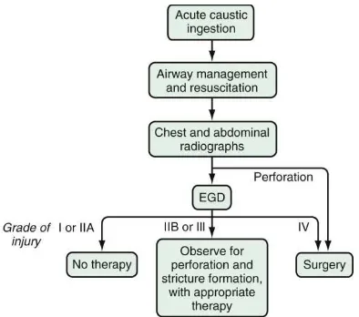
Figure 6 - A step wise approach to a patient with history of corrosive
[image:51.595.93.487.361.709.2]39
Strictures occur due to scar formation in the injured tissues through the
production of collagen. Strictures are a debilitating complication of
corrosive ingestions usually occurring over a period of weeks or months.
Several treatment strategies have been suggested to prevent stricture
formation and consequent esophageal obstruction. These include the use
of corticosteroids, antibiotics or the placement of esophageal stents or
nasogastric tubes.
Role for steroids:
The role of steroids in preventing stricture in patients with grade II b and
grade III continues to be a controversial issue. It has been hypothetised
that steroid therapy arrests the process of inflammatory repair and hence
can potentially prevent stricture formation. Corticosteroids affect the
ability of a wound (i.e., burn) to heal by decreasing the amount of collagen
in the tissue and inhibiting the inflammatory process initiated early after
the burn injury. Cellular collagen content is diminished through the
inhibition of enzymes affecting synthesis (i.e., prolyl hydroxylase) and
crosslinking (i.e., lysyl oxidase), as well as stimulation of degradative
activity through collagenase activation. Corticosteroids also exert their
effect on the inflammatory process by diminishing the amount of
40
formation taking place within the membranes of the cells. Based on these
mechanisms, their use in the prevention of stricture formation after acid
ingestion seems rational.
In studies evaluating the efficacy of steroid use for the prevention of
stricture, the results were not supportive for the use of steroids. They
noted an increase in the adverse effects of steroids such as increased
susceptibility to infection and increased risk of GI bleeding26. Most studies
find that first-degree burns of the esophagus do not progress to strictures
whether treated with corticosteroids or not, whereas third-degree burns
do progress to stricture formation regardless of corticosteroid therapy. In a
prospective, randomized, controlled trial conducted in children with history
of corrosive ingestion showed that corticosteroids were not effective in
decreasing the occurrence of strictures though the need for surgery (total
esophagectomy) was lesser in the group treated with steroids (four vs.
seven untreated patients).
Methyl prednisolone is the agent of choice at a dose of 40 to 60 mg/ day
intravenously given usually for duration of at least 3 weeks and then
tapered off over a period of 4 – 6 weeks. Ampicillin is usually added to
mitigate secondary inflammation due to bacterial invasion of injured
41
Role of antibiotics:
The use of antibiotics alone for preventing strictures has not been majorly
investigated. It is generally agreed to use an antibiotic (usually Ampicillin)
in patients who are treated with steroids. The prophylactic use of
antibiotic, in the absence of steroid therapy, is not advocated.
Esophageal stents and nasogastric tubes
Intraluminal silicone rubber stents or nasogastric tubes can be used for
maintaining the esophageal luminal patency. They can be retained for a
period of 3 weeks following which the esophageal lumen can be reassessed
for the presence of strictures. However esophageal stents are not without
potential disadvantages, which interfere with healing, in the form of
mechanical trauma at the site and increased reflux. Nasogastric tubes
likewise can be placed early in the course of treatment for ensuring the
patency of the lumen. But NG tubes also carry the risk of increasing fibrosis
and stricture formation by causing irritation and increasing inflammation of
the healing esophagus. Nevertheless an NG tube can be placed as it allows
42
Proton pump inhibitors (PPIs) and H2-blockers:
Parenteral PPIs and H2 blockers also have a role in limiting ongoing injury
to the injured GI mucosa. Hence they are being routinely recommended in
corrosive ingestion31.
Additionally, several xenobiotics have been studied in various animal
models which can prevent stricture formation by interfering with collagen
synthesis or enhancing its destruction. Some examples are β-Amino
propionitrile (BAPN), heparin, halofuginone, N-acetylcysteine (NAC),
vitamin E, epidermal growth factor (EGF), penicillamine, caffeic acid
phenethyl ester (CAPE) and colchicine. There have also been reports
advocating the effects of other agents like sucralfate and the use of TPN
(Total Parenteral Nutrition) in decreasing the incidence of stricture
formation but more studies are needed to delineate their beneficial action.
Early dilatation is associated with increased risk of perforation and is hence
discouraged. The presence of co existent GERD accelerates the formation
of stricture by worsening the corrosive insult to the esophagus and hence
periodic screening for GERD is recommended in patients with corrosive
43
Management of long term complications
Esophageal stricture can develop in as much as one third of corrosive
ingestion patients after initial recovery. The most common period for
stricture formation to present is at two months after injury but it can occur
over a wide period ranging from two weeks to as many as years after the
initial injury. Stricture formation is more common following more severe
(grade IIB or III) injuries. Commonly, the management of esophageal
strictures includes early endoscopic dilation using various types of
dilators32.
Maximal esophageal wall thickness, as determined by contrast CT is one
parameter which has several applications such as determining long-term
follow-up, type of nutritional support, predicting the response to dilatation
and the number of sessions required to achieve adequate dilation, in
identifying those patients in whom dilations should be done under
fluoroscopy to limit the risk of perforation and finally in assessing the
potential need for surgical repair as an alternative to dilations. Multiple
dilations may often be necessary.
The initial esophageal dilation is best delayed for at least 4 weeks
post-ingestion, to allow for the esophagus to heal, and allow remodelling, and
44
reducing the risk of perforation associated with dilatation. When
perforation occurs during dilatation, patients may complain of sudden
onset of shortness of breath or chest pain and show signs of subcutaneous
emphysema or pneumomediastinum. Diagnostic imaging may be needed
to identify the perforation and provide information for emergent surgical
repair if the diagnosis is unclear. Long-term endoscopic follow-up is
required in patients with stricture formation for assessing the occurence of
neoplastic changes of the esophagus that may occur with a delay of several
decades.
Antral and pyloric strictures may also occur after corrosive injury usually at
one to six weeks after ingestion, but can also occur years later with the risk
of being directly related to the degree of injury. Antral strictures can be
successfully managed in many patients with endoscopic dilation and acid
suppression. However surgical treatment in the form of antrectomy may
45
MATERIALS AND METHODS
SUBJECTS
The study was conducted on 50 cases admitted at the toxicology ward in
Rajiv Gandhi Government General Hospital, Chennai, with alleged history
of consumption of corrosive ingestion.
PERIOD OF STUDY
6 months
DESIGN OF STUDY
Cross-sectional study
CONSENT
Informed consent from all the patients
METHOD OF COLLECTION OF DATA
Inclusion criteria
Patients age > 12 yrs
Patients with history of corrosive ingestion presenting within 24
hours of ingestion
46
Exclusion criteria
Patients presenting after 24 hours of corrosive ingestion
Patients with respiratory distress
Patients with suspected perforation either radiologically or
endoscopically (grade III b injury)
Patients with normal findings in Upper GI endoscopy (no evidence of
initial injury)
METHODOLOGY
All patients who were admitted with history of corrosive ingestion
underwent thorough history taking and detailed clinical examination after
initial stabilisation of Airway, Breathing and Circulation. The parameters
taken into consideration were history regarding amount consumed, type of
corrosive, and duration since consumption, symptomatology, physical
signs, upper GI endoscopy findings and they were correlated with
outcome. Laboratory investigations including Complete blood counts, renal
and liver function tests were done in all patients. Chest and Abdomen X
rays were taken to rule out perforation. Patients were kept Nil per oral and
subjected to Upper GI endoscopy within 24 hours of admission. The
findings were noted and patients were managed accordingly (oral feeds
47
placement for grade II b – III a injuries and feeding jejunostomy for
duodenal injuries).
The patients were serially followed and were subjected for a re-look upper
GI endoscopy after 6 weeks and the findings were compared and the
outcome was graded into 2 categories.
Category I – normal endoscopy study
Category II – stricture esophagus or stricture antri or pylori.
INVESTIGATION DETAILS
Complete history and thorough physical examination in all patients with
history of corrosive ingestion and routine blood investigations, chest and
abdomen X rays and Upper GI endoscopy.
DATA COLLECTION AND METHODS
Collection of data as per proforma with consent from patients with history
of corrosive ingestion in Toxicology ward, Rajiv Gandhi Govt. General
hospital.
ANALYSIS
Data analysed using statistical package-SPSS software
Conflict of interest: Nil.
48
Table 6
GRADING OF INJURY AS PER ZARGAR GRADING SYSTEM:
Grade 0 Normal endoscopy findings
Grade I Mucosal edema and hyperaemia
Grade II a Friability, blisters, hemorrhages, erosions, whitish
membranes, exudates, superficial ulcerations
Grade II b Grade 2a plus deep discrete or circumferential ulceration
Grade III a Small scattered areas of multiple ulcerations and areas of
necrosis (brown-black or grayish
discoloration)
49
STATISTICAL ANALYSIS
Statistical analysis was carried out for 50 patients with history of corrosive
ingestion after categorizing each variable – age sex, type of corrosive
consumed, duration since consumption, amount consumed, circumstance
of consumption, symptomatology, presence of physical signs and upper GI
endoscopy findings. Datas were analysed using Statistical package- SPSS
software version 11.5.The values are presented as mean, standard error of
mean, standard deviation, standard error of mean and median.
Percentages were used to describe the proportions of discrete variables.
The significance of difference between the proportions was indicated by
the Chi square (x2) statistic. The significance of difference in mean
between the groups was calculated by Fisher exact test. Variables were
50
OBSERVATIONS AND RESULTS
50 cases with history of corrosive ingestion and with positive findings on
endoscopy formed the study group. In these patients age wise distribution,
sex wise distribution, circumstances of poisoning (suicidal/accidental),
agent of exposure (acid/alkali), symptomatology, physical findings and
endoscopy findings were analysed.
The upper GI endoscopy findings were compared with the final outcome.
The agent exposed to and the circumstances of poisoning were compared
with the final outcome. Other independent variables were entered into the
51
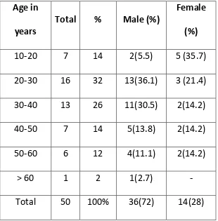
[image:66.595.155.465.203.518.2]AGE AND SEX DISTRIBUTION
TABLE 7:
Age in
years
Total % Male (%)
Female
(%)
10-20 7 14 2(5.5) 5 (35.7)
20-30 16 32 13(36.1) 3 (21.4)
30-40 13 26 11(30.5) 2(14.2)
40-50 7 14 5(13.8) 2(14.2)
50-60 6 12 4(11.1) 2(14.2)
> 60 1 2 1(2.7) -
Total 50 100% 36(72) 14(28)
The mean age among the patients was 32.88 ±12.74. The youngest age was
15 years and the oldest was 67 years. There was no significant difference in
TABLE 8
Age in years
10 20 30 40 50 > 60 Figure 7 6 12 1 4 0 2 4 6 8 10 12 14 16 18
10 - 20 years 20 - 30 years
[image:67.595.91.509.487.729.2]52
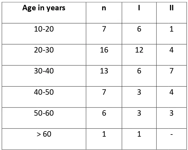
TABLE 8: Age wise analysis of outcome
Age in years n I
10-20 7 6
20-30 16 12
30-40 13 6
40-50 7 3
50-60 6 3
> 60 1 1
7 – Graph showing age wise distribution
12 6 3 3 4 7 4 3
30 years 30 - 40 years 40 - 50 years 50 - 60 years
Normal Stricture II 1 4 7 4 3 -
showing age wise distribution
1
Sex Male Female Figure 8 0 5 10 15 20 25 30 35 40 Male 53
[image:68.595.96.496.369.629.2]SEX WISE ANALYSIS
Table 9
Sex distribution with outcome
n I (%) II (%)
36 24 (66.7%) 12 (33.3%)
14 7 (50%) 7 (50%)
Figure 8: Graph showing sex wise distribution
Male Female 24 7 12 7 I II II (%) 12 (33.3%) 7 (50%)
Type of corrosive Acid Alkali Figure 0 2 4 6 8 10 12 14 16 18 20 acid 19 54
CORROSIVE WISE ANALYSIS
Table 10
n (%) I
32 (64%) 19
18 (36%) 12
Figure 9 – type of corrosive versus outcome
acid alkali 12 13 6 normal stricture II 13 6
55
CIRCUMSTANCES OF CONSUMPTION
Table 11 – Frequencies of the circumstances of consumption
Circumstance n (%) I II
Suicidal 38 (76) 21 (55.2%) 17 (44.8%)
Accidental 8 (16) 8(100%) 0
Accidental (under
alcohol influence)
4 (8) 2 (50%) 2 (50%)
In our study though majority of strictures occurred in the suicidal
comsumption group, the difference was not statistically significant (p value
56
Figure 10 – Circumstance of poisoning versus outcome.
55.20%
100%
50% 44.80%
0
50%
0% 10% 20% 30% 40% 50% 60% 70% 80% 90% 100%
Suicidal Accidental Accidental under the influence of alcohol
57
DURATION SINCE CONSUMPTION
Figure 11 – distribution of the duration since consumption when the
patient was subjected to upper GI endoscopy.
The mean duration for performing the endoscopy was 14.06 ± 3.48 hours.
The least duration was 8 hours and the maximum duration was 20 hours.
The incidence of strictures was higher in the patients who underwent
endoscopy later than 12 hours but the difference was not statistically
significant (p value > 0.05). 0 1 2 3 4 5 6 7 8 9
8 9 10 12 13 14 15 16 17 18 19 20
n o . o f p a ti e n ts
[image:72.595.95.530.227.429.2]Table12 – Analysis
Duration since
consumption
≤ 12 hours
> 12 hours
Figure 12 – Analysis
0% 10% 20% 30% 40% 50% 60% 70% 80% 90% 100%
≤ 12 hours
58
Analysis of duration since consumption versus outcome
Frequency I
20 14
30 17
Analysis of duration since consumption versus outcome
≤ 12 hours > 12 hours
14
17 6
13
Normal Stricture
of duration since consumption versus outcome
II
6
13