MINI-FOCUS ISSUE: EXERCISE PERFORMANCE AND CARDIAC REHABILITATION
Rate-Response Programming Tailored
to the Force-Frequency Relationship
Improves Exercise Tolerance in
Chronic Heart Failure
John Gierula, PHD,aMaria F. Paton, MSC,aJudith E. Lowry, MSC,aHaqeel A. Jamil, PHD,aRowenna Byrom, RN,a
Michael Drozd, MB,aJack O. Garnham, MSC,aRichard M. Cubbon, PHD,aDavid A. Cairns, PHD,bMark T. Kearney, MD,a Klaus K. Witte, MDa
ABSTRACT
OBJECTIVESThis study sought to examine whether the heart rate (HR) at which the force-frequency relationship (FFR) slope peaks (critical HR) could be used to tailor HR response in chronic heart failure (CHF) patients with cardiac pacemakers and whether this favorably influences exercise capacity.
BACKGROUNDCHF secondary to left ventricular (LV) systolic dysfunction is characterized by blunting of the positive relationship between HR and LV contractility known as the FFR.
METHODSThis observational study was carried out in patients with CHF and healthy subjects with pacemaker devices. The study assessed the 3 important features of the FFR (critical HR, peak contractility, and the FFR slope), and their reproducibility was measured noninvasively using echocardiography. The investigators then undertook a double-blind, randomized, controlled crossover study comparing the effects of tailored pacemaker rate-response programming on the basis of the FFR with conventional rate-response programming on exercise time and maximal oxygen consumption.
RESULTSThe study enrolled 90 patients with CHF into the observational cohort study: mean age, 73.68.9 years; mean left ventricular ejection fraction (LVEF), 33.510.9%. The study investigated 15 control subjects with normal LV function (LVEF, 55.65.3%). The critical HR (10322 beats/min vs. 12615 beats/min; p¼0.0002), peak contractility (3.83.7 SBP/LVESVI vs. 9.84.1 SBP/LVESVI; p¼0.0001), and the slope of the FFR (p<1015) were lower in patients with CHF than in control subjects. A total of 52 patients, with a mean LVEF of 3211% on optimal therapy, took part in the crossover study. Rate-response settings limiting HR rise to below the critical HR led to greater exercise time (475189 s vs. 425196 s; p¼0.003) and higher peak oxygen consumption (17.34.6 ml/kg/min vs. 16.64.7 ml/kg/min; p¼0.01).
CONCLUSIONSA personalized approach to rate-response programming, determined using a reproducible noninvasive method for assessing the FFR, improves exercise time in patients with CHF and pacemaker devices. (Bowditch Revisited: Defining the Optimum Heart Rate Range in Chronic Heart Failure;NCT02563873) (J Am Coll Cardiol HF 2018;6:105–13) © 2018 The Authors. Published by Elsevier on behalf of the American College of Cardiology Foundation. This is an open access article under the CC BY-NC-ND license (http://creativecommons.org/licenses/by-nc-nd/4.0/).
ISSN 2213-1779 https://doi.org/10.1016/j.jchf.2017.09.018
C
haracterized by reduced exercise tolerance, chronic heart failure (CHF) secondary to left ventricular (LV) systolic dysfunction (LVSD) has a dele-terious impact on quality of life in millions of individuals worldwide (1). Advances in our understanding of the pathophysiology of CHF have informed the development of drugs and devices, including cardiac resynchronization therapy (CRT), that have substantially improved life expectancy (2–5). Nonetheless, many patients with CHF and CRT remain significantly limited(6).An important determinant of exercise performance is thought to be LV contrac-tility, which in 1871 was shown to be posi-tively coupled to increments in heart rate (HF)(7). As HR increases in healthy humans, LV contractility and stroke volume increase simultaneously (8). Using invasive measurements, we and other investigators have shown that this critical physiological response, described as the force-frequency relationship (FFR), becomes flattened in patients with CHF, with a decline in LV contractility occurring above a certain HR (9,10). We hypothesized that by defining an in-dividual’s FFR noninvasively (and in particular the peak of this response before the decline in contrac-tility characteristic in CHF), it may be possible to tailor individual pacing algorithms to exploit a pa-tient’s unique physiology. Here we show that it is possible to use a noninvasive, reproducible approach to define the peak of the force-frequency curve of patients with CHF with CRT devices in situ and use this information to improve exercise capacity.
METHODS
This paper describes thefindings of 1 observational and 1 interventional study.
OBSERVATIONAL STUDY.S u b j e c t s .Patients with CHF secondary to LVSD (LV ejection fraction#50%) with persistent symptoms on exertion and a CRT device, implantable cardioverter-defibrillator, or standard pacemaker for at least 3 months were
recruited (Leeds NIHR Cardiovascular Clinical Research Facility, Leeds Teaching Hospitals NHS trust, Leeds, United Kingdom). Patients also had to be taking optimally tolerated medical therapy with no change in medication or other invasive cardiac pro-cedures for at least 3 months. We also enrolled an unselected consecutive group of patients with a standard pacemaker, normal atrioventricular con-duction, no evidence of heart failure, and normal echocardiographicfindings as a control group.
Information was collected on comorbidities, past medical history, medication, pacemaker settings, and New York Heart Association functional class. All pa-tients gave written informed consent, and the study was approved by the local ethics committee and registered on clinicaltrials.gov (Bowditch Revisited: Defining the Optimum Heart Rate Range in Chronic Heart Failure;NCT02563873).
Echocardiographic techniques.Full baseline echocardi-ography was carried out with gray-scale and tissue Doppler images recorded in 2- and 4-chamber views by using harmonics to improve border definition if necessary (GE Vivid E95, GE Healthcare, Milwaukee, Wisconsin). Further images were recorded at each 15-beat frequency increase during the incremental pac-ing protocol. Images were stored in the EchoPAC digital imaging system (GE Healthcare) and analyzed off-line. This analysis included a calculation of LV end-diastolic volume and LV end-systolic volume (LVESV) that used the biplane disks (modified Simp-son) method by tracing the endocardial border, excluding the papillary muscles(11). An average of 3 measurements was used in the final analysis. The frame at the R-wave was taken as end-diastole, and the frame with the smallest LV cavity was considered to represent end-systole. The LVESV index (LVESVI) was calculated at each stage as LVESV/body surface area, where body surface area was calculated using the Mosteller equation(12).
B l o o d p r e s s u r e m e a s u r e m e n t .Calculation of the end-systolic pressure-volume relationship requires measurement of the LV pressure at end-systole (13,14). Systolic blood pressure (SBP) measured with a manual blood pressure cuff was used as a surrogate for end-systolic LV pressure. Blood pressure re-cordings were made using a sphygmomanometer and SEE PAGE 114
A B B R E V I A T I O N S A N D A C R O N Y M S
CHF= chronic heart failure
CRT= cardiac resynchronization therapy
FFR= force-frequency relationship
HR= heart rate
LV= left ventricular
LVESV= left ventricular end-systolic volume
LVESVI= left ventricular end-systolic volume index
LVSD= left ventricular systolic dysfunction
SBP= systolic blood pressure
received a research grant from Medtronic. Dr. Paton has received an NIHR-Integrated Clinical Academic fellowship award. Dr. Lowry is funded by a Leeds Charitable Foundation fellowship. Dr. Cubbon has received a British Heart Foundation intermediate fellowship. Dr. Kearney has received research grants from Medtronic; and is also a British Heart Foundation Professor of Cardi-ology. Dr. Witte has received an NIHR Clinician Scientist Award. All other authors have reported that they have no relationships relevant to the contents of this paper to disclose.
a standard stethoscope coinciding with echocardio-graphic images at each HR stage. SBP was recorded at the point where the first tapping sound (phase 1 Korotkoff) occurred for 2 consecutive beats(15). P a c i n g p r o t o c o l .Echocardiographic images were collected at rest, after which atrial pacing was initi-ated in the DDD-mode (or VVI in patients with atrial
fibrillation) for CRT-treated patients and in the AAI-mode (or DDD with long atrioventricular delays to avoid right ventricular pacing, or VVI for patients in atrial fibrillation) for subjects without CRT, at the lowest multiple of 10 above baseline. After 4 min, a further set of echocardiographic images was recor-ded, and subsequently the pacing rate increased in stepwise 15-beat intervals with images recorded after every 4 min. This was repeated until the maximum predicted HR predicted by Åstrand (220 age) was reached. At this point peak data were collected, and pacing was returned to baseline settings. For safety, subjects were asked to remain in the research facility for a further 30 min.
F o r c e - f r e q u e n c y c a l c u l a t i o n .Dividing the SBP by the LVESVI (SBP/LVESVI) gives a surrogate of contractility (16–20), which has been validated against invasive methods(15,19). In our protocol, we repeated these measures at a series of HR stages (induced by pacing) to allow us to plot the FFR. The slope of this relationship was then calculated as the ratio between SBP/LVESVI change from baseline and HR increase from baseline. We defined the HR at which, in a biphasic pattern, the SBP/LVESVI reached maximum value or that beyond the SBP/LVESVI declined by 5% as the“critical HR.”In a negative FFR (in cases where there was no increase in contractility with increments in HR), baseline HR was deemed the critical HR(21).
R e p r o d u c i b i l i t y .Each image set was anonymized and reported by a second echocardiographer for interoperator reproducibility. For intraoperator reproducibility, each image set was also reported a second time by the initial reporter. For each dataset, we documented the HR at which peak contractility was reached (the critical HR), peak contractility itself, and the slope of the FFR as described earlier.
RANDOMIZED CONTROLLED TRIAL. Patients with
CHF who were included in the observational phase of the study were subsequently invited to participate in a double-blind, randomized, crossover treadmill-based study comparing exercise time under conventional rate-response settings with rate-response settings taking into account the data from the FFR assessment. An unselected subgroup was invited to attend a third (blinded) exercise test withfixed-rate pacing.
E x c l u s i o n c r i t e r i a .For this part of the investiga-tion, we invited patients who had participated in the
first study and who did not have peripheral vascular disease or noncardiac conditions such as significant airway disease and musculoskeletal abnormalities that could restrict walking on a treadmill.
L a b o r a t o r y a r r a n g e m e n t a n d e x e r c i s e p r o t o c o l . Subjects recruited to this randomized crossover study were present on 2 (or 3) consecutive occasions at the same time of day 1 week apart. Before each test, the pacemaker was interrogated, and patients were then randomly assigned to the following: rate-adaptive pacing with conventional age-determined settings (22); optimized settings on the basis of the results of their FFR assessment; specifically limiting the rate-response algorithm to the critical HR; or, for pa-tients agreeing to do a third test, fixed-rate pacing with rate-response settings to“off.”
Subjects were exercised using the ramping tread-mill protocol (23). Expired air was collected, and metabolic gas exchange analysis was performed (Ultimo CardO2, Medical Graphics, St. Paul, Minne-sota) throughout the test. HR (beats/min), oxygen uptake (VO2) (ml/kg/min), and carbon dioxide output
(VCO2) (ml/kg/min) were recorded as 15-s averages.
Anaerobic threshold was calculated using the V-slope method.
The cardiopulmonary exercise test equipment was recalibrated using manufacturer-recommended vol-ume and gas calibration techniques before each test. All test subjects were encouraged to exercise to exhaustion, and no further motivation or instructions were given. The arrangement of the laboratory to ensure double blinding has been described previously (24). To maintain blinding, the continuous 12-lead electrocardiogram monitor was obscured throughout the test (and recovery phase) from subjects and the supervising physician. Only the unblinded cardiac physiologist was aware of the programming mode or testing arm. The unblinded cardiac physiologist monitored the electrocardiogram throughout the study, communicated only with the other team members unless there were safety concerns, and reprogrammed the pacemaker to its original settings at the end of every visit.
with a secondary endpoint of peak oxygen con-sumption. On the basis of guidelines for pilot studies (26,27), and accounting for a dropout rate of 20%, we aimed to recruit 28 patients to achieve 20 participants with complete data.
S t a t i s t i c a l a n a l y s i s .Data were analyzed using the Statistical Package for the Social Sciences SPSS version 21 (IBM Corp., Armonk, New York), R: A Language and Environment for Statistical Computing version 3.2.3 (R Development Core Team, Vienna, Austria), and SAS version 9.4 (SAS Institute, Inc., Cary, North Carolina).
Normality for all continuous variables was tested using the Shapiro-Wilk test. Normally distributed continuous variables were reported as mean and meanSD, and non-normally distributed continuous variables were reported as median (interquartile range). Subsequently, associations between groups or interventions and baseline characteristics were assessed using either analysis of variance and the 2-sample Student t test for normally distributed values or the Kruskal-WallisHtest (1-way analysis of variance of ranks) for non-normally distributed data. Similar associations with categorical variables were analyzed using the chi-squared test for contingency tables.
Once a familiarization test has been performed, a peak exercise test is not a training stimulus. We pre-viously performed up to 5 exercise tests in consecu-tive weeks in patients with CHF and controls, with no longitudinal effects(28). However, to account for any carryover effects, the interventional crossover study was analyzed using a linear mixed model with a random effect for subject. For each endpoint Yak(e.g.,
exercise time) under consideration in the study:
Yak ¼
m
þdiþp
jþinþa
kþinkwhere inkwN (0,
s
2ε),a
kwN (0,s
2a) andm
is the overall mean,s
is the treatment effect,p
is the period effect, andl
is the carryover effect (which is mathe-matically identical to an interaction term between treatment and period). This model was estimated using PROC MIXED in SAS, and least squares means were estimated for each of these terms and their differences.All statistical tests were 2-sided, and any p value<0.05 was called statistically significant.
Ethical approval for both phases of the investiga-tion was granted by the Health Research Authority (National Research Ethics Service Centre: Yorkshire and the Humber REC: 12/YH/0097). Written informed consent was obtained from all participants. The crossover study was registered on clinicaltrials.gov (NCT02563873) before any patient enrollment.
RESULTS
OBSERVATIONAL STUDY. We enrolled 90 patients
with CHF and 15 control subjects with normal LV TABLE 1 Subject Demographics at Baseline Visit
Non-HF (n¼15)
HF Patients
(n¼90) p Value*
Male 14 (93) 79 (88) 0.53
Age, yrs 71.116.0 73.58.9 0.39
Ischemic heart disease 2 (13) 54 (60) 0.0008
Diabetes mellitus 4 (27) 28 (31) 0.73
BSA, m2 1.90.1 2.00.2 0.07
NYHA functional class 0.0001
I 15 (100) 1 (1)
II n/a 70 (78)
III n/a 19 (21)
Beta-blockers 3 (20) 82 (91) 0.0001
ACE inhibitor/ARB 3 (20) 77 (86) 0.0001
Furosemide dose, mg/day 0 47 (26)
Digoxin 0 (0) 15 (17) 0.084
AA 0 (0) 38 (42) 0.001
Device (PPM/ICD/CRT) n/a 14/9/67 (16/10/74)
Atrialfibrillation 3 (20) 25 (28) 0.51
LVEF, % 55.65.3 33.310.8 0.0001
Critical heart rate, beats/min 12615 10322 0.0002
Peak contractility, SBP/LVESVI 9.84.1 3.83.7 0.0001 Force-frequency relationship† 0.0540.042 0.0110.028 <0.0001
Values are n (%) or meanSD. *p values are from unpaired Studentttests or chi-squared tests as appropriate. †From likelihood ratio test in linear mixed model.
AA¼aldosterone antagonist; ACE¼angiotensin-converting enzyme; ARB¼aldosterone receptor blocker; BSA¼body surface area; CRT¼cardiac resynchronization therapy; HF¼heart failure; ICD¼implantable cardioverter-defibrillator; LVEF¼left ventricular ejection fraction; LVESVI¼left ventricular systolic volume index; n/a¼not applicable; NYHA¼New York Heart Association functional class; PPM¼permanent pacemaker; SBP¼systolic blood pressure.
FIGURE 1 Force-Frequency Relationship in Patients and Controls
function into thefirst phase of the study. Baseline clinical, echocardiographic, and pacemaker variables are shown inTable 1. We were able to establish the 3 key variables of peak contractility, critical HR, and the slope of the relationship between HR and contractility in all patients.
Patients with CHF had lower mean peak contrac-tility, lower mean critical HR, and a lower slope of the relationship between HR and contractility below the critical HR (the FFR) than control subjects (Table 1, Figure 1).
Separate models were required for each group, as compared with considering the data overall. This was confirmed by a likelihood ratio test for a saturated model compared with a simple additive model for HR, HR2, and patient group (chi-square test ¼ 214.63;
p < 1015). Further examining the relationship in linear mixed effects models showed that there was little evidence of a quadratic relationship in each group of patients:
Controls: 0.2915604þ0.0844503HR 0.00017HR2
CHF: 1.971þ7.170e03þ2.510e05HR2
In the controls, the linear term was significant (p < 0.05), and the quadratic term showed weak evidence of being required (p¼0.15). However, for CHF the linear term was not significant (p¼0.334), and the quadratic term was not significant (p¼0.525). Considering the linear terms, in controls, for every 10 beats/min increase in HR there was a 0.8-unit increase in contractility. For the patients with CHF, this relationship was significantly less, at<0.02.
The Strand formula calculated a higher peak HR than the calculated critical HR for all except 1 CHF patient (mean HR 1469.0 vs. 10322; p<0.0001), and all but 3 control subjects (14916 vs. 12615; p¼0.002). The inaccuracy of the Strand formula in beats per minute was greater in patients than in control subjects (4324 vs. 23 24; p¼0.0039). Age was unrelated to any of the measures of cardiac function including contractility, and there was no relationship between critical HR and resting LV function in either group. However, in patients with CHF, there was a strong correlation between peak contractility and baseline ejection fraction (0.50; 95% confidence in-terval: 0.33 to 0.64; p<0.0001) (Figure 2).
R e p r o d u c i b i l i t y .The linear mixed effects models allowed direct assessment of the reproducibility of the reading of the echocardiograms by different operators through components of variance. The per-centage of variation in the models for controls and CHF attributed to the contractility measurement by
different operator or repeated assessment by the same operators was<1% of variance.
INTERVENTIONAL STUDY. A total of 52 patients were enrolled in this study (Table 2). Baseline clinical and echocardiographic variables of this subgroup were not different from those of patients enrolled in the
FIGURE 2 Peak Contractility by Levels of Left Ventricular Dysfunction
[image:5.576.260.467.467.714.2]The greater impairment of peak contractility (as described by the ratio of SP to LVESVI) in response to heart rate rise in patients with more severe left ventricular systolic dysfunction (1-way analysis of variance p<0.0001). Abbreviations as inFigure 1.
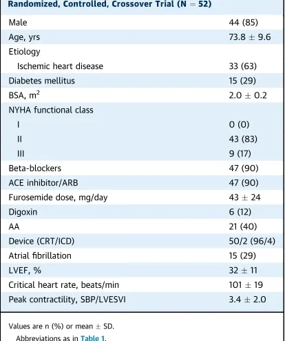
TABLE 2 Baseline Variables of Patients Enrolled in the Randomized, Controlled, Crossover Trial (N¼52)
Male 44 (85)
Age, yrs 73.89.6
Etiology
Ischemic heart disease 33 (63)
Diabetes mellitus 15 (29)
BSA, m2 2.00.2
NYHA functional class
I 0 (0)
II 43 (83)
III 9 (17)
Beta-blockers 47 (90)
ACE inhibitor/ARB 47 (90)
Furosemide dose, mg/day 4324
Digoxin 6 (12)
AA 21 (40)
Device (CRT/ICD) 50/2 (96/4) Atrialfibrillation 15 (29)
LVEF, % 3211
Critical heart rate, beats/min 10119
Peak contractility, SBP/LVESVI 3.42.0
TABLE 3 Exercise Variables in 52 Patients With Heart Failure During Conventional and Optimized Heart Rate Programming
Programming Mean
95% Confidence Interval
Mean Difference
95% Confidence
Interval p Value
Exercise time, s Tailored 474.74 (420.69 to 528.79) 49.85 (18.41 to 81.29) 0.0025 Not tailored 424.89 (370.84 to 478.94)
Peak VO2, ml/kg/min Tailored 17.31 (16.00 to 18.62) 0.75 (0.16 to 1.34) 0.0134
Not tailored 16.56 (15.25 to 17.87)
O2pulse Tailored 13.39 (12.33 to 14.45) 3.21 (2.33 to 4.08) <0.0001
Not tailored 10.19 (9.13 to 11.25)
VEVCO2slope Tailored 31.80 (29.81 to 33.78) 1.89 (3.38 to0.40) 0.0139 Not tailored 33.69 (31.70 to 35.67)
Peak RER Tailored 1.01 (0.99 to 1.04) 0.00 (0.03 to 0.02) 0.7456
Not tailored 1.02 (0.99 to 1.05)
Peak heart rate, beats/min Tailored 109.11 (106.01 to 112.21) 28.88 (32.83 to24.93) <0.0001
Not tailored 137.99 (108.87 to 167.12)
O2¼oxygen; RER¼respiratory exchange ratio; VE/VCO2slope¼relationship between ventilation and carbon dioxide output; VO2¼oxygen consumption.
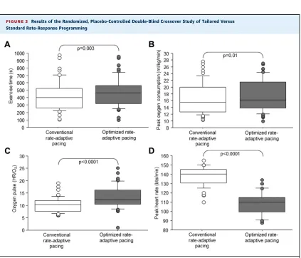
FIGURE 3 Results of the Randomized, Placebo-Controlled Double-Blind Crossover Study of Tailored Versus Standard Rate-Response Programming
[image:6.576.113.536.316.678.2]observational study. Of these 52 patients, 12 under-went a third test withfixed-rate pacing.
Table 3 andOnline Table 1show the exercise vari-ables for conventional and optimized HR rise tests. Optimized settings were associated with improved exercise time, peak oxygen consumption, oxygen pulse, and lower VET/VCO2 slope, whereas HR was
lower and respiratory exchange ratio was the same despite a small period effect (Figures 3A to 3D).
Fixed-rate pacing in 12 unselected patients led to similar exercise times as Strand-guided conventional rate-response pacing (Online Figures 1A to 1C). Furthermore, there was no heterogeneity in the benefits of tailored programming between patients with and without diabetes mellitus, those with and without atrialfibrillation, and those with and without ischemic heart disease (Online Tables 2 to 4).
DISCUSSION
Our study demonstrates that patients with CHF have an impaired force-frequency curve compared with controls that can easily be assessed using a nonin-vasive, reproducible echocardiographic method and that peak contractility is related to the baseline cardiac function. We have also shown that using the data from the force-frequency curve in patients with CHF allows us to tailor pacing algorithms tar-geting the critical HR and leads to a significant improvement in exercise time and peak oxygen consumption.
Rate-adaptive cardiac pacing, whereby HR is increased in response to movement or ventilation detected by internal device sensors, was developed as an attempt to treat exercise intolerance thought to result from chronotropic incompetence(29,30). The settings are broadly based on a series of experiments to describe maximal HR and age in healthy adults (31–33). Although there are numerous published datasets describing maximal HR changes with aging (34–37), the “Strand formula” (220 age), which in itself is an extrapolation of data from various sour-ces, remains the most frequently used. However, all datasets are taken from healthy individuals exer-cising to physiological maximum. In patients without CHF who are receiving standard pacemakers for bradycardia, rate-adaptive programming on the basis of a simple, age-related algorithm is associated with an increase in cardiac output during exercise (38), and better quality of life (39–41), but inconsistent improvements in exercise capacity(42,43), compared withfixed-rate pacing.
We have demonstrated that chronotropic incom-petence as determined by the standard equation
seems not to be a limiting factor in maximal perfor-mance in patients with CHF(17,44,45), because con-ventionalrate-adaptive pacing in patients with CHF does not improve exercise capacity compared with
fixed-rate pacing(17). Furthermore, we and other in-vestigators have shown that rate-adaptive pacing may worsen prognosis and cardiac function(46,47).
The reason for the failure of increments in HR to increase exercise capacity in patients with CHF may be that conventional rate-adaptive pacing algorithms do not take into account the altered cardiac contrac-tile function in CHF.
Increased contractility during exercise is thought to be important to maintain stroke volume in the presence of impaired filling during higher HRs. The FFR is abnormal in CHF such that contractility does not rise normally with increases in HR. In agreement with this, in our cohort of unselected, optimally managed patients with CHF secondary to LVSD, we demonstrated a consistent and reproducible impair-ment of the FFR with a lower critical HR, lower peak contractility, and a lower FFR slope than in subjects without CHF.
We hypothesized that noninvasive assessment of the FFR could provide a physiologically relevant descriptor of the optimal HR range and peak HR that could be used to optimize rate-response programming patients with CHF. To examine this we performed a randomized, double-blind, placebo-controlled cross-over study of optimized rate-response programming versus conventional rate-response programming in patients with CHF to examine this concept. Our study showed that this precision-approach rate-adaptive programming can acutely improve treadmill exercise time and peak oxygen consumption. This approach represents a paradigm in rate-responsive pacing for patients with CHF in which the pace-maker rate-response settings are personalized to work in synergy with and take advantage of intrinsic car-diac physiology.
STUDY LIMITATIONS. Our observational study has
biases that are common in studies of this type. There is a degree of patient selection in that those patients who are too unwell with advanced CHF or who have other comorbidities may be less keen to participate in clinical research.
contractile response to increased HRs than patients without a pacemaker device.
We cannot exclude the possibility of systematic differences in the level of motivation or encourage-ment from the technicians running the tests at different time points. However, we believe that the randomization and double-blind study design will have removed any significant bias. Our blinding pro-cedure has worked well in previous reports (24). Although we defined a pre-specified significance level for rejecting hypotheses for the primary endpoint, no multiple testing control was applied to the secondary endpoints. Therefore all results determined on the basis of secondary endpoints should be considered as hypothesis generating.
We used the ramping treadmill protocol to be consistent for all patients, to allow us to compare exercise times rather than just metabolic gas analysis data, and because treadmill-based activity is associ-ated with greater upper body movement required for activation of the rate-response algorithms in pace-makers. We acknowledge that this exercise modality and protocol may not have been ideal for all of our patients, but on balance we doubt that the protocol choice materially altered our results. The early, low-workload stage allowed even those patients with the greatest limitation in exercise capacity to complete at least thefirst stage, thereby reducing the bias toward less limited patients.
Our FFR data were collected with patients in the supine position, and the exercise testing on the treadmill was upright. This positioning has the po-tential to alter loading conditions, which could have an effect on the critical HR. However, upright echo-cardiography has an adverse effect on image quality and patient comfort(48,49). Furthermore, the Bow-ditch phenomenon as originally described and its mechanism are purported to be loading independent. Finally, our data were collected immediately following pacemaker reprogramming during 2 (3) visits to the NIHR Leeds Cardiovascular Clinical Research Facility at Leeds Teaching Hospitals NHS Trust, and pacing was reset to nominal values for each patient at the end of the visit. Whether
longer-term rate-response optimization tailored for the FFR data is safe and has beneficial effects on ex-ercise capacity remains to be proven.
CONCLUSIONS
We have demonstrated that a reproducible, nonin-vasive assessment of the FFR is possible in patients with a pacemaker and that using these data to personalize the rate-response settings of the pace-maker can improve exercise time in patients with CHF and LVSD.
ACKNOWLEDGMENT The authors acknowledge the
consistent administrative support provided by Mrs. Andrea Marchant.
ADDRESS FOR CORRESPONDENCE: Dr. Klaus K.
Witte, Leeds Institute of Cardiovascular and Meta-bolic Medicine, LIGHT Building, University of Leeds, Clarendon Way, Leeds LS2 9JT, United Kingdom. E-mail:[email protected].
R E F E R E N C E S
1.Hobbs FD, Kenkre JE, Roalfe AK, Davis RC, Hare R, Davies MK. Impact of heart failure and left ventricular systolic dysfunction on quality of life: a cross-sectional study comparing common chronic cardiac and medical disorders and a representative adult population. Eur Heart J 2002;23:1867–76.
2.Cubbon RM, Gale CP, Kearney LC, et al.
Changing characteristics and mode of death
associated with chronic heart failure caused by left ventricular systolic dysfunction: a study across therapeutic eras. Circ Heart Fail 2011;4:396–403.
3.Cleland JG, Daubert JC, Erdmann E, et al., Cardiac Resynchronization-Heart Failure (CARE-HF) Study Investigators. The effect of cardiac resynchronization on morbidity and mortality in heart failure. N Engl J Med 2005;352:1539–49.
4.Moss AJ, Hall WJ, Cannom DS, et al.,
MADIT-CRT Trial Investigators.
Cardiac-resynchronization therapy for the prevention of heart-failure events. N Engl J Med 2009;361: 1329–38.
5.Cubbon RM, Witte KK. Cardiac resynchronisa-tion therapy for chronic heart failure and con-duction delay. BMJ 2009;338:b1265. PERSPECTIVES
COMPETENCY IN MEDICAL KNOWLEDGE: Noninvasive assessment of contractility is
reproducible and identifies a physiological HR range target to which to program a pacemaker. This precision programming improves exercise time in patients with CHF. Optimizing pacemaker settings on the basis of a simple and reproducible noninvasive assessment of the FFR in patients with heart failure can improve exercise capacity.
6.Bowen TS, Cannon DT, Begg G, Baliga V, Witte KK, Rossiter HB. A novel cardiopulmonary exercise test protocol and criterion to determine maximal oxygen uptake in chronic heart failure. J Appl Physiol (1985) 2012;113:451–8.
7.Bowditch HP. Uber die Eigenthϋmlichkeiten der Reizbarkeit, welche die Muskelfasern des Herzens zeigen. Ber Sachs Ges (Akad) Wiss 1871;23: 652–89.
8.Mattera GG, Vanoli E, Martinez V, Luciani M, Falco T, Borsini F. Adrenergic effects on force-frequency relationship: a pivotal role for the car-diac intrinsic systems. Acta Physiol (Oxf) 2011;202: 141–9.
9.Kayhan N, Bodem JP, Vahl CF, Hagl S.
The positive staircase (force-frequency
rela-tionship) and the Frank-Starling mechanism
are altered in atrial myocardium of patients in end-stage heart failure transplanted for
dilative cardiomyopathy. Transplant Proc
2002;34:2185–90.
10.Cotton JM, Kearney MT, MacCarthy PA, et al. Effects of nitric oxide synthase inhibition on basal function and the force-frequency relationship in the normal and failing human heart in vivo. Cir-culation 2001;104:2318–23.
11.Lang RM, Badano LP, Mor-Avi V, et al. Rec-ommendations for cardiac chamber quantification by echocardiography in adults: an update from the American Society of Echocardiography and the European Association of Cardiovascular Imaging. J Am Soc Echocardiogr 2015;28:1–39.
12.Mosteller RD. Simplified calculation of body surface area. N Engl J Med 1987;317:1098.
13.Grossman W, Braunwald E, Mann T,
McLaurin LP, Green LH. Contractile state of the left ventricle in man as evaluated from end-systolic pressure-volume relation. Circulation 1977;56:845–52.
14.Mehmel HC, Stockins B, Ruffmann K, von
Olshausen K, Schuler G, Kubler W. The linearity of the end systolic pressure volume relation in man and its sensitivity for the assessment of left ven-tricular function. Circulation 1981;63:1216–22.
15.Nutter D. Measuring and recording systemic blood pressure. In: Hurst JW, Logue RB, Schlant R, Wenger NK, editors. The Heart. 4th edition. New York, NY: McGraw-Hill; 1978:220–2.
16.Bombardini T, Agrusta M, Natsvlishvili N, et al.
Noninvasive assessment of left ventricular
contractility by pacemaker stress echocardiogra-phy. Eur J Heart Fail 2005;7:173–81.
17.Ginzton LE, Laks MM, Brizendine M, Conant R, Mena I. Noninvasive measurement of the rest and exercise peak systolic pressure/end systolic vol-ume ratio: a sensitive two-dimensional echocar-diographic indicator of left ventricular function. J Am Coll Cardiol 1984;4:509–16.
18.Picano E, Pasanisi E, Venneri L, Agrusta M, Mottola G, Sicari R. Stress echocardiography. Curr Pharm Des 2005;11:2137–49.
19.Colonna P, Montisci R, Galiuto L, Meloni L, Lliceto S. Effects of acute myocardial ischemia on intramyocardial contraction heterogeneity: a study performed with ultrasound backscatter during
transesophageal atrial pacing. Circulation 1999; 100:1770–6.
20.Bombardini T, Correia MJ, Cicerone C,
Agricola E, Ripoli A, Picano E. Force-frequency relationship in the echocardiography laboratory: a noninvasive assessment of Bowditch treppe? J Am Soc Echocardiogr 2003;16:646–55.
21.Inagaki M, Yokota M, Izawa H, et al. Impaired force-frequency relations in patients with hyper-tensive left ventricular hypertrophy. Circulation 1999;14:1822–30.
22.Fox SM, Naughton JP, Haskell WL. Physical activity and the prevention of coronary heart dis-ease. Ann Clin Res 1971;3:404–32.
23.Porszasz J, Casaburi R, Somfay A,
Woodhouse LJ, Whipp BJ. A treadmill ramp pro-tocol using simultaneous changes in speed and grade. Med Sci Sports Exerc 2003;35:1596–603.
24.Jamil HA, Gierula J, Paton MF, et al. Chrono-tropic incompetence does not limit exercise ca-pacity in chronic heart failure. J Am Coll Cardiol 2016;67:1885–96.
25.Bombardini T. Myocardial contractility in the echo lab: molecular, cellular and pathophysiolog-ical basis. Cardiovasc Ultrasound 2005;3:27.
26.Julious SA. Sample size of 12 per group rule of thumb for a pilot study. Pharm Stat 2005;4: 287–90.
27.Lancaster GA, Dodd S, Williamson PR. Design and analysis of pilot studies: recommendations for good practice. J Eval Clin Pract 2004;10:307–12.
28.Witte KK, Thackray SD, Nikitin NP, Cleland JG, Clark AL. The effects of alpha and beta blockade on ventilatory responses to exercise in chronic heart failure. Heart 2003;89:1169–73.
29.Alt E, Schleg M, Matula M. Intrinsic HR response as a predictor of rate-adaptive pacing benefit. Chest 1995;107:925–30.
30.McElroy P, Janicki J, Weber K. Physiologic correlates of the HR response to upright isotonic exercise: relevance to rate-responsive pace-makers. J Am Coll Cardiol 1988;11:94–9.
31.Åstrand P. Experimental Studies of Physical Working Capacity in Relation to Sex and Age. Copenhagen, Denmark: Musksgaard, 1952.
32.Åstrand I, Åstrand PO, Halback I, Kilbom A. Reduction in maximal oxygen uptake with age. J Appl Physiol 1973;35:649–54.
33.Åstrand PO, Bergh U, Kilbom A. A 33-yr
follow-up of peak oxygen uptake and related variables of former physical education students. J Appl Physiol 1997;82:1844–52.
34.Robinson S. Experimental studies of physical fitness in relation to age. Arbeitsphysiol 1938;10: 251–323.
35.Cotes JE, Berry G, Burkinshaw L, et al. Cardiac frequency during submaximal exercise in young adults; relation to lean body mass, total body potassium and amount of leg muscle. Q J Exp Physiol Cogn Med Sci 1973;58:239–50.
36.Bassey EJ. Age, inactivity and some physio-logical responses to exercise. Gerontology 1978; 24:66–77.
37.Robergs RA, Landwehr R. The surprising his-tory of the“HRmax¼220age”equation. J Exerc Physiol Online 2002;5:1–10.
38.McMeekin JD, Lautner D, Hanson S,
Gulamhusein SS. Importance of HR response dur-ing exercise in patients usdur-ing atrioventricular synchronous and ventricular pacemakers. Pacing Clin Electrophysiol 1990;13:59–68.
39.Lau CP, Rushby J, Leigh-Jones M, et al. Symptomatology and quality of life in patients with rate-responsive pacemakers: a double-blind, randomized, crossover study. Clin Cardiol 1989; 12:505–12.
40.Trappe HJ, Klein H, Frank G, Lichtlen PR. Rate-responsive pacing as compared tofixed-rate VVI pacing in patients after ablation of the atrio-ventricular conduction system. Eur Heart J 1988; 9:642–8.
41.Smedgard P, Kristensson BE, Kruse I, Ryden L. Rate-responsive pacing by means of activity sensing versus single rate ventricular pacing: a double-blind cross-over study. Pacing Clin Elec-trophysiol 1987;10:902–15.
42.Galtes I, Lamas GA. Cardiac pacing for
brady-cardia support: evidence-based approach to
pacemaker selection and programming. Curr Treat Options Cardiovasc Med 2004;6:385–95.
43.Osswald S, Leiggener C, Buser PT,
Pfisterer ME, Burckhardt D, Burkart F. Benefits and limitations of rate adaptive pacing under labora-tory and daily life conditions in patients with minute ventilation single chamber pacemakers. Pacing Clin Electrophysiol 1996;19:890–8.
44.Witte KK, Cleland JG, Clark AL. Chronic heart failure, chronotropic incompetence, and the effects of beta blockade. Heart 2006;92: 481–6.
45.Al-Najjar Y, Witte KK, Clark AL. Chronotropic incompetence and survival in chronic heart failure. Int J Cardiol 2012;157:48–52.
46.Nagele H, Rodiger W, Castel MA.
Rate-responsive pacing in patients with heart failure: long-term results of a randomized study. Euro-pace 2008;10:1182–8.
47.Thackray SD, Ghosh JM, Wright GA, et al. The effect of altering HR on ventricular function in patients with heart failure treated with beta-blockers. Am Heart J 2006;152:713. e9–13.
48.Dagianti A, Penco M, Bandiera A, Sgorbini L, Fedele F. Clinical application of exercise stress echocardiography: supine bicycle or treadmill? Am J Cardiol 1998;81:62–7G.
49.Badruddin S, Ahmad A, Mickelson J, et al. Supine bicycle versus post-treadmill exercise echocardiography in the detection of myocardial ischemia: a randomized single-blind crossover trial. J Am Coll Cardiol 1999;33:1485–90.
KEY WORDS exercise capacity, force-frequency relationship, heart failure, heart rate
APPENDIX For supplemental tables and a