This is a repository copy of
Correlating catalytic activity of Ag-Au nanoparticles with 3D
compositional variations.
.
White Rose Research Online URL for this paper:
http://eprints.whiterose.ac.uk/86233/
Version: Published Version
Article:
Slater, TJA, Macedo, A, Schroeder, SLM et al. (4 more authors) (2014) Correlating
catalytic activity of Ag-Au nanoparticles with 3D compositional variations. Nano Letters, 14
(4). 1921 - 1926. ISSN 1530-6984
https://doi.org/10.1021/nl4047448
[email protected] https://eprints.whiterose.ac.uk/
Reuse
Unless indicated otherwise, fulltext items are protected by copyright with all rights reserved. The copyright exception in section 29 of the Copyright, Designs and Patents Act 1988 allows the making of a single copy solely for the purpose of non-commercial research or private study within the limits of fair dealing. The publisher or other rights-holder may allow further reproduction and re-use of this version - refer to the White Rose Research Online record for this item. Where records identify the publisher as the copyright holder, users can verify any specific terms of use on the publisher’s website.
Takedown
If you consider content in White Rose Research Online to be in breach of UK law, please notify us by
Thomas J. A. Slater,
Alexandra Macedo,
Sven L. M. Schroeder,
M. Grace Burke,
Paul O
’
Brien,
Pedro H. C. Camargo,
*
,‡and Sarah J. Haigh
*
,††
School of Materials, The University of Manchester, Manchester, M13 9PL, United Kingdom
‡
Departamento de Química Fundamental, Instituto de Química, Universidade de São Paulo, São Paulo 05508-000, Brazil
§
School of Chemical Engineering and Analytical Science, The University of Manchester, Manchester, M13 9PL, United Kingdom ∥
School of Chemistry, The University of Manchester, Manchester, M13 9PL, United Kingdom
*
S Supporting InformationABSTRACT: Significant elemental segregation is shown to exist
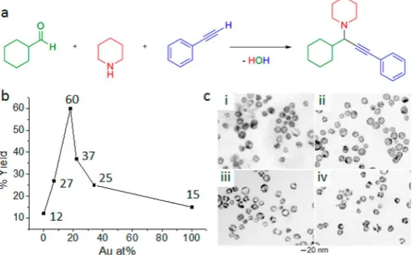
within individual hollow silver−gold (Ag−Au) bimetallic nanoparticles obtained from the galvanic reaction between Ag particles and AuCl4−. Three-dimensional compositional mapping using energy dispersive X-ray (EDX) tomography within the scanning transmission electron microscope (STEM) reveals that nanoparticle surface segregation inverts from Au-rich to Ag-rich as Au content increases. Maximum Au surface coverage was observed for nanoparticles with approximately 25 atom % Au, which correlates to the optimal catalytic performance in a three-component coupling reaction among cyclohexane carboxyalde-hyde, piperidine, and phenylacetylene.
KEYWORDS: Galvanic replacement reaction, hollow bimetallic nanoparticles, heterogeneous catalysis, scanning transmission electron microscopy, energy dispersive X-ray spectroscopy, electron tomography
T
he exceptional catalytic properties of Au nanoparticles as compared to the inert nature of the bulk element are well-known.1The emergence of catalytic activity as particles’critical dimensions approach nanometer scale is in part due to the increase in the particle’s surface-to-volume ratio.1 Synthetic routes that maximize this property, especially in distinct geometries such as hollow shells or cages,2,3 have been extensively investigated. Bimetallic noble metal nanoparticles are known to have excellent catalytic performance in a wide range of reactions.4 The extent of alloying in these systems isknown to influence catalytic activity and selectivity, but an
understanding of the origin of this effect is lacking.5 Hollow
bimetallic nanoparticles of accurately tunable size, shape, and composition can be readily prepared by a galvanic replacement reaction.6−8 The Ag−Au bimetallic system offers the
opportunity to both reduce the cost of the Au catalysts and enhance catalytic performance.9,10 Hollow Ag−Au nanostruc-tures of various compositions and shapes have been synthesized by galvanic reactions between Au metal salts, such as AuCl4−, and Ag nanoparticle sacrificial templates.10,11 Previous
low-resolution transmission electron microscope (TEM) imaging and optical spectroscopy observations suggest that this process initiates with the creation of Ag ions at the surface and the formation of a gold surface layer which immediately transforms into an Au−Ag alloy because of the fast interdiffusion between
gold and silver at 100 °C.7,12 As the replacement reaction proceeds, dealloying and processes such as Ostwald ripening
induce morphological reconstruction, as well as the formation, and enlargement, of pinholes in the walls. However, the alloying in individual particles, as well as the effect of elemental
segregation on catalytic properties, was previously unknown. Details of elemental distribution have now been made clear by analytical EDX analysis within the STEM that has only become possible due to recent advances in EDX detector design.
Ag−Au nanoparticles with controlled compositions and structures (solid vs hollow interiors) were synthesized by the galvanic replacement reaction between Ag nanoparticles and different amounts of AuCl4−. The average compositions as
determined by flame atomic absorbance spectrometry were
Ag93Au7, Ag82Au18, Ag78Au22, and Ag66Au34. The catalytic activity of the Ag−Au nanoparticles as a function of composition and structure was investigated for the three-component (A3) coupling reaction among cyclohexanecarbox-yaldehyde, piperidine, and phenylacetylene to form propargyl-amines (Figure 1a). Propargylpropargyl-amines are useful intermediates in the synthesis of heterocyclic compounds and represent structural elements of several natural products and drugs, which has led to their use as intermediates in the total synthesis of such molecules.13,14 Recently, it has been shown that this transformation can be catalyzed by nanostructured Au via a C−
Received: December 20, 2013
Revised: February 24, 2014
H activation mechanism.15,16The yield of propargylamines as a function of the Au atomic percentage in the Ag−Au nanoparticles is given in Figure 1b. All of the bimetallic and hollow Ag−Au nanoparticles produce higher yields of propargylamine than their solid and monometallic counterparts (Ag or Au nanoparticles). This observation could be related to a change in electronic structure on account of the bimetallic composition or to the increased surface area due to the formation of hollow interiors as shown in the TEM images (Figure 1c).
More surprising is an exceptionally high peak in yield of 60% at a catalyst composition of 18 at. % Au (Figure 1b). TEM imaging shows that the sizes and morphologies of the nanoparticles are broadly similar for all bimetallic compositions (Figure 1c), suggesting that the observed peak in the catalytic
results is not explained by size, shape, or surface area differences
alone.
Nanoscale compositional variations have been observed to strongly affect the catalytic activity of platinum (Pt) alloy
nanoparticles.17,18 Au and Ag form a miscible solid solution over the full range of compositions; the lattice parameter of Au is just 0.2% larger than that of Ag. Therefore, structurally sensitive techniques, such as X-ray diffraction, electron
diffraction, and high resolution TEM imaging, are unable to
distinguish among monometallic, alloyed, or composite nano-particles in the Ag−Au system. UV−visible spectroscopy19,20
[image:3.625.164.463.72.257.2]and X-ray photoelectron spectroscopy (XPS)20have been used to elucidate surface composition for bimetallic nanoparticles but cannot provide information on the local distribution of elements or on the variation of alloying inside the volume of a nanoparticle. XPS data for our system suggests an enhancement
Figure 1.Ag−Au bimetallic nanoparticles improve propargylamine yield. (a) Three-component coupling reaction to form propargylamines. (b) Propargylamine yield as a function of the composition of the nanoparticle catalysts demonstrating a spike in yield for an average composition of 18
atom % Au (numerical percentage yields are noted on the graph). (c) TEM images of the synthesized Ag−Au nanoparticles with labels
corresponding to the average bimetallic compositions plotted in b, (i) Ag93Au7, (ii) Ag82Au18, (iii) Ag78Au22, and (iv) Ag66Au34.
Figure 2.STEM EDX mapping showing the inversion of surface segregation behavior for Ag−Au nanoparticles as the Au content is increased. EDX
maps for representative nanoparticles from populations with average stoichiometry (a) Ag93Au7, (b) Ag82Au18, (c) Ag78Au22, and (d) Ag66Au34. The
actual composition of each nanoparticle shown inset was measured using the summed EDX spectrum for the wholefield of view of each spectrum
image. The chemistry of 20 nanoparticles with a range of compositions was mapped and the structure classified as to whether the particle appeared
to show a gold-rich surface, a silver-rich surface, or no surface segregation (alloyed composition). The average nanoparticle composition was then measured from the summed spectral image, and the results are shown in e. The change from gold to silver surface segregation is found to correlate with the gold content of the nanoparticle.
Nano Letters Letter
dx.doi.org/10.1021/nl4047448|Nano Lett.2014, 14, 1921−1926
[image:3.625.100.522.313.498.2]in Au composition at the surface which decreases as the total Au content increases up to 34 at. % (see Supporting Information), but the polydispersity of the population makes deeper interpretation difficult. High-angle annular dark-field
(HAADF) scanning transmission electron microscope (STEM) imaging is able to reveal local compositional variation if there are sufficiently large differences in the average atomic number
of constituent elements. However, the nonuniform cross section of our particles meant that HAADF STEM imaging of the nanoparticles in this study was inconclusive (Supporting Information Figure S1a).
Through the combination of STEM imaging with electron energy loss (EEL) or EDX spectroscopy, it is possible to obtain chemical information with a spatial resolution that approaches 0.1 nm for bulk oxides.21,22 Using high efficiency EDX
spectroscopy, coupled with aberration corrected STEM imaging, high-resolution chemical spectrum images for >20 of the Ag−Au bimetallic nanoparticles of different compositions
were obtained (Figure 2). These images are the first
observation of a reversal in the surface segregation within individual nanoparticles with increased Au content. Nano-particles with a low Au content (average composition Ag93Au7) have an approximately 1 nm Au-rich surface layer, whereas many particles from the sample with the highest Au concentration (average composition Ag66Au34) exhibited Ag surface segregation. Nanoparticles of intermediate average composition Ag82Au18 displayed mixed segregation; most appeared homogeneous in composition but a small fraction (∼20%) showed an Au-rich surface layer. Nanoparticles with intermediate average composition Ag78Au22 were also mostly homogeneously alloyed except for a small fraction that displayed Ag surface segregation. The apparent surface segregation behavior for an individual nanoparticle correlates
well with its actual Au composition measured by processing the EDX sum spectra obtained over the entirefield-of-view for each
spectrum image (Figure 2e). To gain a better understanding of the surface segregation behavior, line profiles have been
extracted perpendicular to the surface of the elemental maps (Figure 2a−d). Such two-dimensional elemental images can be difficult to interpret because different compositions overlap
when projected along the electron beam direction. For example, Figure 3c shows a line compositional profile extracted
from the 2D elemental maps in Figure 3a,b in which Ag appears to be clearly segregated to the surfaces for the left-hand side of the nanoparticle ring but not for the right-hand side.
STEM imaging can be extended to provide three-dimen-sional information through electron tomography, in which two-dimensional images are acquired at a range of sample tilts in a method similar to X-ray tomography. Electron tomography using HAADF STEM imaging has proven a useful tool in the analysis of nanoparticle size, shape, and distribution.23Energy
filtered transmission electron microscopy (EFTEM)24 and
STEM imaging with EDX spectroscopy24,25 can provide chemical sensitivity to electron tomographic reconstructions, but both approaches are challenging. New EDX detector designs (see Supporting Information) combined with the increased currents and small probe size obtainable in aberration corrected STEM instruments mean that EDX tomography is now an increasingly viable tool for nanoparticle character-ization.
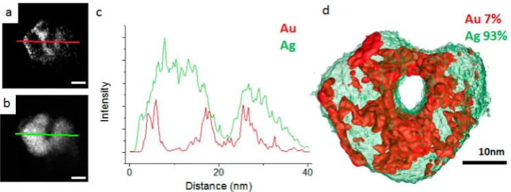
[image:4.625.94.528.70.303.2]To better understand the surface segregation behavior of the nanoparticles, EDX tomograms were compared for a nano-particle exhibiting Ag-surface segregation with another showing evidence of Au-rich surface segregation. The Ag surface-segregated nanoparticle reveals a fairly uniform surface layer approximately 2 nm thick (Figure 3d,e). The compositional line
Figure 3.3D chemical mapping of an Ag−Au nanoparticle with a high Au content showing Ag surface segregation. Two-dimensional EDX maps acquired at zero degrees of tilt showing the distribution of (a) Au and (b) Ag. (c) Compositional variation extracted right to left from the 2D maps at the position of the line shown in a and b. 2D EDX maps extracted from the 3D tomograms showing the distribution of (d) Au and (e) Ag. (f)
Compositional variation extracted right to left from the 2D slices shown in d and e. The line profile extracted from the tomographic data slice more
clearly reveals the silver surface segregation due to the improved resolution in thezdirection. (g) Surface visualization of the Au (red) and Ag
profile from the tomogram (Figure 3f) reveals the benefit of the
three-dimensional data set for nanoparticle surface analysis, more clearly demonstrating the segregated surface compared to the profiles obtained from the two-dimensional data (Figure
3c). This is due to the improved resolution in thez-direction of the three-dimensional data, removing the contribution of the top and bottom surfaces from the line profile data. Processing
of the EDX sum spectrum revealed an average composition of Ag60Au40for the particle shown in Figure 3, close to the average composition of the whole sample (Ag66Au34). Figure 3f shows a surface visualization of the Au and Ag tomograms together, which clearly demonstrate the surface segregation of Ag (see Supporting Video S1 for a rotating 3D render). In contrast, the tomogram of a nanoparticle taken from the low Au sample Ag93Au7 shows clear evidence of a thinner (∼1 nm) nonuniform Au surface segregation present on both the inner and outer surfaces of the hollow particle (Figure 4) (see Supporting Video S2 for a rotating 3D render).
The 2D compositional mapping and 3D tomographic data show a clear inversion in the surface composition for these particles as the Au content is increased and is in broad agreement with XPS data for large ensembles of nanoparticles (see the Supporting Information). Previous theoretical and lower spatial resolution experimental studies have suggested that particles are either homogeneously alloyed (due to the lower Ag−Au bond energy compared to either Au−Au or Ag− Ag6,7,26,27) or have Ag segregated to the surface as a result of
this element’s lower surface energy.28
Our results demonstrate that the Ag and Au distribution in the nanoparticles strongly depends upon the composition of the particle. At low Au content (<18 at. %) galvanic replacement initially proceeds to give Au substitution for Ag at the surface, forming a noncontinuous Au coating. As more Ag is replaced by Au, there is an increased driving force to minimize the particle’s intermetallic bond energy and surface energy which produces alloying and finally, at higher
concentrations of Au, Ag surface segregation. The maximum catalytic yield is observed at 18 at. % Au, close to the point at which the particles change from Au surface segregation to a homogeneously alloyed composition (Figure 2e). This is consistent with the observation that Au provides a greater yield than Ag for A3coupling reactions.29It is feasible that the
greater electronegativity of Au compared to Ag allows electrons to be transferred from Ag to Au, increasing the electron density of the surface relative to that which would be found in either Ag
or Au monometallic systems and improving C−H bond activation of alkynes. Indeed, an XPS analysis of the nanoparticles (Supporting Information, figure and table)
revealed that the surfaces of the nanoparticles are enriched in Au relative to their bulk. Moreover, the Au 4f core level binding energy of the materials with the lowest Au content (7 and 18 at. % Au) was found to be somewhat reduced relative to that at higher Au contents (Supporting Information, table), supporting the view that Au is present in ultrasmall metallic aggregates with electronic structure different from metallic bulk Au.30−32
The Au 4f binding energy of the catalytically most active sample (18 at. % Au) is intermediate (83.44 eV) between those of the most dilute (83.21 eV) and the more Au-rich alloys (83.66 and 83.69 eV), suggesting that high activity is associated with achieving the right balance between the influence of small
particle and bulk electronic structure effects. The Ag 3d binding
energies detected by XPS vary only slightly and are in the range expected for metallic Ag and its alloys with Au.33−35
Our results provide direct 3D observation of an inversion in the segregation of silver and gold for the first time. This
approach is not confined to galvanic replacement reactions but
could be applied to nanomaterials synthesized via many other chemical and physical processing routes. There are other examples of bimetallic systems which have shown a peak in catalytic performance for a specific composition. In the absence
of suitable local compositional information a peak in mass specific activity has been attributed solely to a change in the
bulk composition of the nanoparticle.36 Our results demon-strate that segregation in bimetallic particles is influenced by the
synthetic route and is not simple to predict. Furthermore, the experimental results from this study provide direct experimental evidence that nanoscale surface segregation can influence
nanoparticle properties. The approach could be extended to analyze the significance of different shape geometries and the
facet dependence of enrichment, providing insights for the improved design and understanding of nanoparticle synthesis and catalytic activities.
■
ASSOCIATED CONTENT*
S Supporting InformationDetails of synthesis, the catalysis of the three-component coupling-reaction, STEM imaging, tomography and XPS analysis;figures demonstrating the single/polycrystalline nature
[image:5.625.128.500.67.207.2]of nanoparticles, EDX spectra, and XPS spectra; and videos of aligned tilt series and rotating views of isosurface renders. This
Figure 4.EDX tomogram of nanoparticle with average composition Ag93Au7 with linescan. (a) Au and (b) Ag slices through the respective
tomograms. Surface segregation of Au can be seen in the slice through the Au tomogram. (c) Linescan through slices a and b showing the distribution of Ag and Au. Surface segregation of Au is observed although there appears to be a thin surface of Ag outside of the Au. (d) Surface visualization of the Au (red) and Ag (green) tomograms displaying the surface segregation of Au. The scale bars in a and b are 10 nm.
Nano Letters Letter
dx.doi.org/10.1021/nl4047448|Nano Lett.2014, 14, 1921−1926
wrote the paper. T.J.A.S. performed the 3D reconstructions. S.L.M.S. performed the XPS measurements and data analysis. All authors contributed to the interpretation and commented on the manuscript.
Notes
The authors declare no competingfinancial interest.
■
ACKNOWLEDGMENTST.J.A.S., P.O.B., and S.J.H. thank the UK Engineering and Physical Sciences Research Council, NowNANO doctoral training centre for funding support. S.J.H. thanks the USA Defense Threat Reduction Agency (grant number HDTRA1-12-1-0013) and Gates Foundation for funding support. P.H.C.C. and A.M. thank FAPESP and CNPq for funding support (grant numbers 2011/06847-0, 2013/19861-6, and 471245/2012-7, respectively). The authors wish to acknowl-edge the support from HM Government (UK) for the provision of the funds for the FEI Titan G2 80-200 S/TEM associated with research capability of the Nuclear Advanced Manufacturing Research Centre.
■
ABBREVIATIONSEDX, energy dispersive X-ray; STEM, scanning transmission electron microscope; HAADF, high-angle annular dark field;
EFTEM, energy filtered transmission electron microscopy;
XPS, X-ray photoelectron spectroscopy
■
REFERENCES(1) Herzing, A. A.; Kiely, C. J.; Carley, A. F.; Landon, P.; Hutchings, G. J. Identification of active gold nanoclusters on iron oxide supports
for CO oxidation.Science2008,321(5894), 1331−1335.
(2) Kim, S. W.; Kim, M.; Lee, W. Y.; Hyeon, T. Fabrication of hollow palladium spheres and their successful application to the recyclable
heterogeneous catalyst for Suzuki coupling reactions.J. Am. Chem. Soc.
2002,124, 7642−7643.
(3) Lou, X. W.; Archer, L. A.; Yang, Z. C. Hollow Micro-/
Nanostructures: Synthesis and Applications. Adv. Mater. 2008, 20
(21), 3987−4019.
(4) Sankar, M.; Dimitratos, N.; Miedziak, P. J.; Wells, P. P.; Kiely, C. J.; Hutchings, G. J. Designing bimetallic catalysts for a green and
sustainable future.Chem. Soc. Rev.2012,41(24), 8099−8139.
(5) Toshima, N.; Yonezawa, T. Bimetallic nanoparticles - novel
materials for chemical and physical applications.New J. Chem.1998,22
(11), 1179−1201.
(6) Petri, M. V.; Ando, R. A.; Camargo, P. H. C. Tailoring the structure, composition, optical properties and catalytic activity of
Ag-Au nanoparticles by the galvanic replacement reaction. Chem. Phys.
Lett.2012,531, 188−192.
(7) Sun, Y. G.; Xia, Y. N. Mechanistic study on the replacement reaction between silver nanostructures and chloroauric acid in aqueous
medium.J. Am. Chem. Soc.2004,126(12), 3892−3901.
(12) Sun, Y. G.; Xia, Y. N. Alloying and dealloying processes involved in the preparation of metal nanoshells through a galvanic replacement
reaction.Nano Lett.2003,3(11), 1569−1572.
(13) Miura, M.; Enna, M.; Okuro, K.; Nomura, M. Copper-catalyzed
reaction of terminal alkynes with nitrones−selective synthesis of
1-aza-1-buten-3-yne and 2-azetidinone derivatives.J. Org. Chem. 1995,
60(16), 4999−5004.
(14) Jenmalm, A.; Berts, W.; Li, Y. L.; Luthman, K.; Csoregh, I.; Hacksell, U. Stereoselective epoxidation of phe-gly and phe-phe vinyl
isosteres.J. Org. Chem.1994,59(5), 1139−1148.
(15) Gonzalez-Bejar, M.; Peters, K.; Hallett-Tapley, G. L.; Grenier, M.; Scaiano, J. C. Rapid one-pot propargylamine synthesis by plasmon mediated catalysis with gold nanoparticles on ZnO under ambient
conditions.Chem. Commun.2013,49(17), 1732−1734.
(16) Kidwai, M.; Bansal, V.; Kumar, A.; Mozumdar, S. The first Au-nanoparticles catalyzed green synthesis of propargylamines via a
three-component coupling reaction of aldehyde, alkyne and amine.Green
Chem.2007,9(7), 742−745.
(17) Cui, C.; Gan, L.; Heggen, M.; Rudi, S.; Strasser, P. Compositional segregation in shaped Pt alloy nanoparticles and their
structural behaviour during electrocatalysis.Nat. Mater.2013,12(8),
765−771.
(18) Stamenkovic, V. R.; Mun, B. S.; Arenz, M.; Mayrhofer, K. J. J.; Lucas, C. A.; Wang, G.; Ross, P. N.; Markovic, N. M. Trends in electrocatalysis on extended and nanoscale Pt-bimetallic alloy surfaces. Nat. Mater.2007,6(3), 241−247.
(19) Chen, D. H.; Chen, C. J. Formation and characterization of
Au-Ag bimetallic nanoparticles in water-in-oil microemulsions. J. Mater.
Chem.2002,12(5), 1557−1562.
(20) Wang, A. Q.; Chang, C. M.; Mou, C. Y. Evolution of catalytic activity of Au-Ag bimetallic nanoparticles on mesoporous support for
CO oxidation.J. Phys. Chem. B2005,109(40), 18860−18867.
(21) Muller, D. A. Structure and bonding at the atomic scale by
scanning transmission electron microscopy.Nat. Mater.2009,8(4),
263−270.
(22) D’Alfonso, A. J.; Freitag, B.; Klenov, D.; Allen, L. J.
Atomic-resolution chemical mapping using energy-dispersive X-ray
spectros-copy.Phys. Rev. B2010,81(10), 100101.
(23) Midgley, P. A.; Dunin-Borkowski, R. E. Electron tomography
and holography in materials science.Nat. Mater.2009,8 (4), 271−
280.
(24) Mobus, G.; Doole, R. C.; Inkson, B. J. Spectroscopic electron
tomography.Ultramicroscopy2003,96(3−4), 433−451.
(25) Genc, A.; Kovarik, L.; Gu, M.; Cheng, H.; Plachinda, P.; Pullan, L.; Freitag, B.; Wang, C. XEDS STEM tomography for 3D chemical
characterization of nanoscale particles.Ultramicroscopy2013,131(0),
24−32.
(26) Deng, L.; Hu, W.; Deng, H.; Xiao, S.; Tang, J. Au-Ag Bimetallic
Nanoparticles: Surface Segregation and Atomic-Scale Structure.J. Phys.
Chem. C2011,115(23), 11355−11363.
(27) Wei, L. Y.; Qi, W. H.; Huang, B. Y.; Wang, M. P. Surface
segregation of Au-Ag bimetallic nanowires.Comput. Mater. Sci.2013,
69, 374−380.
(28) Gong, H. R. Electronic structures and related properties of
(29) Huang, B.; Yao, X.; Li, C.-J. Diastereoselective synthesis of alpha-oxyamines via gold-, silver- and copper-catalyzed, three-component couplings of alpha-oxyaldehydes, alkynes, and amines in
water.Adv. Synth. Catal.2006,348(12−13), 1528−1532.
(30) Radnik, J.; Mohr, C.; Claus, P. On the origin of binding energy shifts of core levels of supported gold nanoparticles and dependence of
pretreatment and material synthesis.Phys. Chem. Chem. Phys.2003,5,
172−177.
(31) Willneff, E. A.; Braun, S.; Rosenthal, D.; Bluhm, H.; Havecker,̈
M.; Kleimenov, E.; Knop-Gericke, A.; Schlögl, R.; Schroeder, S. L. M.
Dynamic electronic structure of a Au/TiO2 catalyst under reaction
conditions.J. Am. Chem. Soc.2006,128, 12052−12053.
(32) Mason, M. G. Electronic Structure of Supported Small Metal
Clusters.Phys. Rev. B1983,27, 748−762.
(33) Tyson, C. C.; Bzowski, A.; Kristof, P.; Kuhn, M.; Sammy-Naiken, R.; Sham, T. K. Charge Redistribution in Au-Ag Alloys from a
Local Perspective.Phys. Rev. B1992,45, 8924−8928.
(34) Bzowski, A.; Kuhn, M.; Sham, T. K.; Rodriguez, J. A.; Hrbek, J. Electronic structure of Au-Ag bimetallics: Surface alloying on Ru(001). Phys. Rev. B1999,59, 13379−13393.
(35) Seah, M. P.; Gilmore, L. S.; Beamson, G. XPS: Binding energy calibration of electron spectrometers 5 - Re-evaluation of the reference
energies.Surf. Interface Anal.1998,26, 642−649.
(36) Zhang, H.; Jin, M. S.; Wang, J. G.; Li, W. Y.; Camargo, P. H. C.; Kim, M. J.; Yang, D. R.; Xie, Z. X.; Xia, Y. A. Synthesis of Pd-Pt Bimetallic Nanocrystals with a Concave Structure through a
Bromide-Induced Galvanic Replacement Reaction.J. Am. Chem. Soc.2011,133
(15), 6078−6089.
Nano Letters Letter
dx.doi.org/10.1021/nl4047448|Nano Lett.2014, 14, 1921−1926