This is a repository copy of The effect of ozone on progression or regression of artificial caries-like enamel lesions in vitro.
White Rose Research Online URL for this paper: http://eprints.whiterose.ac.uk/80736/
Version: Accepted Version
Article:
Tahmassebi, JF, Chrysafi, N and Duggal, MS (2014) The effect of ozone on progression or regression of artificial caries-like enamel lesions in vitro. Journal of Dentistry, 42 (2). 167 - 174. ISSN 0300-5712
https://doi.org/10.1016/j.jdent.2013.11.011
eprints@whiterose.ac.uk https://eprints.whiterose.ac.uk/ Reuse
Unless indicated otherwise, fulltext items are protected by copyright with all rights reserved. The copyright exception in section 29 of the Copyright, Designs and Patents Act 1988 allows the making of a single copy solely for the purpose of non-commercial research or private study within the limits of fair dealing. The publisher or other rights-holder may allow further reproduction and re-use of this version - refer to the White Rose Research Online record for this item. Where records identify the publisher as the copyright holder, users can verify any specific terms of use on the publisher’s website.
Takedown
If you consider content in White Rose Research Online to be in breach of UK law, please notify us by
The Effect of Ozone on Progression or Regression of Artificial Caries-like enamel
Lesions in vitro
Authors: J.F Tahmassebi, N Chrysafi, M.S Duggal,
Department of Paediatric Dentistry, Leeds Dental Institute, Leeds UK
Key words: Ozone, caries, microhardness
Short title: Effect of Ozone on the dental hard tissue
Correspondence:
Dr. Jinous Tahmassebi
Dept of Paediatric Dentistry
Leeds Dental Institute
Clarendon Way
Leeds LS2 9LU
Tel: 01133433955
Abstract
Ozone therapy combines the use of ozone gas and adjunct products called Reductant
and the patient kit, which all contain fluoride. This study investigated the effect of
Ozone on the progression or regression of artificial caries-like lesions on enamel
following pH cycling conditions in vitro. A randomised, single blind, four legs design
was used. 20 full thickness enamel slabs were allocated to each of the four groups
which were: Fluoride free toothpaste (control); Ozone alone; Reductant/Patient Kit
alone and a combination of both Ozone/Reductant/Patient Kit. Artificial lesions were
created and subjected to the pH cycling regime for a 14 days period. Assessments were
carried out before and after the pH cycling on the slabs using the Microhardness testing
and Quantitative Light-induced Fluorescence (QLF).Statistical significant difference
were found in the percentage change of enamel microhardness before and after pH
cycling between Ozone/Reductant/Patient Kit group and all the other three groups of the
study, as well as between Reductant/Patient Kit group and control There was a
statistical significant difference in the change of size and severity of the lesion ( Q)
between all the three regimes tested and the control with a trend favouring
Ozone/Reductant/Patient Kit group. In our model, it appeared that Ozone treatment
alone is not effective in protecting the enamel against demineralisation or promoting
remineralisation, unless combined with the reductant/patient kit, which contain high
Introduction
Ozone (O3) is a very powerful oxidizing compound whose antimicrobial effects have
been known for many years. Direct application of Ozone gas to the coronal or root tooth
surface is claimed to have a sterilising effect by disrupting the cell walls of
microorganisms within seconds, leading to immediate functional cessation.1 and 2 It is
consequently alleged to be able to reverse arrest or slow down the progression of dental
caries. It is also maintained that Ozone is useful for reducing the microbial flora in
cavitated lesions, before fillings are inserted.
HealOzone is a certified Medical Device [Conformité Europèene (CE) marked] for the
management of occlusal pit and fissure caries, and root caries. According to the
manufacturer, HealOzone devices are currently in use in dental practices in the UK and
more than one million people have already received HealOzone treatment.2 The
HealOzone technology has not yet received Food and Drug Administration (FDA)
approval in the USA.
There are a few published studies concerning the clinical effectiveness of Ozone
treatment. One of the earliest published studies in this field was by Baysan et al3, who
reported that Ozone application for either 10 or 20 seconds was effective to kill the
great majority of micro-organisms in primary root carious lesions (PRCLs) in vitro. In a
later study Baysan and Lynch4, showed that after nine months 45% of PRCLs reversed
from severity index 2 to 0 in the Ozone only group whilst none of the lesions became
hard in the control group with similar findings reported by.1 Also Abu-Salem5, reported
statistically significant effect of treatment upon clinical severity scores with time.
However in contrast to the above studies, Abu-Naba’a6 reported no significant
difference between the two intervention groups - Ozone plus Reductant group versus air
treatment plus Reductant only group at 12 months follow up. Similarly Baysan and
Beighton7 found Ozone treatment of non-cavitated occlusal lesions for 40 seconds failed
to significantly reduce the numbers of viable bacteria in infected dentine beneath the
demineralised enamel.
A recent in situ study by Duggal et al8 investigated the effect of Ozone on inhibition of
mineral loss from human enamel and dentine under a cariogenic challenge (14 days
has no additional effect on the inhibition of dental hard tissue demineralisation as
compared with the use of Reductant and Patient Kit.
Cochrane review concluded that given the high risk of bias in the available studies and
lack of consistency between different outcome measures, there was no reliable evidence
that application of Ozone to the surface of carious teeth stops or reverses the carious
process.9They concluded that there is not enough high quality evidence to support the
use of Ozone gas in a primary care setting. There is insufficient evidence on the
effectiveness of HealOzone treatment for this technology to be recommended, except as
part of well-designed RCTs.
Similarly, Brazelli et al 10 in a systematic review that aimed to assess the effectiveness
and cost-effectiveness of HealOzone for the management of pit and fissure caries, and
root caries, concluded that the current evidence on HealOzone is insufficient to
conclude that it is a cost-effective addition to the management and treatment of occlusal
and root caries. They also stated that in order to make a decision on whether HealOzone
is a cost-effective alternative to current preventive methods for the management of
dental caries, further research into its clinical effectiveness is required.
There are limited studies in the literature about the effects of Ozone on enamel and its
potential to inhibit demineralisation and enhance remineralisation in vitro. Furthermore,
as the use of ozone is always accompanied by the use of a Reductant and a Patient Kit
for home use, both of which contain high concentrations of fluoride, it is possible that
the benefit reported in some studies on caries is a result of these fluoride containing
products rather than ozone alone.
From the preceding literature review it seems that the effect of Ozone on the
progression of dental caries remains controversial. Therefore, it would be important to
study the effect of Ozone and the fluoride containing adjunct therapies that accompany
Material and Methods
Ethical approval was not required as this was an in vitro study. Approval was obtained
from the Leeds Dental Institute Tissue Bank where all the teeth used in the study were
collected. Statistical advice was sought and the sample size was calculated by making
estimations using data from similar study carried out by Al-Mullahi.11 Using the Stata/
SE 10.1 software it was found that for 90% power and significance level of 0.05 the
sample size of 2 slabs per group was required.
Inclusion and exclusion criteria
Intact first or/and second, upper and lower premolars extracted for orthodontic reason
under general or local anaesthesia at Leeds Dental Institute were selected. Teeth with
signs of caries, trauma, erosion, restorations or any malformation were excluded from
the study.
Enamel Slab Preparation
Enamel slabs that were used in the study were from human premolars extracted for
orthodontic reasons and stored in a solution of distilled water and 0.1% thymol (Sigma
Aldrich) at room temperature. The teeth were carefully screened by transillumination
and transmitted light using low–power microscopy for the detection of cracks (Leitz,
Wetzlar®, Germany), caries or any malformations. Suitable teeth were selected and
lightly abraded with fine paper to remove the outermost enamel and any remnants of
pellicle. A Well Diamond Wire Saw, water cooled, cutting machine for sectioning was
used (Well® Walter EBNER, CH-2400 Le Loche). The buccal and lingual surface of
each tooth was separated and was then cut into two slabs each, according to the relevant
standard operating procedures (about 2mm wide, 4mm length, and 2mm depth). Each
slab was used for one of the four legs, in order to standardise slabs with the same origin
throughout the entire study. After cutting, the slabs were polished whilst wet using fine
grit abrasive paper (P1000 Weodry paper, 3M) in combination with 5 m and 1 m
aluminia paste to remove the outermost enamel and dentine and any remnants of the
pellicle and to achieve a flat surface. Care was taken not to fully abrade the enamel.
Once the enamel slabs had been prepared, they were kept moist in water with thymol, in
micro-centrifuge tubes sealed with Parafilm to prevent leakage of the thymol solution
of the University of Liverpool, where they were exposed to gamma irradiation
(4080Gy). This level of exposure provides sterilisation without altering the structural
integrity of the enamel.
Surface Microhardness measurements
Baseline Surface Microhardness measurements were performed prior to any acid
exposure. Microhardness was assessed using a computer-aided Duramin Indenter
Machine (Struers A/S, DK 26-10, Denmark) and expressed as the length of the indent
(µm). The indentations were made using a Knoop diamond under 100 g load for 15
seconds. The depth of indenter penetration was measured by means of an image analysis
system. Five indentations, spaced 50 m apart, were made for each slab and the mean
was estimated. The same procedure was followed after the creation of the acid lesions
(before the pH cycling) and then at the end of the 14th day..12 In total, there were 3
measurements of microhardness for each slab during the study.
Creation of artificial caries-like lesions and QLF measurement
For creation of caries-like lesions in the enamel slabs, a rectangular window
(approximately 2 mm × 3 mm) was marked on each enamel slab using a carbon pencil.
The rest of the enamel slab apart from the marked area was coated with two coats of
transparent nail varnish (Max Factor “Infinity”) and was placed in plastic containers
with an acidified lactic acid homogenous gel in room temperature.
The slabs were left in this gel for 48 hours in room temperature in total, after which a
white spot was visible on each enamel slab.
Enamel slabs were assessed after the creation of white spot lesions and at the end of the
14 day test period using the QLF. All the slabs were dried for 5 seconds with
compressed air prior to imaging, and were examined in a dark room.
Enamel slabs were exposed to approximately 10 mW/cm2 violet-blue light (wavelength:
290-450 nm). The images were acquired using a miniature CCD camera kept inside the
hand piece through a 520 nm high-pass filter, transmitting only light at wavelengths
over 520 nm. The QLF camera hand piece was fixed at a position that provided
optimum illumination of the enamel block surface. A patch was drawn around the
carious lesion site by the study examiner with its borders on sound enamel. Inside this
fluorescence radiance of the surrounding sound enamel. The percentage difference
between the reconstructed and the original fluorescence levels was calculated.
Data were collected, stored and analysed by custom-made software (Inspektor Research
Systems BV, Amsterdam, The Netherlands). Demineralised areas appeared as dark
spots. Fluorescence radiance levels less than 95% of reconstructed sound fluorescence
radiance levels were considered to be artificial early caries lesions. Three metrics were
obtained: F (average change in fluorescence, in %), lesion area (mm2), and Q
(multiplication of F and area).
To ensure that images of the enamel slab were always captured in the same camera
positions and from the same angles, the software uses video-repositioning techniques.
The video-repositioning technique displays baseline and live images simultaneously and
computes their correlation based on similar geometry of the fluorescence intensities.
The enamel slabs were coded using block randomisation based on a random table of
numbers, according to computer programme of random allocation (S-Plus, insightful
corporation, Seattle WA) and were randomly allocated to each of the four study groups.
Each of the four slabs of each premolar was allocated to one of the four study groups.
The code was kept with another member of staff. As the products used in the study were
not indistinguishable the study investigator could not be blinded during the treatment
period. However, the investigator was blinded while obtaining the final measurements
using the microhardness testing and QLF at the end of the 14 days pH cycling period.
Protocol for the study
The study was carried out in accordance with principles of Good Clinical Practice
(GCP/ICH) and Good Lab Practice.
A prospective randomised, single blind, four legs design was used. Twenty full
thickness enamel slabs were randomly allocated to each of the four groups which were:
1. Fluoride free toothpaste (control) 2. Ozone alone
3. Reductant/Patient Kit alone
PH Cycling Regime
In each leg of the study the enamel specimens were dipped in 50 ml of demineralisation
solution (acetic acid buffer) for 2 minutes.13After the demineralisation challenge the
enamel specimens were rinsed with distilled water for 1 minute and then placed in 50
ml of day-time artificial saliva for 1 hour. Using this process enamel specimens were
subjected to 5 demineralisation challenges daily. For the control group, after the first
and last demineralisation challenge the enamel slabs were dipped for 5 minutes in
fluoride free toothpaste, rinsed with distilled water and following that they were dipped
in slurry 1:3 (F free toothpaste/ day-time artificial saliva) for 10 minutes. During the
night the enamel specimens were kept in night-time artificial saliva. In all instances
when the enamel slabs were taken off from the saliva, demineralisation solution and
treatment regimens were rinsed with distilled water. The enamel slabs were kept in an
incubator at 37oC in between dipping and overnight. Artificial saliva (day/night-time)
was changed daily to prevent any contamination or bacteria growth. Also a new volume
of acetic acid was used for each demineralisation challenge. All the solutions were
prepared on daily basis.
The daily cycling protocol for the three treatment groups as in control group, the only
difference being that for Ozone group, at day 1, 4, 8 and 11, Ozone was applied for 60
seconds on each enamel slab. In Reductant plus Patient Kit group at day 1, 4, 8 and 11,
Reductant was dropped manually on the enamel lesions using a small vehicle (supplied
by the manufacture), the slabs were then dipped in F- toothpaste (from Patient Kit) and
in slurry made from 1 part of F- toothpaste (from Patient Kit) and 3 parts day-time
artificial saliva. Also, after the 2nd, 3rd and 4th daily demineralisation challenge the
enamel slabs were dipped for 2 minutes in F- rinse which was provided in the Patient
Kit. Finally in the Ozone plus Reductant plus Patient Kit group Ozone was applied for
60 seconds on each enamel slab at day 1, 4, 8 and 11, Then Reductant was dropped
manually on the enamel lesions using a small vehicle (supplied by the manufacture).
The daily cycling protocol was as in Reductant plus Patient Kit group. The summary of
Statistical analysis
The SPSS statistical software package for Windows version 15.0 (SPSS Inc. Illinois)
was used for data analysis, calculation of ‘P’ values and confidence intervals. A
significance level of <0.05 was adopted. The data were initially tested using the
Kolmogorov-Smirnov test in order to check their normality.
In cases that the One Way ANOVA test showed statistical significant difference
between the groups, then we conducted multiple comparisons in order to find out where
the differences lay. However as the sample in this study was small in order to prevent
false positive result the Bonferroni adjustment was used.
Using the Kolmogorov-Smirnov test it was found that the data for the difference in
white spot lesion area and the difference in Q before and after pH cycling were not
normally distributed; therefore non-parametric tests (Kruskal-Wallis and Mann-Whitney
U tests) were used. The Bland-Altman plots were used in order to assess the
intra-examiner reproducibility.14
Results
Results for Microhardness
The primary parameters were the degree of progression or regression of artificial
caries-like lesions on enamel which were defined as the changes in microhardness and
Quantitative Light-induced Fluorescence (QLF)measurements from the start of
treatment period (baseline value).
Irrespective of the sequence of treatment,
microhardness measurements showed that Ozone/Reductant/Patient Kit, and
Reductant/Patient Kit groups showed similar enamel and dentine softening. Fluoride
free group showed the most softening of the enamel and dentine compared to baseline
(Figure 2).
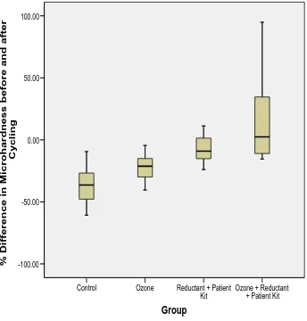
The boxplot for the distribution of the percentage (%) difference (change) in enamel
microhardness showed that the Control group had the smallest median value followed
by Ozone group. The Ozone plus Reductant plus Patient Kit group had the highest
median (2.44%). Also it was obvious that there was a wider variation in the percentage
differences in enamel microhardness in Ozone plus Reductant plus Patient Kit group
It was found that the data were normally distributed according to Kolmogorov-Smirnov
test (P=0.3). The One Way ANOVA test showed that there was a statistically significant
difference (P<0.05) in the mean % differences (changes) in enamel microhardness
between the groups. The Bonferroni method was then applied and it revealed no
statistical significant difference in the mean % changes (differences) in enamel
microhardness between Control group and Ozone group, and between Ozone group and
Reductant plus Patient Kit group. However, there was a clear trend in less reduction in
enamel microhardness after the pH cycling in the Reductant plus Patient Kit group
compared with Ozone alone group.
Results for QLF
For the QLF, the three main parameter that were statistically analysed were the
difference in white spot lesion area, the difference in F and the difference in Q.
Difference in white spot lesion area
The comparison between the groups revealed that the Control group showed the most
profound increase of white spot lesion area after pH cycling compared with baseline,
whereas the Ozone plus Reductant plus Patient Kit group showed the most significant
decrease of white spot lesion area compared with baseline.
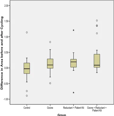
The boxplot for the distribution of the difference in white spot lesion area showed that
there was a wider variation in differences in area in Control group and Ozone group
compared to Reductant plus Patient Kit group and Ozone plus Reductant plus Patient
Kit group. Also the median values for the Ozone group and the Ozone plus Reductant
plus Patient Kit group were almost the same (Figure 3). In these data 3 outliers were
present in Control group, 2 in Ozone group, 2 in Reductant plus Patient Kit group and 4
in Ozone plus Reductant plus Patient Kit group.
The data were not normally distributed according to Kolmogorov-Smirnov test
(P=0.004). The Kruskal-Wallis test showed that there was a statistically significant
difference (P=0.019) in the changes in area between groups. So, the Mann-Whitney U
Test was used to compare between groups, and it was found statistical significant
difference in the area changes (differences) between Control and Ozone group
as well as between Control and Ozone plus Reductant plus Patient Kit group (P=0.009).
No statistical significant difference was found between the other groups (P>0.05).
Difference in F
The F difference (change) (%) was measured using the following formula:
F before pH cycling - F after pH cycling
The comparison between the groups revealed that the Control group showed the most
profound increase in loss in fluorescence between the demineralised area and the sound
enamel in the end of pH cycling compared to baseline, whereas the Reductant plus
Patient Kit group showed the most significant decrease in fluorescence loss between the
white spot lesion area and the sound enamel in the end of pH cycling compared to
baseline.
The data were normally distributed after testing using the Kolmogorov-Smirnov test
(P=0.28). The One Way ANOVA test showed that there was not a statistically
significant difference (P=0.13) in the mean differences (changes) in F between the
groups. Since One Way ANOVA test indicated no significant difference, no pairwise
comparisons of groups were conducted.
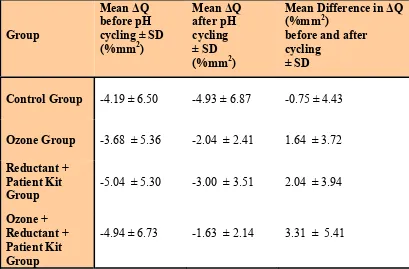
Difference in Q
The Q difference (change) (%mm2) was measured using the following formula:
Q before pH cycling - Q after pH cycling
The comparison between the groups revealed that the Control group showed the most
profound increase in the volume of the lesion in the end of pH cycling compared to
baseline, whereas the Ozone plus Reductant plus Patient Kit group showed the most
significant decrease in the volume of the lesion in the end of pH cycling compared to
baseline measurements (Table 1).
The data were not normally distributed according to Kolmogorov-Smirnov test
(P=0.007). The Kruskal-Wallis test showed that there was a statistically significant
difference in the differences in Q between groups (P=0.025). So, the Mann-Whitney
U Test was used to compare between groups, and it was found statistical significant
difference in the Q changes (differences) between Control group and Ozone group
as well as between Control and Ozone plus Reductant plus Patient Kit group (P=0.017).
No statistical significant difference was found between the other groups.
Intra-examiner reproducibility
The intra-examiner reproducibility was tested using the Bland-Altman plot. The study
investigator (NC) randomly retested 15% of the enamel slabs at the end of the study
(after the pH cycling) for microhardness, white spot lesion, F and Q measurements.
For the enamel microhardness measurements the bias, which indicates the level of
agreement on ‘average’, was -1.57 KHN. This value was very close to 0 indicating that
on average there was a good level of agreement. For the white spot area measurements
the bias, which indicates the level of agreement on ‘average’, was -0.01 mm2. This
value was very close to 0 indicating that on average there was a good level of
agreement. For the F measurements the bias, which indicates the level of agreement
on ‘average’, was -0.14%. This value was very close to 0 indicating that on average
there was a good level of agreement. For the Q measurements the bias, which
indicates the level of agreement on ‘average’, was 0.03%mm2. This value was very
close to 0 indicating again that on average there was a good level of agreement.
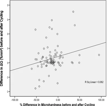
Correlation between Microhardness testing and QLF
The Pearson correlation coefficient was used to measure the correlation between the
percentage difference in enamel microhardness (%) and difference in Q (%mm2)
before and after pH cycling. A positive correlation between the two variables existed
(P=0.026). Nevertheless, the correlation between the values of two variables was weak
(r=0.25) (Figure 4).
Discussion
There is conflicting evidence regarding the clinical effectiveness of Ozone treatment. A
recent Cochrane review9 identified only three RCT’s.4 and 5 The remaining articles were
excluded for reasons such as lack of blinding, randomisation or controls, less than 6
months follow-up, or lack of investigation of extracted teeth. Furthermore the included
studies were judged to be at high risk of bias. In addition, given the statistically
imperative that the effects of ozone are evaluated in the way its clinical use is
prescribed, which is always alongside the fluoride containing products, i.e Reductant
and patient kit.Therefore the present study aimed to investigate additional effect of
Ozone, in combination with the fluoride containing adjuncts used with it on the
progression or regression of artificial caries-like lesions on enamel following pH
cycling conditions in vitro. This was achieved by using demineralised enamel slabs,
where artificial lesions had already been created in vitro. This has the benefit of
allowing both demineralisation and remineralisation to be studied. In order to create the
artificial lesions we used an acidified hydroxyethyl cellulose gel. It has been proposed
that it is preferable to use hydroxyethyl cellulose in order to create artificial lesions as it
is easier to be produced, the formation of lesions is consistent, there are uniform areas
of demineralisation and the produced lesions are more rigid, compared with the lesions
produced with acetic acid buffer.17 To avoid excessive loss of minerals during the
lesions formation and in order for the enamel slabs to be able to withstand further
demineralisation challenge during the pH cycling, an acidified hydroxyethyl cellulose
gel was used for two days in this study.
The method of pH cycling simulates the pH changes taking place in the plaque by
subjecting the enamel to a series of de- and remineralisation challenges.18 A 14 days
period was chosen in this study in order to have sufficient time to produce changes in
the pre-demineralised enamel slabs. The same pH cycling period has been used
successfully in many previous studies in order to assess de- and remineralisatio.18-20
In most of the previous studies Ozone has been applied for the duration of 10 to 120
seconds per tooth. In this study Ozone was delivered for 60 seconds on each enamel
slab per application. This was carried out four times in the 14 days pH cycling period.
The number of Ozone applications was increased in the current study as this was an in
vitro study and Ozone could exhibit only its oxidizing properties against acids, and no
antimicrobial effect could be demonstrated in an in vitro model such as the one we used.
After the pH cycling in order to monitor the progression or regression of the carious
lesions, a second image with the QLF was obtained. It is critical that the new image
obtained to be superimposed as accurately as possible over the original image of the
demineralised area. In this study we found some difficulties to superimpose the second
image over the first. To overcome this problem the camera’s position and angle from
it is unlikely that this had a major effect on the accuracy of the results as the QLF
software uses video repositioning techniques and the new image is recorded only if its
correlation with the initial image is higher than 0.98.
To our knowledge no other studies in the literature have used Ozone alone to assess its
effect on de- and remineralisation of enamel. Our results indicated that when used
alone, ozone did not have a significant effect on preventing further demineralisation or
promoting remineralisation as seem from the results of rthe microhardness and QLF
respectively. . In addition to other well published mechanisms of proposed action of
ozone, it is known that Ozone has a severely disruptive effect on the bacterial
population in the carious lesion and obliterates the cariogenic bacteria, thereby swinging
the equilibrium in favour of remineralisation. No more acid can be produced within the
lesion when the acid-producing bacteria are eliminated. As this was an in vitro study the
effect of Ozone on elimination of bacteria in the lesion could not be studied. On the
other hand, it is possible that Ozone because of its powerful oxidizing properties
decreased carbohydrates and acids within the lesions and so enhanced remineralisation.
Also, Ozone has the ability to remove organic material in the carious lesions and
enables calcium and phosphate ions to diffuse through the lesions, which might account
for the small differences comparted to control group in our study
Although in the present study we failed to prove a statistically significant difference
between Reductant plus Patient Kit group and Ozone group, there was a very clear trend
in less reduction in enamel microhardness after the pH cycling in favour of the
Reductant plus Patient Kit group. The beneficial effect of fluoride is showed in many
reports from previous studies, where the resistance of enamel to acid demineralisation
and reduction of mineral loss and lesion depth in the presence of fluoride in solution
was evident.21 and 22 It is possible that these differences may have been statistically
evident if the sample size had been even larger.
The significant remineralisation in Ozone plus Reductant plus Patient Kit group shows
the synergistic effect of Ozone and fluoride. As it is already mentioned, it has been
theorized that Ozone can oxidize organic material within the carious lesion. This
reportedly opens up “channels” within the dental tissue to allow the penetration of
calcium, phosphate, and fluoride ions to allow remineralisation of the surface. These
Generally, conflicting data exist about the parameter "lesion severity" as a determinant
for lesion repair. Strang et al24 showed mineral deposition during in situ
remineralisation to be (positively) correlated to the amount of mineral lost during the
formation of the lesions. Also, in situ studies have shown that smaller lesions were more
prone to demineralisation than larger ones.25 and 26 White et al, 27 on the contrary,
demonstrated in vitro that deeper lesions remineralised less rapidly.
The results of the current study are in accordance with ten Cate et al22 who found that
when shallow (‘early’) lesions were studied the predominant effect of fluoride was to
inhibit demineralisation up to 50% in the ‘classical’ fluoride range 0–1,500 ppm, and
this increased to 70% when 3,000 ppm F- pastes were given.
The synergistic effect of Ozone and fluoride may be explained by the suggestion that
Ozone can inhibit hypermineralisation. Baysan and Lynch4 demonstrated that it is
possible to remineralise or arrest primary root carious lesions and that a dentifrice
containing 5,000 ppm F- was significantly more effective than that containing 1,100
ppm F-. On the other hand, it has been suggested that the remineralisation of primary
root carious lesions following the use of a dentifrice with high fluoride content may
result in the hypermineralisation on the surface of the lesions and this would prevent
further remineralisation occurring through the lesion depth.
The results of the current study indicated that ozone alone has a minimal effect but this
was enhanced when fluoride containing products were used in combination with use of
ozone and this is in agreement with recently published study that also disputed the
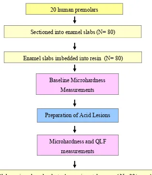
Figure 1. The flow chart of the study protocol
20 human premolars
Sectioned into enamel slabs (N= 80)
Enamel slabs imbedded into resin (N= 80)
Baseline Microhardness
Measurements
Preparation of Acid Lesions
Microhardness and QLF
measurements
Slabs assigned randomly to 4 experimental groups ( N= 20/group)
14 Days pH cycling
Microhardness and QLF
measurements
Figure 2: Boxplot for the distribution of the percentage (%) difference (change) in enamel microhardness in all groups (error bars represent SD, the line in the box of Box-and-whisker plot is the median value of the data).
Group
Ozone + Reductant + Patient Kit Reductant + Patient
Kit Ozone
Control
%
Difference in Microhardness before and after
Cycling
100.00
50.00
0.00
-50.00
Figure 3: Boxplot for the distribution of the difference in white spot lesion area (mm2) in all treatment groups (error bars represent SD, the line in the box of Box-and-whisker plot is the median value of the data).
Group
Ozone + Reductant + Patient Kit Reductant + Patient Kit
Ozone Control
Difference in Area befor
e
and
after Cyclin
g
2.00
1.50
1.00
0.50
0.00
-0.50
Table 1: The mean difference in the volume of lesion ( Q) before and after pH cycling in all groups.
Group
Mean Q before pH cycling ± SD (%mm2)
Mean Q after pH cycling ± SD (%mm2)
Mean Difference in Q (%mm2)
before and after cycling
± SD
Control Group -4.19 ± 6.50 -4.93 ± 6.87 -0.75 ± 4.43
Ozone Group -3.68 ± 5.36 -2.04 ± 2.41 1.64 ± 3.72
Reductant + Patient Kit Group
-5.04 ± 5.30 -3.00 ± 3.51 2.04 ± 3.94
Ozone + Reductant + Patient Kit Group
Figure 4: Plot of Difference in Q (%mm2) related to percentage difference in enamel microhardness (%) before and after pH cycling.
% Difference in Microhardness before and after Cycling
100.00 50.00
0.00 -50.00
-100.00 20.00
15.00
10.00
5.00
0.00
-5.00
-10.00
R Sq Linear = 0.062
References
1. Holmes J. Clinical reversal of root caries using Ozone, double-blind
randomized controlled 18- month trial. Gerodont 2003; 20(2):106-114
2. Lynch E. Ozone: the revolution in dentistry. Quintessence Publishing Co.
Ltd; 2004
3. Baysan A, Whiley RA, Lynch E. Antimicrobial effect of a novel Ozone-
generating device on micro-organisms associated with primary root carious
lesions in vitro. Caries Res 2000; 34:498–501
4. Baysan A, Lynch E. Effect of Ozone on the oral microbiota and clinical
severity of primary root caries. Am J Dent 2004; 17:56–60
5. Abu-Salem O. Reversal of occlusal caries in primary teeth. MPhil Thesis:
Queen's University Belfast; 2004
6. Abu-Naba'a L.A. Management of primary occlusal pit and fissure caries
using Ozone. PhD Thesis: Queen's University Belfast; 2003
7. Baysan A, Beighton D. Assessment of the Ozone-mediated killing of
bacteria in infected dentine associated with non-cavitated occlusal carious
lesions. Caries Res 2007; 41(5):337-341
8. Duggal MS, Nikolopoulou A, Tahmassebi JF. The additional effect of ozone
in combination with adjunct remineralisation products on inhibition of
demineralization of the dental hard tissue in situ. J Dent 2012; 40:934-940
9. Rickard GD, Richardson R, Johnson T, McColl D, Hooper L. Ozone therapy
for the treatment of dental caries. The Cochrane Database of Systematic
Reviews, Issue 3 2004; DOI:10.1002/14651858.CD0041
10.Brazzelli M, McKenzie L, Fielding S, Fraser C, Clarkson J, Kilonzo M,
Waugh N. Systematic review of the effectiveness and cost-effectiveness of
HealOzone for the treatment of occlusal pit/fissure caries and root caries.
Healt Technol Asses 2006; 10(16):iii-iv, ix-80
11.Al-Mullahi AM. Effect of slow-release fluoride devices and
casein-phosphopeptide nanocomplexes on enamel remineralisation in vitro.
MDenSci Thesis: University of Leeds, Leeds; 2008
12.Zero DT, Fu J, Anne KM, Cassata S, McCormack SM, Gwinner LM. An
improved intra-oral enamel demineralization test model for the study of
13.Bland JM, Altman DG. Statistical methods for assessing agreement between
two methods of clinical measurement. Lancet 1986; 1:307-310
14.Marinho VC, Higgins JP, Logan S, Sheiham A. Fluoride toothpastes for
preventing dental caries in children and adolescents. Cochrane Database of
Systematic Reviews; Issue 1 DOI: 10.1002/14651858.CD002278; 2003 a
15.Marinho VC, Higgins JP, Logan S, Sheiham A. Fluoride mouthrinses in
preventing dental caries in children and adolescents. Cochrane Database of
Systematic Reviews; Issue 3 DOI: 10.1002/14651858.CD002284 ; 2003 b
16.Marinho VC, Higgins JP, Logan S, Sheiham A. Topical fluoride
(toothpastes, mouthrinses, gels or varnishes) for preventing dental caries in
children and adolescents. Cochrane Database of Systematic Reviews; Issue
4 DOI: 10.1002/14651858.CD002782 ; 2003 c
17.Issa AI. The effect of intrinsic and extrinsic sugars on enamel
demineralisation and plaque pH as determined using intraoral cariogenicity
tests and plaque pH telemetry. PhD Thesis: University of Leeds, Leeds,
2004
18.ten Cate JM, Duijsters PPE. Alternating demineralization and
remineralization of artificial enamel lesions. Caries Res 1982; 16(3):201-210
19.Malinowski M, Toumba KJ, Strafford SM, Duggal MS. Effect of Varying
Concentrations of Fluoride in Milk on Enamel Remineralisation in vitro.
(ORCA Congress 2007, Abstract no. 29).Caries Res 2007;41:278
20.Gkourtsogianni S. The effect of various preventive agents on dentine
hardness and surface loss in vitro. MDenSci Thesis: University of Leeds,
Leeds, 2008
21.Duggal MS, Toumba KJ, Amaechi BT, Kowash MB, Higham SM. Enamel
demineralization in situ with various frequencies of carbohydrate
consumption with and without fluoride toothpaste. J Dent Res 2001;
8:1721-1724
22.ten Cate JM, Exterkate RAM, Buijs MJ. The relative efficacy of fluoride
toothpastes assessed with pH cycling. Caries Res 2006; 40(2):136-141
23.Hodson N. Dunne SM. Using ozone to treat dental caries. J Esthetic &Rest
24.Strang R, Damato FA, Creanor SL, Stephen KW. The effect of baseline
lesion mineral loss on in situ remineralization. J Dent Res 1987;
66:1644-1646
25.Mellberg JR. Relationship of original mineral loss in caries-like lesions to
mineral changes in situ. Caries Res 1991; 25:459–461
26.Schafer F, Raven SJ, Parr TA. The effect of lesion characteristic on
remineralization and model sensitivity. J Dent Res 1992; 71: 811–813
27.White DJ, Chen WC, Nancollas GH. Kinetic and physical aspects of enamel