RESEARCH
Parasite fauna of wild Antillean manatees
(
Trichechus manatus manatus
) of the Andean
Region, Colombia
Juan Vélez
1,2*, Jörg Hirzmann
1, Katerin Arévalo‑González
3,4,5, Malin K. Lange
1, Anika Seipp
6, Ulrich Gärtner
6,
Anja Taubert
1, Susana Caballero
3and Carlos Hermosilla
1Abstract
Background: Antillean manatees (Trichechus manatus manatus) are large herbivorous aquatic mammals living in lim‑ ited areas of South, Central and North America. As with other aquatic mammals, Antillean manatees can be infected by a variety of protozoan and metazoan parasites, some of them with zoonotic potential, which affect not only their welfare but also population health status. Therefore, we conducted the first epidemiological survey in Colombian free‑ranging Antillean manatees to estimate their actual gastrointestinal parasite status.
Results: In total, 69 faecal samples were collected from free‑ranging individual manatees during ecology field studies in the rivers Carare and San Juan and in two associated wetlands in the Andean region of Colombia. Parasite diversity encompassed six different endoparasite species. The highest prevalence was found for protozoan infections with Eimeria nodulosa (47.8%) and Eimeria manatus‑like species (type A, B; 43.4%), followed by Entamoeba sp. (14.49%) and Giardia sp. (1.4%) infections. In addition, infections with the trematode Chiorchis fabaceus were detected at a high prevalence (33.3%). Molecular characterization of sirenian Eimeria species led to the distinction of three species, E. nodulosa and two E. manatus‑like species (type A, B). Phylogenetic analyses indicated a host‑specific adaptation of sirenian Eimeria species as previously reported for Eimeria species from other mammalian hosts.
Conclusions: This study provides the first record of Antillean manatee infection with Giardia and Entamoeba species in Colombia, representing two important anthropozoonotic parasite genera. This survey should serve as a baseline investigation for future monitoring on parasitic zoonoses in this mammal and encourage for investigations on their impact on both public health and wild manatee welfare.
Keywords: Manatees, Trichechus manatus manatus, Eimeria, Phylogeny, Chiorchis, Entamoeba, Giardia
© The Author(s) 2019. This article is distributed under the terms of the Creative Commons Attribution 4.0 International License (http://creat iveco mmons .org/licen ses/by/4.0/), which permits unrestricted use, distribution, and reproduction in any medium, provided you give appropriate credit to the original author(s) and the source, provide a link to the Creative Commons license, and indicate if changes were made. The Creative Commons Public Domain Dedication waiver (http://creat iveco mmons .org/ publi cdoma in/zero/1.0/) applies to the data made available in this article, unless otherwise stated.
Background
The Antillean manatee (Trichechus manatus manatus) is the only sirenis, which ranges from the northeast of South America through the Caribbean Sea up to Mexico. It inhabits costal, freshwater fluvial and wetland environ-ments in Colombia [1, 2]. In contrast to other aquatic/ marine mammals, which all exhibit carnivorous/piscivo-rous diets, sirenians are considered as pure herbivocarnivorous/piscivo-rous species. In Colombia, Antillean manatees reside in the
Orinoco and the Caribbean basins with the Magdalena riparian system representing the largest habitat area with the highest capture [1–4]. The Antillean manatee is listed as a seriously endangered species in Colombia experienc-ing a threatenexperienc-ing population decrease accordexperienc-ing to the International Union for the Conservation of the Nature (IUCN) [1, 5]. The latest assessment referred to a popu-lation of approximately 500 animals left in these Colom-bian regions [6].
There are several reports highlighting the devastating impact of anthropogenic and environmental pressure on wild manatee populations due to hunting activities [1, 2], watercraft collisions [5, 7–11], sewage pollution,
Open Access
*Correspondence: juan.velez@vetmed.uni‑giessen.de
1 Institute of Parasitology, Biomedical Research Center Seltersberg (BFS),
brevetoxicosis, [12–15], accidental death in fishing nets [16] and loss of natural habitats [1]. Unfortunately, the Colombian wild manatee populations are still exposed to all these adverse factors and therefore urgently require ongoing national protection measures. Besides the factors mentioned above, parasitoses also have a critical impact on wildlife population health, including that of aquatic mammals [17–20], and data on actual infections are needed to assess environmental risk fac-tors for endangered species [21]. As such, monitoring studies will allow gaining an improved knowledge on pathogen diversity and relevance for manatee welfare, on a potential spillover of human parasites and on para-site reservoirs [20]. All these aspects are important, not only for conservation purposes, but also for the pres-ervation of important ecological dynamics and human health protection.
So far, several reports on metazoan and protozoan parasites of manatees from different geographical regions have been published [4, 11, 22–28]. Nonetheless, detailed knowledge on manatee parasites, i.e. their biology, epide-miology, pathogenesis and immunity, is still limited and restricted to studies on captive animals or carcasses [4, 11, 29]. Consequently, these parasitological reports may not necessarily reflect true parasite diversity of mana-tee populations living within their natural habitats. Few investigations have been performed on South American wild manatee populations due to the elusive behaviour of these animals and the turbid waters they inhabit [21, 30, 31]. Overall, the implementation of ‘non-invasive’ sampling techniques (e.g. by collecting faeces, vomitus, expirations), of photographic records in combination with sonar-based manatee tracing [32] and of molecu-lar approaches [21], may not only contribute to improve sampling efficacy but will also change the scope of future parasitological studies on manatees within natural eco-systems. Whilst there are vast amounts of molecular data available for parasites affecting terrestrial mammals, there is still a lack of molecular analyses on parasites occurring in wild manatees. Currently, sequences from only three helminth species of manatees are available [21, 33]. The option to identify and characterize para-site diversity via DNA amplification from faeces, nasal/ ocular secretions and tissue samples opens a wide range of future tasks that may help to protect this unique mammal.
The present study represents the first large-scale survey on gastrointestinal parasites of wild, living and free-rang-ing Antillean manatees (n = 69) in Colombia and pro-vides first report of the genus Entamoeba in sirenians in South America. Furthermore, it adds a novel molecular characterization on monoxenous Eimeria species infect-ing these endangered aquatic mammals.
Results
Parasite infections
Overall, 72% (50/69) of Antillean manatees were infected with at least one parasite species (Table 1). In total, six different gastrointestinal parasite species belonging to protozoan and metazoan taxa were diagnosed. Five dif-ferent protozoan and one metazoan parasitic stages (i.e. cysts, oocysts and eggs) were detected. Metazoan para-sites were represented by one trematode species ( Chior-chis fabaceus). No stages of cestodes, nematodes or acanthocephalans were found in manatee faecal samples. A list of known parasitic stages and respective preva-lences from manatees is presented in Additional file 1: Table S1. In addition, selected illustrations of parasitic stages are given in Fig. 1.
The most prevalent parasitic stages were oocysts of
Eimeria nodulosa (47.8%; Fig. 1b), followed by Eimeria manatus-like oocysts (43.4%; Fig. 1a), eggs of C. faba-ceus (33.3%; Fig. 1d) and cysts of Entamoeba sp. (14.5%; Fig. 1c). One faecal sample proved positive for Giardia
antigen (1.4%) in coproantigen-ELISA. None of the identified parasites revealed as core species (preva-lence > 50%) and the diplomonadid protozoan Giardia sp. was found as component species (prevalence < 10%). Two parasite genera have anthropozoonotic potential, namely
Entamoeba and Giardia.
Referring to parasite genus level, the present findings include one new host record on Entamoeba for Antillean manatees (T. manatus manatus) in South America. In addition, giardiasis has not previously been reported to occur in Colombian manatees.
Overall, two water-borne parasitic infections (i.e. giar-diasis and entamoebiasis), one gastropod-borne disease (chiorchiosis) and two monoxenous infections (coccidi-osis) were detected in this epidemiological survey.
[image:2.595.305.539.642.724.2]Morphometric and morphological analyses of coccid-ian oocysts and trematode eggs revealed the following data: E. nodulosa oocysts had a mean size of 12.55 × 11.72 (7.0–14.9 × 7.2–13.97) µm and showed characteristic knob-like structures on the surface (Fig. 1b). Scanning electron microscopy (SEM) analyses illustrated these
Table 1 Prevalence of parasites in wild Antillean manatees (T. manatus manatus) (n= 69) from the Carare River, Santander
Species Prevalence (%) Technique
Eimeria nodulosa 47.8 SAF/flotation
Eimeria sp. A and Eimeria sp. B 43.4 SAF/flotation
Entamoeba sp. 14.5 SAF
Giardia sp. 1.4 Copro‑ELISA
knob-like structures in more detail (Fig. 2). E. manatus -like oocysts [9.82 × 9.24 (8.9–11.95 × 8.0–11.31) µm] were slightly smaller than E. nodulosa oocysts and lacked any knob-like structures on oocyst wall. SEM analyses showed a micropyle-like cap structure in the E. mana-tus-like oocysts (data not shown). Interestingly, such a structure was not reported before for manatee-specific oocysts [25, 26]. Entamoeba sp. cysts had a mean size of 14.19 × 12.0 (10.45–18.57 × 8.17–15.89) µm and pre-sented at least more than two spherical nuclei (Fig. 1c).
Chiorchis fabaceus eggs had a mean size of 151 × 111
(139–157 × 99–133) µm, an ovoid shape, a unipolar operculum and a brownish morula being delimited by a smooth capsule (Fig. 2d). All these morphological char-acteristics agree with previous descriptions [25].
Molecular analyses of Eimeria species
In total, 62% (43/69) of Antillean manatee faecal sam-ples contained Eimeria oocysts, which were diagnosed morphologically as E. nodulosa and E. manatus-like spe-cies. A subset of samples (n= 17) with single and mixed
Eimeria oocyst specimen was characterized molecu-larly by copro-PCR and consecutive sequencing of the almost entire SSU rDNA. Overall, three different Eimeria
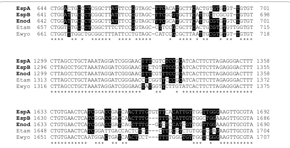
sequences were identified with an interspecies identity of 98.3–98.7% (Fig. 3, partial alignment). The highest homology obtained by BLAST search of the GenBank database was related to Eimeria sequences from rodents showing 96–97% identity. Out of the phylogeny DNA sequences, one could directly be assigned to E. nodulosa
based on microscopic diagnostics on samples showing mono-infections with E. nodulosa. The remaining two sequences corresponded to E. manatus-like oocysts. These appear to represent two distinct Eimeria species (designated here as E. manatus-like type A and B) which were indistinguishable on the level of oocyst morphol-ogy. The partial SSU rRNA gene sequences of E. nodu-losa and the two E. manatus-like species were deposited in the GenBank database under the accession numbers MG652357–MG652359.
A simplified phylogenetic tree showing representative
Eimeria species from bovines, rodents and chickens was generated based on BLAST search data including mana-tee Eimeria sequences (Fig. 4). Statistical analyses from series of likelihood-ratio tests obtained in MrModeltest v.2 [34] allowed to select SYM+G as the most appro-priate mathematical model for our phylogenetic analy-sis which was carried out using MrBayes v.3.2 [35]. The Bayesian maximum posterior probability tree with corre-sponding clade-credibility values is shown in Fig. 4. The inferred phylogenetic tree revealed a host-specific adap-tation of the sirenian Eimeria species as also shown for
Eimeria species from the other host groups. The closest neighbour to the cluster of sirenian Eimeria species was represented by the rodent lineage.
Discussion
Overall, manatees were infected with five different intes-tinal protozoan parasites (i.e. E. nodulosa, E. manatus -like type A and B, Entamoeba sp. and Giardia sp.) and one intestinal trematode parasite, i. e. C. fabaceus. Thus, manatees were found to be infected with E. nodulosa
(47.8%), E. manatus-like specimens (43.4%), Entamoeba
sp. (14.5%) and C. fabaceus (33.3%). Giardia sp. revealed Fig. 1 Illustration of manatee gastrointestinal parasites. aEimeria
manatus‑like oocyst, bEimeria nodulosa oocyst, cEntamoeba sp. cyst, dChiorchis fabaceus egg. Scale-bars: a, c, d 20 µm; b 10 µm
[image:3.595.56.292.87.306.2] [image:3.595.57.291.353.534.2]as a component species (prevalence < 10%), which is in agreement with previous published data [31].
In the present study, Eimeria spp. revealed as the most prevalent parasites and these findings are in agreement with data on manatees inhabiting Florida and Puerto
Rico (Additional file 1: Table S1) [25, 36]. DNA sequenc-ing-based analyses identified three Eimeria species in Colombian manatee samples, i.e. E. nodulosa and two
E. manatus-like species. Although microscopic analyses revealed smaller sizes of E. manatus-like oocysts when compared to measures recently reported for E. mana-tus [25], the present size characteristics almost matched those described elsewhere [37]. Based on oocyst size we could also exclude the presence of E. trichechi, which was described in T. inunguis in Brazil [24].
Interestingly, the Antillean manatee population stud-ied here was found to be infected with only one trema-tode species, i.e. C. fabaceus, thereby showing a different trematode spectrum than Antillean manatees from Cór-doba, Colombia, which had Nudacotyle undicola infec-tions [21]. This might be due to the relative geographical separation of the different manatee populations. Consist-ently, the Carare River is localized between the Andes Mountains of Colombia and therefore far away from shores of the Atlantic Ocean where other wild Antillean manatees reside. Trematodes have indirect life-cycles and need adequate gastropod intermediate hosts to fulfil their development. Therefore, the presence or absence of specific molluscs (snails/slugs) represents a further factor for trematode diversity in manatees. However, the cur-rent lack of knowledge on epidemiology, transmission and pathogenicity of almost all parasitoses of manatees
EspA 644 CTGGACTGCCCTCGGCTTATTTCCGGTAGC-TTCACATGCTTCACTGTGTGTGT-TGTGT 701 EspB 641 CTGGATTGCCCTCGGCTTATTTCCGGTAGC-TTCGCGTGCTTCATTGCTCGCGT-TGTGT 698 Enod 642 CTGGATTGCCCTCGGCTTATTTCCGGTAGCTTTCGCATGCTTCATTGCGTGTGTCTGTGT 701 Etam 657 CTGGTTTGCCCTCGGCTTTCTTCCGGTAGC-CTTCCGCGCTTCACTGCGTGTGTTGGTGT 715
Ewyo 661 CTGGTTTGGCTGCGGCTTTATTCCTGTAGC-CATCCGCGCTTAATTGCGTGTGT-TGTGT 718
EspA 1299 CTTAGCCTGCTAAATAGGATCGGGAACTTTCGTCTCCGCATCACTTCTTAGAGGGACTTT 1358
EspB 1296 CTTAGCCTGCTAAATAGGATCGGGAACTTCTGTTTCCGCATCACTTCTTAGAGGGACTTT 1355
Enod 1299 CTTAGCCTGCTAAATAGGATCGGGAACTTTCGTTTCCGCATCACTTCTTAGAGGGACTTT 1358
Etam 1313 CTTAGCCTGCTAAATAGGATCGGGAACCTCGGTTCCCGCATCACTTCTTAGAGGGACTTT 1372
Ewyo 1316 CTTAGCCTGCTAAATAGGATCGGGAACTATGGTTCTTGTATCACTTCTTAGAGGGACTTT 1375
EspA 1633 CTGTGAACTCATCGGACCGATCAACTTTGTGTTTCACATTGTTGGTTGGGAAGTTGCGTA 1692
EspB 1630 CTGTGAACTCATCGGACCGATCAACTTTG---TTTACATTGTTGGCTGGAAAGTTGCGTA 1686
Enod 1633 CTGTGAACTCATCGGACCGATCAGCTTTG--GTCTCCATCGTTGGTTGGAAAGTTGCGTA 1690 Etam 1648 CTGTGAACTCATCGGATTGACCACTCTCG---CTTGCGTCTGTGGTCGAGAAGTTGCGTA 1704
Ewyo 1651 CTGTGAACTCAATGGACTGATCAACTCCT---TTTTGGGTGTTGGTCGGGAAGTTGCGTA 1707
**** ** * ****** **** ***** * **** * ** * ** ****
*************************** ** * *********************
*********** *** ** ** *** * **********
Fig. 3 Alignment of Eimeria nodulosa, Eimeria manatus‑like type A, Eimeria manatus‑like type B, E. tamiasciuri (squirrel) and E. wyomingensis (bovines) SSU rRNA gene sequences. Positions with identical nucleotides in all five sequences are denoted by a star; positions with more than 50% agreement are highlighted by black shading
[image:4.595.58.538.86.332.2] [image:4.595.56.290.399.575.2]impair the establishment of proper international and national conservation politics. Indeed, the pathogenic-ity of manatee intestinal trematode infections, e.g. nuda-cotylosis, could be of importance, especially for this studied isolated manatee population which may be naïve for different manatee-specific trematode infections [25]. Additionally, the probability of a low genetic diversity in confined and remote manatee populations, a product of inbreeding, may impair the host immune system, ren-dering manatees more susceptible to disease as demon-strated for other wild animals [38–40].
The present parasitological findings represent the first host record for Entamoeba sp. in T. manatus manatus
in South America (prevalence: 14.5%). So far, neither the species nor the zoonotic potential of this pathogen is known. To our best knowledge, Entamoeba infections in aquatic mammals have so far only been reported in certain whale species such as sperm whales, blue whales, fin whales and sei whales [41], and there is one report for manatees [36]. Nonetheless, the Entamoeba-like cysts reported in Florida manatees [36] present a larger size and differ in the number and form of the nuclei.
Therefore, future parasitological research in manatees requires a broader approach, e.g. incorporating molec-ular analysis [21, 33]. In general, Entamoeba spp. are water-borne parasites and their transmission commonly occur in developing countries where drinking water quality is poor and open water often is contaminated by human faeces, which are still used as a fertilizer [42]. Besides some non-pathogenic species, such as E. coli,
E. hartmanni and E. polecki [43], the genus Entamoeba
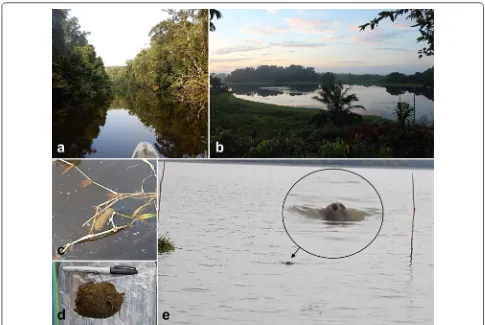
also includes the worldwide occurring species E. histol-ytica which is considered as one of the major causes of human deaths induced by parasitic pathogens [44]. As such, contamination of water and shores with human fae-ces might pose a risk on local manatee health. Addition-ally, one Giardia antigen-positive Antillean manatee was identified in the present study thereby representing, to our best knowledge, the first report for Colombia. Along-side this, there is only one other report on giardiasis in manatees from Brazil [31]. Giardia spp. are also consid-ered as water-borne zoonotic parasites which are trans-mitted by highly resistant cysts which are orally ingested by hosts [20]. Given that no cyst stages were detected in Fig. 5 Illustration of sampling sites and specimen: a Carare River in the Andean region; b “Cienaga La San Juana”, a wetland ecosystem; c, d
[image:5.595.55.540.86.411.2]the antigen-positive animal one may question an active infection and thereby the zoonotic potential of this posi-tive sample. In fact, we cannot exclude that Giardia
stages were simply representing intestinal passers-by. As also tackled in the present study, water-borne zoonoses clearly need more attention by public health authorities worldwide as suggested elsewhere [45]. A fun-damental aspect of entamoebiasis/giardiasis control is to identify reservoirs and routes of transmission in diverse climatic and geographical areas. This is highly important in the case of wild Antillean manatees, considering that this species inhabits shallow waters close to populated marine shores which renders them highly vulnerable to classical water-borne parasitic infections [23, 30, 46].
Conclusions
The present study adds novel data on clearly neglected anthropozoonotic parasitoses [44, 47] and calls for more integrated investigation to avoid exposure of Antillean manatees or human beings to these intestinal pathogens. It is of particular importance to strengthen interdiscipli-nary health agendas which favour ‘One Health’ concept considering ecosystem, domestic animal, wild animal and human health as a single unit [47–50]. This study emphasizes the relevance of the sentinel role of manatees [51] and the regular manatee monitoring programmes being supported by both Colombian authorities for pub-lic health issues and biologists/ecologists responsible for conservation programmes.
Methods
Study area, sample collection and coprological analyses
Wild Antillean manatees (T. manatus manatus) inhab-iting river-, swamp- and wetland-waters within the Andean region of the Santander Department of Colom-bia were sampled. The study area has an average annual precipitation of 2955 mm and a temperature range between 26.8 and 30.3 °C. Faecal samples were collected during dry and rainy seasons of the years 2015 and 2016. In detail, a boat-based line transect survey was conducted to search for faecal samples along the banks and floating plant patches in the San Juan River, the San Juana swamp and in the Carare river basin. In total, 69 individual fae-cal samples were collected by tracing individual animals during boat excursions according to the Guidelines for the Treatment of Marine Mammals in Field Research of the Society for Marine Mammalogy. Whenever defeca-tion occurred, floating faecal samples were immediately collected from water surfaces or from floating vegetation patches using a net (Fig. 5). Thereafter, faecal samples were transferred into 10-ml plastic tubes (Sarstedt, Nüm-brecht, Germany) containing 70% ethanol for fixation and stored at 4 °C until further diagnosis. This survey
comprised a total of 130 boat trips covering a distance of 585 km and resulted in 288 h of sampling effort.
For parasitological examination, faecal samples of manatees were submitted and analysed using Sheather’s sedimentation, flotation (SSF)-technique [52] and the standard sodium acetate acetic formalin (SAF)-tech-nique modified with ethyl acetate [53]. Whilst the SSF-technique was applied for trematode egg diagnosis, the SAF-technique was used for detection of helminth eggs and protozoan stages (trophozoites, cysts, sporocysts, oocysts). Samples were analysed by light microscopy (BH-52® microscope equipped with a SC30® digital cam-era, both Olympus, Hamburg, Germany) and CellSens® imaging software (Olympus) for illustration (Fig. 1) and specimen measurements. Additionally, carbol-fuchsin-stained faecal smears were performed for Cryptosporid-ium spp. oocyst detection [53, 54]. As shown for other marine mammals [41, 55–57], coproantigen-ELISAs (ProSpecTTM, Thermo ScientificTM, Schwerte, Germany)
were applied to detect Cryptosporidium and Giardia
antigens in manatee faecal samples.
Molecular analyses
Amplification of Eimeria-specific DNA via PCR and amplicon sequencing was performed to characterize
Eimeria oocysts in manatee faecal samples (n =17) and to elucidate phylogenetic relationships.
DNA extraction of Eimeria‑oocysts from faecal samples
DNA was extracted from faecal samples using a QIAamp DNA Stool Mini kit® (Qiagen, Hilden, Germany) follow-ing glass bead homogenization [58]. First, the ethanol used for sample preservation was removed through evap-oration by opening the collection tubes at room tem-perature for 30 min. Then, 6 ml of ASL buffer (Stool lysis buffer, QIAamp DNA Stool Mini kit®) and 30 sterile glass beads (4 mm diameter, Carl Roth, Karlsruhe, Germany) were added to 1 g of faeces. Samples were mixed by horizontal vortexing (Vortex Genie 2®, Scientific Indus-tries Inc, New York, USA; equipped with a 13000-V1-15 adapter, MO BIO Labs, Qiagen, Hilden, Germany) and incubated at 70 °C for 15 min. Afterwards, samples (2 ml) were transferred to a reaction tube (Eppendorf, Berzdorf, Germany), incubated at 95 °C for 10 min and then pel-leted (14,000×rpm, 1 min). Subsequently, an InhibitEX Tablet® (Qiagen) was added to 1.2 ml of the superna-tant. DNA isolation was then performed according to the manufacturer’s protocol.
Eimeria‑specific PCR, cloning and sequencing
TCA-3′ for PCR1 and TK1: 5′-AGT AGT CAT ATG CTT GTC TC-3′ together with 18S-14R: 5′-ACG GAA ACC GTG TTA CGA CT-3′ for PCR2, according to [59]. The nested PCR produced a fragment (~1800 bp) of the small subunit ribosomal DNA (SSU rDNA). For PCR1, the reaction volume of 50 µl contained 0.2 µM of each primer (TK2, ETS2), 10 µl of 5× HOT FIREPol Blend Master Mix with 7.5 mM MgCl2 (Solis BioDyne,
Tartu, Estonia) and 5 µl of copro-DNA template. The cycling programme was: 95 °C for 15 min (initial dena-turation) followed by 30 cycles of 95 °C (20 s, denatura-tion), 67 °C decreasing 1 °C per cycle until 60 °C (30 s, annealing) and finally 72 °C for 2 min 30 s. For PCR2, 2 µl of amplificate (PCR1) was used as a template using the following conditions: 95 °C for 15 min (initial dena-turation), followed by 35 cycles of 95 °C, 20 s (denatur-ation), 56 °C for 30 s (annealing) and 72 °C for 2 min. PCR-derived DNA samples were analysed in a 1% aga-rose gel. Subsequently, DNA-amplicons were purified from a preparative agarose gel (1%) using HiYield® Gel/ PCR DNA Extraction Kit (Süd-Laborbedarf, Gauting, Germany). Afterwards, the amplicons were cloned into pDrive vector (Qiagen) and the isolated recombinant plasmid DNA with amplicons was sequenced in both directions by LGC Genomics (Berlin, Germany).
Phylogenetic analysis on manatee Eimeria spp.
The SSU rDNA sequence-based phylogenetic analysis was conducted using a reduced dataset. Nine Eimeria
sequences (Fig. 4) from three different host groups (bovines, rodents and chickens) were selected after BLAST search in GenBank. Cystoisospora canis was chosen as the outgroup-member to prove the mono-phyly of the ingroup-members. Sequences were aligned using ClustalX v.2.1 software [60] and the alignment was corrected manually. For phylogenetic analyses, the best-fit models of sequence evolution were deter-mined with JModelTest v.2.1.10 [61, 62] and MrMod-eltest v.2 [34] software applying Akaike’s criterion. For maximum likelihood (ML) and Bayesian inference (BI) analyses, the following methods were used: ML analysis was performed using PhyML v.3.0 software [62] using the TIM3 + I + G model. BI analysis was conducted using MrBayes v.3.2 software [35] applying a SYM+G model for 20,000 generations. Burn-in was deter-mined according to the indications implemented in the MrBayes software [deviation of split frequencies below 0.01, a potential scale reduction factor (PSRF) close to 1.0 for all parameters]. The phylogenetic trees were visualized in FigTree v.1.4.3 software [63] and adjusted using Adobe Illustrator CS5 v.15.0 (Adobe Systems Inc., San Jose, USA).
Scanning electron microscopy (SEM) of sirenian Eimeria
oocysts
Droplets of Eimeria spp. oocyst-positive faecal samples were deposited on circular, poly-L-lysine (Merck,
Darm-stadt, Germany) pre-coated glass coverslips (10 mm diameter; Nunc). Thereafter, samples were fixed in 2.5% glutaraldehyde (Merck), post-fixed in 1% osmium tetrox-ide (Merck), washed in distilled water, dehydrated, dried by CO2 treatment and afterwards sputtered with gold
particles as described for faecal probes of other marine mammals [64]. SEM samples were analysed using an XL30® scanning electron microscope (Philips, Hillsboro, USA) at the Institute of Anatomy and Cell Biology, Justus Liebig University Giessen, Germany.
Additional file
Additional file 1: Table S1. Prevalence of gastrointestinal and respiratory parasites in wild manatees; present study and literature review.
Abbreviations
(SAF)‑technique: sodium acetate acetic acid formalin; ELISA: enzyme‑linked immunosorbent assay; SSU rDNA: small‑subunit ribosomal RNA gene; IUCN: International Union for the Conservation of the Nature; PCR: polymerase chain reaction; DNA: deoxyribonucleic acid; rDNA: ribosomal deoxyribonucleic acid; rRNA: ribosomal ribonucleic acid; SEM: scanning electron microscopy; WCS: Wildlife Conservation Society; CAS: Corporación Autónoma Regional de Santander.
Acknowledgements
We are thankful to “Grupo de Guardianes del Manatí de Carare y Riberas del San Juan”, WCS Colombia, Ecopetrol, “Fundación Julio Mario Santo Domingo” and “Cabildo Verde Sabana de Torres”, which organized the project “Wildlife” and have supported the sample collection of the study. We also extend our thanks to Dr Iván Conejeros (Institute of Parasitology, Justus Liebig University Giessen) for his contributions provided in the phylogenetic analysis.
Funding
The sample collection in this research was supported by “Grupo de Guardi‑ anes del Manatí de Carare y Riberas del San Juan”, through the “Proyecto Vida Silvestre”, funded by Ecopetrol, Wildlife Conservation Society, “Fundación Mario Santo Domingo” and “Cabildo Verde Sabana de Torres”. The processing of samples, analysis of results and the publication of the present study did not receive any specific grant from funding agencies in the public, commercial, or not‑for‑profit sectors.
Availability of data and materials
All relevant data supporting the conclusions of this study are included within the article.
Authors’ contributions
Conceived and designed the study: CH and SC. Performed the sample collec‑ tion: KAG. Processed and analysed the samples: JV, JH and MKL. Produced the SEM images: AS and UG. Supervised laboratory work: CH and AT. Wrote the paper: JV, CH, MKL, JH, KA‑G and AT. All authors read and approved the final manuscript.
Ethics approval and consent to participate
Guardianes del Manatí de Carare y Riberas del San Juan” funded by Ecopetrol, Wildlife Conservation Society, “Fundación Mario Santo Domingo” and “Cabildo Verde Sabana de Torres” and Wildlife Conservation Society (WCS). The “Wildlife” Project was conducted with the permission of the “Corporación Autónoma Regional de Santander ‑ CAS” to access the samples.
Consent for publication Not applicable.
Competing interests
The authors declare that they have no competing interests.
Publisher’s Note
Springer Nature remains neutral with regard to jurisdictional claims in pub‑ lished maps and institutional affiliations.
Author details
1 Institute of Parasitology, Biomedical Research Center Seltersberg (BFS), Justus
Liebig University Giessen, Schubertstr. 81, 35392 Giessen, Germany. 2 CIBAV
Research Group, Veterinary Medicine School, University of Antioquia, Calle 70 No. 52‑21, Medellín, Colombia. 3 LEMVA, Laboratory of Molecular Ecology
of Aquatic Vertebrates, University of Los Andes, Carrera 1 No. 18A‑12, Bogotá, Colombia. 4 Universidad Veracruzana, km. 7,5 Carretera Túxpan‑Tampico,
Túxpan, México. 5 Cabildo Verde Sabana de Torres, Carrera 11 No 14‑75, Sabana
de Torres, Colombia. 6 Institute of Anatomy and Cell Biology, Justus Liebig
University Giessen, Aulweg 123, 35392 Giessen, Germany.
Received: 20 January 2019 Accepted: 17 April 2019
References
1. Trujillo F, Gartner A, Caicedo D. Diagnóstico del estado de conocimiento y conservación de los mamíferos acuáticos en Colombia. Bogota: Ministe‑ rio de Ambiente y Desarrollo Sostenible FO, Conservación Internacional y WWF; 2013.
2. Montoya‑Ospina RA, Caicedo‑Herrera D, Millán‑Sánchez SL, Mignucci‑ Giannoni AA, Lefebvre LW. Status and distribution of the West Indian manatee, Trichechus manatus manatus, in Colombia. Biol Conserv. 2001;102:117–29.
3. Bertram G, Bertram CR. The modern Sirenia: their distribution and status. Biol J Linn Soc. 1973;5:297–338.
4. Beck C, Forrester DJ. Helminths of the Florida manatee, Trichechus mana-tus latirostris, with a discussion and summary of the parasites of sirenians. J Parasitol. 1988;74:628–37.
5. Deutsch C, Self‑Sullivan C, Mignucci‑Giannoni A. Trichechus manatus. The IUCN Red List of Threatened Species. Version 2014.2. 2008. http://dx.doi. org/10.2305/IUCN.UK.2008.RLTS.T2210 3A935 6917.en. Accessed 3 Nov 2018.
6. Quintana‑Rizzo E, Reynolds J. Regional management plan for the West Indian manatee (Trichechus manatus). Gosier, Guadeloupe, France: Carib‑ bean Environment Programme, United Nations Environment Programme; 2008.
7. O’Shea TJ, Beck CA, Bonde RK, Kochman HI, Odell DK. An analysis of manatee mortality patterns in Florida, 1976–1981. J Wildlife Manag. 1985;49:1–11.
8. Dierauf L, Gulland FM. CRC handbook of marine mammal medicine: health, disease, and rehabilitation. Boca Raton: CRC Press; 2001. 9. Gomes Borges JC, Câmara Alves L, Gloria Faustino MAD. Criptosporidi‑
ose: uma revisão sobre a sua implicação na conservação dos mamíferos aquáticos. Biota Neotrop. 2007;7:3.
10. Bossart GD. The Florida manatee: on the verge of extinction? J Am Vet Med Assoc. 1999;214:1178–83.
11. Mignucci‑Giannoni AA, Williams EH, Toyos‑González GM, Pérez‑Padilla J, Rodrı́guez‑López MA, Vega‑Guerra MB, et al. Helminths from a stranded manatee in the Dominican Republic. Vet Parasitol. 1999;81:69–71. 12. Bossart GD, Baden DG, Ewing RY, Roberts B, Wright SD. Brevetoxicosis
in manatees (Trichechus manatus latirostris) from the 1996 epizootic:
gross, histologic, and immunohistochemical features. Toxicol Pathol. 1998;26:276–82.
13. O’Shea TJ, Rathbun GB, Bonde RK, Buergelt CD, Odell DK. An epizootic of Florida manatees associated with a dinoflagellate bloom. Mar Mam‑ mal Sci. 1991;7:165–79.
14. Flewelling LJ, Naar JP, Abbott JP, Baden DG, Barros NB, Bossart GD, et al. Brevetoxicosis: red tides and marine mammal mortalities. Nature. 2005;435:755–6.
15. Landsberg J, Flewelling L, Naar J. Karenia brevis red tides, brevetoxins in the food web, and impacts on natural resources: decadal advance‑ ments. Harmful Algae. 2009;8:598–607.
16. Castelblanco‑Martínez DN, Bermúdez‑Romero AL, Gómez‑Camelo IV, Rosas FCW, Trujillo F, Zerda‑Ordoñez E. Seasonality of habitat use, mor‑ tality and reproduction of the vulnerable Antillean manatee Trichechus manatus manatus in the Orinoco River, Colombia: implications for conservation. Oryx. 2009;43:235–42.
17. Hudson PJ, Dobson AP, Newborn D. Do parasites make prey vulnerable to predation? Red grouse and parasites. J Anim Ecol. 1992;61:681–92. 18. Daszak P, Cunningham AA, Hyatt AD. Emerging infectious diseases
of wildlife–threats to biodiversity and human health. Science. 2000;287:443.
19. Møller AP, Nielsen JT. Malaria and risk of predation: a comparative study of birds. Ecology. 2007;88:871–81.
20. Thompson R, Lymbery A, Smith A. Parasites, emerging disease and wildlife conservation. Int J Parasitol. 2010;40:1163–70.
21. Vélez J, Hirzmann J, Lange M, Chaparro‑Gutiérrez J, Taubert A, Her‑ mosilla C. Occurrence of endoparasites in wild Antillean manatees (Trichechus manatus manatus) in Colombia. Int J Parasitol Parasites Wildl. 2018;7:54–7.
22. Bonde RK, Oʼshea T, Beck C. Manual of procedures for the salvage and necropsy of carcasses of the West Indian manatee (Trichechus manatus). Springfield, Virginia, USA: National Technical Information Service; 1983. 23. Bossart GD, Mignucci‑Giannoni AA, Rivera‑Guzman AL, Jimenez‑Marrero
NM, Camus AC, Bonde RK, et al. Disseminated toxoplasmosis in Antillean manatees Trichechus manatus manatus from Puerto Rico. Dis Aquat Organ. 2012;101:139–44.
24. Lainson R, Naiff R, Best R, Shaw J. Eimeria trichechi n. sp. from the Ama‑ zonian manatee, Trichechus inunguis (Mammalia: Sirenia). Syst Parasitol. 1983;5:287–9.
25. Bando M, Larkin IV, Wright SD, Greiner EC. Diagnostic stages of the parasites of the Florida manatee, Trichechus manatus latirostris. J Parasitol. 2014;100:133–8.
26. Upton SJ, Odell DK, Bossart GD, Walsh MT. Description of the oocysts of two new species of Eimeria (Apicomplexa: Eimerüdae) from the Florida manatee, Trichechus manatus (Sirenia: Trichechidae). J Protozool. 1989;36:87–90.
27. Carvalho V, Meirelles A, Motta M, Maia D, Campello M, Bevilaqua C. Occur‑ rence of Pulmonicola cochleotrema (syn. Cochleotrema cochleotrema) (Digenea: Opisthotrematidae) in Antillean manatees (Trichechus manatus manatus) in Brazil. Lat Am J Aquat Mamm. 2009;7:47–52.
28. Hernández‑Olascoaga A, Olivera‑Gómez LD, Morales‑Vela B. Helmintos parásitos en heces de manatí Antillano Trichechus manatus manatus (Sire‑ nia: Tricheachidae) en México: Golfo de México y Caribe. Hidrobiológica. 2017;27:39–44.
29. Colón‑Llavina MM, Mignucci‑Giannoni AA, Mattiucci S, Paoletti M, Nascetti G, Williams EH. Additional records of metazoan parasites from Caribbean marine mammals, including genetically identified anisakid nematodes. Parasitol Res. 2009;105:1239.
30. Borges JCG, Alves LC, da Gloria Faustino MA, Marmontel M. Occurrence of Cryptosporidium spp. in Antillean manatees (Trichechus manatus) and Amazonian manatees (Trichechus inunguis) from Brazil. J Zoo Wildl Med. 2011;42:593–6.
31. Borges JCG, dos Santos Lima D, da Silva EM, de Oliveira Moreira AL, Marmontel M, Carvalho VL, et al. Cryptosporidium spp. and Giardia sp. in aquatic mammals in northern and northeastern Brazil. Dis Aquat Organ. 2017;126:25–31.
32. Buaka Muanke P, Niezrecki C. Locating manatee position with an acoustic array. J Acoust Soc Am. 2006;119:3405.
•fast, convenient online submission •
thorough peer review by experienced researchers in your field • rapid publication on acceptance
• support for research data, including large and complex data types •
gold Open Access which fosters wider collaboration and increased citations maximum visibility for your research: over 100M website views per year •
At BMC, research is always in progress.
Learn more biomedcentral.com/submissions
Ready to submit your research? Choose BMC and benefit from: 34. Nylander JAA. MrModeltest 2.3. Program distributed by the author.
Evolutionary Biology Centre, Uppsala University. 2004. http://www.abc. se/~nylan der/. Accessed 2 Mar 2018.
35. Ronquist F, Huelsenbeck J, Teslenko M. Draft MrBayes version 3.2 manual: tutorials and model summaries. 2011. http://evolu tion.genet ics.washi ngton .edu/phyli p/softw are.old.html Accessed 22 Feb 2018.
36. Wyrosdick H, Chapman A, Mignucci‑Giannoni AA, Rivera‑Pérez CI, Bonde RK. Internal parasites of the two subspecies of the West Indian manatee Trichechus manatus. Dis Aquat Organ. 2018;130:145–52.
37. Wyrosdick HL. Epidemiology of Toxoplasma gondii and a survey of other parasites in the West Indian manatee (Trichechus manatus). 2016. https :// trace .tenne ssee.edu/utk_gradt hes/4274/. Accessed 12 Jan 2018. 38. Hale K, Briskie J. Decreased immunocompetence in a severely bot‑
tlenecked population of an endemic New Zealand bird. Anim Conser. 2007;10:2–10.
39. Pedersen AB, Jones KE, Nunn CL, Altizer S. Infectious diseases and extinc‑ tion risk in wild mammals. Conser Biol. 2007;21:1269–79.
40. Peeler EJ, Oidtmann BC, Midtlyng PJ, Miossec L, Gozlan RE. Non‑native aquatic animals introductions have driven disease emergence in Europe. Biol Invasions. 2011;13:1291–303.
41. Hermosilla C, Silva LM, Kleinertz S, Prieto R, Silva MA, Taubert A. Endo‑ parasite survey of free‑swimming baleen whales (Balaenoptera musculus, B. physalus, B. borealis) and sperm whales (Physeter macrocephalus) using non/minimally invasive methods. Parasitol Res. 2016;115:889–96. 42. Marshall MM, Naumovitz D, Ortega Y, Sterling CR. Waterborne protozoan
pathogens. Clin Microbiol Rev. 1997;10:67–85.
43. Matsubayashi M, Kanamori K, Sadahiro M, Tokoro M, Abe N, Haritani M, et al. First molecular identification of Entamoeba polecki in a piglet in Japan and implications for aggravation of ileitis by coinfection with Lawsonia intracellularis. Parasitol Res. 2015;114:3069–73.
44. Salles JM, Salles MJ, Moraes LA, Silva MC. Invasive amebiasis: an update on diagnosis and management. Expert Rev Anti Infect Ther. 2007;5:893–901.
45. Ponce‑Gordo F. Jirků‑Pomajbíková, K. Balantidium coli. Global water pathogen project part three. specific excreted pathogens: environmental and epidemiology aspects. 2017. https ://www.resea rchga te.net/publi catio n/32570 1431_Balan tidiu m_coli. Accessed 16 Aug 2018.
46. Jones J, Dubey J. Waterborne toxoplasmosis ‑ recent developments. Exp Parasitol. 2010;124:10–25.
47. Thompson RA. Parasite zoonoses and wildlife: one health, spillover and human activity. Int J Parasitol. 2013;43:1079–88.
48. Schwabe CW. Cattle, priests, and progress in medicine. Minneapolis, Min‑ nesota, USA: University of Minnesota Press; 1978.
49. Cook R, Karesh W, Osofsky S. The Manhattan principles on ‘One World One Health’. One World, One Health: building interdisciplinary bridges
to health in a globalized world. New York: Wildlife Conservation Society; 2004.
50. Schurer J, Mosites E, Li C, Meschke S, Rabinowitz P. Community‑based surveillance of zoonotic parasites in a ‘One Health’ world: a systematic review. One Health. 2016;2:166–74.
51. Bossart GD. Marine mammals as sentinel species for oceans and human health. Oceanography. 2006;19:134–7.
52. Yang J, Scholten T. A fixative for intestinal parasites permitting the use of concentration and permanent staining procedures. Am J Clin Pathol. 1977;67:300–4.
53. Untersuchungsmethoden Bauer C. In: Schnieder T, editor. Veterinär‑ medizinische parasitologie. 3rd ed. Stuttgart: Parey; 2006. p. 84–104. 54. Heine J. Eine einfache Nachweismethode für Kryptosporidien im Kot. ZBL
Vet Med B. 1982;29:324–7.
55. Kleinertz S, Hermosilla C, Ziltener A, Kreicker S, Hirzmann J, Abdel‑Ghaffar F, et al. Gastrointestinal parasites of free‑living Indo‑Pacific bottlenose dolphins (Tursiops aduncus) in the Northern Red Sea, Egypt. Parasitol Res. 2014;113:1405–15.
56. de Vos A, Faux CE, Marthick J, Dickinson J, Jarman SN. New determination of prey and parasite species for northern Indian Ocean Blue Whales. Front Mar Sci. 2018;5:104.
57. Hermosilla C, Silva LM, Navarro M, Taubert A. Anthropozoonotic endopar‑ asites in free‑ranging “urban” South American sea lions (Otaria flavescens). J Vet Med. 2016;2016:1–7.
58. Nunes CM, Lima LGF, Manoel CS, Pereira RN, Nakano MM, Garcia JF. Fecal specimens preparation methods for PCR diagnosis of human taeniosis. Rev Inst Med Trop São Paulo. 2006;48:45–7.
59. Kokuzawa T, Ichikawa‑Seki M, Itagaki T. Determination of phylogenetic relationships among Eimeria species, which parasitize cattle, on the basis of nuclear 18S rDNA sequence. J Vet Med Sci. 2013;75:1427–31. 60. Larkin M, Blackshields G, Brown N, Chenna R, McGettigan P, McWilliam H.
Clustal W and Clustal X version 2.0. Bioinformatics. 2007;23:2947–8. 61. Darriba D, Taboada GL, Doallo R, Posada D. jModelTest 2: more models,
new heuristics and parallel computing. Nat Methods. 2012;9:772. 62. Guindon S, Gascuel O. A simple, fast, and accurate algorithm to estimate
large phylogenies by maximum likelihood. Syst Biol. 2003;52:696–704. 63. Andrew Rambaut. Molecular evolution, phylogenetics and epidemiology.
Andrew Rambaut´s research group: FigTree. 2007. http://tree.bio.ed.ac.uk/ softw are/figtr ee/. Accessed 18 Feb 2019.


