REVIEW
Malaria and the ‘last’ parasite: how can
technology help?
Ngoc Minh Pham
1, Walter Karlen
1, Hans‑Peter Beck
2,3*and Emmanuel Delamarche
4*Abstract
Malaria, together with HIV/AIDS, tuberculosis and hepatitis are the four most deadly infectious diseases globally. Progress in eliminating malaria has saved millions of lives, but also creates new challenges in detecting the ‘last para‑ site’. Effective and accurate detection of malaria infections, both in symptomatic and asymptomatic individuals are needed. In this review, the current progress in developing new diagnostic tools to fight malaria is presented. An ideal rapid test for malaria elimination is envisioned with examples to demonstrate how innovative technologies can assist the global defeat against this disease. Diagnostic gaps where technology can bring an impact to the elimination cam‑ paign for malaria are identified. Finally, how a combination of microfluidic‑based technologies and smartphone‑based read‑outs could potentially represent the next generation of rapid diagnostic tests is discussed.
Keywords: Malaria, Rapid diagnostic tests, Elimination, Microfluidics, Smartphones
© The Author(s) 2018. This article is distributed under the terms of the Creative Commons Attribution 4.0 International License
(http://creat iveco mmons .org/licen ses/by/4.0/), which permits unrestricted use, distribution, and reproduction in any medium,
provided you give appropriate credit to the original author(s) and the source, provide a link to the Creative Commons license, and indicate if changes were made. The Creative Commons Public Domain Dedication waiver (http://creat iveco mmons .org/
publi cdoma in/zero/1.0/) applies to the data made available in this article, unless otherwise stated.
The burden of malaria
The first record of malaria fevers dates back to the 5th century BC [1]. Today, malaria remains one of the four most life-threatening infectious diseases worldwide, together with tuberculosis, HIV/AIDS and hepatitis [2]. Latest data published by the World Health Organiza-tion (WHO) are staggering: more than 216 million cases in 91 countries and more than 400,000 deaths occurred globally in 2016 [3]. These figures are the same as in 2015, indicating that despite the unprecedented efforts in recent years, progress has stalled. This calls for more effective tools to reduce malaria and finally to eliminate this scourge. If this historical milestone can be accom-plished, it could save the global economies $2 trillion by 2040 [4].
Current diagnostic technologies
and the challenges of detecting the ‘last’ parasite
This review only focuses on relevant innovative diag-nostic technologies for malaria elimination settings where the malaria transmission is low; therefore, there
is a critical need to detect asymptomatic individuals. Together with other effective interventions, ultra-sen-sitive rapid diagnostic tests are much needed to iden-tify the invisible reservoirs. The role of innovative tools becomes crucial in the fight against malaria and the WHO identifies three strategic pillars (universal access to prevention, drugs and diagnosis, elimination and surveil-lance), of which accurate and effective diagnostics at the point-of-care (POC) is the first step towards appropriate diagnosis and treatment for malaria infection [5, 6].
Table 1 compares the performance of currently avail-able malaria diagnostic tests for case management and surveillance. The landscape for malaria diagnosis can be divided into two main groups, POC methods in case management and laboratory-based methods for surveil-lance [7]. In case management, microscopy and RDTs are the two diagnostic methods that are recommended in primary settings whilst highly sensitive RDTs and molecular diagnostics [polymerase chain reaction (PCR) and loop mediated isothermal amplification (LAMP)] are often used in laboratory settings [8]. While present-ing ultra-sensitivity (less than 2 parasites/μL for both Pan and Pf-LAMP) in the field [9, 10], implementing malaria diagnostic tools in the field still requires address-ing of several critical challenges such as simplified sample preparation steps, ready to use kits that require no cold
Open Access
*Correspondence: hans‑[email protected]; [email protected] 2 Swiss Tropical and Public Health Institute, Socinstrasse 57, 4051 Basel, Switzerland
chain [11]. Further, there is no reported literature refer-ring to the use of malaria LAMP as a diagnostic tool in populations, or of being endorsed and procured by any programs or governments. In the meantime, also being less sensitive, conventional RDTs are at much lower cost of approximately 1 $USD per test [12]. Field studies have shown that POC methods such as microscopy and rapid diagnostic tests (RDTs) are effective in low-resource set-tings (LRS) [10, 13–25].
Microscopy
Microscopy is the reference standard for visualization of parasites in blood smears with an analytical sensitivity under normal circumstances approximately tenfold infe-rior than that of molecular testing [26]. Microscope has been commonly used as a diagnostic tool in peripheral health centres for various reasons, including availability [27]. However, the quality of such diagnosis depends on the availability and skills of trained microscopists, which might not always be available in the LRS, where malaria is endemic.
Rapid diagnostic tests
Field studies have confirmed the benefits of introducing RDTs into routine testing such as better case manage-ment, improved adherence to test results, and having more rational treatments [28, 29]. Characteristics of current malaria RDTs are summarized in Table 2. Key advantages of RDTs are the ease to use and quick result
delivery time (15–20 min). Unlike PCR or microscopy, RDTs detect circulating antigen; therefore they can also be used to detect placental malaria [30]. Diagnosis of malaria in pregnancy is challenging because of placental sequestration, which is specific to Plasmodium falcipa-rum infections, can make microscopy detection of para-sites difficult.
Table 1 Characteristics of current malaria diagnostic tools used in case management and surveillance
p/µL parasites/µL, LoD limit of detection, CI confidence interval LoD (p/µL
or ng mL−1) Sensitivity (%) (95% CI) Specificity (%) (95% CI)
Cost ($US/test) Time Other requirements
Instrument Test
Case management
Microscopy Expert: 4–20 [18] Depends on microscopist ~ 3000 0.12–0.40 [19] 60 min [18] Trained personnel, microscope, Giemsa stain [18]
Average: 50–200 [19]
RDTs Existing RDTs: 100 p/µL [22] Latest product: 80 pg/mL for PfHRP2 [21]
> 85% depending
on species [19] > 99% [19] No need for expensive instrument 0.55–1.50 [18] 20 min [20] Test kit, appropriate storage conditions [18]
Surveillance
RDTs Latest product: 80 pg/mL for PfHRP2 [21]
> 85% depending
on species [19] > 99% [19] No need for expensive instrument 0.55–1.50 [18] 20 min [20] Test kit, appropriate storage conditions [18]
PCR 26 (real‑time) [10] 100% [23] > 99% [10] Real‑time instru‑
ment > 20,000 [25] 1.5–4.0 [24] Standard > 6 h Thermocycler, cold chain, power, reagent grade, water − 0.5 to 5. 0 [24]
LAMP 47 (real‑time) [10] 83.3% [22] > 99% [22] Conventional PCR and
LAMP ~ 5000 [25] 0.40–0.70 [24] 60 min Heat source for ampli‑fication and DNA extraction ≥ 1 [23] 97.3% [24] > 85% [23]
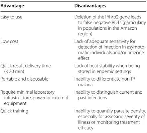
Table 2 Advantages and disadvantages of current malaria RDTs
Advantage Disadvantages
Easy to use Deletion of the Pfhrp2 gene leads to false negative RDTs (particularly in populations in the Amazon region)
Low cost Lack of adequate sensitivity for detection of infection in asympto‑ matic individuals and/or prozone effect
Quick result delivery time
(< 20 min) Lack of heat stability when being stored in endemic settings Portable and disposable Inability to differentiate non‑Pf
malaria Require minimal laboratory
infrastructure, power or external equipment
Inability to distinguish current and past infections
[image:2.595.56.541.104.337.2] [image:2.595.304.540.513.726.2]Although using the same technology of lateral flow immunoassays, the performance of malaria RDTs varies greatly from brand-to-brand, and lot-to-lot, especially with specimens having low parasite density (< 200 para-sites/μL). In a collaboration between the Foundation for Innovative New Diagnostics (FIND), the WHO and the Centers for Disease Control and Prevention, 293 malaria RDTs were evaluated from 2008 to 2016 [31]. Most of the evaluated malaria RDTs detect P. falciparum histi-dine-rich protein 2 (PfHRP2) or P. falciparum lactase dehydrogenase (PfpLDH). In the last round of evalua-tion, anomalies that interfered with result interpretation were also recorded [31]. The most common anomalies were incomplete clearing and red background, which were observed in 48 and 24% of products. The second most common anomalies were failed migration of liquid, incomplete migration and patchy broken test lines, which occurred in 15, 11 and 11% of the products, respectively.
The performance of lateral flow-based RDTs depends on two main factors: the sensitivity and specificity of antibody-antigen combinations, and the ability to facili-tate reliable liquid migration on the nitrocellulose mem-brane. Much research has focused on new biomarker discovery [32–34], and only limited attention has been paid to reduce limitations imposed by the inhomoge-neous migration of liquid across porous nitrocellulose membranes [35].
Figure 1 illustrates how unstructured the flow paths could be in a nitrocellulose membrane [36]. As the migration of liquid occurs in a porous network and is not actively controlled, a number of limitations arise: large volumes of sample needed, accumulation of reagents at the leading edge of the liquid flow, and increased cross-reactivity [37]. It is, therefore, time to consider alterna-tive options to facilitate a more precise liquid migration, hence more accurate test results.
Promising and alternative technologies for malaria detection
Table 3 summarizes six major classes of technologies used for detecting malaria and indicates their maturity levels. These technologies are individually reviewed in depth elsewhere [38] and most of them rely on stand-ard concepts using immunoassays [39, 40], molecular diagnostics [41–49] and the visualization of parasites [50–53]. Table 4 provides specifications of some recently entered market malaria diagnostic [38]. Of those market-ready products, four of them are molecular diagnostics, three are immunoassays and one is based on automated microscopy. Several promising proof-of-concepts for the next generation of malaria RDTs are emerging. For exam-ple, prototypes have been built to detect the presence of haemozoin in blood sample [54–57]. Haemozoin crystals
are produced by Plasmodium parasites as a final non-toxic compound of haemoglobin metabolism. In a spe-cific example, a portable light meter was built to image crystalized haemozoin pigment [58]. These pigments are birefringent, so the detection of haemozoin is based on rotating a plane of polarized light through them and observing anisotropic output of the light. The minimum concentration of haemozoin that could be detected with this polarized light system was 15 pg/mL, equivalent to 30 parasites/μL of blood. Applications in the field are to be tested.
Another example utilizes a portable breath analyzer: breaths of malaria-infected patients were found to con-tain terpenes, a family of aromatic chemicals that are produced by parasites that can further attract mosquitoes [59, 60]. A pilot study in Malawi confirmed that these aromatic compounds could be transported into the lungs and hence could be detected in the exhalation of infected patients [61].
Despite being unquestionably novel, these abovemen-tioned methods of detection still need to prove their practicality for POC in LRS and demonstrate a clini-cally relevant limit of detection (LOD). For instance, in the breath analyzer, it would be useful to be able to con-vert the level of terpenes detected in breath into parasite density.
Specifications for a new generation of malaria RDTs
Different settings require different target product pro-files (TPP) [8]. Unlike previous malaria control cam-paigns, the key characteristics of malaria elimination efforts are to interrupt endemic transmission and to prevent its re-establishment [62]. The Program for
[image:3.595.309.538.87.259.2]Appropriate Technology in Health (known as PATH) and FIND are pioneering the development and valida-tion of sensitive rapid tests for mass screening in LRS.
They also proposed a TPP for malaria RDTs in elimina-tion settings, stating specific requirements for the ideal rapid tests according to concept of Affordable, Sensitive, Table 3 Examples of promising technologies for point-of-care diagnostics. table based on information contained in Ref [38]
Technology Early stage of R&D Design and development Evaluation Regulatory approval(s) Piloting Post market surveillance Laboratory Field application
Microscopy
Autoscope - 2015 [50]
Intellectual Ventures Laboratory
Foldscope [51]
Stanford University Parasight - 2014 [52]Sight Diagnostics Commercially available malaria microscopy
Cellphone-based microscopy [53]
CellMic
Antigen detection
Highly sensitive Pf
RDTs - 2017 [39]
Alere
Fluorescent-based urine malaria test -2015 [40]
Fyodor
Commercially available HRP2, pLDH and pan -malaria RDTs (lateral flow assays)
Nucleic acid detection
NALFIA DIGMAL [41]
Diagmal Consortium
Saliva based test -2015 [42] John Hopkins & Ceres Nanosciences
Truelab - 2013 [43]
Molbio Diagnostics
Commercially available PCR & LAMP for research purposes
Accutas [44]
Auilia
Illumigence LAMP -2016 [45]
Meridian
LabDisk - 2015 [46]
DiscoGnosis NINA LAMP [47]PATH LAMP - 2012 [48]Eiken & FIND
NANOMAL Q-POC [49]
QuantuMdx
Hemozoin detection
MRR -2015 [54]
Singapore - MIT
MOT - 2008 [55]
University of Exeter
VNB - 2015 [56]
Rice University
Magneto Optical -2014 [57]
Budapest Univeristy
Spectroscopy Breath test [61]University of Washingon
Commercially available spectrometer
Serology ELISAn/a
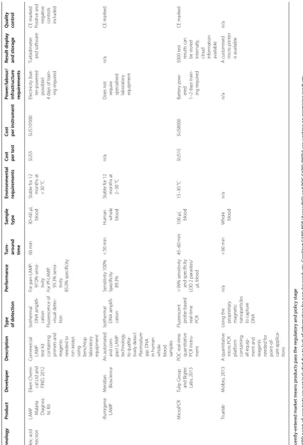
[image:4.595.59.537.101.656.2]Table 4 Sp ecific ations of r ec en tly -en ter ed mar ket* t echnolo gies f or malaria diagnosis
. table based on inf
orma
tion c
on
tained in Ref [
38 ] Technology Pr oduc t De veloper Description
Type of det
ec
tion
Per
formanc
e
Turn- around time Sample type
En vir onmen tal requir emen ts
Cost per t
est
Cost per instrumen
t Po w er/labour/ infr astruc tur e requir emen ts Result displa y and st or age Q ualit y con tr ol M icr oscop y Parasight Sight Diag ‑ nostics L td , 2014 A ut omat ed micr oscop y suitable f or pr ocessing
of multiple malar
ia Slide r eading Under wa y n/a
Blood smear
n/a n/a n/a n/a M alar ia RD Ts** Fio ‑net Fio C or pora ‑ tion, 2012 Univ ersal RD T r eader
and cloud infor
mation ser vices t o impr ov e malar ia RD T qualit y
assurance and malar
ia
sur
veillance
Combination of mobile diag
nostics
(mobile uni
‑
versal r
eader)
with cloud infor
mation ser vices A ut omat ed and cust omising repor ts Sensitivit y and specificit y ar e func tions
of the RD
Ts
being r
ead
RD
Ts processing time is depend
‑
ent on manu
‑ fac tur er ’s recom ‑ menda ‑ tion
Data upload within minut
es Daily qualit y contr ol needed D epend ‑
ing on RDTs
’ manu ‑ fac tur ers Subjec t t o RD Ts ma nuf ac tu rers ’ recommenda ‑ tions 5–40 °C Similar t o pr e‑ paid cellphone plans Batt er y po w ‑ er ed
Basic 1 da
y
training needed
On scr een and w eb por tal CE mar ked U MT Fy
odor Biot
ech ‑ nolog ies , 2015 A senstitiv e
and specific lateral flow assa
y det ec t‑ ing no vel
Plasmodium prot
eins
shed in the ur
ine of f ebr il malar ia patients
Dipstick technology (lat
eral flo
w
assa
y)
LOD 125 para
‑ sit es/µL ~ 20 min 100 µL ur ine n/a n/a Usable b y la y people n/a n/a
Holomic Rapid Diag
‑
nostic Reader
Holomic LL C, 2013 Univ ersal RD T r eader
attachment for smar
t‑
phones and sof
twar
e t
o
read RD
Ts
and transmit result t
o
a secur
e
cloud infor
mation ser vice Por table , smar t‑ phone ‑based lat eral flo w immunoas ‑ sa y r eader Quantitativ e and qualita ‑ tiv e RD
Ts processing time is depend
‑
ent on manu
‑ fac tur er ’s recom ‑ menda ‑ tion
Data upload within seconds
D
epend
‑
ing on RDTs
’ manu ‑ fac tur ers Subjec t t o RD Ts manufac tur ‑ ers ’ 5–40 °C Cust omis ‑ able $US500 Batt er y po w ‑ er ed Basic < 0.5 da y training needed User int er face
of the smar
tphones
application
Class I medi
‑
cal de
Table 4 (c on tinued) Technology Pr oduc t De veloper Description
Type of det
ec
tion
Per
formanc
e
Turn- around time Sample type
En vir onmen tal requir emen ts
Cost per t
est
Cost per instrumen
t Po w er/labour/ infr astruc tur e requir emen ts Result displa y and st or age Q ualit y con tr ol
Nucleic acid det
ec
tion
LA
MP Malar
ia Diag nos ‑ tic K it Eik en Chemi ‑ cal L td and FIND , 2012 Commer cial LA MP test k it
containing primers and reagents needed t
o
run assa
ys
using bencht
op laborat or y equipment Isother mal DNA amplifi ‑ cation Fluor escence of visual det ec ‑ tion For pan ‑LA MP : 97.0% sensi ‑ tivit y For Pf ‑LA MP : 93.3% sensi ‑ tivit y 85.0% specificit y 60 min 30–60 µL blood Stable f or 12
months at < 30
°C $US5 $US10’000 Elec tr icit y (bat ‑ ter ‑po w er ed possible) 4 da
ys of train
‑ ing r equir ed Tur bidimet er and sof twar e CE mar ked Positiv e and negativ e contr ols included
illumigene LA
MP M er idian Bioscience An aut omat ed and com ‑ pac t LA MP
technology to qualita
‑
tiv
ely det
ec
t
Plasmodium spp
. DNA
in human whole blood samples
Isother mal DNA amplifi ‑ cation Sensitivit y 100% Specificit y 89.3% < 50 min
Human whole blood
Stable f
or 12
months at 2–30
°C
n/a
D
oes not requir
e
specialised laborat
or y equipment n/a CE mar ked M icr oPCR Tulip Gr oup and Bigt ec Labs , 2013 POC r eal ‑time quantitativ e PCR instru ‑ ment Fluor escent pr obe ‑based real ‑time PCR > 99% sensitivit y and specificit y
LOD 2 parasit
es/ µL blood 45–60 min 100 µL blood 15–30 °C $US15 $US8000 Batt er y po w ‑ er ed 1–2 da ys train ‑ ing r equir ed 5000 t est
results can be st
or ed int er nally ,
cloud infor
mation available CE mar ked Truelab M olbio , 2013 A quantitativ e micr o PCR platf or m
containing all equip
‑
ment and reagents needed f
or point ‑of ‑ car e applica ‑ tions
Using the pr
opr ietar y mag netic nanopar ticles to captur e DNA n/a < 60 min
Whole blood
n/a n/a A cust omised micr o pr int er is a vailable n/a * R ec en tly -en ter ed mar
ket means pr
oduc
ts pass the r
egula
tor
y and polic
y stage
** G6PD poin
t-
of-car
e t
ests ar
e not included due t
o lack of inf
or
ma
tion f
or popular pr
oduc ts . C ar eS tar
t G6PD RD
T (
Ac
cessBiO
) and POC G6PD (P
ATH) ar
e w
or
king on pr
omising pr
oduc
[image:6.595.67.495.86.713.2]Specific, User-friendly, Equipment-free and Deliverable (ASSURED) [63]. The desired LOD is 5 parasites/µL or less, or concentration range of 6–12 ng/mL PfHRP2 [63]. For RDT developers it is important to note the caveat of the prozone phenomenon that might prevent detec-tion of high parasite density [64]. Poor specificity could lead to over-treatment, thus depreciation of the intended value of RDTs (from public health perspectives); there-fore, the required specificity for effective malaria diagno-sis is at least 97% or ideally 99% [63].
Additional requirements for ideal RDTs are suitability and appropriateness for LRS where most malaria cases occur. To make an impact simplicity and affordability are of utmost importance. Simplicity means, the system should be equipment-free and should require very little resources [65]. A simple and automated test could obvi-ate false results caused by user-errors [66]. Affordability is difficult to measure and depends on the cost–benefit equation of a specific situation. Also, tests should be designed to minimize impact of inappropriate storage conditions (2–40 °C) on reagent stability and usability of the devices [67].
Microfluidic technology for malaria POC testing
Microfluidics enable the miniaturization and simplifica-tion of complicated analytical processes while consuming less reagents, minimizing waste, and requiring less sup-porting instrumentation [68]. This stems out from the predictable behaviour of liquids at the microscale where flow is typically laminar. At microscale, minute amounts
of liquids can be manipulated using microstructures, such as microvalves, micromixers or micropumps [69]. Low volumes of reagents, fast reaction times, compact and portable platforms are just a few advantages that make microfluidics technology attractive for POC appli-cations [70, 71]. Figure 2 shows several examples demon-strating the archetype of microfluidic-based diagnostics for POC applications, which is an integrated system com-posed of a disposable unit (where analysis takes place) and a signal acquisition and processing module to pro-cess the results. (a) [72], (b) [73], (c) [74].
Currently, microfluidic-based diagnostic devices can be divided into two categories: non-paper-based “tradi-tional” microfluidics and paper-based microfluidics [75,
76]. Research on traditional microfluidics often focuses on miniaturizing conventional techniques. For example, a collection of passive and active mixing elements were designed to facilitate mixing processes on chips [77]. Recent work in developing microfluidic-based diagnostic devices has focused on integrating all necessary elements into stand-alone platforms [78, 79] because such inte-grated systems can operate without bulky accessories and do not require water, buffer, or a constant supply of elec-tricity [80]. There are many ways to control liquid flows on microfluidic platforms, for instance, acoustic forces, mechanical forces, magnetic forces, as well as capillary and centrifugal forces [81–85]. To satisfy the stringent requirements for LRS, devices based on capillary and centrifugal forces have shown promising results. Table 5
presents some examples of microfluidic-based systems
a Microfluidic-based point-of-care dongle b Lab-on-a-disk diagnostic platform c Paper-based diagnostics
Antibody holder
1. Blood in cassette inlet
Reagent cassette
Test cassette 2.
Gold-labelled antibodies
3. Washes Venting port 4. Silver B
4. Silver A
[image:7.595.58.539.469.688.2]that have been designed to detect PfHRP2 and PfpLDH antigens or genetic materials from the parasites using on-chip molecular testing, cell deformation mechanism, electrical, optical, and magnetic detections amongst oth-ers [54, 58, 79, 81, 86–94].
Immunodiagnostics on microfluidic platforms for malaria detection
Standard protocols to perform immunodiagnostics on microfabricated platforms require sample pre-concentra-tion, flow control and detection of biomarkers (analytes
Table 5 Performance of proof-of-concept platforms based on microfluidics for malaria detection
RBC red blood cell, iRBC infected red blood cell
Application Concept/detection
principle Biomarker/target Limit of detection Performance Time (min) Refs
Sensitivity (%) Specificity (%)
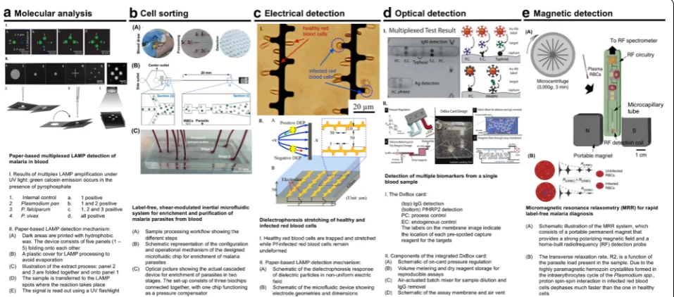
Molecular analysis Paper‑based LAMP P. falciparum 5 p/µL 61% 98% 45 min [81]
P. vivax 81% 98%
P. pan > 80% > 98%
Continuous flow PCR P. falciparum 2 p/µL 97.40% 93.80% n/a [86]
< 1 p/µL n/a n/a 2.5 h [87]
Cell deformation
mechanism Inertial focusing
P. falciparum 2–10 p/µL n/a n/a 400 µL/min [88]
Inertial microfluidics P. falciparum iRBCs 2 cells/min n/a [89] Non‑inertial lift effect P. falciparum ring
stage iRBCs Enrichment factor of 4.3 n/a [90] Throughput
12,000 cells/h Electrical detection Electrical conductivity
of iRBCs is signifi‑ cantly higher than healthy RBCs
P. falciparum ring
stage n/a n/a [91]
Optofluidic‑flow analyser that can measure the optical absorption of RBCs in P. falciparum
infected blood sample
P. falciparum 1712 RBCs/s n/a 3 min [92]
2.96% parasite density
Naked‑eye screening of in‑meso detec‑ tion of hemozoin crystallites based on birefringence
Hemozoin crystals produced by P. falciparum
n/a ~ 12 min [58]
Optical detection Visual detection of colored assay spot on a disposable microfluidic card based on a flow‑ through membrane immunoassay
Malaria PfHRP2 10–20 ng/mL n/a 1–5 min [79]
Paper‑based catridge containing detec‑ tion areas for both thin and thick smears
P. falciparum 100 p/µL n/a 30 min [93]
Magnetic detection Cell enrichment microfluidics com‑ bined with mag‑ netic relaxometry detection
P. falciparum ring
stage parasites 5% parasite density n/a 15 min [54]
Detection of hemo‑ zoin in iRBCs by magnetic resonance relaxometry
Hemozoin in iRBCs in
P. falciparum infec‑
tions
[image:8.595.57.541.100.625.2]and/or parasites). These multi-step protocols can benefit greatly from miniaturization, and in fact, microfluidic-based immunoassays have demonstrated their potential for reliable and accurate performance [95, 96]. Figure 3
presents some examples to illustrate how microfluid-ics technology can be used to detect malaria by different methods of detection, such as molecular testing, size-based cell sorting, electrical differentiation of healthy and infected red blood cells, optical detection of antigen and magnetic detection of haemozoin. (a) [97], (b) [88], (c) [91], (d) [79], (e) [94].
Sample pre-concentration
Low antigen concentration is a common problem in diagnostic immunoassays and malaria antigen detection is not an exception. To overcome this challenge, several prototypes of analyte concentrator have been developed to enrich biomarkers hence improve LOD. To illustrate how analyte enrichment prior to analysis can improve sensitivity of ELISA, Cheow et al. reported a prototype that can enhance the LOD of prostate-specific-antigen assay up to 1.85 pg/mL [98]. The significant enhancement of 100-fold was achieved by trapping the charged fluores-cent product of standard ELISA (analyte-bound enzyme complex) using a multiplex electrokinetic preconcentra-tion technique without modifying the immunobinding process.
Blood is the most common type of specimen for POC testing. However, the cellular components in whole blood often cause non-specific background. To address this problem, a continuous microfluidic device was developed
to filter the cells, making plasma available for on-chip analysis [99].
Healthy and P. falciparum-infected red blood cells exhibit different ionic permeability of their plasma mem-brane, with infected cells being more permeable. There-fore, when healthy and infected cells are suspended in a low conductivity medium, infected cells lose internal ions and acquire a different dielectrophoretic mobility than healthy ones [100]. Several groups have developed microfluidic chips using dielectrophoresis and variants of it to separate cells successfully leading to promising pro-totypes for detecting infected red blood cells thus malaria infections [101–103].
Flow control
Controlling flow on microfabricated devices often introduces a great degree of complexity. For example, a combination of screws, pneumatic and solenoid valves was integrated into a microfluidic platform to actuate flow and control chemical gradients in microchannels [104]. This design might be suitable for laboratory-based tests, but may not lead to robust systems for LRS. Nonetheless, the uses of centrifugation and capillary forces to transport liquids are excellent examples of stand-alone systems [105, 106]. Extensive reviews dis-cussing how to engineer flow path in microscale using capillary and centrifugal forces for POC applications exist [69, 107]. Libraries of microfluidic elements such as valves, mixers and pumps have also been developed [77, 108, 109].
[image:9.595.60.539.478.689.2]Detection
Sensitive detection remains one of the biggest hurdles for clinical diagnosis at the onset of infection. The bottleneck is the limited amount of detectable analytes in a very limited volume of sample. One strategy is to amplify the signal, then convert it into quantitative measurements such as electrical and/or optical signals [96]. The detec-tion strategy is therefore critical for the overall design and fabrication of a device. Optical detection is consid-ered as the ideal read-out for POC applications of micro-fluidics owing to the simple design and potentially low cost [110, 111]. There are five main categories of optical detection based on the type of generated optical signals: fluorescence, luminescence, absorbance, surface plas-mon resonance, and surface-enhanced Raman scattering [112–116]. Detailed discussions about detection strate-gies for microfluidics systems also exist in the literature [117].
Molecular testing on microfluidic platforms for malaria detection
At the moment, PCR and LAMP are the most sensitive technique for identification of asymptomatic individuals, for example, in 130 clinical samples presenting no para-sites based on microscopy, as low as 3.6 × 10⁻⁴ parasite/ μL could be identified in 117 samples by a highly sensi-tive genus-specific quantitasensi-tive reverse transcriptase real-time PCR (qPCR) [118]. This low LOD was achieved by amplifying and detecting the total nucleic acids of the 18S rRNA genes, which increased the analytical sensitiv-ity of the assay by more than 1 log unit compared to DNA only. However, current applications of PCR and LAMP are still restricted to well-equipped laboratories and thus not suitable for LRS [119]. Miniaturized PCR and/or LAMP is desirable, but developing such devices is a more challenging task than that for biomarkers detection for three reasons: (1) sample pre-treatment is essential for extracting DNA of parasites for downstream analysis, (2) the critical signal amplification step highly depends on temperature control, and (3) robust, low cost, and porta-ble detection techniques are required for remote settings [120].
Sample pre-treatment
The PCR/LAMP process requires isolation of genetic materials from infected cells, pre-concentration, as well as signal amplification and analysis. All steps need to be integrated seamlessly in a closed process to overcome time consuming laboratory-like processing steps. Ear-lier studies have demonstrated successful prototypes that could sequentially perform cell isolation and lysis
for messenger RNA purification [121]. On this device, a unique valving system was designed to facilitate liquid migration and analysis. Microfluidics with “macrofluid-ics” can also be combined to precisely reconstitute rea-gents, and automated filling liquids for multiplex PCR technique. A successful story is the Cepheid GeneXpert instrument, where all steps from sample preparation, nucleic acid extraction, to thermal cycling for amplifica-tion and eventually detecamplifica-tion can be integrated into one platform [122]. A review of microfluidic-based DNA analysis systems is available here [123].
Heating systems
The major challenge of miniaturizing bench-top PCR instruments is the requirement of numerous heating cycles for thermal reactions. To overcome this challenge, micromixers and microchambers were designed to allow thermal reactions to take place rapidly [124]. To speed up DNA amplification by improving thermal transfer through interfaces, microfluidic elements, such as mix-ers, heaters and temperature controlling units were inte-grated into glass and silicon substrates [125]. Another strategy to enable different heating regions using con-tinuous flow was investigated using a Peltier element to regulate the temperature for thermal cycling [86]. On this platform, as few as to 2 P. falciparum parasites/μL could be detected. This device offered a simplified sam-ple processing step using desiccated hydrogel, reagents and a camera to detect amplicons. When analysing 188 archived, frozen samples collected in Uganda, this proto-type achieved 97.4% sensitivity and 93.8% specificity.
One of the most promising development for stand-alone integrated systems for DNA analysis perhaps was an elegant combination of an exothermic reaction with phase change materials to regulate the heat for thermal cycling [126]. In this prototype, downstream processes such as purification and concentration of sample were integrated seamlessly into the same platform.
Paper-based microfluidics
Paper-based microfluidics was proposed by Whitesides and colleagues [127]. Since then, this technology has been growing fast with great promises for global health applications [128]. Unlike its sister products of paper test strips, paper-based microfluidic analytical devices offer well-defined, millimetre-sized microchannels to trans-port liquids in a controlled manner, yet with low cost for production (< $0.01) [129]. Using hydrophobic “inks” to define areas on hydrophilic paper, it is possible to per-form multiple immunodiagnostic assays on the same test strip. To illustrate how complex analytical processes can be simplified and transformed into a paper-based micro-fluidic device, Pereira et al. integrated concentration and detection steps into a single step assay [130]. The analyte PfpLDH in low abundance was first accumulated using a micellar aqueous two-phase system (ATPS). The micellar ATPS consisted in a nonionic Triton X-114 surfactant, which was used to concentrate biomarkers in a sample and enhance the LOD. In this system, a tenfold improved LOD of 10 ng/μL PfpLDH was achieved. In an alternative development of a foldable, card-like test device, PfHRP2 could be detected and quantified [131]. The generated signal in presence of PfHRP2 was amplified by gold nan-oparticles, yielding a LOD of 1.2 ng/mL PfHRP2, which is four times higher than that of the unamplified case. These studies serves as excellent examples for low cost, non-instrumented analysis systems without compro-mised performance. Many other innovative approaches to control liquid flows such as selective hydrophobic ren-dering or origami in which folding of multiple paper lay-ers to trigger reactions were also investigated successfully [132–134].
Interfacing microfluidic-based analysis with networked mobile devices
Mobile health applications have rapidly been growing in recent years and there is a trend in interfacing con-sumer electronics such as smartphones with lateral flow RDTs or microfluidic-based devices [135, 136]. Such combination is expected to deliver increased objectivity of test result interpretation and improved connectivity of the entire healthcare systems. The automation and digitized test results can be more easily combined with other health related parameters and combined with medical decision support systems. User-friendly inter-faces, automated result analyses, remote-monitoring and data aggregation, increased storage conditions, and active quality assurance are just a few additional ben-efits of this approach [137].
In 2008, paper-based microfluidics were integrated with a smartphone camera to perform immunoassays
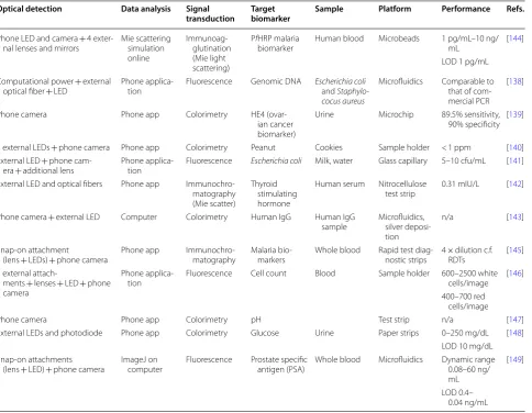
[128]. The camera of the phone was used to take a photograph of the detection zone before and after the deposition of specimen. Since then, many groups have started to develop and enhance capabilities of phone-based low cost diagnostic readers [136]. Table 6
presents an overview of recent work in developing phone-based prototypes that can be used to detect vari-ety of biomarkers for a wide range of diseases with clin-ically relevant performance. Devices are designed for a broad spectrum of applications, from genetic testing, cancer detection to personalized food allergen moni-toring [136, 138–140]. A wide range of strategies are also derived to enhance signal strength, for instance, using Quantum dots, Rayleigh/Mie scatter or gold nan-oparticles [141–143]. At present, applications of smart-phone-based diagnostics for malaria detection can be divided into two categories: phone-based RDT readers, which provides automatic interpretation of results, and phone-based brightfield microscopes, which allow sim-ple and portable means to visualize parasites in blood samples [138–149].
Phone-based RDT readers
A smartphone was used for quantitative reading of the Optimal-IT test, a commercially available malaria RDT with a snap-on unit as reader that is suitable for both Android and iPhone [145]. Images of RDTs were acquired, in either transmission or reflection, and then processed in real time to deliver test results within 10 min. The spatio-temporal information collected by this device can document prevalence of many infec-tious diseases and would allow efficient tracking of epidemics. Another approach to integrate a custom microfluidic-based immunoassay detecting PfHPR2 with phone-based detection was the development of a microfluidic chip, which can be connected to a phone camera to analyze signals and deliver results in 10 min. The opto-mechanical unit in this case consisted of opti-cal fibers, microfluidic chips and mirrors, and could be easily removed from the back camera of the phone. The principle was to quantify changes in fluorescent inten-sity upon capturing of PfHPR2 on the sensing region, yielding a LOD of 1 pg/mL of PfHRP2 in 10% diluted blood [144].
Phone-based bright-field microscope
simplified imaging techniques suitable for smartphone apps: (1) lens-free holographic imaging, and (2) on-lens devices.
Holography is an image-constructing technique using scattering and interference of light and pixel super-res-olution to enhance optical images [151]. An automated lens-less holography was developed with a sufficient field of view of 24 mm2 to visualize and capture images of P.
falciparum in blood smears [152].
Phone-based microscopy can also be engineered to be a field-ready polarized light microscope without compro-mised fidelity and resolution [153]. The principle was to detect light birefringence caused by the crystallization of haemozoin. This field-based, modular microscope could magnify Plasmodium chabaudi parasites up to 50 times, gaining a comparable performance compared to conven-tional polarized microscope. Addiconven-tional benefits of this prototype are simple operations and low cost per test. Further work using clinical samples could confirm the
full potential of this novel phone-based polarized light microscope.
Conclusion
Accurate and effective diagnosis is the first step to fur-ther pursue efforts to eliminate and reduce the global burden of malaria by 90% in 2030. Current diagnostic methods can detect malaria symptomatic infections, but often miss out asymptomatic cases. The rise in pro-portion of asymptomatic infections in low transmission areas calls for a new generation of rapid diagnostic tests that can detect the hidden parasite reservoir. Technology is advanced nowadays to (at least theoretically) be able to track down the last parasite carriers. While malaria case management has improved, other causes of fever need to be detected and treated accordingly. Therefore, the ideal RDT should come in as a complete package with ultra-high sensitivity and specificity, meet the ASSURED standards for LRS, and also provide additional diagnostic
Table 6 Examples of lab-on-a-phone applications
Optical detection Data analysis Signal
transduction Target biomarker Sample Platform Performance Refs.
Phone LED and camera + 4 exter‑
nal lenses and mirrors Mie scattering simulation online
Immunoag‑ glutination (Mie light scattering)
PfHRP malaria
biomarker Human blood Microbeads 1 pg/mL–10 ng/mL [144] LOD 1 pg/mL
Computational power + external
optical fiber + LED Phone applica‑tion Fluorescence Genomic DNA Escherichia coliand Staphylo-cocus aureus
Microfluidics Comparable to that of com‑ mercial PCR
[138]
Phone camera Phone app Colorimetry HE4 (ovar‑ ian cancer biomarker)
Urine Microchip 89.5% sensitivity, 90% specificity [139]
2 external LEDs + phone camera Phone app Colorimetry Peanut Cookies Sample holder < 1 ppm [140] External LED + phone cam‑
era + additional lens Phone applica‑tion Fluorescence
Escherichia coli Milk, water Glass capillary 5–10 cfu/mL [141]
External LED and optical fibers Phone app Immunochro‑ matography (Mie scatter)
Thyroid stimulating hormone
Human serum Nitrocellulose
test strip 0.31 mIU/L [142]
Phone camera + external LED Computer Colorimetry Human IgG Human IgG
sample Microfluidics, silver deposi‑ tion
n/a [143]
Snap‑on attachment
(lens + LEDs) + phone camera Phone app Immunochro‑matography Malaria bio‑markers Whole blood Rapid test diag‑nostic strips 4 ×RDTs dilution c.f. [145] 3 external attach‑
ments + lenses + LED + phone camera
Phone applica‑
tion Fluorescence Cell count Blood Sample holder 600–2500 white cells/image [146] 400–700 red
cells/image
Phone camera Phone app Colorimetry pH Test strip n/a [147]
External LEDs and photodiode Phone app Colorimetry Glucose Urine Paper strips 0–250 mg/dL [148] LOD 10 mg/dL Snap‑on attachments
(lens + LED) + phone camera ImageJ on computer Fluorescence Prostate specific antigen (PSA) Whole blood Microfluidics Dynamic range 0.08–60 ng/ mL
[149]
[image:12.595.60.545.101.477.2]capabilities. Microfluidic devices coupled to phone-based readouts offer a unique opportunity to not only reduce the burden of infectious diseases, such as malaria, but also could provide tools for monitoring epidemics and elimination progress on very large scales.
Authors’ contributions
NMP drafted the manuscript; NMP and EMD wrote the manuscript with contri‑ butions from HPB and WK. All authors read and approved the final manuscript.
Author details
1 Department of Health Sciences and Technology, ETH Zürich, Lengghalde 5, 8092 Zurich, Switzerland. 2 Swiss Tropical and Public Health Institute, Socinstrasse 57, 4051 Basel, Switzerland. 3 University of Basel, Petersgraben 1, 4001 Basel, Switzerland. 4 IBM Research‑Zurich, Säumerstrasse 4, 8803 Rüsch‑ likon, Switzerland.
Acknowledgements
Not applicable.
Competing interests
The authors declare that they have no competing interests.
Availability of data and materials
Not applicable.
Consent for publication
Not applicable.
Ethics approval and consent to participate
Not applicable.
Funding
NMP receives doctoral scholarship funding from the Engineering for Develop‑ ing Program at ETH Zürich (Sawiris Foundation).
Publisher’s Note
Springer Nature remains neutral with regard to jurisdictional claims in pub‑ lished maps and institutional affiliations.
Received: 2 March 2018 Accepted: 3 July 2018
References
1. Cox FEG. History of human parasitology. Clin Microbiol Rev. 2002;15:595–612.
2. WHO. Accelerating progress on HIV, tuberculosis, malaria, hepatitis and neglected tropical diseases : a new agenda for 2016–2030. Geneva: World Health Organization; 2015. p. 64.
3. WHO. World malaria report 2017. Geneva: World Health Organization; 2017.
4. Gates Foundation. From aspiration to action: what will it take to end malaria. Seattle: Bill & Melinda Gates Foundation; 2015.
5. WHO. Global technical strategy for malaria 2016–2030. Geneva: World Health Organization; 2015.
6. Mabey D, Peeling RW, Ustianowski A, Perkins MD. Diagnostics for the developing world. Nat Rev Microbiol. 2004;2:231–40.
7. Tangpukdee N, Duangdee C, Wilairatana P, Krudsood S. Malaria diagno‑ sis: a brief review. Korean J Parasitol. 2009;47:93–102.
8. The malERA Consultative Group on Diagnoses and Diagnostics. A research agenda for malaria eradication: diagnoses and diagnostics. PLoS Med. 2011;8:1–10.
9. Vallejo AF, Martínez NL, González IJ, Arévalo‑Herrera M, Herrera S. Evalu‑ ation of the loop mediated isothermal DNA amplification (LAMP) kit for malaria diagnosis in P. vivax endemic settings of Colombia. PLoS Negl Trop Dis. 2015;9:e3453.
10. Cook J, Aydin‑Schmidt B, González IJ, Bell D, Edlund E, Nassor MH, et al. Loop‑mediated isothermal amplification (LAMP) for point‑of‑care detection of asymptomatic low‑density malaria parasite carriers in Zanzibar. Malar J. 2015;14:43.
11. Lucchi NW, Ndiaye D, Britton S, Udhayakumar V. Expanding the malaria molecular diagnostic options: opportunities and challenges for loop‑ mediated isothermal amplification tests for malaria control and elimina‑ tion. Expert Rev Mol Diagn. 2018;18:195–203.
12. WHO. Determining cost effectiveness of malaria rapid diagnostic tests in rural areas with high prevalence. World Health Organization Western Pacific Region. http://www.wpro.who.int/sites /rdtPA GE. Accessed 21 May 2018.
13. WHO. New perspective in malaria diagnosis. Geneva: World Health Organization; 2000.
14. Das S, Peck RB, Barney R, Jang IK, Kahn M, Zhu M, et al. Performance of an ultra‑sensitive Plasmodium falciparum HRP2‑based rapid diagnostic test with recombinant HRP2, culture parasites, and archived whole blood samples. Malar J. 2018;17:118.
15. Das S, Jang IK, Barney B, Peck R, Rek JC, Arinaitwe E, et al. Perfor‑ mance of a high‑sensitivity rapid diagnostic test for Plasmodium falciparum malaria in asymptomatic individuals from Uganda and Myanmar and naive human challenge infections. Am J Trop Med Hyg. 2017;97:1540–50.
16. Program for Appropriate Technology in Health. Project diameter— enhanced visual parasite detection; 2014.
17. BioCat GmbH. Loop mediated isothermal amplification (LAMP). https ://www.bioca t.com/genom ics/rna‑ampli ficat ion/loop‑media ted‑isoth
ermal ‑ampli ficat ion‑lamp. Accessed 21 May 2018.
18. Hathiwala R, Mehta PR, Nataraj G, Hathiwala S. LED fluorescence micros‑ copy: novel method for malaria diagnosis compared with routine methods. J Infect Public Health. 2016;10:1–5.
19. Wongsrichanalai C, Barcus MJ, Muth S, Sutamihardja A, Wernsdorfer WH. A review of malaria diagnostic tools: microscopy and rapid diag‑ nostic test (RDT). Am J Trop Med Hyg. 2007;77:119–27.
20. Feleke DG, Tarko S, Hadush H. Performance comparison of CareStart™ HRP2/pLDH combo rapid malaria test with light microscopy in north‑western Tigray, Ethiopia: a cross‑sectional study. BMC Infect Dis. 2017;17:399.
21. Jimenez A, Rees‑Channer RR, Perera R, Gamboa D, Chiodini PL, Gonza‑ lez IJ, et al. Analytical sensitivity of current best‑in‑class malaria rapid diagnostic tests. Malar J. 2017;16:128.
22. UNITAID. Malaria diagnostics technology and market landscape 2015; 2015.
23. Hopkins H, Gonzalez IJ, Polley SD, Angutoko P, Ategeka J, Asiimwe C, et al. Highly sensitive detection of malaria parasitemia in a malaria‑endemic setting: performance of a new loop‑mediated isothermal amplification kit in a remote clinic in Uganda. J Infect Dis. 2013;208:645–52.
24. Cordray MS, Richards‑Kortum RR. Emerging nucleic acid‑based tests for point‑of‑care detection of malaria. Am J Trop Med Hyg. 2012;87:223–30. 25. Erdman LK, Kain KC. Molecular diagnostic and surveillance tools for
global malaria control. Travel Med Infect Dis. 2008;6:82–99.
26. Hofmann N, Mwingira F, Shekalaghe S, Robinson LJ, Mueller I, Felger I. Ultra‑sensitive detection of Plasmodium falciparum by amplification of multi‑copy subtelomeric targets. PLoS Med. 2015;12:e1001788. 27. WHO. New perspectives malaria diagnosis. Geneva: World Health
Organization; 2000.
28. Ezennia IJ, Nduka SO, Ekwunife OI. Cost benefit analysis of malaria rapid diagnostic test: the perspective of Nigerian community pharmacists. Malar J. 2017;16:7.
29. Bisoffi Z, Sirima SB, Meheus F, Lodesani C, Gobbi F, Angheben A, et al. Strict adherence to malaria rapid test results might lead to a neglect of other dangerous diseases: a cost benefit analysis from Burkina Faso. Malar J. 2011;10:226.
30. Kattenberg JH, Tahita CM, Versteeg IAJ, Tinto H, Traoré Coulibaly M, D’Alessandro U, et al. Evaluation of antigen detection tests, microscopy, and polymerase chain reaction for diagnosis of malaria in peripheral blood in asymptomatic pregnant women in Nanoro, Burkina Faso. Am J Trop Med Hyg. 2012;87:251–6.
32. Krampa F, Aniweh Y, Awandare G, Kanyong P. Recent progress in the development of diagnostic tests for malaria. Diagnostics. 2017;7:54. 33. Mathema VB, Na‑Bangchang K. A brief review on biomarkers and
proteomic approach for malaria research. Asian Pac J Trop Med. 2015;8:253–62.
34. Jain P, Chakma B, Patra S, Goswami P. Potential biomarkers and their applications for rapid and reliable detection of malaria. BioMed Res Int. 2014;2014:852645.
35. Sajid M, Kawde AN, Daud M. Designs, formats and applications of lateral flow assay: a literature review. J Saudi Chem Soc. 2015;19:689–705. 36. Credou J, Volland H, Berthelot T. Photolinker‑free photoimmobiliza‑
tion of antibodies onto cellulose for the preparation of immunoassay membranes. J Mater Chem B. 2015;3:1079–88.
37. Posthuma‑Trumpie GA, Korf J, Van Amerongen A. Lateral flow (immuno) assay: its strengths, weaknesses, opportunities and threats. A literature survey. Anal Bioanal Chem. 2009;393:569–82.
38. UNITAID. Malaria diagnostics technology and market landscape. Geneva: World Health Organization; 2016.
39. Alere. Alere Launches the Alere™ Malaria Ag P.f, the first‑ever rapid test to screen malaria infection in asymptomatic individuals; 2017. http:// news.alere .com/~/media /Files /A/Alere ‑Newsr oom‑V2/press ‑relea se/
Alere _Malar ia_Ag_Pf_launc h_relea se_42520 17_FINAL .pdf. Accessed 10
Jan 2018.
40. Fyodor Biotechnologies Corp. Urine Malaria Test (UMT); 2015. http://
www.fyodo rbio.com/produ cts/umt/. Accessed 1 Aug 2015.
41. DIAGMAL. Diagmal rapid malaria diagnostic. http://www.diagm al.eu/. Accessed 10 Jan 2018.
42. John Hopkins researchers to evaluate malaria saliva test; 2015. https :// globa lbiod efens e.com/2015/03/11/johns ‑hopki ns‑resea rcher s‑to‑evalu
ate‑malar ia‑saliv a‑test/. Accessed 10 Jan 2018.
43. MolBio Diagnostics. TrueLab. http://www.molbi odiag nosti cs.com/
produ cts_pcr_works tatio n.html. Accessed 10 Jan 2018.
44. Aquila. Accutas for malaria. http://www.aquil adiag nosti cs.com/Accut
as/for/Malar ia. Accessed 10 Jan 2018.
45. Meridian Biosciences. illumigene Malaria. http://www.merid ianbi oscie
nce.eu/em‑stron g‑illum i‑stron g‑gene‑em‑reg‑malar ia.html. Accessed
10 Jan 2018.
46. Labdisk: The portable lab bringing EU technology to Low‑ and Middle‑ Income countries; 2015. https ://ec.europ a.eu/digit al‑singl e‑marke t/ en/blog/labdi sk‑porta ble‑lab‑bring ing‑eu‑techn ology ‑low‑and‑middl
e‑incom e‑count ries. Accessed 10 Jan 2018.
47. PATH. Non‑instrumented nucleic acid amplification (NINA) platform. Program for appropriate technology in health. https ://sites .path.org/dx/
hiv‑stis/nina/. Accessed 10 Jan 2018.
48. FIND. Foundation for innovative new diagnostics. https ://www.findd
x.org/news/high‑throu ghput ‑lamp‑for‑diagn osis‑of‑malar ia/. Accessed
10 Jan 2018.
49. QuantuMdx. NANOMAL Q‑POC. http://quant umdx.com/appli catio ns/
malar ia. Accessed 10 Jan 2018.
50. Intellectual Ventures Laboratory. Automated optical diagnostics micros‑ copy; 2015. http://www.intel lectu alven tures lab.com/inven t/autom
ated‑optic al‑diagn ostic s‑micro scopy . Accessed 10 Jan 2018.
51. Foldscope Instrument. Magnify your curiosity. https ://www.folds cope. com/. Accessed 10 Jan 2018.
52. Becton Dickinson and Company. BD collaborates with Sight diagnostics to introduce parasight malaria detection device in India; 2016. http://
www.bd.com/conte ntman ager/b_artic le.asp?Item_ID=28030 .
Accessed 10 Jan 2018.
53. CellMic. http://www.cellm ic.com/conte nt/dxall dxlab /. Accessed 10 Jan 2018.
54. Fook Kong T, Ye W, Peng WK, Wei Hou H, Marcos Preiser PR, et al. Enhancing malaria diagnosis through microfluidic cell enrichment and magnetic resonance relaxometry detection. Sci Rep. 2015;5:11425. 55. Newman DM, Heptinstall J, Matelon RJ, Savage L, Wears ML, Bed‑
dow J, et al. A magneto‑optic route toward the in vivo diagnosis of malaria: preliminary results and preclinical trial data. Biophys J. 2008;95:994–1000.
56. Lukianova‑Hleb E, Bezek S, Szigeti R, Khodarev A, Kelley T, Hurrell A, et al. Transdermal diagnosis of malaria using vapor nanobubbles. Emerg Infect Dis. 2015;21:1122–7.
57. Orbán Á, Butykai Á, Molnár A, Pröhle Z, Fülöp G, Zelles T, et al. Evalua‑ tion of a novel magneto‑optical method for the detection of malaria parasites. PLoS ONE. 2014;9:1–8.
58. Vallooran JJ, Handschin S, Pillai SM, Vetter BN, Rusch S, Beck H‑PP, et al. Lipidic cubic phases as a versatile platform for the rapid detec‑ tion of biomarkers, viruses, bacteria, and parasites. Adv Funct Mater. 2015;26:1–10.
59. Kelly M, Su CY, Schaber C, Crowley JR, Hsu FF, Carlson JR, et al. Malaria parasites produce volatile mosquito attractants. Am Soc Microbiol. 2015;6:1–6.
60. Berna AZ, McCarthy JS, Wang RX, Saliba KJ, Bravo FG, Cassells J, et al. Analysis of breath specimens for biomarkers of Plasmodium falciparum infection. J Infect Dis. 2015;212:1120–8.
61. Roberts M. Malaria breath test shows promise. http://www.bbc.com/
news/healt h‑41820 346. Accessed 3 Jan 2018.
62. Moonen B, Cohen JM, Snow RW, Slutsker L, Drakeley C, Smith DL, et al. Operational strategies to achieve and maintain malaria elimination. Lancet. 2010;376:1592–603.
63. PATH. Target product profile : point‑of‑care malaria infection detection test. Program for Appropriate Technology in Health; 2014. http://sites
.path.org/dx/files /2012/11/DIAME TER_IDT_TPP_FINAL _forwe bsite .pdf.
Accessed 10 Jan 2018.
64. Gillet P, Scheirlinck A, Stokx J, De Weggheleire A, Chaúque HS, Can‑ hanga ODJV, et al. Prozone in malaria rapid diagnostics tests: how many cases are missed? Malar J. 2011;10:166.
65. Weigl BH, Boyle DS, de los Santos T, Peck RB, Steele MS. Simplicity of use: a critical feature for widespread adoption of diagnostic technolo‑ gies in low‑resource settings. Expert Rev Med Devices. 2009;6:461–4. 66. Seidahmed OME, Mohamedein MMN, Elsir AA, Ali FT, Malik EFM, Ahmed
ES. End‑user errors in applying two malaria rapid diagnostic tests in a remote area of Sudan. Trop Med Int Health. 2008;13:406–9. 67. Albertini A, Lee E, Coulibaly SO, Sleshi M, Faye B, Mationg ML, et al.
Malaria rapid diagnostic test transport and storage conditions in Bur‑ kina Faso, Senegal, Ethiopia and the Philippines. Malar J. 2012;11:406. 68. Whitesides GM. The origins and the future of microfluidics. Nature.
2006;442:368–73.
69. Gervais L, De Rooij N, Delamarche E. Microfluidic chips for point‑of‑care immunodiagnostics. Adv Healthc Mater. 2011;23:151–76.
70. Yager P, Edwards T, Fu E, Helton K, Nelson K, Tam MR, et al. Micro‑ fluidic diagnostic technologies for global public health. Nature. 2006;442:412–8.
71. Hawkins K, Weigl B. Microfluidic diagnostics for low‑resource settings. In: Proceedings of the SPIE; 2010. p. 75930L–75930L‑15.
72. Laksanasopin T, Guo TW, Nayak S, Sridhara AA, Xie S, Olowookere OO, et al. A smartphone dongle for diagnosis of infectious diseases at the point of care. Sci Transl Med. 2015;7:273.
73. Zhao Y, Czilwik G, Klein V, Mitsakakis K, Zengerle R, Paust N. C‑reactive protein and interleukin 6 microfluidic immunoassays with on‑chip pre‑stored reagents and centrifugo‑pneumatic liquid control. Lab Chip. 2017;17:1666–77.
74. Ebola epidemic: medical and military technologies converge. http:// afric aheal thitn ews.com/ebola ‑epide mic‑medic al‑and‑milit ary‑techn
ologi es‑conve rge/. cited 28 Nov 2017.
75. Sia SK, Kricka LJ. Microfluidics and point‑of‑care testing. Lab Chip. 2008;8:1982.
76. Yamada K, Shibata H, Suzuki K, Citterio D. Toward practical application of paper‑based microfluidics for medical diagnostics: state‑of‑the‑art and challenges. Lab Chip. 2017;17:1206–49.
77. Meijer HEH, Singh MK, Kang TG, Den Toonder JMJ, Anderson PD. Passive and active mixing in microfluidic devices. Macromol Symp. 2009;279:201–9.
78. Dimov IK, Basabe‑Desmonts L, Garcia‑Cordero JL, Ross BM, Park Y, Ricco AJ, et al. Stand‑alone self‑powered integrated microfluidic blood analysis system (SIMBAS). Lab Chip. 2011;11:845–50. 79. Lafleur L, Stevens D, McKenzie K, Ramachandran S, Spicar‑Mihalic P,
Singhal M, et al. Progress toward multiplexed sample‑to‑result detec‑ tion in low resource settings using microfluidic immunoassay cards. Lab Chip. 2012;12:1119–27.
81. Bourquin Y, Reboud J, Wilson R, Zhang Y, Cooper JM. Integrated immunoassay using tuneable surface acoustic waves and lensfree detection. Lab Chip. 2011;11:2725–30.
82. Chen C‑F, Liu J, Chang C‑C, DeVoe DL. High‑pressure on‑chip mechanical valves for thermoplastic microfluidic devices. Lab Chip. 2009;9:3511–6.
83. Nam J, Huang H, Lim H, Lim C, Shin S. Magnetic separation of malaria‑infected red blood cells in various developmental stages. Anal Chem. 2013;85:7316–23.
84. Juncker D, Schmid H, Drechsler U, Wolf H, Wolf M, Michel B, et al. Autonomous microfluidic capillary system. Anal Chem. 2002;74:6139–44.
85. Hugo S, Land K, Madou M, Kido H. A centrifugal microfluidic platform for point‑of‑care diagnostic applications. S Afr J Sci. 2014;110:1–7. 86. Taylor BJ, Howell A, Martin KA, Manage DP, Gordy W, Campbell SD,
et al. A lab‑on‑chip for malaria diagnosis and surveillance. Malar J. 2014;13:179.
87. Juul S, Nielsen CJF, Labouriau R, Roy A, Tesauro C, Jensen PW, et al. Droplet microfluidics platform for highly sensitive and quantitative detection of malaria‑causing Plasmodium parasites based on enzyme activity measurement. ACS Nano. 2012;6:10676–83.
88. Warkiani ME, Tay AKP, Khoo BL, Xiaofeng X, Han J, Lim CT. Malaria detection using inertial microfluidics. Lab Chip. 2015;15:1101–9. 89. Birch CM, Hou HW, Han J, Niles JC. Identification of malaria parasite‑
infected red blood cell surface aptamers by inertial microfluidic SELEX (I‑SELEX). Sci Rep. 2015;5:11347.
90. Geislinger TM, Chan S, Moll K, Wixforth A, Wahlgren M, Franke T. Label‑free microfluidic enrichment of ring‑stage Plasmodium falciparum‑infected red blood cells using non‑inertial hydrodynamic lift. Malar J. 2014;13:375.
91. Du E, Dao M, Suresh S. Quantitative biomechanics of healthy and diseased human red blood cells using dielectrophoresis in a micro‑ fluidic system. Extrem Mech Lett. 2014;1:35–41.
92. Banoth E, Kasula VK, Jagannadh VK, Gorthi SS. Optofluidic single‑cell absorption flow analyzer for point‑of‑care diagnosis of malaria. J Biophotonics. 2016;9:610–8.
93. Horning MP, Delahunt CB, Singh SR, Garing SH, Nichols KP. A paper microfluidic cartridge for automated staining of malaria para‑ sites with an optically transparent microscopy window. Lab Chip. 2014;14:2040.
94. Peng WK, Kong TF, Ng CS, Chen L, Huang Y, Bhagat AAS, et al. Micro‑ magnetic resonance relaxometry for rapid label‑free malaria diagnosis. Nat Med. 2014;20:1069–73.
95. Baratchi S, Khoshmanesh K, Sacristán C, Depoil D, Wlodkowic D, McIntyre P, et al. Immunology on chip: promises and opportunities. Biotechnol Adv. 2014;32:333–46.
96. Han KN, Li CA, Seong GH. Microfluidic chips for immunoassays. Annu Rev Anal Chem. 2013;6:119–41.
97. Xu G, Nolder D, Reboud J, Oguike MC, van Schalkwyk DA, Sutherland CJ, et al. Paper‑origami‑based multiplexed malaria diagnostics from whole blood. Angew Chem Int Ed. 2016;55:15250–3.
98. Cheow LF, Ko SH, Kim SJ, Kang KH, Han J. Increasing the sensitivity of enzyme‑linked immunosorbent assay using multiplexed electrokinetic concentrator. Anal Chem. 2010;82:3383–8.
99. Yang S, Undar A, Zahn JD. A microfluidic device for continuous, real time blood plasma separation. Lab Chip. 2006;6:871–80.
100. Gascoyne P, Mahidol C, Ruchirawat M, Satayavivad J, Watcharasit P, Becker FF. Microsample preparation by dielectrophoresis: isolation of malaria. Lab Chip. 2002;2:70–5.
101. Wang XB, Yang J, Huang Y, Vykoukal J, Becker FF, Gascoyne PRC. Cell separation by dielectrophoretic field‑ flow‑fractionation. Anal Chem. 2000;72:832–9.
102. Rousselet J, Markx GH, Pethig R. Separation of erythrocytes and latex beads by dielectrophoretic levitation and hyperlayer field‑flow frac‑ tionation. Colloids Surfaces. 1998;140:209–16.
103. Macounová K, Cabrera CR, Yager P. Concentration and separation of proteins in microfluidic channels on the basis of transverse IEF. Anal Chem. 2001;73:1627–33.
104. Hulme SE, Shevkoplyas SS, Whitesides GM. Incorporation of prefabri‑ cated screw, pneumatic, and solenoid valves into microfluidic devices. Lab Chip. 2009;9:79–86.
105. Park J, Sunkara V, Kim TH, Hwang H, Cho YK. Lab‑on‑a‑disc for fully integrated multiplex immunoassays. Anal Chem. 2012;84:2133–40. 106. Gervais L, Delamarche E. Toward one‑step point‑of‑care immunodiag‑
nostics using capillary‑driven microfluidics and PDMS substrates. Lab Chip. 2009;9:3330–7.
107. Strohmeier O, Keller M, Schwemmer F, Zehnle S, Mark D, von Stetten F, et al. Centrifugal microfluidic platforms: advanced unit operations and applications. Chem Soc Rev. 2015;44:6187–229.
108. Oh KW, Ahn CH. A review of microvalves. J Micromechanics Microengi‑ neering. 2006;16:R13–39.
109. Laser DJ, Santiago JG. A review of micropumps. J Micromechanics Microengineering. 2004;14:R35–64.
110. Kuswandi B. Nuriman, Huskens J, Verboom W. Optical sensing systems for microfluidic devices: a review. Anal Chim Acta. 2007;601:141–55. 111. Wu J, Zheng G, Lee LM. Optical imaging techniques in microfluidics and
their applications. Lab Chip. 2012;12:3566.
112. Dittrich PS, Manz A. Single‑molecule fluorescence detection in microfluidic channels‑the Holy Grail in μTAS? Anal Bioanal Chem. 2005;382:1771–82.
113. Mirasoli M, Guardigli M, Michelini E, Roda A. Recent advancements in chemical luminescence‑based lab‑on‑chip and microfluidic platforms for bioanalysis. J Pharm Biomed Anal. 2014;87:36–52.
114. Chin CD, Laksanasopin T, Cheung YK, Steinmiller D, Linder V, Parsa H, et al. Microfluidics‑based diagnostics of infectious diseases in the developing world. Nat Med. 2011;17:1015–9.
115. Petryayeva E, Krull UJ. Localized surface plasmon resonance: nanostructures, bioassays and biosensing‑A review. Anal Chim Acta. 2011;706:8–24.
116. Li M, Cushing SK, Wu N. Plasmon‑enhanced optical sensors: a review. Analyst. 2014;140:386–406.
117. Baker CA, Duong CT, Grimley A, Roper MG. Recent advances in micro‑ fluidic detection systems. Bioanalysis. 2009;1:967–75.
118. Kamau E, Tolbert LS, Kortepeter L, Pratt M, Nyakoe N, Muringo L, et al. Development of a highly sensitive genus‑specific quantitative reverse transcriptase real‑time PCR assay for detection and quantitation of Plasmodium by amplifying RNA and DNA of the 18S rRNA genes. J Clin Microbiol. 2011;49:2946–53.
119. Batista‑dos‑Santos S, Raiol M, Santos S, Cunha MG, Ribeiro‑dos‑Santos Â. Real‑time PCR diagnosis of Plasmodium vivax among blood donors. Malar J. 2012;11:345.
120. Liu P, Mathies RA. Integrated microfluidic systems for high‑performance genetic analysis. Trends Biotechnol. 2009;27:572–81.
121. Hong JW, Studer V, Hang G, Anderson WF, Quake SR. A nanoliter‑scale nucleic acid processor with parallel architecture. Nat Biotechnol. 2004;22:435–9.
122. Timoney CF, Felder RA. Feature Article Cepheid: Expanding the bounda‑ ries for practical applications of microinstrumentation and microfluid‑ ics. J Assoc Lab Autom. 1998;3:22–6.
123. Bruijns B, van Asten A, Tiggelaar R, Gardeniers H. Microfluidic devices for forensic DNA analysis: a review. Biosensors. 2016;6:1–35.
124. Krishnan M, Ugaz VM, Burns MA. PCR in a Rayleigh‑Bénard convection cell. Science. 2002;298:793.
125. Burns MA, Johnson BN, Brahmasandra SN, Handique K, Webster JR, Krishnan M, et al. An integrated nanoliter DNA analysis device. Science. 1998;282:484–7.
126. Mcmahon T, Van Zijl PCM, Gilad AA. Instrument‑free exothermic heat‑ ing with phase change temperature control for paper microfluidic devices. Proc SPIE. 2013;27:320–31.
127. Martinez AW, Phillips ST, Butte MJ, Whitesides GM. Patterned paper as a platform for inexpensive, low‑volume, portable bioassays. Angew Chem Int Ed. 2007;46:1318–20.
128. Martinez AW, Phillips ST, Carrilho E, Thomas SW III, Sindi H, Whitesides GM. Simple telemedicine for developing regions: camera phones and paper‑based microfluidic devices for real‑time, off‑site diagnosis. Anal Chem. 2008;80:3699–707.
129. Martinez AW, Phillips ST, Whitesides GM, Carrilho E. Diagnostics for the developing world: microfluidic paper‑based analytical devices. Anal Chem. 2010;82:3–10.
![Fig. 1 Scanning electron micrograph showing the porousity of nitrocellulose membrane (Reprinted with permission from [36] copyright 2014 Royal Society of Chemistry)](https://thumb-us.123doks.com/thumbv2/123dok_us/8314738.294342/3.595.309.538.87.259/scanning-micrograph-porousity-nitrocellulose-reprinted-permission-copyright-chemistry.webp)
![Table 3 Examples of promising technologies for point-of-care diagnostics. table based on information contained in Ref [38]](https://thumb-us.123doks.com/thumbv2/123dok_us/8314738.294342/4.595.59.537.101.656/table-examples-promising-technologies-point-diagnostics-information-contained.webp)
![Fig. 2 Examples of microfluidic‑based diagnostics for low resource settings. Reprinted with permission: a from [72], copyright 2015 The American Association for the Advancement of Science, b from [73], copyright 2017 Royal Society of Chemistry, c from [74]](https://thumb-us.123doks.com/thumbv2/123dok_us/8314738.294342/7.595.58.539.469.688/examples-microfluidic-diagnostics-reprinted-permission-association-advancement-chemistry.webp)