Open Access
Research
Test characteristics of two rapid antigen detection tests (SD FK50
and SD FK60) for the diagnosis of malaria in returned travellers
Mirna Van der Palen
1, Philippe Gillet
2, Emmanuel Bottieau
2,
Lieselotte Cnops
2, Marjan Van Esbroeck
2and Jan Jacobs*
1,2Address: 1Faculty of Health, Medicine and Life Sciences (FHML), Maastricht, The Netherlands and 2Department of Clinical Medicine, Institute of
Tropical Medicine (ITM), Antwerp, Belgium
Email: Mirna Van der Palen - [email protected]; Philippe Gillet - [email protected]; Emmanuel Bottieau - [email protected]; Lieselotte Cnops - [email protected]; Marjan Van Esbroeck - [email protected]; Jan Jacobs* - [email protected]
* Corresponding author
Abstract
Background: Two malaria rapid diagnostic tests were evaluated in a travel clinic setting: the SD FK50 Malaria Ag Plasmodium falciparum test (a two-band test) and the SD FK60 Malaria Ag P. falciparum/Pan test (a three-band test).
Methods: A panel of stored whole blood samples (n = 452 and n = 614 for FK50 and FK60, respectively) from returned travellers was used. The reference method was microscopy with PCR in case of discordant results.
Results: For both tests, overall sensitivity for the detection of P. falciparum was 93.5%, reaching 97.6% and 100% at parasite densities above 100 and 1,000/μl respectively. Overall sensitivities for
Plasmodium vivax, Plasmodium ovale and Plasmodium malariae for the FK60 test were 87.5%, 76.3% and 45.2%, but they reached 92.6% and 90.5% for P. vivax and P. ovale at parasite densities above 500/μl. Specificities were above 95% for all species and both tests when corrected by PCR, with visible histidine-rich protein-2 lines for P. malariae (n = 3) and P. vivax and P. ovale (1 sample each). Line intensities were reproducible and correlated to parasite densities. The FK60 tests provided clues to estimate parasite densities for P. falciparum below or above 1,000/μl.
Conclusion: Both the FK50 and FK60 performed well for the diagnosis of P. falciparum in the present setting, and the FK60 for the diagnosis of P. vivax and P. ovale at parasite densities > 500/μl. The potential use of the FK60 as a semi-quantitative estimation of parasite density needs to be further explored.
Background
Microscopic diagnosis of malaria requires considerable training and experience. Most diagnostic laboratories in non-endemic countries lack sufficient samples to enable building-up and maintenance of microscopic expertise. In addition, many returned travellers suspected of malaria
present outside office hours, when expert microscopy may not be at hand [1,2]. Rapid diagnostic tests (RDTs) offer a simple and rapid complement to microscopic malaria diagnosis. The earlier two-band tests were designed to detect Plasmodium falciparum. They display a control line and a test line, which targets either histidine-rich protein-Published: 5 May 2009
Malaria Journal 2009, 8:90 doi:10.1186/1475-2875-8-90
Received: 23 November 2008 Accepted: 5 May 2009 This article is available from: http://www.malariajournal.com/content/8/1/90
© 2009 Palen et al; licensee BioMed Central Ltd.
2 (HRP-2) or P. falciparum-specific parasite lactate dehy-drogenase (pLDH). Newer generation three-band tests display a control line and two test lines, one for detection of P. falciparum-specific antigen and another for detection of antigens common to the four species, such as pan- Plas-modium-specific parasite lactate dehydrogenase or aldo-lase. Simplified "one-step" malaria RDTs have been marketed. Unlike their predecessors, the one-step RDTs only require one or two manipulations, i.e. application of blood and a running buffer. It is to be expected that they will increase performance by laboratory and clinical staff who are using the RDTs on an incidental base. Indeed, multistep RDTs have been demonstrated to require con-siderable training to reach optimal sensitivity [2]. Many brands are marketed, but published reports are only avail-able for a small number of them [3-6]. The World Health Organization, through the Regional Office for the Western Pacific, lists a number of malaria RDTs, which are pro-duced in compliance with ISO 13485:2003 [7].
The SD FK50 Malaria Ag P. falciparum test (Standard Diag-nostics, Hagal-Dong, Korea) and the SD FK60 Malaria Ag
P. falciparum/Pan (Standard Diagnostics) are one-step malaria diagnostic tests in a cassette format, in a two- and three-band design, respectively. In this study, their per-formance was assessed when challenged with a collection of stored blood samples of returned travellers in a refer-ence centre. For convenirefer-ence, these tests will be referred to as FK50 and FK60, respectively.
Methods
Study design
Both kits were retrospectively evaluated in a reference lab-oratory on a panel of stored blood samples obtained in returned travellers suspected of malaria. The reference method was microscopy, performed at presentation of the patient. All discordant results were subsequently analysed by PCR, and test characteristics were recalculated accord-ing to the PCR-corrected results.
Patients and Materials
EDTA-blood samples from patients attending the outpa-tient clinic of the Institute of Tropical Medicine (ITM), Antwerp, Belgium, or those that were sent by Belgian lab-oratories to ITM for confirmation in the scope of the national reference function were used. Patients included European travellers returning from malaria-endemic areas and, to a lesser extent, natives of endemic countries returning from visiting friends and relatives. The samples were submitted as part of the diagnostic protocol for sus-pected malaria. Samples had been collected from January 1996 to October 2007 and had been stored at -70°C. Diagnosis was based on standard microscopy. Among these samples, a panel was selected based on relevant rep-resentation of the four malaria species (P. falciparum, Plas-modium vivax, Plasmodium ovale and Plasmodium malariae)
and different parasite densities. For the FK60, additional samples of non-falciparum species were included. Sam-ples without malaria parasites (negative samSam-ples) were collected prospectively during the period of September and October 2007 from returned travellers attending the outpatient clinic of ITM and for whom a thick film, requested as part of work-up of suspected malaria, did not show any malaria parasites.
Reference method
Diagnosis of malaria, species identification and determi-nation of parasite density were done by microscopy. According to standard practice at the ITM, thick and thin blood films were prepared, stained with Giemsa (pH 8.0) and examined by light microscopy using a × 500 magnifi-cation. An examination of 15 minutes for a thick film, with a minimum of 200 fields read, was performed before the blood film was reported negative. Parasite densities were estimated by counting asexual parasites against 200 white blood cells (WBC) in thick blood films, converting this number to parasites/μl using the actual WBC count or, when this was not available, the standard 8,000 WBC/
μl value [4]. Parasite densities are further in this text expressed as counts (of asexual parasites)/μl (of whole blood).
Test platforms
The FK50 is a two-band RDT targeting HRP-2 antigen. Results are expressed as positive or negative for P. falci-parum. The FK60 is a three-band test targeting HRP-2 and pLDH. The presence of a HRP-2 line together with a pLDH line indicates an infection with P. falciparum or a mixed infection with P. falciparum and one or more of the other
Plasmodium species. The presence of a unique HRP-2 line refers to an infection with P. falciparum, whereas a unique pLDH line indicates infection with one or more of the other Plasmodium species. Both assays are lateral flow immunochromatographic antigen detection tests in a cas-sette format.
Test procedure
the one who performed the test procedure invariably was the first. Observers were blinded to the results of micros-copy and to each others' readings. Readings were carried out at daylight assisted by a standard electricity bulb, between 20 and 30 minutes after application of the sam-ple and buffer. The results of the readings considered were based on consensus agreement, which means that the same result was observed by at least two out of three dif-ferent observers. Where there was no consensus (in rare cases for the FK60), results of the first observer were con-sidered. To assess inter-observer agreement, results of pos-itive and negative readings as well as line intensity readings were considered. To assess test reproducibility, a panel of six positive samples for P. falciparum with various parasite densities (116, 200, 1,123, 2,900, 138,000 and 275,000/μl) was tested on five occasions.
Statistical analysis
For the FK50, true positive results were defined as those with a HRP-2 line visible in samples with P. falciparum
seen at microscopy, and true negative results as those with no HRP-2 line visible in microscopy-negative samples. Incorrect test results included false-negative samples (those with a microscopic diagnosis of P. falciparum but no test line visible), false-positive samples (microscopic negative samples showing a HRP-2 line) and species mis-idenfications (non-falciparum species showing a HRP-2 line). For the FK60, samples infected with P. falciparum
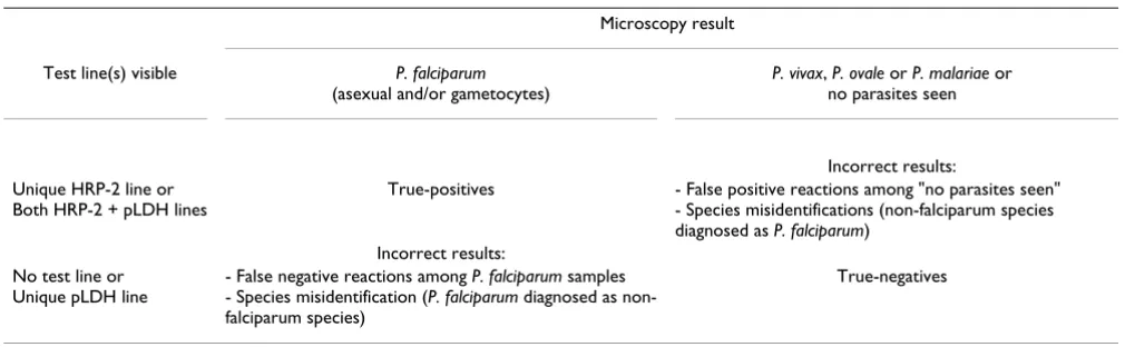
and the non-falciparum species were considered sepa-rately and the control panels included both microscopy negative samples and samples infected by the non-falci-parum species and P. falciparum respectively, as shown in Tables 1 and 2. Of note is that, for both tests, samples with pure gametocytaemia were included among the positive
P. falciparum samples. To avoid complex problems of interpretation, microscopically identified mixed infec-tions were considered separately and not included in the calculations of test characteristics.
Sensitivity and specificity were calculated with 95% confi-dence intervals (C.I.) and differences were tested for sig-nificance using the chi-square test or, in case of small sample sizes, a two-tailed Fisher's exact test. A p value < 0.05 was considered as significant. In addition, positive and negative likelihood ratios (LHR+ and LHR-) were cal-culated. Likelihood ratios provide direct information on the tests power to include (LHR+ > 10) or exclude (LHR-< 0.1) a disease without being influenced by its prevalence [3]. Inter-observer agreements were assessed using the kappa statistic for paired observers and percentage agree-ments for all three observers combined. Associations between line intensity readings and parasite densities were assessed for strength of association with Cramer's V
for categorical variables, using interpretative criteria pub-lished previously [9].
Analysis of discordant results
For discordant results (i.e. false-negative and false-positive results and species misidentifications) a species-specific polymerase chain reaction (PCR) was performed. Primers and probes sequences detecting small subunit 18S rRNA genes were selected according to Rougemont et al [10].
Ethical review
The study was reviewed and approved by the Institutional Review Board of ITM and by the Ethical Committee of Antwerp University, Belgium.
Results
SD FK50 Malaria Ag P. falciparum test
Sample collection
[image:3.612.53.558.556.712.2]A total of 452 samples were assessed, including those infected with P. falciparum (n = 324), P. vivax (n = 12), P. ovale (n = 11), P. malariae (n = 6), mixed species (n = 4) and negative samples (n = 95). The majority (272/324, 84.0%) of P. falciparum samples had been acquired in sub-Saharan Africa.
Table 1: Interpretation of test results of the FK60 for the detection of P. falciparum infection
Microscopy result
Test line(s) visible P. falciparum
(asexual and/or gametocytes)
P. vivax, P. ovale or P. malariae or no parasites seen
Incorrect results: Unique HRP-2 line or
Both HRP-2 + pLDH lines
True-positives - False positive reactions among "no parasites seen" - Species misidentifications (non-falciparum species diagnosed as P. falciparum)
Incorrect results: No test line or
Unique pLDH line
- False negative reactions among P. falciparum samples - Species misidentification (P. falciparum diagnosed as non-falciparum species)
Sensitivity, specificity and likelihood ratios
No invalid test results were observed. Additional file 1 shows the overall and detailed test characteristics matched for parasite densities. Sensitivity increased marginally when samples with pure gametocytaemia were subtracted. Sensitivity was related to parasite density, with values at parasite densities < 100/μl significantly lower compared to those at parasite densities > 100/μl (78.9% and respec-tively 97.6%, p < 0.001). Fifteen out of 21 false-negative samples had parasite densities < 100/μl (including three samples with pure gametocytaemia), the remaining six had parasite densities ranging from 122 to 400/μl. All these infections had been acquired in Africa. Of the four samples with mixed infection, a single one (with P. falci-parum and P. ovale) gave a false-negative result. The overall specificity was 94.4%, with seven samples that were incor-rectly identified: a visible test line was observed among four microscopic negative samples and three P. malariae
samples. The exclusion power was excellent (LHR- < 0.10) except for parasite densities less than 100/μl, the inclusion power was also excellent (LHR+ > 10).
Line intensity readings
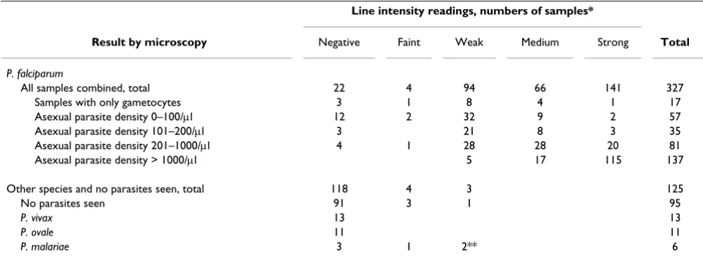
Table 3 lists the line intensity readings related to parasite density. Line intensity readings were significantly related to parasite densities with a substantial correlation (V = 0.434, p < 0.001), but there was considerable overlap between categories. Faint or weak line intensities occurred in 98/305 (32.1%) true positive results, mostly but not exclusively at low parasite densities and among all seven false positive results and misidentifications.
Inter-observer agreement and reproducibility
[image:4.612.49.558.99.250.2]The inter-observer agreement for positive and negative readings was high, with 97.3% overall agreement between the three observers and kappa values between 0.95–0.98 Table 2: Interpretation of test results of the FK60 for the detection of Plasmodium non-falciparum infection
Microscopy result
Test line(s) visible P. vivax, P. ovale or
P. malariae
P. falciparum or no parasites seen
Incorrect results:
Unique pLDH line True-positives - False positive reactions among "no parasites seen" - Species misidentifications (P. falciparum diagnosed as non-falciparum species)
Incorrect results: No test line or
Unique HRP-2 line or Both pLDH + HRP-2 lines
- False negative reactions among non-falciparum samples - Species misidentification (non-falciparum species diagnosed as P. falciparum)
[image:4.612.54.555.506.692.2]True-negatives
Table 3: Line intensity consensus readings for the FK50 according to parasite densities
Line intensity readings, numbers of samples*
Result by microscopy Negative Faint Weak Medium Strong Total
P. falciparum
All samples combined, total 22 4 94 66 141 327
Samples with only gametocytes 3 1 8 4 1 17
Asexual parasite density 0–100/μl 12 2 32 9 2 57
Asexual parasite density 101–200/μl 3 21 8 3 35
Asexual parasite density 201–1000/μl 4 1 28 28 20 81
Asexual parasite density > 1000/μl 5 17 115 137
Other species and no parasites seen, total 118 4 3 125
No parasites seen 91 3 1 95
P. vivax 13 13
P. ovale 11 11
P. malariae 3 1 2** 6
* Total number of samples = 452, including two P. falciparum/P. ovale and one P. falciparum/P. malariae mixed infections (allocated under P. falciparum) and one P. vivax/P. malariae mixed infection (allocated under P. vivax)
**PCR confirmed microscopic diagnosis for all discordant samples except for one sample that was diagnosed as a mixed P. malariae/P. falciparum
for each pair out of three observers. All discordances in line intensity readings were limited to one category of dif-ference (e.g. a line scored as weak by one observer was scored as medium or faint but not as strong by the other observer(s)). For line intensity readings, overall agree-ment was 86.9% and kappa values were between 0.86 and 0.90. Reproducibility testing showed consistent readings by all three observers on all occasions for three out of six samples. For two other samples discrepancies were lim-ited to differences within one category of line intensity and had no impact on final test result. A single sample (with parasite density 1,123/μl) provided weak, faint and negative readings upon repeat testing.
Analysis of discordant results
PCR analysis confirmed the discrepant results in favour of the reference microscopy except for a single sample that was microscopically identified as P. malariae and while showing a HRP-2 line, this sample was diagnosed by PCR as a mixed P. falciparum/P. malariae infection (Additional file 1). Correcting for this result increased the specificity from 94.4% to 95.2% (C.I. 89.6%–98.0%).
SD FK60 Malaria Ag P. falciparum/Pan test
Sample collection
Compared to the panel for the FK50, the numbers of sam-ples with non-falciparum species were increased up to 80 for P. vivax and P. ovale each and 31 for P. malariae, result-ing in a total of 614 samples.
Sensitivity, specificity and likelihood ratios
No invalid test results were recorded. A total of 303 out of 324 P. falciparum samples were correctly identified. The remaining 21 samples showed no test line. Among the control samples, there was one out of 95 microscopy neg-ative samples that generated a HRP-2 line, and there were six out of 191 non-falciparum samples showing cross-reaction with HRP-2 (one sample each with P. ovale and P. vivax and four samples with P. malariae). The resulting test characteristics are displayed in Additional file 2, with an overall sensitivity and a specificity of 94.1% and 97.6% respectively. As for the FK50, sensitivities were lower at decreasing parasite densities with significant difference below and above 100/μl (p < 0.001).
For the P. non-falciparum species, there were 145 out of 191 samples correctly identified (unique pLDH line visi-ble), six samples had a visible HRP-2 line and 40 samples showed no test line. The resulting overall sensitivities were 87.5%, 76.3% and 45.2% for P. vivax, P. ovale and P. malariae respectively (Additional file 3). Sensitivity for P. malariae was significantly lower compared to the two other species (p < 0.001). As for P. falciparum, sensitivities declined at lower parasite densities, and dipped at parasite densities below 500/μl. Differences between sensitivities
in samples above and below parasite densities of 500/μl reached statistical significance in the case of P. ovale (p < 0.05). Among the microscopic negative samples, there were no false-positive pLDH readings neither were there
P. falciparum samples that generated a unique pLDH line. The specificity for non-falciparum species was 100%, resulting in high LHR+ values.
Of the four samples with mixed infection, a single one (with P. falciparum and P. ovale at a parasite density of 700/μl) showed only a pLDH line, the other mixed infec-tions were correctly identified.
Line intensity readings
As for the FK50, faint and weak line intensities for the HRP-2 line occurred mostly but not exclusively at low par-asite densities. The pLDH line performed worse in terms of visibility, with 224/353 (63.4%) of true positive read-ings in the faint or weak categories, as opposed to 90/305 (29.5%) for the HRP-2 line (p < 0.001). The pLDH line showed lowest intensities among the non-falciparum samples, with only 28 out of 80 P. vivax samples, 5 of 80
P. ovale samples and none of the P. malariae samples showing medium or strong line intensities. Line intensity readings for HRP-2 and for pLDH were related to parasite densities (HRP-2: V = 0.387, p < 0.05; pLDH: V = 0.457, p < 0.05) but there was considerable overlap between the different categories. Interestingly, in the case of P. falci-parum infection, the unique presence of a HRP-2 line pointed almost exclusively (98.1%, 103/105 of samples) to a parasite density below 1,000/μl (Table 4). In addi-tion, the presence of medium or strong pLDH line inten-sity was invariably associated with parasite densities exceeding 1,000/μl in the case of P. falciparum and 500/μl in the case of the non-falciparum species, except for one P. falciparum sample with pure gametocytaemia (12,700/μl). Of interest, among the 17 P. falciparum samples with pure gametocytaemia, there were 14 with HRP-2 lines visible compared to six with pLDH lines visible. HRP-2 lines in samples with non-falciparum species gave faint or weak line intensities except for a single P. malariae sample with strong line intensity. This latter sample however proved to be a mixed infection with P. falciparum/P. malariae upon PCR analysis (see below).
Inter-observer agreement and reproducibility
occa-sions for four out of six samples. Two other samples (with parasite density 116/μl and 200/μl) provided weak, faint and negative readings upon repeat testing.
Analysis of discordant results
PCR analysis of all discordances resulted in the following corrections: two samples that were microscopically diag-nosed as P. ovale and that did not show any test line proved to be mixed P. falciparum/P. ovale infections, and one P. malariae sample that showed cross-reaction with the HRP-2 line proved to be a mixed P. falciparum/P. malariae infection. When correcting for these results, spe-cificity for the detection of P. falciparum and sensitivity for the diagnosis of P. malariae increased slightly (Additional files 2 and 3).
Side by side comparison of the SD FK50 and SD FK60 assays
A total of 324 P. falciparum samples were assessed by both the FK50 and FK60 tests. Two of them provided different results: one was uniquely positive by FK50 and the other by FK60. Both samples had parasite densities below 100/
μl, and displayed faint and weak HRP-2 line intensity readings without a visible pLDH line. Since both samples belonged to the same parasite density category (< 100/μl), data on sensitivity for P. falciparum were identical for both assays. In addition to the three P. malariae samples that showed HRP-2 lines with the FK50 test, the FK60 showed HRP-2 lines for three other samples, one sample of P.
malariae, P. vivax and P. ovale respectively. Among micro-scopic negative samples, a single sample gave a false-pos-itive HRP-2 line by the FK60 as opposed to four samples for the FK50, resulting in slightly better specificity and LHR+ for the FK60 as compared to the FK50. Agreement between both tests for positive and negative readings and line intensities was excellent (kappa value 0.95) and sub-stantial (kappa value 0.68) respectively.
Discussion
In this study, the performance of two one-step malaria rapid diagnostic tests, the SD FK50 Malaria Ag P. falci-parum test (a two-band HRP-2 test) and the SD FK60 Malaria Ag P. falciparum/Pan test (a three-band HRP-2 and pLDH test) was evaluated on large panels of stored sam-ples obtained from international travellers. For both tests, overall sensitivity for the detection of P. falciparum was 93.5%, reaching 97.6% and 100% at parasite densities above 100 and 1,000/μl respectively. Overall sensitivities for P. vivax, P. ovale and P. malariae for the FK60 test were 87.5%, 76.3% and 45.2%, but they reached 92.6% and 90.5% for P. vivax and P. ovale at parasite densities > 500/
μl. Specificities were above 95% for all species. Inter-observer agreement and reproducibility were high for both tests.
[image:6.612.53.558.100.214.2]One of the limitations of the present study was its retro-spective design, which made it difficult to trace back causes of discordant results such as previous therapy and Table 4: Reactivity of test lines for the FK60 for P. falciparum samples in relation to parasite density
SD FK 60 result: test lines visible Total
Parasite densities for P. falciparum samples None Unique HRP-2 line Both HRP-2 and pLDH lines
Pure gametocytaemia 3 8 6 17
0 – 100/μl 12 42 3 57
101 – 200/μl 3 18 14 36
201 – 1000/μl 3 35 42 80
> 1000/μl 0 2 133 135
Total 21 105 198 324
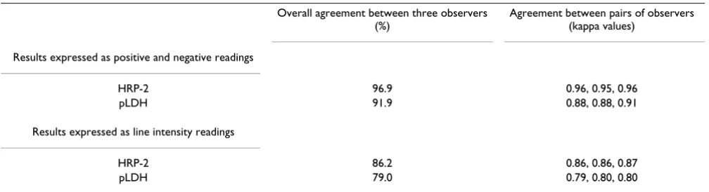
Table 5: Overall agreement and inter-observer agreement between pairs of observers for the FK60 assay
Overall agreement between three observers (%)
Agreement between pairs of observers (kappa values)
Results expressed as positive and negative readings
HRP-2 96.9 0.96, 0.95, 0.96
pLDH 91.9 0.88, 0.88, 0.91
Results expressed as line intensity readings
HRP-2 86.2 0.86, 0.86, 0.87
[image:6.612.54.558.593.730.2]the presence of interfering factors (e.g. rheumatoid factor). Further, the study population was not completely homog-enous and the small numbers of semi-immune immi-grants (who may tolerate low-level parasite densities [5]) were not identified. Another possible limitation is the fact that stored blood samples were used for analysis. Although, on theoretical grounds, there have been con-cerns about the stability of the target antigens under these conditions [11], previous evaluations of RDTs have been performed on stored samples [12,13] and a prospective evaluation of fresh and stored samples revealed similar results in case of the HRP-2 antigen [14]. In the present study, no obvious differences in test performance were found for samples stored for several (> 2) years compared to those stored for a shorter period (results not shown), and the samples had not been exposed to repeat thawing and freezing. Further, it should be realised that the present study design did not consider the performance of these RDTs when applied in clinical diagnosis by laboratory technicians in non-endemic settings, who have few expo-sure to malaria positive samples [1,2]. Assessing samples with different parasite densities should be part of the lab-oratory validation when introducing RDTs in clinical practice, for instance, to train the occasional reader to interpret faint line intensities as positive results. Finally, it should be realized that the present study was performed in a reference setting, with the availability of expert micro-scopy, trained observers and optimal environmental con-ditions. Likewise, a calibrated transfer pipette was used instead of the manufacturer's transfer device, in order to ensure correct volume transfer [11]. However, an evalua-tion of such a test in a reference setting is a logic first step preceding in-depth evaluations and field trials [11].
For both tests and the diagnosis of P. falciparum, the sen-sitivities were in line with those reported in other HRP-2 tests in returned travellers, with sensitivities ranging from 80% to 99%, depending on the setting and parasite den-sities [3,15-22]. However, most of these studies, in partic-ular the systematic reviews, addressed the multistep RDTs that are available on the market for a long time [3,5,6,23]. By contrast, evaluations of most of the other RDTs dis-played on the WHO website are pending [7]. For the non-falciparum species, the reported sensitivities vary, with decreasing sensitivities for P. vivax followed by either P. ovale or P. malariae [3,4,15,19,24]. In a recent meta-anal-ysis on malaria RDTs in international travellers [3], sensi-tivities for P. ovale and P. malariae ranging from 36 – 95% were mentioned. Part of this wide range in sensitivities is probably due to low sample sizes in different studies. The sample sizes in the present study enabled us to calculate test characteristics with narrower confidence intervals, and consequently this study demonstrates a significantly lower sensitivity for detection of P. malariae, as compared to P. ovale and P. vivax, even at parasite densities above
500/μl. Considering the present methods, two other remarks are to be made. First, in the case of P. falciparum, samples with only gametocytes were considered as part of the microscopic positive samples. From the standpoint of travel medicine, this is a recommended choice, but game-tocytes may be present even after successful eradication of the asexual forms [3,5]. Moving these pure gametocytae-mia samples to the "non-malaria" category in the present study collection would add slightly to the sensitivity and the LHR- of both tests (Additional files 1 and 2), at a con-siderable cost of specificity (87.0% and 93.3% for FK50 and FK60 respectively), but with LHR+ still above 10. Sec-ond, among our control panel not only microscopy nega-tive samples were included, but also samples from other
Plasmodium species and we consequently scored species misidentifications as incorrect diagnosis. One could argue that species misidentification can be tolerated as long as the diagnosis of malaria is not missed. Competent malaria diagnosis however requires distinction between at least P. falciparum and the other species, as prognosis, therapy, follow-up and epidemiology are different. With regard to both tests, it is reassuring that among the present samples,
P. falciparum was not erroneously misidentified as a non-falciparum species (with the exception of a single failure of HRP-2 reactivity in a mixed P. falciparum/P. ovale infec-tion), and that misidentification only occurred in the other direction, i.e. non-falciparum species (especially P. malariae) were misidentified as P. falciparum. The impact of adding other species to the control group however was low in terms of test characteristics: limiting the control panel to exclusively the negative samples would result in a slight increase in specificity (95.8% and 98.9% for the FK50 and FK60 in case of P. falciparum respectively), a slight increase in sensitivity for P. vivax and P. ovale
(88.8% and 77.5% respectively) and a moderate increase in sensitivity for P. malariae (58.1%). Of note are the false positive reactions for the HRP-2 line among the non-falci-parum species, in particular among 10% of our P. malariae
samples. HRP-2 cross-reaction have been reported for P. vivax and P. malariae, but not for P. ovale [25,26].
For all species, declining sensitivities at lower parasite densities were observed. For P. falciparum this is a well-known phenomenon [3-6,11]. The present study demon-strated this decline for the non-falciparum species as well, indicating a breakpoint at 500/μl. In line with the results from the meta-analysis mentioned above [3], most of the
Further, the results did not show any relation between geographic distribution and false-negative results by HRP-2 due to possible genetic variations in HRP-HRP-2 target [HRP-27], but the majority of samples were acquired in Africa, and more samples should be tested from the Asia-Pacific to rule out an influence of such variations.
In contrast to most other studies, reproducibility and inter-observer agreements of both tests were presently assessed. Line intensity readings (and consequently test results) showed high inter-observer agreements and were also reproducible upon repeat testing, but performances were clearly better for the HRP-2 line as compared to the pLDH line. For the latter line, the preponderance of weak and faint readings for the non-falciparum species is of concern. Furthermore, for the detection of P. falciparum, the three-band FK60 test performed as well as the two-band FK50. This is of note, as one could expect the three-band test, which has to meet two optimums of antigen-antibody interactions, would perform somewhat less than the two-band test. Although the present devices are not designed to use line intensities as a tool for grading para-site densities, this study also explored their possible diag-nostic value. In line with other findings, line intensities were related to parasite density [17,26,28] but considera-ble overlaps precluded their use as a semi-quantitative estimation of parasite density. However, the FK60 test provided interesting clues to parasite densities below or above 1,000/μl for P. falciparum (the unique presence of a HRP-2 line and the presence of medium or strong pLDH line respectively). A similar approach has been described for a HRP-2 and aldolase three-band RDT, for which co-reactivity of both test lines pointed to parasite densities of = 40,000/μl [29]. Further product research might refine and expand these possibilities, thereby enlarging the scope of malaria RDTs application [6].
Conclusion
Taking into account their sensitivity and specificity, inter-observer agreement and reproducibility, it is clear that the FK50 and the FK60 tests devices are a valuable adjunct to microscopy for the diagnosis of malaria in a non-endemic setting. They share the limitations of other malaria rapid diagnostic tests, in particular the limited exclusion power for P. falciparum malaria at low parasite densities and a lower sensitivity for the non-falciparum species, especially
P. malariae. Possible test improvements – apart from the sensitivity – would be an increase in intensity of the pLDH line, and the exploration of the semi-quantitative estima-tion of the parasite densities.
Abbreviations
SD FK50: SD FK50 Malaria Ag P. falciparum test (Standard Diagnostics): a two-band "one-step" malaria rapid
diag-nostic test for the detection of P. falciparum, targeting HRP-2 antigen; SD FK60: SD FK60 Malaria Ag P. falci-parum/Pan test (Standard Diagnostics): a three-band "one-step" malaria rapid diagnostic test for the detection of P. falciparum and P. non-falciparum species, targeting HRP-2 and pLDH antigen; Ag: Antigen; P.: Plasmodium;
FHML: Faculty of Health Medicine and Life Sciences, Maastricht, The Netherlands; ITM: Institute of Tropical Medicine, Antwerp, Belgium; PCR: Polymerase Chain Reaction; RDT(s): Rapid Diagnostic Test(s); HRP-2: Histi-dine Rich Protein-2; pLDH: Parasite lactate dehydroge-nase; EDTA: Ethylene diamine tetra-acetic acid; WBC: White Blood Cells; C.I.: Confidence interval; LHR+: Posi-tive likelihood ratio; LHR-: NegaPosi-tive likelihood ratio; rRNA: Ribosomal ribonucleic acid; ∞: Infinite;
Competing interests
The authors declare that they have no competing interests.
Authors' contributions
MvdP, PG and JJ designed the study protocol. MvE and EB organized prospective sample collection. MvdP and PG carried out the test evaluations, LC performed PCR analy-sis. MvdP, PG and JJ analyzed and interpreted the results and drafted the manuscript, PG performed statistical anal-ysis. All authors contributed to the discussion of the results and the redaction of the manuscript, they all approved the final manuscript.
Additional material
Acknowledgements
We would like to thank Veronique Guns and the staff of the Central Labo-ratory of Clinical Biology for technical support.
Additional file 1
Test characteristics of the FK50 related to parasite densities.
Click here for file
[http://www.biomedcentral.com/content/supplementary/1475-2875-8-90-S1.doc]
Additional file 2
Test characteristics of the FK60 for P. falciparum samples according to parasite densities.
Click here for file
[http://www.biomedcentral.com/content/supplementary/1475-2875-8-90-S2.doc]
Additional file 3
Test characteristics of the FK60 for non-falciparum species according to parasite densities.
Click here for file
Publish with BioMed Central and every scientist can read your work free of charge "BioMed Central will be the most significant development for disseminating the results of biomedical researc h in our lifetime."
Sir Paul Nurse, Cancer Research UK
Your research papers will be:
available free of charge to the entire biomedical community
peer reviewed and published immediately upon acceptance
cited in PubMed and archived on PubMed Central
yours — you keep the copyright
Submit your manuscript here:
http://www.biomedcentral.com/info/publishing_adv.asp
BioMedcentral
References
1. Hänscheid T: Current strategies to avoid misdiagnosis of malaria. Clin Microbiol Infect 2003, 9:497-504.
2. Wiese L, Bruun B, Baek L, Friis-Moller A, Gahrn-Hansen B, Hansen J, Heltberg O, Hojbjerg T, Hornstrup MK, Kvinesdal B, Gomme G, Kurtzhals JA: Bedside diagnosis of imported malaria using the Binax Now malaria antigen detection test. Scand J Infect Dis 2006, 38:1063-1068.
3. Marx A, Pewsner D, Egger M, Nuesch R, Bucher HC, Genton B, Hatz C, Juni P: Meta-analysis: accuracy of rapid tests for malaria in travelers returning from endemic areas. Ann Intern Med 2005, 142:836-846.
4. Moody AH, Chiodini PL: Non-microscopic method for malaria diagnosis using OptiMAL IT, a second-generation dipstick for malaria pLDH antigen detection. Br J Biomed Sci 2002, 59:228-231.
5. Murray CK, Gasser RA Jr, Magill AJ, Miller RS: Update on Rapid Diagnostic Testing for Malaria. Clin Microbiol Rev 2008, 21:97-110.
6. Wongsrichanalai C, Barcus MJ, Muth S, Sutamihardja A, Wernsdorfer WH: A review of malaria diagnostic tools: microscopy and rapid diagnostic test (RDT). Am J Trop Med Hyg 2007, 77:119-127.
7. World Health Organization: List of known commercially-availa-ble antigen-detecting malaria RDTs: information for national public health services and UN Agencies wishing to procure RDTs. [http://www.wpro.who.int/NR/rdonlyres/ 9C624A45-3554-4695-8C11-233DB743FC7A/0/
MD_table23_ISO131485criteriarev041207.pdf].
8. Bell DR, Wilson DW, Martin LB: False-positive results of a Plas-modium falciparum histidine-rich protein 2-detecting malaria rapid diagnostic test due to high sensitivity in a community with fluctuating low parasite density. Am J Trop Med Hyg 2005, 73:199-203.
9. Stamatopoulos B, Meuleman N, Haibe-Kains B, Duvillier H, Massy M, Martiat P, Bron D, Lagneaux L: Quantification of ZAP70 mRNA in B cells by real-time PCR is a powerful prognostic factor in chronic lymphocytic leukemia. Clin Chem 2007, 53:1757-1766. 10. Rougemont M, Van Saanen M, Sahli R, Hinrikson HP, Bille J, Jaton K:
Detection of four Plasmodium species in blood from humans by 18S rRNA gene subunit-based and species-specific real-time PCR assays. J Clin Microbiol 2004, 42:5636-5643.
11. Bell D, Peeling RW: Evaluation of rapid diagnostic tests: malaria. Nat Rev Microbiol 2006, 4:S34-S38.
12. Craig MH, Bredenkamp BL, Williams CH, Rossouw EJ, Kelly VJ, Klein-schmidt I, Martineau A, Henry GF: Field and laboratory compar-ative evaluation of ten rapid malaria diagnostic tests. Trans R Soc Trop Med Hyg 2002, 96:258-265.
13. Piper R, Lebras J, Wentworth L, Hunt-Cooke A, Houze S, Chiodini P, Makler M: Immunocapture diagnostic assays for malaria using
Plasmodium lactate dehydrogenase (pLDH). Am J Trop Med Hyg 1999, 60:109-118.
14. Mayxay M, Pukrittayakamee S, Chotivanich K, Looareesuwan S, White NJ: Persistence of Plasmodium falciparum HRP-2 in suc-cessfully treated acute falciparum malaria. Trans R Soc Trop Med Hyg 2001, 95:179-182.
15. De Monbrison F, Gerome P, Chaulet JF, Wallon M, Picot S, Peyron F: Comparative diagnostic performance of two commercial rapid tests for malaria in a non-endemic area. Eur J Clin Micro-biol Infect Dis 2004, 23:784-786.
16. Durand F, Crassous B, Fricker-Hidalgo H, Carpentier F, Brion JP, Grillot R, Pelloux H: Performance of the Now Malaria rapid diagnostic test with returned travellers: a 2-year retrospec-tive study in a French teaching hospital. Clin Microbiol Infect 2005, 11:903-907.
17. Farcas GA, Zhong KJ, Lovegrove FE, Graham CM, Kain KC: Evalua-tion of the Binax NOW® ICT test versus polymerase chain reaction and microscopy for the detection of malaria in returned travelers. Am J Trop Med Hyg 2003, 69:589-592. 18. Gatti S, Gramegna M, Bisoffi Z, Raglio A, Gulletta M, Klersy C, Bruno
A, Maserati R, Madama S, Scaglia M: A comparison of three diag-nostic techniques for malaria: a rapid diagdiag-nostic test (NOW Malaria), PCR and microscopy. Ann Trop Med Parasitol 2007, 101:195-204.
19. Grobusch MP, Hanscheid T, Zoller T, Jelinek T, Burchard GD: Rapid immunochromatographic malarial antigen detection
unreli-able for detecting Plasmodium malariae and Plasmodium ovale. Eur J Clin Microbiol Infect Dis 2002, 21:818-820.
20. Grobusch MP, Hanscheid T, Gobels K, Slevogt H, Zoller T, Rogler G, Teichmann D: Comparison of three antigen detection tests for diagnosis and follow-up of falciparum malaria in travellers returning to Berlin, Germany. Parasitol Res 2003, 89:354-357. 21. Jelinek T, Grobusch MP, Schwenke S, Steidl S, von Sonnenburg F,
Nothdurft HD, Klein E, Loscher T: Sensitivity and specificity of dipstick tests for rapid diagnosis of malaria in nonimmune travelers. J Clin Microbiol 1999, 37:721-723.
22. Ende J Van den, Vervoort T, Van Gompel A, Lynen L: Evaluation of two tests based on the detection of histidine rich protein 2 for the diagnosis of imported Plasmodium falciparum malaria. Trans R Soc Trop Med Hyg 1998, 92:285-288.
23. Cruciani M, Nardi S, Malena M, Bosco O, Serpelloni G, Mengoli C: Systematic review of the accuracy of the Para Sight™-F test in the diagnosis of Plasmodium falciparum malaria. Med Sci Monit 2004, 10:MT81-MT88.
24. Playford EG, Walker J: Evaluation of the ICT Malaria P.f/P.v and the OptiMal rapid diagnostic tests for malaria in febrile returned travellers. J Clin Microbiol 2002, 40:4166-4171. 25. Iqbal J, Hira PR, Sher A, Al Enezi AA: Diagnosis of imported
malaria by Plasmodium lactate dehydrogenase (pLDH) and histidine-rich protein 2 (PfHRP-2)-based immunocapture assays. Am J Trop Med Hyg 2001, 64:20-23.
26. Pieroni P, Mills CD, Ohrt C, Harrington MA, Kain KC: Comparison of the Para Sight™-F test and the ICT Malaria Pf™ test with the polymerase chain reaction for the diagnosis of Plasmo-dium falciparum malaria in travellers. Trans R Soc Trop Med Hyg 1998, 92:166-169.
27. Baker J, McCarthy J, Gatton M, Kyle DE, Belizario V, Luchavez J, Bell D, Cheng Q: Genetic diversity of Plasmodium falciparum Histi-dine-Rich Protein 2 (PfHRP2) and its effect on the perform-ance of PfHRP2-Based rapid diagnostic tests. J Infect Dis 2005, 192:870-877.
28. Mason DP, Kawamoto F, Lin K, Laoboonchai A, Wongsrichanalai C: A comparison of two rapid field immunochromatographic tests to expert microscopy in the diagnosis of malaria. Acta Trop 2002, 82:51-59.
29. Richter J, Gobels K, Muller-Stover I, Hoppenheit B, Haussinger D: Co-reactivity of plasmodial histidine-rich protein 2 and aldo-lase on a combined immuno-chromographic-malaria dip-stick (ICT) as a potential semi-quantitative marker of high