City, University of London Institutional Repository
Citation
:
Qassem, M. & Kyriacou, P. A. (2012). In vitro spectrophotometric near infrared
measurements of skin absorption and dehydration. Conference proceedings : ... Annual
International Conference of the IEEE Engineering in Medicine and Biology Society. IEEE
Engineering in Medicine and Biology Society., 2012, pp. 6044-6047. doi:
10.1109/EMBC.2012.6347372
This is the accepted version of the paper.
This version of the publication may differ from the final published
version.
Permanent repository link:
http://openaccess.city.ac.uk/13281/
Link to published version
:
http://dx.doi.org/10.1109/EMBC.2012.6347372
Copyright and reuse:
City Research Online aims to make research
outputs of City, University of London available to a wider audience.
Copyright and Moral Rights remain with the author(s) and/or copyright
holders. URLs from City Research Online may be freely distributed and
linked to.
Abstract— The application of Near Infrared Spectroscopy
(NIRS) for measuring skin water content has long been established, and has gained wide interest as a precise, safe, fast and noninvasive technique for determining skin hydration. This paper reports near infrared spectrophotometric measurements using a highly sophisticated spectrophotometer in the region of 1000-2500 nm to study the water uptake and dehydration properties of skin in vitro using samples of porcine skin. Initial results of pure liquid water and skin samples have clearly displayed the prominent bands associated with water content, and desorption tests have been able to verify changes in these bands associated with water content, although a clear correlation between the rates of weight loss and absorbance loss at various hydration periods has not yet been established. These preliminary results are expected to further explain the relationship between water and skin, and its role within, in hope to aid the future development of a portable instrument based on near infrared spectroscopy that would be capable of directly measuring skin hydration and/or water content in a fast and noninvasive manner.
I. INTRODUCTION
Without a doubt, the skin is the most complex organ of the human body, and one that requires constant care and attention [1]. In its widely appreciated context, the skin barrier function refers to the epidermal barrier to water permeability, and is also the largest organ of the body, an approximately two square meter, covering its entire surface [1, 2]. In essence, it acts as a means for adjusting to variations in environmental temperatures through elegant controls that regulate the microcirculation, and as an interact impermeable barrier that precipitates the organism to intake water without flooding its internal organs, all whilst prohibiting the intrusion of various xenobiotics, and even includes touch, pain and pressure receptors. Large-scale damage to this part would make human life impossible, and its maintenance enhances our physical and mental health [1, 2]. The outermost layer of the skin, known as the Stratum Corneum (SC) acts as an essential permeable barrier and its water content or degree of hydration can have profound effects on its transport and mechanical properties. However, although water plays an important role in the well- functioning of the skin, it is potentially a strong irritant and can cause skin damage [3, 4]. Skin exposure to water can disrupt its barrier function by altering its physiological behaviour through extraction of water-soluble substances,
*Research supported by City University London.
P. A. Kyriacou. School of Engineering and Mathematical sciences, City University London, Northampton square, London, EC1V 0HB USA (phone: 020 7040 8131; e-mail: p.kyriacou@city.ac.uk).
M. Q. is with City University London, Northampton square, London, EC1V 0HB. (e-mail: ct719@city.ac.uk).
Or Natural Moisturizing Factors (NMFs) from the skin, or by facilitating the penetration of foreign substances, and can even cause skin disease [3, 4].
So far, various technologies have been implemented to assess the water content of the SC and other skin parameters noninvasively for moisture evaluation and/or for detecting abnormalities in the skin that may serve as indicators for underlying illnesses or skin conditions [5]. Previous measuring techniques have included those based on thermal, mechanical, imaging and spectroscopic methods, but with electrical probe-based measurements being the most commonly utilized today. Unfortunately though, the latter suffers several principal drawbacks due to the absence of a direct correlation between skin conductivity and water content, and that the flow of current can be influenced by variations in ion movements, and re-orientation of protein dipole moments [6]. Alternatively, Near Infrared Spectroscopy (NIRS) has the ability to measure the amount of water in skin directly using the intensities of overtone and combination bands of OH and HOH water bonds occurring in the NIR region, that are good indicators of the state of skin hydration [7]. Hence, this allows a more precise method of measurement, and with an added advantage in that their instrumentation can easily be equipped with fibre optic probes for in vivo measurements in various anatomical sites. Furthermore, NIR spectra have shown to provide further details with regards to other skin constituents such lipids and proteins, as well as information about the types of water present within the skin [1, 8, 9]. Together, this has made the technique very attractable as a safe and noninvasive alternative, and whilst several earlier studies have applied it to study skin hydration and the efficacy of cosmetic products [4, 6, 8-10], many others continue to do so, and consecutive developments and advancements are being made in its instrumentation and chemometric analysis processes, thus extending its applications and utility in this field.
Although there has been interest in improving this technique for the purpose of skin hydration and water content measurements [11-13], a reliable handheld or portable measuring instrument of this type does not yet exist. The main objective of this study is to compare spectroscopic measurements in the NIR region of pure liquid water against samples of porcine skin using a highly sophisticated spectrophotometer, and to conduct in vitro desorption experiments on porcine skin to investigate the mechanisms of water uptake and dehydration by the skin at various hydration periods, in order to aid the future development of a portable device based on NIR spectroscopy that would be capable of measuring skin water content in a safe, fast and noninvasive manner.
In vitro
spectrophotometric near infrared
measurements of skin absorption and dehydration
II. MATERIALS AND METHODS A. Optical properties of liquid water
The absorption and scattering of liquid water belong to its physical chemistry, but are of great interest here since water is a major constituent of the skin and its overtones and combinations are clearly distinguishable in skin spectra. So, by presuming the intrinsic absorption properties of water will remain the same even when measured in the skin, the absorption spectra of water is measured separately. This assumption is almost always true as water dominates NIR absorption spectra of various high water content intact soft tissues, such as the skin. Therefore, by determining the water spectrum, NIR spectroscopy can then be used to detect its changes in skin and the change in skin water content under certain physiological conditions.
In order to obtain a spectrum of liquid water, the Lambda 1050 dual beam UV/Vis/NIR spectrophotometer (Perkin Elmer Corp, Massachusetts, USA) was used with increment steps of 1 nm, in the spectral region between 200-3300 nm. The source was a deuterium lamp between 250-319.20 nm, and beyond that was a halogen tungsten lamp up to 3300 nm. The detector system was set to use a photomultiplier tube detector (PMT) for the shortest wavelengths from 250 nm until 860.80 nm, and an indium gallium arsenide detector (InGaAs) between 860.80- 2500 nm. For the rest of the spectral region up to 3300 nm, a lead sulfide detector (PbS) was applied. Slits settings for the PMT detector were fixed at 2 nm, whilst InGaAs and PbS detectors were set on "servo mode", whereby the system monitors the reference beam energy and adjusts the slits accordingly to avoid over saturation of the detectors. Gain settings for the three detectors, which are applied for energy measurement in single beam mode, must be set to generate a useable spectrum. After a few trials, a gain of 3 was selected for the InGaAs and PbS detectors, whereas the PMT gain was set on auto to allow the instrument to determine the appropriate value. The response time for these detectors was set at 0.2 seconds for all. The front and rear attenuators enclosed in this instrument are used to select the attenuation in the sample and reference beam, and in this case were set at 100% for the sample beam and reduced to 1 % for the reference beam. Prior to placing the sample of liquid water through the spectrum, baseline corrections were performed on the spectrophotometer at 100 %T/0A and 0 %T/blocked beam to remove background noise. The sample of liquid water was contained in a rectangular cell made of quartz SUPRASIL, made of high purity synthetic fused silica material to ensure top performance, and with a light path length of 1 mm (P/N: B0631013, Perkin Elmer Corp, Massachusetts, USA). Liquid water was inserted into the cell through a syringe, which was then placed into the sample compartment whilst a blank duplicate cell was inserted into the reference compartment.
B. In vitro experiment on porcine skin
Prior to performing any measurements, ethics approval was sought and accepted from the university to allow testing on
animal samples. The previous settings were maintained on the spectrophotometer and only the interval steps of measurements were changed to increments of 2 nm and the operating spectral region was reduced to 1000-2500 nm. Fresh porcine meat was acquired and kept frozen until ready for testing. Porcine skin samples were cut using a scalpel into a rectangular shape, of roughly 1.0 mm and 1.5 mm thickness. For the first part of the experiment, both samples were not hydrated or treated in any way, and were tested in their normal state to compare against the spectrum of liquid water. The 1 mm sample was then placed in a jar containing pure liquid water to measure its dehydration rate when it was placed in water for different periods of time. For the dehydration rate testing, it was chosen to leave the sample in water for 30 minutes, 60 minutes, 3 hours, 12 hours, 18 hours and 24 hours. Once the sample reached the required time in water, it was removed, dried, and weighed on the TR-214 model analytical balance (Denver Instruments GmbH, Gottingen, Germany), before placing it in the spectrophotometer. Measurements were then taken every 20 minutes from the initial reading, and lasted 3 hours. The porcine sample was weighed each time before the next measurement.
In order to directly place skin samples into the cell compartment of the spectrophotometer, a disposable polystyrene cell was used throughout the entire experiment which had a hole cut on both the front and rear of it in the area of the beam target, so that radiation from the spectrophotometer source was directly incident on the skin sample without striking the windows of the cell. The reference compartment was left blank.
All spectra obtained during these experiments were available immediately at the end of each test, and were displayed on the UVWinlab software (Perkin Elmer Corp, Massachusetts, USA) that accompanies the Lambda 1050 spectrophotometer. The software was also used to perform quant scanning of data, but for further analysis, spectra were transferred to the UVWinlab Data Processor and Viewer program to carry out processes such as smoothing, calculating derivative spectra, and exporting data in excel format.
III. RESULTS AND DISCUSSION
The smoothed spectrum of 1mm and 1.5mm thickness of porcine skin between 1300-2500nm is shown in Fig. 1 along with that of liquid water. The smoothing process was carried out using the UVWinlab software which performs this function by detecting the sample data points from the spectrophotometer prior to storing it in an array in a data station, and then allows a pre-selected smoothing bandwidth to be entered into the data station from the keyboard. A smoothing factor of 25 was selected here.
Fig 1. Near infrared absorption spectra of porcine skin of 1 mm and 1.5 mm thickness, and that of liquid water.
bands of CH bonding at 2300 nm, 1760 nm and 1730 nm, NH bonding at 2050 nm and 1500 nm, and combination C=O bonding at 2180 nm from lipids and proteins within the skin, as well as a shoulder-like character at 2000 nm which indicates the presence of bound water [8, 9]. The spectrum shown in Fig. 2 of 1 mm thickness porcine skin highlights these points and shows the slightly different results obtained here.
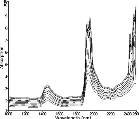
As for the evaporation and dehydration tests where the 1 mm sample of porcine skin was placed in water for different amounts of time (30 minutes, 60 minutes, 3 hours, 12 hours, 24 hours and 46 hours) then weighed and measured on the spectrophotometer over 3 hours, only the spectra of porcine skin left in water for 30 minutes have been included in this paper and are shown in full in Fig. 3. For all the tests performed, both the weight and peak intensity of the sample decreased over time. However, this reduction in intensity bands is more distinguishable at 1450 nm as oppose to the 1920-1940 nm band, and so further calculations were based on this particular peak.
Moreover, the second derivative spectra were calculated for
[image:4.612.315.553.58.262.2]Fig 2. Spectrum of the 1 mm thick sample of porcine skin with its distinguishing features highlighted. Values included for OH, NH, CH, and CO combination and overtone bands.
Fig 3. Near infrared spectra of porcine skin left to hydrate for 30 minutes in water undergoing natural water desorption during a weight loss experiment.
the spectra undergoing dehydration in Fig. 3, and are shown in Fig. 4. Here, a clear shift can be seen from 1949.8 nm to 1967.73 nm of the water combination band as the content of water within the skin sample is reduced although another study [ 6 ] indicates that the shift occurs from 1915 nm to 1940 nm. The spectrum with the largest peaks is the second derivative of the absorption spectrum obtained earlier where the sample had the highest water content or least dehydration period, whereas that with the smallest peaks belong to the absorption spectrum obtained when the sample was least hydrated. So, it follows that intensity decreased as the water content lessened but a wavelength shift is only apparent between 1949.8 and 1967.7 nm, and not on any of the other bands associated with NH, CH or CO bonds. As mentioned earlier, the weight of the sample declined with increased desorption time. So for each test of different hydration period, the weight of the sample was greatest when measured immediately after removing it from the jar containing water and this value decreased over time, and was
[image:4.612.318.552.513.697.2] [image:4.612.57.296.523.698.2]lowest at the end of the 3 hour testing period. This directly correlates with the resulting spectra since the magnitude or intensity of the 1450 nm band also decreased over time, but however, there does not seem to be a direct relationship between the duration of time spent in water by the sample and differences in weight, or intensity of the absorption peaks.
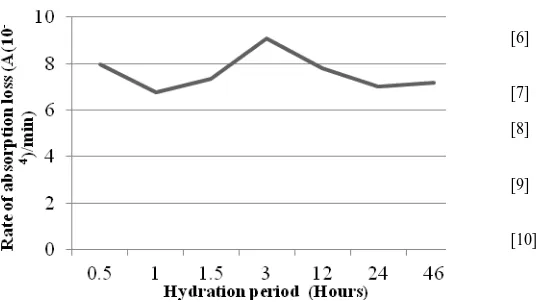
[image:5.612.54.325.261.631.2] [image:5.612.57.316.278.445.2]Furthermore, the rate of weight loss over the 3 hour testing period was calculated for each hydration time measurement, and is shown in Fig. 5, as well as the rate of absorption loss that occurred during the same interval which is shown in Fig. 6. Looking at both plots, there seem to be a conflicting relationship between the rate of weight loss and absorbance loss, as hydration periods whose rate of weight loss were higher compare to others had a reduced rate of absorption loss at the 1450 nm band and vice versa. However, it is not yet clear at this point whether a connection exists between the two rates and whether the duration of time spent in water had an effect on these results.
Fig 5. A graph showing the rate of weight loss by the sample of porcine skin versus hydration periods.
Fig 6. A graph showing the rate of absorbance loss by the sample of porcine skin versus hydration periods.
IV. CONCLUSION AND FUTURE WORK
Using a highly advanced and accurate spectrophotometer, it has been possible to obtain direct absorption spectra of porcine skin samples in vitro that evidently showed the dominant combination and overtone bands around 1920 and at 1450 nm, and weaker bands belonging to CH, NH and CO
bonds. The desorption tests were able to verify changes in these in accordance with water content but a clear relationship has not yet been established between hydration periods and the intensity of water absorption bands. Also, there seem to be a conflicting or rather a slightly nonlinear correlation in relation to rates of weight loss experienced by the sample and rates of loss in intensities of water bands. Future work will focus on further analyzing these data to provide clearer understanding of this relationship and the contribution of barrier function to these results, then to employ this method along with fibre optic technology to conduct in vivo experiments on human participants that will possibly investigate the effects of certain skin pathologies and external conditions on the water content of skin. Finally, the preliminary results obtained here will be used to aid in the development of a handheld optical system which can be assembled either by building its individual components or by choosing ones available already in the market, that could reliably measure the hydration level of skin in vivo, or more specifically, the hydration in the SC in a fast and noninvasive manner, and that would be smaller in size to eliminate the use of current bench-top instruments, yet perform to the same standard, and could even be portable for wider use in various applications.
REFERENCES
[1] A. V. Rawlings and J. J. Leyden, Skin moisturization. New York: Marcel Dekker, 2002.
[2] E. N. Marieb and J. Mallatt, Human Anatomy: Benjamin-Cummings Pub Co, 1996.
[3] T. F. Tsai and H. I. Maibach, "How irritant is water? An overview," Contact Dermatitis, vol. 41, pp. 311-4, Dec 1999. [4] C. Duch Lynggaard, D. Bang Knudsen, and G. B. Jemec, "Stratum
corneum damage and ex vivo porcine skin water absorption - a pilot study," Skin pharmacology and physiology, vol. 22, pp. 295-8, 2009.
[5] I. Bodén, D. Nilsson, P. Naredi, and B. Lindholm-Sethson, "Characterization of healthy skin using near infrared spectroscopy and skin impedance," Medical & Biological Engineering &
Computing, vol. 46, pp. 985-995, 2008.
[6] L. T. Kilpatrick-Liverman, P. Kazmi, E. Wolff, and T. G. Polefka, "The use of near-infrared spectroscopy in skin care applications,"
Skin Research and Technology, vol. 12, pp. 162-169, 2006.
[7] P. G. Agache, P. Humbert, and H. I. Maibach, Measuring the skin: Springer, 2004.
[8] P. L. Walling and J. M. Dabney, "Moisture in skin by near-infrared reflectance spectroscopy," J. Soc. Cosmet. Chem, vol. 40, pp. 151-171, 1989.
[9] K. A. Martin, "Direct measurement of moisture in skin by NIR spectroscopy," Journal-society of cosmetic chemists", vol. 44, pp. 249-249, 1993.
[10] J. De Rigal, M. J. Losch, R. Bazin, C. Camus, C. Sturelle, V. Descamps, and J. L. Leveque, "Near infrared spectroscopy: a new approach to the characterization of dry skin," Journal-society of cosmetic chemists", vol. 44, pp. 197-197, 1993.
[11] Y. A. Woo, J. W. Ahn, I. K. Chun, and H. J. Kim, "Development of a method for the determination of human skin moisture using a portable near-infrared system," Analytical chemistry, vol. 73, pp. 4964-4971, 2001.
[12] A. F. Omar and M. Z. MatJafri, "Optical Fiber Near Infrared Spectroscopy for Skin Moisture Measurement."
[13] Mohamad, A. R. Msabbri, and M. Z. MatJafri, "Conceptual design of near infrared spectroscopy instrumentation for skin moisture measurement," 2011 IEEE Colloquium on Humanities Science and
[image:5.612.58.326.475.626.2]