City, University of London Institutional Repository
Citation
:
Pelah, A., Barbur, J. L., Thurrell, A. and Hock, H. S. (2015). The coupling of vision with locomotion in cortical blindness. Vision Research, 110(PB), pp. 286-294. doi: 10.1016/j.visres.2014.04.015This is the accepted version of the paper.
This version of the publication may differ from the final published
version.
Permanent repository link:
http://openaccess.city.ac.uk/12512/Link to published version
:
http://dx.doi.org/10.1016/j.visres.2014.04.015Copyright and reuse:
City Research Online aims to make research
outputs of City, University of London available to a wider audience.
Copyright and Moral Rights remain with the author(s) and/or copyright
holders. URLs from City Research Online may be freely distributed and
linked to.
City Research Online: http://openaccess.city.ac.uk/ [email protected]
The Coupling of Vision with Locomotion in Cortical Blindness
Adar Pelaha, John Barburb, Adrian Thurrellc, and Howard S. Hockd
a. Department of Electronics, University of York, York, Y010 5DD, UK
b. School of Health Sciences, City University London, London EG1V 0HB, UK
c. Girton College, University of Cambridge, Cambridge CB3 0JG, UK
d. Department of Psychology and the Center for Complex Systems and Brain Science, Florida Atlantic University, Boca Raton FL 33486, USA
Corresponding Author: Adar Pelah
Department of Electronics University of York
York, Y010 5DD, UK +44 7808 707766 [email protected]
Short title: Visuo-Locomotor Coupling in Cortical Blindness
Abstract
Maintaining or modifying the speed and direction of locomotion requires the
coupling of the locomotion with the retinal optic flow that it generates. It is shown that
this essential behavioral capability, which requires on-line neural control, is preserved
in the cortically blind hemifield of a hemianope. In experiments, optic flow stimuli were
presented to either the normal or blind hemifield while the patient was walking on a
treadmill. Little difference was found between the hemifields with respect to the
coupling (i.e. co-dependency) of optic flow detection with locomotion. Even in the
cortically blind hemifield, faster walking resulted in the perceptual slowing of detected
optic flow, and self-selected locomotion speeds demonstrated behavioral discrimination
between different optic flow speeds. The results indicate that the processing of optic
flow, and thereby on-line visuo-locomotor coupling, can take place along neural
pathways that function without processing in Area V1, and thus in the absence of
conscious intervention. These and earlier findings suggest that optic flow and object
motion are processed in parallel along with correlated non-visual locomotion signals.
Extrastriate interactions may be responsible for discounting the optical effects of
locomotion on the perceived direction of object motion, and maintaining visually guided
self-motion.
1.0 Introduction
The relationship between perception and action has been of long-standing interest to
researchers concerned with both visual processing and motor control. Indicative of
their co-dependence is evidence that locomotion can induce changes in the perceived
speed of concurrent optic flow (Pelah & Barlow, 1996; Thurrell, Pelah & Distler, 1998;
Thurrell & Pelah, 2002, 2005; Durgin, Gigone & Scott, 2005), that changes in optic flow
speed while walking at a constant speed can signal an impending collision (Lee, 1980),
and that locomotion can change involuntarily in response to changes in optic flow
(Prokop, Schubert & Berger, 1997; Dong, Pelah, Cameron & Lasenby, 2008).
Perhaps the most important aspect of the on-line coupling of optic flow detection
and locomotion is that they interact recursively. That is, locomotion generates an optic
flow pattern on the retina, changes in the optic flow pattern produce changes in the
speed and/or direction of locomotion, which in turn changes the optic flow pattern, and
so on. The function of this dynamic co-dependence is to maintain (or modify) walking
speed and/or walking direction (heading) in response to stability (or change) in the
locomotion-generated optic flow pattern (Held & Freedman, 1963; Gibson, 1950;
Warren & Hannon, 1988).
A noteworthy feature of locomotion in normally sighted individuals is that
retrospectively (and introspectively) episodes of walking seem to have taken place
without conscious awareness or attention to the optic flow pattern that had been
generated by the locomotion. This effect, together with observations that a surprising
degree of visual control of locomotion can be retained in cortical blindness (Humphrey,
be processed without access to the pathways mediating conscious visual awareness.
The objective of this study is therefore to determine whether behavior requiring the
detection of optic flow and its inherent coupling with locomotion are preserved, despite
the absence of processing in Area V1 and the accompanying loss of conscious
awareness.
This objective was addressed by testing a hemianope, an individual for whom
unilateral damage to the striate cortex (Area V1) has resulted in the loss of
object/shape perception and conscious awareness for stimuli presented in the
contralateral hemifield (Barbur, Ruddock & Waterfield, 1980; Weiskrantz, 1986; Barbur
et al. 1993), the ipsilateral hemifield having remained normally sighted and thus acting
as a control. During trials, optic flow stimuli were presented to either the normally
sighted or the cortically blind hemifield whilst the hemianope was walking on a
treadmill. Evidence for partial sparing of direction discrimination for a variety of moving
stimuli (e.g., Barbur et al. 1993; Azzopardi & Cowey, 2001) led to the expectation that
optic flow motion could also be processed in the cortically blind hemifield. What is
unique about the current study is that rather than direction discrimination, as in earlier
studies, it is aimed at showing that this kind of unconsciously detected motion is
coupled with an essential behavior, locomotion.
The further possibility that there are independent pathways for the processing of
optic flow and object motion was suggested by evidence for qualitative differences in
the stimulus information that serves as a basis for direction discrimination in the
cortically blind and normally sighted hemifields. That is, Azzopardi and Hock (2011)
the detection of spatio-temporal changes in “raw” luminance (Chubb & Sperling, 1989),
or more generally, 1st-order motion energy (Adelson & Bergen, 1985), whereas
direction discrimination within his normally sighted hemifield relied on the detection of
changes in shape (although motion energy extraction remained possible as well).
Treadmill walking was essential for this study because it disrupts the normal
correlation between locomotion speed and optic flow speed (Pelah & Barlow, 1996); on
a treadmill, faster walking no longer automatically results in faster optic flow. Under
these open loop conditions, optic flow on the retina is not affected by walking speed,
and thus, the lack of conscious awareness of an optic flow pattern cannot be attributed
to compensatory mechanisms that discount or cancel the retinal motion signal via
matching walking-speed determined efferent or afferent motor information (Andersson
et al. 1981; Thurrell & Pelah, 2005; Tcheang, Gilson & Glennerster, 2005), nor to an
internal template of the optic flow pattern for different locomotion speeds (Perrone,
1992).
Obtaining evidence for visuo-locomotor coupling when optic flow stimuli are
presented within the hemianope’s cortically blind hemifield, where there is no feed
forward projection to Area V1, and no conscious awareness of the stimuli, would then
indicate that retinal optic flow signals have reached extrastriate areas via neural
pathways that by-pass Area V1. In the macaque, these pathways involve the superior
colliculus of the midbrain (Gross, 1991; Mohler & Wurtz, 1977) and/or direct
connections from the lateral geniculate nucleus (Cowey & Stoerig,1989; Schmid et al.
The extrastriate targets for pathways through Area V1 and pathways that
by-pass Area V1, include directionally selective motion detectors in macaque Area MT
(Newsome, Mikami & Wurtz, 1986). Cooling or lesioning Area V1 leaves a high
proportion of MT neurons active, and the additional destruction of the superior
colliculus completely eliminates MT activation (Rodman, Gross & Albright, 1989, 1990).
Significantly, directionally selective Area MT motion detectors project onto optic flow
detectors in Area MSTd (Tanaka & Saito, 1989; Yu et al. 2010).
Three experiments are described in which the cortically blind and normally
sighted hemifields of the hemianope were compared in order to determine behaviorally
whether visuo-locomotor coupling could be based on the processing of optic flow along
neural pathways that by-pass Area V1, independently of conscious awareness of the
optic flow stimulus, and independently of motion processing along the geniculostriate
pathway.
2.0 Methods
2.1 General method
Testing was done with an individual, denoted as GY, who suffered damage to
his occipital lobe following an automobile accident at the age of 8 years that resulted in
unilateral loss of function in his left primary visual cortex (Area V1). He is functionally
hemianopic, with less than 3 deg macular sparing, probably due to spared tissue in the
occipital pole (Barbur, Ruddock & Waterfield, 1980). As illustrated in Figure 1a, testing
was done with a locomotion simulator composed of a Woodway Exo43 treadmill facing
a large translucent screen (Pelah et al. 1998). Optic flow stimuli were rear-projected
pixels and a refresh rate of 70 Hz, updated on alternate frames (the projection covered
a visual area of 93 x 77 deg). Movement on the treadmill was not motorized. Its belt
was composed of low-friction rolling slats, so GY’s self-generated locomotion required
minimal exertion. Whether walking or standing, the viewing distance to the center of
the screen was approximately 90 cm.
The optic flow stimulus was composed of a set of 15 nested, concentric square
frames that radiated outward to create the appearance of walking through a corridor.
Consistent with the laws of perspective, the frames varied in diameter and thickness as
the inverse tangent of their simulated distance from the observer. The innermost frame
intercepted a visual angle of 19.0 deg and was 1.2 deg thick. The outer-most frame
intercepted a visual angle of 77.0 deg and was 2.4 deg thick. The radially expanding
motion was faster for the outer than the inner squares (as measured in the plane of the
display). The optic flow speeds indicated for each experiment were characterized by
the speed measured at the mid-hemifield position of the stimulus, approximately 27.8
deg from fixation (indicated by the broken line in Figure 1b).
The luminance values of the nested squares varied with eccentricity. It was
dimmest (0.1 cd/m2) for the innermost frame, simulating it being the furthest square
from the perceiver when expanding optic flow results from forward walking. As the
frames radiated outward, their luminance gradually increased to 1.9 cd/m2 at their
mid-hemifield location, and gradually decreased to the background luminance of 0.01 cd/m2
as the square frames continued radiating outward toward the display’s periphery.
When the outer-most frame disappeared, it was immediately replaced by the
changes in luminance minimized luminance transients, and in particular, edge flicker in
the far periphery. In different experimental conditions, luminance values were reduced
from the above values by placing neutral density filters in front of the lens of the LCD
projector. Goggles were worn in order to shield GY’s left eye and occlude peripheral
distractions.
The left and right halves of the nested squares stimulus were presented during
separate blocks of trials, directed respectively at either the normally sighted or the
cortically blind visual hemifield. The experiments were conducted following 30 min of
dark adaptation. GY was instructed to maintain fixation on a small square (0.28 x 0.28
deg; luminance = 1.5 cd/m2) at eye level in the center of the display. Self-propelled
walking speeds were measured with a sensor attached to the treadmill. The time series
of walking speeds was low-pass filtered and the average speed determined over the
last 5 sec of each walking interval. No part of the stimulus was presented within a 3.5
deg radius circular arc surrounding the fixation square in order to ensure that the
0 5 10 15 20 25
0 10 20 30
Eccentricity (deg) O p ti c F lo w S p e e d ( d e g /s e c ) (b) (a) Figure 1
stimulus was outside GY’s spared macula region of the retina, which responds to visual
information in both the blind and sighted hemifields (Barbur et al. 1980).
2.2 Monitoring eye fixation
GY previously participated in numerous psychophysical studies that required
fixation at a specified location, most of which confirmed fixation by visual inspection.
Quantitative measurements by Weiskrantz, Harlow and Barbur (1991) indicated that he
could maintain fixation to within approximately +/- 0.5 deg. Whether fixation could also
be maintained while GY was walking on a treadmill was determined in this study with a
head-mounted Epic 1-Diamond IR Limbus Eye Tracker, which detected horizontal eye
movements with respect to the fixation point. These measurements were made while
GY’s head was placed in a chin rest while walking on the treadmill. Despite the head
movements produced by the locomotion, GY maintained fixation to within +/- 2.0 deg,
well enough that random fluctuations in eye position were too small to displace portions
of the optic flow onto his spared macular region. Fixation was monitored by the visual
inspection of GY’s eyes throughout all three experiments.
2.3 Conscious awareness
After each trial, GY indicated whether or not he was aware of the optic flow
stimulus. He reported full awareness when it was presented in his normally sighted
hemifield, but not in his cortically blind hemifield. His reports for blind hemifield
presentations may have reflected both Type 1 blindsight, for which there is no
conscious awareness whatsoever, and Type 2 blindsight, for which there is no
conscious awareness of the stimulus, but there is an awareness that “something is
that GY can exhibit both types of blindsight, depending on the stimulus discrimination
required. In Experiment 1 of the current study, no discrimination was required of GY
when he was walking on the treadmill while optic flow was presented in either his
normally sighted or cortically blind hemifield. Type 1 blindsight, with no conscious
awareness whatsoever, is therefore possible for his blind hemifield. In Experiments 2
and 3, GY was required to discriminate between different optic flow speeds by walking
at a speed that matched the optic flow speed; Type 2 blindsight was therefore possible
here. However, at the start of each trial in each of the last two experiments GY had to
be told when to start walking, even though the optic flow stimulus was already
presented in his cortically blind hemifield. He also had to be told when to stop walking
at the end of a trial after the stimulus was gone. He was unable to distinguish the optic
flow stimulus from a blank screen, was indicative of Type 1 blindsight. Because GY’s
reports of no awareness of the optic flow stimulus could have reflected either type of
blindsight, we have taken the conservative position that the results reflect Type 2
blindsight.
3.0 Experiment 1: Locomotion and Judgments of Optic Flow Speed
Most experimental and computational analyses of optic flow processing have
been concerned with the distortion of locomotion-generated optic flow patterns by eye
movements, and its resulting effect on the perception of heading (e.g., Warren &
Hannon, 1988; Warren et al. 2001). Much less frequent are studies examining the
relationship between optic flow and the motor-related signals of locomotion. Many of
walking speed and gait patterns (Pailhous, et al. 1990; Konzak, 1994; Prokop et al.
1997; Dong, et al. 2008). Experiment 1 of the current study was concerned with the
reverse, i.e., the effect of locomotion speed on the perception of optic flow. That is,
while open-loop treadmill walking speed does not affect the retinal speed of an
independently presented optic flow stimulus, it does affect its perceived speed. The
‘speeding-up’ of perceived optic flow occurs while walking in a normal environment
following a period of treadmill walking in the absence of optic flow (Pelah & Barlow,
1996), and the ‘slowing down’ of perceived optic flow occurs during treadmill walking in
the presence of optic flow (Thurrell et al. 1998). For the latter, the more rapid the
treadmill walking, the slower the physically constant optic flow appears (Thurrell let al.
1998; Thurrell & Pelah, 2002, 2005; Durgin et al. 2005).
It was determined in this experiment whether the slowing effect of walking speed on
perceived optic flow speed, an indicator of visuo-locomotor coupling, would be
observed in GY’s cortically blind as well as his normally sighted hemifield. This was
determined by presenting optic flow stimuli to either hemifield while he was walking on
a self-propelled treadmill at one of six self-selected speeds.
3.1 Method
Each trial began with a written instruction on the screen indicating the subjective
walking speed required of GY for that trial: either ‘stationary’, ‘very slow’, ‘slow’,
‘normal’, ‘fast’, or ‘very fast’. Five sec was provided for GY to reach his self-selected
walking speed according to the instruction. This was followed by a 10 sec interval
during which a vertically split expanding optic flow stimulus with a speed of 2.7 deg/sec
cortically blind or normally sighted hemifield. Immediately after the 15 sec interval,
while now standing stationary on the treadmill, GY adjusted the speed of an optic flow
stimulus presented in his normally sighted hemifield so that it reproduced the
remembered speed of the optic flow stimulus during the preceding walking episode.
The initial setting for each 10 sec test was at a randomly selected optic flow speed.
The average speed-matching setting was determined over the final 1 sec of the 10 sec
speed-setting interval. There were a total of 18 randomly ordered trials, 3 for each of
the 6 subjective walking speeds, presented first in GY’s normally sighted hemifield, and
then for two blocks of 18 trials in his cortically blind hemifield. Before testing, GY
practiced walking on the treadmill in response to the six different speed instructions.
3.2 Results
The effect of locomotion speed on the perceived speed of accompanying
optic flow was measured by the post-locomotion reproduction of that optic flow speed.
Remarkably, faster walking resulted in the perceived slowing of optic flow, even when
the optic flow was presented in GY’s cortically blind hemifield, Moreover, the slowing
effect in the blind hemifield was similar to that obtained in the normally sighted
hemifield; for both the fastest walking resulted in the constant-speed optic flow stimulus
appearing to be stationary. The negative correlation between walking speed and
perceived optic flow speed was significant for the normally sighted hemifield, r(16) =
-0.92, p < 0.001 (Figure 2a), as well as for the first block, r(16) = -0.78, p < 0.001, and
second block, r(16) = -0.91, p < 0.001, of trials for the cortically blind hemifield (Figures
2b and 2c). However, the hemifields differed in their sensitivity to the differences in
for the two blocks of blind-hemifield trials (slope = -0.63 and -0.60) than for the
normally sighted hemifield trials (slope = -0.89). The reduced sensitivity to differences
in speed in the blind hemifield was not surprising given the substantial loss in
spatiotemporal contrast sensitivity in GY’s cortically blind hemifield (Cowey, 2010). y = -0.8926x + 5.1121
R = 0.85431
!" #" $" %" &" '" ("
!" #" $" %" &" '" ("
0 1 2 3 4 5 6
Walking Speed (km/hour) 0 6 4 2 O p ti c F lo w S p e e d ( d e g /s e c )
(Normally Sighted Hemifield) Perceived Speed of Expansive Optic Flow
correlation = -0.92 slope = -0.89
[image:14.612.96.504.110.500.2](a)
Figure 2
(Caption on last page of the manuscr ipt) EXPERIMENT 1 !"#"$%&'%()*"+",&))-" ./"#"%&0,(11" !" #" $" '" ("
!" #" $" %" &" '" ("
0 1 2 3 4 5 6
Walking Speed (km/hour) 0 6 4 2 O p ti c F lo w S p e e d ( d e g /s e c )
Perceived Speed of Expansive Optic Flow (Cortically Blind Hemifield - Block 2)
correlation = -0.91 slope = -0.60
(c) (b) !" #" $" %" &" '" ("
!" #" $" %" &" '" ("
0 1 2 3 4 5 6
Walking Speed (km/hour) 0 6 4 2 O p ti c F lo w S p e e d ( d e g /s e c )
Perceived Speed of Expansive Optic Flow (Cortically Blind Hemifield - Block 1)
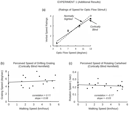
3.3 Additional results – verbal ratings of optic flow speed
The results of Experiment 1 are indicative of optic flow being detected, under the
influence of walking speed, in GY’s blind hemifield. Alternatively, it might be argued
that GY detected nothing useful in his cortically blind hemifield, and that instead his
post-locomotion judgments of optic flow speed in his blind hemifield were derived from
visual memories from earlier judgments of optic flow speed in his sighted hemifield.
That is, the apparent slowing effect of locomotion speed on the perceived speed of the
optic flow presented in GY’s cortically blind hemifield may have been due to visual
memories associated with similar locomotion speeds during earlier testing in his
normally sighted hemifield.
Contrary to this possibility, there is clear evidence that differences in stimulus
speed can be discriminated in cortically blind hemifields (Barbur, Ruddock &
Waterfield, 1980; Morland et al. 1999). The purpose of this additional experiment was
to confirm these earlier findings with the optic flow stimuli tested in the current study.
GY judged four randomly ordered speeds of expanding optic flow (4.0, 7.0, 10.0
and 13.0 deg/sec) while standing stationary on the treadmill. After each 10 sec
presentation, he verbally rated the speed of the optic flow on a scale from 1 to 4, with 4
denoting the fastest and 1 the slowest speed. There were 160 trials (40 for each of the
4 optic flow speeds) separately for his sighted and blind hemifields. The luminance of
the square frame near the mid-hemifield location was 0.005 deg/m2. Consistent with
previous studies, these judgments were made while GY was stationary. Behavioral
discrimination of optic flow speeds in his cortically blind hemifield, as indicated by
It can be seen in Figure 3a that GY was able to verbally discriminate optic flow
speeds approximately equally well in his cortically blind and normally sighted
hemifields. With detected optic flow speeds differentially encoded in his blind hemifield
only moments before the sighted-hemifield reproduction test for a trial, it is very unlikely
that GY instead based his judgments of optic flow speed in his blind hemifleld on
perceived speeds recalled from a preceding block of sighted-hemifield trials, which
occurred six or twelveminutes prior to the two blocks of blind-hemifield trials. It can be
!"#"$%$&'()"*"+%,-'." /0"#"$%$,, .," !" #" %" &"
!" '" #" (" $" )" %"
!"#"$%&%%'()"*"%&++,-" ./"#"%&%'%-0" !" !#$" !#&" !#'"
!" $" %" &" '" (" )"
0 1 2 3 4 5 6
Walking Speed (km/hour)
0 1 2 3 4 5 6
Walking Speed (km/hour) 0 6 4 2 G ra ti n g S p e e d ( d e g /s e c ) 8 R o ta ti o n S p e e d ( / s e c ) 0.4 0.3 0.2 0.1 0
(Cortically Blind Hemifield) (Cortically Blind Hemifield)
Perceived Speed of Drifting Grating Perceived Speed of Rotating Cartwheel EXPERIMENT 1 (Additional Results)
correlation = 0.11 slope = 0.08
correlation = -0.17 slope = -0.03 (c)
[image:16.612.93.518.75.451.2](b)
Figure 3
(Caption is on last page of the manuscr ipt)
Cortically Blind Normally Sighted 1 2 3 4 V e rb a l S p e e d R a ti n g s
Optic Flow Speed (deg/sec)
3 5 7 9 11 13
concluded, therefore, that locomotion does indeed slow the perceived speed of
detected optic flow more than slower locomotion (as indicated by the negative slopes in
Figure 2), regardless of whether the optic flow is presented in the normally sighted or
cortically blind hemifield.
3.4 Additional results – locomotion with non-optic flow stimuli
A further experiment determined whether the slowing effect of locomotion on the
perception of speed is specific to optic flow stimuli, as would be expected if this
evidence for visuo-locomotor coupling were relevant to visually guided locomotion in
the natural environment. To determine whether this was the case, testing for
locomotion induced slowing was done by presenting, in the cortically blind hemifield, a
downward drifting, horizontally-oriented rectangular grating, and a rotating cartwheel
stimulus, neither of which can be generated as optic flow by forward locomotion.
As in the main experiment, nothing but the fixation mark was presented within a
3.5 deg diameter arc surrounding the macula. The grating had a fundamental spatial
frequency of approximately 0.4 cycles/deg and a speed of approximately 2.5 deg/sec.
It was composed of 15 equally spaced, anti-aliased bars (luminance = 1.5 cd/m2)
presented against a dark (0.006 cd/m2) background. In order to minimize luminance
transients and edge flicker, as each bar appeared at the top of the display and then
drifted downward, its luminance gradually increased to a constant level, and then
gradually decreased until the bar disappeared at the bottom of the screen. The
cartwheel was composed of 15 spokes (luminance = 1.5 cd/m2), rotating
counterclockwise at a speed of 2.7 deg/sec. There were 18 randomly ordered trials for
As in Experiment 1, after each locomotion episode, GY, standing stationary on the
treadmill, adjusted the grating (or cartwheel) speed presented in his sighted hemifield
so that it matched the remembered speed of the drifting grating (or rotating cartwheel)
during the immediately preceding locomotion episode. It was found that there was no
effect of locomotion speed on the perceived speed of either the drifting grating or the
rotating cartwheel (Figures 3b and 3c). Thurrell and Pelah (2002) have reported similar
results with unimpaired subjects.
These additional results indicate that the slowing effect is specific to the
engagement of locomotion with optic flow stimuli. It is not a general bias due to
1 2 3 4
3 5 7 9 11 13
W a lk in g S p e e d ( k m /h o u r)
3 5 7 9 11 13
Normally Sighted
Cortically Blind
3 5 7 9 11 13
Normally Sighted Cortically Blind Normally Sighted Cortically Blind 1 2 3 4
3 5 7 9 11 13
Normally Sighted
Cortically Blind
3 5 7 9 11 13
Normally Sighted
Cortically Blind
3 5 7 9 11 13 Optic Flow Speed (deg/sec) Optic Flow Speed (deg/sec) Optic Flow Speed (deg/sec)
Optic Flow Speed (deg/sec) Optic Flow Speed (deg/sec) Optic Flow Speed (deg/sec)
W a lk in g S p e e d ( k m /h o u r) EXPERIMENT 2 Normally Sighted Cortically Blind
Luminance = 0.001 cd/m2 Luminance = 0.021 cd/m2 Luminance = 0.011 cd/m2 Luminance = 0.040 cd/m2
[image:18.612.89.525.82.385.2]Luminance = 0.005 cd/m2 Luminance = 0.003 cd/m2
Figure 4
concurrent locomotion that affects the perception of speed for any moving stimulus,
and in particular, it is not a processing bias peculiar to the cortically blind hemifield.
Because the slowing effect was obtained for optic flow stimuli presented in GY’s
cortically blind hemifield, it could be concluded that visuo-locomotor coupling can occur
without the geniculostriate pathway, and thus, without the associated conscious
awareness of the optic flow stimulus. Because it was obtained under the open loop
conditions of treadmill walking, it indicated that (unconscious) visuo-locomotor coupling
can occur irrespective of matched compensatory mechanisms that discount or cancel
optic flow.
4.0 Experiment 2: Matching Walking to Constant Optic Flow Speed
Experiment 1 showed that locomotion induced perceptual slowing can occur in
both GY’s normally sighted and cortically blind hemifields. On the basis of this
evidence for similar visuo-locomotor coupling in the two hemiflelds, it was next
determined whether GY would be able to match his treadmill walking speed to the optic
flow speed despite the absence of the genicuolostriate projections and Area V1
processing for his cortically blind hemifield.
The luminance of the nested square frames composing the expanding optic flow
stimulus was made progressively dimmer during successive blocks of trials in order to
minimize the possibility that locomotion matches for stimuli presented in GY’s blind
hemifield would benefit from light scatter into his sighted hemifield (King et al. 1996).
4.1 Method
0.011, 0.005, 0.003, and 0.001 cd/m2, as measured for the brightest square frame near
the mid-hemifield location of the expanding nested squares. Although the squares were
dim, they were within the range of visibility following a 30 min dark adaptation period as
confirmed by sighted hemifield controls. Blocks of 24 order-randomized trials were
determined by presenting each of the four optic flow speeds (4.0, 7.0, 10.0 and 13.0
deg/sec) six times. Six blocks of these 24 trials, one for each of the 6 luminance levels
were repeated 5 times in the normally sighted hemifield, then 10 times in the cortically
blind hemifield. During each trial GY was instructed to match his walking speed to the
optic flow speed.
4.2 Results
GY’s ability to match the speed of his walking to the speed of the optic flow
stimulus was similar in the two hemifields (Figure 4). For each hemifield and each
luminance level, GY’s average walking speeds were highly correlated with the physical
speed of the optic flow. With one exception (the lowest luminance level stimulus in the
blind hemifield) the correlations were greater than 0.95 (Footnote 1). The regression lines
for each of the six luminance levels were somewhat flatter for the cortically blind than
the normally sighted hemifield. Thus, as in Experiment 1, optic flow speeds were better
differentiated in the normally sighted hemifield.
5.0 Experiment 3: Matching Walking to Changing Optic Flow Speed
As discussed in Section 1.0, to be functional in the natural environment it is
was determined in this experiment whether this can also occur without the
geniculostriate projections to Area V1.
5.1 Method
Four distinctive stimuli, each repeated five times, were used to test whether GY
as well as his normally sighted hemifield. The changes in speed were either abrupt or
gradual, and either increasing or decreasing. Abrupt changes entailed a steep linear
increase (decrease) from 2 to 19 deg/sec (19 to 2 deg/sec) during a 0.3 sec interval in
the middle of a 24 sec trial. Gradual changes in optic flow speed were sinusoidal,
between 2 and 19 deg/sec over the full 24 sec. As in Experiment 2, GY was instructed
to match his walking speed to the speed of the optic flow stimulus in both his normally
sighted and cortically blind hemifields.
5.2 Results
GY was able to modify his walking speed in approximate correspondence to
both gradual and abrupt changes in optic flow speed, regardless of whether the speed
increased or decreased. In his blind hemifield, the average difference in walking speed
between the fast and slow phases of the changing optic flow stimulus was statistically
significant; t(3) = 12.3, p< .001.Footnote 3. That is, it was reliably obtained despite
differences in the type of optic flow change (gradual-increase, gradual-decrease,
abrupt-increase or abrupt-decrease). This also was the case when these stimuli were
tested in his sighted hemifield; t(3) = 13.1, p< .001.
It can be seen for the individual trials presented in Figure 5 that his normally
sighted and cortically blind hemifields were similar with respect to the magnitude of
change in GY’s walking speed, but the changes in walking speed were relatively
delayed in his blind hemifield. Further research will be required to determine whether
delayed responses to changes in velocity (i.e., changes in speed and/or direction) is a
general characteristic of hemianopic vision that results from the absence of V1
6.0 General Discussion
Visuo-locomotor coupling occurs whenever we walk in a natural environment.
Locomotion creates optic flow on the retina, which in turn is used to maintain or change
locomotion in a selected direction and at a selected speed. Visuo-locomotor coupling
also occurs while walking on a treadmill, where it takes the form of locomotion-induced
slowing of perceived optic flow speed (Experiment 1), and on matching walking speed
to constant or changing optic flow speed (Experiments 2 and 3). The results of the
current study show that during visuo-locomotor coupling neither the slowing effect nor
speed matching requires processing in the geniculostriate pathways that sustain
conscious awareness of visual stimuli. These results are unique in comparison with
other studies of hemianopic vision, which typically involve simple discriminations (e.g.,
upward vs. downward motion). Evidence was obtained here for the sparing of optic
flow detection coupled with an essential behavior, locomotion. It is noteworthy that this
linkage with locomotion seems to be specific to the detection of optic flow. Evidence for
locomotion-induced perceptual slowing was not observed for stimuli (vertically drifting
gratings and rotating propellers) presented in the cortically blind hemifield (Section 3.3;
see Thurrell & Pelah, 2002; 2005; for normally sighted subjects). These stimuli are not
generated by locomotion.
Because of unilateral damage to Area V1 of the hemianope, these results
indicate that visuo-locomotor coupling can be based on the processing of optic flow in
neural pathways from retina to Area MT that by-pass Area V1 (likely through the
flow sensitive neurons in homologous Area MSTd (Tanaka & Saito, 1989; Yu et al.
2010) and the posterior parietal cortex for visuo-motor coordination (Milner & Goodale,
1993; Andersen, Snyder, Bradley & Xing, 1997). Given that processing in Area V1 is
deemed necessary for conscious visual awareness (Lamme, 2001; Silvanto et al.
2005), the evidence obtained without Area V1 indicates that conscious awareness of
the optic flow stimulus is not necessary for its coupling with locomotion. It can be
inferred that introspective reports of lack of awareness or attention to optic flow during
locomotion in natural environments may be due to the predominance of activity in
neural pathways that by-pass Area V1.
6.1 Limitations in hemianopic vision
While the hemianope (GY) tested in these experiments reported no conscious
awareness of the stimulus in his cortically blind hemifield, it remains uncertain whether
these reports reflected a complete absence of conscious awareness (see Section 2.3).
This notwithstanding, it has been well-established for GY and other hemianopes that
their perceptual capabilities are typically very different for stimuli presented in their two
hemifields, in that there are substantial blind-hemifield deficits for the discrimination of
numerous visual attributes (Barbur, Harlow & Weiskrantz, 1994; Cowey, 2010).
Notably, the usual hemifield asymmetry was much reduced in the current study, which
found that visuo-locomotor coupling is similar in the cortically blind and normally
sighted hemifields. The observed similarity of the hemifields suggests that neural
pathways in which locomotion-induced optic flow is processed can function
independently of the (in this case, damaged) geniculostriate pathway to Area V1. The
hemifield may have been due to the hemianopic loss in spatiotemporal contrast
sensitivity in the absence of Area V1 processing (Barbur et al. 1994; Cowey, 2010), or
to the absence of feedback from damaged Area V1 to subcortical nuclei that affect
contrast sensitivity (Przybyszewski et al. 2000; Cudeiro & Sillito, 2006).
Alternatively, it is possible that GY’s speed-matching deficits in his cortically
blind hemifield were due to the absence of Area V1 mechanisms that might also couple
optic flow detection with locomotion (Keller, Bonhoeffer & Hübener, 2012; Niell &
Stryker, 2010). The conscious processing that takes place in Area V1 may become
necessary for visually guided locomotion in cluttered, dynamic environments in order to
avoid collisions with stationary and moving objects. That is, both the neural pathway
that by-passes Area V1 and the neural pathway that passes through Area V1
potentially contribute to visually guided locomotion. Their relative contribution depends
on the complexity of the environment to be navigated.
6.2 Effects of non-visual signals
Effects of non-visual signals on perceived optic flow have been indicated by a
number of studies (e.g., Andersson et al. 1981; Pelah & Barlow, 1996; Warren et al.
2001; Thurrell & Pelah, 2002, 2005; Durgin et al. 2005; Tcheang et al. 2005). For
locomotion, the non-visual signals may originate from the control and movement of the
locomoting limbs (Lappe, 1997), as proprioceptive afferents, signals of spinal origin or
associated corollary discharge signals (sometimes called reafferent or efference copy).
Although such inputs to extrastriate regions associated with limb movement have not
been identified, analogous modulation of optic flow neurons in MSTd by non-visual
vestibular signals (Duffy, 1998; Bremmer, et al, 2001; Gu et al, 2006; Fetsch, et al,
2007). The results of the current study suggest that non-visual locomotor-based
signals, if present, are integrated with the detected optic flow in order to determine its
perceived speed, and thereby signal the speed of locomotion (Pelah and Barlow,
1996). This could occur at or prior to extrastriate processing and operate without
projections to, or feedback from, Area V1.
6.3 Motion detection mechanisms
The results obtained in this study are also relevant to identifying the kind of
motion mechanism that is the basis for the detection of optic flow. As indicated earlier,
it has been found that the discrimination of motion direction in GY’s cortically blind
hemifield, where object perception is severely impaired, is based on the detection of
1st-order motion energy; i.e., stimulus information entailing spatiotemporal changes in
luminance rather than changes in shape (Azzopardi & Hock, 2011; Sperling & Lu,
1998; Hock & Nichols, 2013). The evidence in the current study for visuo-locomotor
coupling in GY’s cortically blind hemifield implies that the detection of
locomotion-induced optic flow in GY’s blind hemifield likewise entails the detection of motion
energy. That is, in contrast with changes in the features of an object that determine
both its shape and direction of motion, motion perception would be based on detected
motion energy, which has been characterized as ‘objectless’ (Sperling & Lu, 1998;
Hock & Nichols, 2011, 2013) because it provides a sense of motion without a sense of
what it is in the environment that has moved (an apt characterization of optic flow
In contrast with optic flow, the processing of object motion is thought to depend
on the detection of changes in the features of the object (e.g., changes in edge
contrast at the object’s boundaries; Hock & Nichols, 2010; 2013)(Footnote 2)
. In normal
vision this would take place along a parallel neural pathway that passes through Area
V1 en route to Area MT and other extrastriate areas, enabling conscious awareness of
the object’s shape and direction of motion (Lamme, 2001; Silvanto et al. 2005).
As we walk or run through a natural environment, the retinal optic flow created
by our locomotion is vectorially combined with the independent retinal motion of objects
in the environment. In light of the above evidence for differences in motion processing
in the parallel pathways to Area MT (whether through or by-passing Area V1), it can be
speculated that mutually inhibitory interactions among Area MT neurons (Snowden et
al. 1991; Recanzone et al. 1997; Heeger et al. 1999; Thiele, Dobkins & Albright, 2000),
some of which are motion energy sensitive and some of which are not (Krekelberg &
Albright, 2005), could form the basis for discounting the optical effects of locomotion on
the perceived direction of object motion. Interactions of “objectless’ optic flow with
non-visual signals would modulate optic flow speed, and remain essential for non-visually
guiding the walkers’ self-motion perception as they move through natural or altered
environments (Pelah & Barlow, 1996).
We are grateful to Hartwig Distler for assistance with technical development and
References
Adelson, E.H., & Bergen, J.R. (1985). Spatiotemporal energy models for the perception of
motion. Journal of the Optical Society of America A, 2, 284-299.
Andersen, R.A., Snyder, L.H., Bradley, D.C., & Xing, J. (1997). Multimodal
representation of space in the posterior parietal cortex and its use in planning
movements. Annual Review of Neurology,20, 303-330.
Andersson, O., Forsberg, H., Grillner, S., & Wallen, P. (1981). Peripheral feedback
mechanisms acting on the central pattern generators for locomotion in fish and
cat. Canadian Journal of Physiology and Pharmacology, 59, 713-726.
Azzopardi, P., & Cowey, A. (2001). Motion discrimination in cortically blind patients.
Brain, 124, 30-46.
Azzopardi, P., & Hock, H.S. (2011). Illusory motion perception in blindsight.
Proceedings of the National Academy of Science USA,108, 876-881.
Barbur, J.L., Harlow, A.J., Weiskrantz, L. (1994). Spatial and temporal response
properties of residual vision in a case of hemianopia. Proceedings Royal Society
of London B. 343, 157-166.
Barbur, J.L., Ruddock, K.H., & Waterfield, V.A. (1980). Human visual responses in the
absence of the geniculo-calcarine projection. Brain,103, 905-928.
Barbur, J.L., Watson, J.D.G., Frackowiak, R.S.J., & Zeki S. (1993). Conscious visual
perception without V1. Brain,116, 1293-1302.
Bremmer, F., Schlack, A., Shah, N., Zafiris, O., Kubischik, M., Hoffmann, K.-P.,
and premotor cortex: a human fMRI study strongly implies equivalencies
between humans and monkeys. Neuron, 29, 287-296.
Bullier, J., Girard, P., & Salin, P.-A. (1994). The role of area 17 in the transfer of
information to extrastriate visual cortex. In Cerebral Cortex Vol 10, Peters, A., &
Rockland, K.S. (Eds.), Plenum Press, New York (pp. 301-330).
Chubb, C., & Sperling, G. (1989). Two motion perception mechanisms revealed
through distance-driven reversal of apparent motion. Proceedings of the
National Academy of Science USA , 86, 2985-2989.
Cowey, A. (2010). The blindsight saga. Experimental Brain Research, 200, 3-24. Cowey, A., & Stoerig, P. (1989). Projection patterns of surviving neurons in the dorsal
lateral geniculate nucleus following discrete lesions of striate cortex:
implications for residual vision. Experimental Bain Research, 75, 631-638.
Cowey, A., & Stoerig, P. (1991). The neurobiology of blindsight. Trends in
Neuroscience,14,140-145.
Cudeiro, J., & Sillito, A.M. (2006). Looking back: corticothalmic feedback and early
visual processing. Trends in Neurosciences, 29,
de Gelder, B., Tamietto, M., Boxtel, G.J.M. van, Goebel, R., Sahraie, A., Van den
Stock, J.B., Stienen, B.M.C., Weiskrantz, L., & Pegna, A. (2008). Intact
navigation skills after bilateral loss of striate cortex. Current Biology, 18,
1128-1129.
Dong, H., Pelah, A., Cameron, J.I., & Lasenby, J. (2008) The perceptual influences on
simulators. Applied Perception in Graphics and Visualization: Proceedings of the
5th Symposium on Applied Perception in Graphics and Visualization, 143-146.
Duffy, C.J. (1998). MST neurons respond to optic flow and translational movement.
Journal of Neurophysiology, 80, 1816–1827.
Durgin, F.H., Gigone, K., & Scott, R. (2005). Perception of visual speed while moving.
Journal of Experimental Psychology: Human Perception and Performance, 31,
339-353.
Fetsch, C.R., Wang, S. Gu, Y,, DeAngelis, G,C,, & Angelaki, D,E, (2007). Spatial
reference frames of visual, vestibular, and multimodal heading signals in the
dorsal subdivision of the medial superior temporal area. Journal of
Neuroscience, 27, 700-712.
Gibson, J.J. (1950). The perception of the visual world. New York: Houghton Mifflin.
Gross, C.G. (1991). Contribution of striate cortex and the superior colliculus to visual
function in area MT, the superior temporal polysensory area and the inferior
temporal cortex. Neuropsychologia,29, 497-515.
Gu, Y., Watkins, P.V., Angelaki, D.E., & DeAngelis, G.C. (2006). Visual and nonvisual
contributions to three-dimensional heading selectivity in the medial superior
temporal area. Journal of Neuroscience, 26,73-85.
Heeger, D.J., Boynton, G.M., Demb, J.B., Seidemann, E., & Newsome, W.T. (1999).
Motion opponency in visual cortex. Journal of Neuroscience, 19, 7162-7174.
Held, R., & Freedman, S.J. (1983).Plasticity in human sensorimotor control. Science,
Hock, H.S., & Nichols, D.F. (2010). The line motion illusion: The detection of
counterchanging edge and surface contrast. Journal of Experimental
Psychology: Human Perception and Performance, 36, 781-796.
Hock, H.S., & Nichols, D.F. (2013). The perception of object vs. objectless motion.
Attention, Perception and Psychophysics,75, 726-737.
Humphrey, N. (1974). Vision in a monkey without striate cortex: a case
study. Perception, 3, 241-55.
Keller, G.B., Bonhoeffer, T., & Hübener, M. (2012). Sensorimotor mismatch signals in
primary visual cortex of the behaving monkey. Neuron,74, 809-815.
King, S.M., Azzopardi, P., Cowey, A., Oxbury, J., & Oxbury, S. (1996). The role of light
scatter in the residual visual sensitivity of patients with complete
hemispherectomy. Visual Neuroscience,13, 1-13.
Konzak, J. (1994). Effects of optic flow on the kinematics of human gait: a comparison
of young and older adults. Journal of Motor Behavior, 16, 225-236.
Krekelberg, B., & Albright ,T.D. (2005). Motion mechanisms in macaque MT. Journal of
Neurophysiology, 93, 2908-2921.
Lamme, V.A.F. (2001). Blindsight: the role of feedforward and feedback corticocortical
connections. Acta Psychologica, 107, 209-228.
Lappe, M. (1997). Analysis of self-motion by parietal neurons. In parietall. Orientation
in 3D Space. P. Thier and O. Karnath (Eds.). Springer Verlag: Berlin-Heidelberg,
597-618.
Lee, D.N. (1980). The optic flow field: the foundation of vision. Philosophical
Milner, A.D., & Goodale, M.A. (1993). Visual pathways to perception and action.
Progress in Brain Research, 95, 317-337.
Mohler, C.W., & Wurtz, R.H. (1977). Role of striate cortex and superior colliculus in
visual guidance of saccadic eye movements in monkeys. Journal of
Neurophysiology, 40, 74-94.
Morland, A.B., Jones, S.R., Finlay, A.L,. Deyzac, E., Lê ,S., & Kemp, S. (1999). Visual
perception of motion, luminance and colour in a human hemianope. Brain, 122,
1183-1198.
Newsome, W.T., Mikami, A., & Wurtz, R.H. (1986). Motion selectivity in macaque visual
cortex. III. Psychophysics and physiology of apparent motion. Journal of
Neurophysiology, 55, 1340-1351.
Newsome, W.T., Wurtz, R.H., & Komatsu, H. (1988). Relation of cortical areas MT and
MST to pursuit eye movements. II. Differentiation of retinal from extraretinal
inputs. Journal of Neurophsiology, 60, 604-620.
Niell, C.M., & Stryker, M.P. (2010). Modulation of visual responses by behavioral state
in mouse visual cortex. Neuron, 65, 472-470.
Pailhous, J., & Bonnard, M. (1990).Steady-state fluctuations of human walking.
Behavioral Brain Research, 47, 181-190.
Pelah, A., & Barlow, H.B. (1996). Visual illusion from running. Nature, 381, 283.
Pelah, A., Secker, B., Bishop, A., & Askham, C. (1998). A wide field simulator for
studying visuo-motor interactions in locomotion. Journal of Physiology, 506,
Perrone, J.A. (1992). Model for the computation of self-motion in biological systems.
Journal of the Optical Society of America A, 9, 177-194.
Prokop, T., Schubert, M., & Berger, W. (1997). Visual influence on human locomotion:
modulation to changes in optic flow. Experimental Brain Research, 114, 63-79.
Przybyszewski et al. (2000). Striate cortex increases contrast gain of macaque LGN
neurons. Visual Neuroscience, 17, 485-494.
Recanzone, G.H., Wurtz, R.H., & Schwarz, U. (1997). Responses of MT and MST
neurons to one and two moving objects in the receptive field. Journal of
Neurophysioogy, 178, 2904-2915.
Rodman, H.R., Gross, C.G., & Albright, T.D. (1989). Afferent basis of visual response
properties in area MT of the macaque. I. Effects of striate cortex removal.
Journal of Neuroscience,9, 2033-2050.
Rodman, H.R., Gross, C.G., Albright, T.D. (1990). Afferent basis of visual response
properties in area MT of the macaque. II. Effects of superior colliculus removal.
Journal of Neuroscience,9, 2033-2050.
Schmid, M.C., Mrowka, S.W., Turchi, J., Saunders, R.C., Wilke, M., Peters, A.J., Ye,
F.Q., & Leopold, D.A. (2010). Blindsight depends on the lateral geniculate
nucleus. Nature,466, 373-377.
Silvanto, J., Cowey, A., Lavie, N., & Walsh, V. (2005). Striate cortex (V1) activity gates
awareness of motion. Nature Neuroscience, 8, 143-144.
Snowden, R., Treue, S., Erickson, R.G., & Andersen, R.A. (1991). The response of
area MT and V1 neurons to transparent motion. Journal of Neuroscience, 11,
Sperling, G., & Lu, Z.-L. (1998). A systems analysis of visual motion perception. In T.
Watanabe (Ed.).High-level motion processing: Computational, neurobiological,
and psychophysical perspectives, MIT Press, Cambridge (pp.154-183).
Tanaka, K., & Saito, H.-A. (1989). Analysis of motion of the visual field by direction,
expansion/contraction, and rotation cells clustered in the dorsal part of the
medial superior temporal area of the macaque monkey. Journal of
Neurophysiology, 62, 643-656.
Thiele, A., Dobkins, K.R., & Albright, T.D. (2000). Neuronal correlates of contrast
detection at threshold. Neuron, 26, 715-724.
Thurrell, A.E.I., & Pelah, A. (2002). Reduction of perceived speed during locomotion:
Effect dependent on stimulus similarity to the visual consequences of
locomotion. Journal of Vision,2, 628.
Thurrell, A.E.I., Pelah, A., Distler, H. (1998). The influence of non-visual signals of
walking on the perceived speed of optic flow. Perception, 27, ECVP.
doi:10.1068/v980053.
Thurrell, A.E.I., & Pelah, A. (2005). Matching visual and non-visual signals: Evidence
for a mechanism to discount optic flow during locomotion. Proceedings of
Electronic Imaging, Science and Technology, Human Vision and Electronic
Imaging. 10, 434-448.
Tcheang, L., Gilson, S.J., & Glennerster, A. (2005). Systematic distortions of
perceptual stability investigated using immersive virtual reality. Vision Research,
Warren, P.A., & Rushton, S.K. (2007). Perception of object trajectory: parsing retinal
motion into self and object movement components. Journal of Vision, 7, 1-11.
Warren, P.A., & Rushton, S.K. (2009). Optic flow processing for the assessment of
object movement during ego movement. Current Biology,19, 1555-1560.
Warren, W.H., and Hannon, D.J. (1988). Direction of self-motion is perceived from
optical flow. Nature, 336, 162–168.
Warren, W.H., Kay, B.A, Zosh, W.D., Duchon, A.P. & Sahuc, S. (2001). Optic flow is
used to control human walking. Nature Neuroscience, 4, 213-216.
Warren, W.H., & Saunders, J.A. (1995). Perceiving heading in the presence of moving
objects. Perception, 24, 315-331.
Weiskrantz, L. (1986). Blindsight. Oxfod University Press, Oxford, UK.
Weiskrantz, L., Harlow, A..& Barbur, J.L. (1991). Factors affecting visual sensitivity in a
hemianopic subject. Brain, 114, 2269-2282.
Weiskrantz, L., Barbur, J.L., Sahraie, A. (1995). Parameters affecting conscious versus
unconscious visual discrimination in a patient with damage to visual cortex (V1).
Proceedings of the National Academy of Science USA, 92. 6122-6126.
Yu, P.C., Page, W.K., Gaborski, R., & Duffy, C.J. (2010). Receptive field dynamics
underlying MST neuronal optical flow selectivity. Journal of Neurophysiology,
103, 2794-2807.
Zeki, S.M. (1974). Functional organization of a visual area in the posterior bank of the
superior temporal sulcus of the rhesus monkey. Journal of Physiology (London),
Footnotes
1. Because the nested squares stimuli were so dim, it is unlikely that the results
for stimuli presented in GY’s blind field were due to light scatter into his sighted
hemifield. This was confirmed by additional blocks of trials in which scatter from the
cortically blind into the normally sighted visual field was masked by stimulating GY’s
normally sighted hemifield with a bright, 27.0 deg x 90.0 deg, field of uniform, 4.7 cd/m2
light (displaced 0.5 deg from fixation). The high correlation between walking speed and
optic flow speed confirmed that the locomotor speed-matching results obtained in GY’s
blind field were not due to leakage from light scatter into the sighted hemifield.
2. Structure-from-motion stimuli that result in the perception of an object (e.g.,
dots on an otherwise transparent rotating sphere) are sometimes referred to as optic
flow stimuli. However, our comments regarding object motion are concerned with
translational motion relative to locomotion-induced optic flow, and not the internal
motions that allow for the recovery of an object’s shape.
3. For the trials with gradually changing optic flow speed, walking speed was
averaged between 4 and 6 secs into the 24 sec trial and the last 2 sec of the trial. For
the trials with abruptly changing optic flow speed, walking speed was averaged
Figure Captions
Figure 1. Presentation of optic flow stimulus while test subject is walking on a
treadmill. (a) Sketch of the testing apparatus. Note the absence of visual information in
the region that would stimulate the macula and (in this case) the left hemifield. (b) Four
optic flow speed values determined over a range of eccentricities, measured at the
mid-hemifield location of the optic flow stimulus (27.8 deg, as indicated by the vertical
broken line).
Figure 2. Results for Experiment 1: (a) Perceived optic flow speed as a function
of walking speed for the expanding optic flow stimulus presented in (a) GY’s normally
sighted hemifield, (b) GY’s cortically blind hemifield (block 1), and (c) GY’s cortically
blind hemifield (block 2).
Figure 3. Additional results for Experiment 1. (a) Verbal ratings on a four point
scale for optic flow stimuli presented in either GY’s normally sighted or cortically blind
hemifield. Perceived speed as a function of walking speed for (b) a vertically drifting
grating, and (c) a rotating cartwheel, both of which were presented only in GY’s
cortically blind hemifield.
Figure 4. Experiment 2. Walking speed matched to constant optic flow speeds
for stimuli presented in GY’s normally sighted and cortically blind hemifields. The six
graphs vary according to the luminance of the frames of the expanding optic flow
stimulus.
Figure 5. Single trials for GY walking to match time-varying optic flow speeds.
The optic flow speeds either increased or decreased, and did so either gradually or