GLUCOSE UPTAKE
IN
SKELETAL MUSCLE
By
Cathryn Kolka, BSc (Hons)
A thesis submitted in requirement for the degree of
Doctor of Philosophy
Division of Biochemistry
University of Tasmania
Table of Contents
TABLE OF CONTENTS I
STATEMENT V
AUTHORITY OF ACCESS V
ABSTRACT VI ACKNOWLEDGMENTS VII ABBREVIATIONS VIII PREFACE X
Publications arising directly from this thesis x
Other publications x
Posters at scientific meetings xi
CHAPTER 1: INTRODUCTION 1
GLUCOSE UPTAKE IN SKELETAL MUSCLE 1 1.1 Metabolic effects of insulin in skeletal muscle 1 1.1.1 Insulin-mediated glucose uptake by muscle requires glucose transporters 2 1.2 Insulin has hemodynamic effects in skeletal muscle 4
1.2.1 The hemodynamic effects of insulin can increase insulin-mediated glucose uptake 6 1.2.2 Possible coupling between insulin-mediated glucose uptake and capillary recruitment 10
1.2.3 Insulin resistance in diabetes 11
1.2.3.1 Insulin resistance leads to endothelial dysfunction 12
1.2.3.2 Flow deficit in diabetes 12
1.2.4 Causes of the hemodynamic effects of insulin 13 1.3 Nitric Oxide is the vasodilator involved in insulin action 14
1.3.1 Nitric Oxide 14
1.3.1.1 Nitric Oxide and Vasodilation 14
1.3.1.2 NO in endothelial dysfunction and disease 15
1.3.2 Nitric Oxide and Insulin 16
1.3.2.1 Nitric oxide is involved in the hemodynamic effects of insulin 16 1.3.2.2 Inhibition of Nitric Oxide on insulin-mediated glucose uptake 17 1.3.2.3 Increasing insulin-mediated glucose uptake with agents that stimulate the production
of Nitric Oxide 18
1.3.3 NO action is impaired in diabetes 19
1.4.2.1 In disease 24 1.4.2.2 Interactions between insulin and ET-1 25
1.5 Summary of Aims 26
CHAPTER 2 : METHODS 28
2.1 Introduction 28
2.2 Perfused rat hindlimb 28
2.2.1 Animals 28
2.2.2 Perfusion Buffer 29
2.2.3 Surgery 30
2.2.4 Perfusion apparatus 31
2.2.5 Perfusion protocols 33
2.2.6 Calculation of oxygen consumption (VO2) 34
2.3 Radiolabelled glucose uptake 35
2.3.1 Infusion solutions 35
2.3.2 Protocol 35
2.4 Contraction 36
2.5 Statistics 37
CHAPTER 3: METABOLIC AND HEMODYNAMIC EFFECTS OF
ENDOTHELIN-1 IN THE PERFUSED RAT HINDLIMB 38
3.1 Introduction 38
3.2 Methods 39
3.2.1 Solutions 39
3.2.2 Perfusion conditions 39
3.2.3 Perfusion protocol 40
3.2.4 Statistical Analysis 40
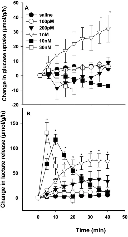
3.3 Results 41
3.3.1 ET-1 dose curve 41
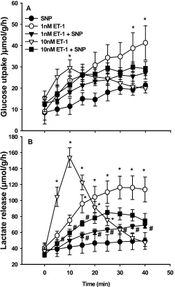
3.3.2 ET-1 and SNP 45
3.4 Discussion 51
CHAPTER 4 : HIGH DOSES OF ET-1 INHIBIT TENSION DEVELOPMENT AND ARE RESISTANT TO EXERCISE-MEDIATED VASODILATION 56
4.1 Introduction 56
4.2 Methods 57
4.2.1 Solutions 57
4.2.2 Perfusion conditions 58
4.2.3 Contraction 58
4.2.4 Perfusion protocol 59
4.2.5 Statistical Analysis 60
4.3 Results 61
4.3.1 Vascular and metabolic effects of ET-1 61 4.3.2 Effects on aerobic tension development 64
4.4 Discussion 69
CHAPTER 5 : INTERACTIONS BETWEEN ET-1 AND INSULIN 72
5.1 Introduction 72
5.2 Methods 73
5.2.1 Solutions 73
5.2.2 Perfusion conditions 73
5.2.3 Perfusion protocol 74
5.2.4 Radiolabelled glucose uptake 75
5.2.5 Statistical Analysis 75
5.3 Results 76
5.4 Discussion 90
CHAPTER 6 : METHACHOLINE-MEDIATED VASODILATION CAN
AFFECT GLUCOSE UPTAKE 97
6.1 Introduction 97
6.2 Methods 98
6.2.1 Solutions 98
6.2.2 Perfusion conditions 98
6.2.3 Perfusion protocol 99
6.2.4 Radiolabelled glucose uptake 100
6.2.5 Statistical Analysis 100
6.3 Results 101
6.4 Discussion 107
CHAPTER 7 : SGLT1 IS NOT INVOLVED IN INSULIN-MEDIATED
GLUCOSE UPTAKE IN RAT MUSCLE 110
7.1 INTRODUCTION 110
7.2 Methods 111
7.2.1 Solutions 111
Phlorizin and phloretin 111
Insulin 112
Vehicle 112
7.2.2 Perfusion conditions 112
7.2.3 Perfusion protocol 112
7.2.4 Radiolabelled glucose uptake 113
7.2.5 Statistical Analysis 113
7.3 Results 114
7.4 Discussion 122
CHAPTER 8 : DISCUSSION 125
8.3 Mechanisms of dose-dependent effects of ET-1 127
8.4 Mechanisms of insulin’s vascular effect – in vivo vs. perfusion 131
8.5 ET-1 in disease 132
8.6 Capillary Recruitment and Glucose Uptake 133
8.7 Conclusion 134
STATEMENT
The work in this thesis has been undertaken exclusively for the use of a Ph.D. in the area of Biochemistry, and has not been used for any other higher degree or graduate diploma in any university. All written and experimental work is my own, except that which has been referenced accordingly.
Cathryn Kolka
AUTHORITY OF ACCESS
This thesis may be available for loan and limited copying in accordance with the Copyright Act 1968.
ABSTRACT
Glucose uptake occurs in skeletal muscle under basal conditions, and increases in response to stimuli such as insulin and exercise. Exercise is known to increase blood flow, and it appears that insulin has similar hemodynamic effects, including increased blood flow and capillary recruitment, which can modify the amount of glucose uptake occurring under each condition. Here we study factors affecting both basal and stimulated myocyte glucose uptake, with a particular focus on vasoactive agents.
Insulin stimulates the release of endothelin-1 (ET-1), a potent vasoconstrictor, from
endothelial cells in culture. As yet it is unknown whether ET-1 is a type A (causing nutritive perfusion) or a type B (non-nutritive) vasoconstrictor, so here we use the pump-perfused rat hindlimb to characterize the distribution effects of ET-1. We show that ET-1 causes a type A vasoconstriction, stimulating basal metabolism at low doses, while at high doses the
distribution of flow changes to become non-nutritive, inhibitory to metabolism. As a general vasodilator prevents both metabolic and hemodynamic effects, the effects on metabolism are due to the redistribution of flow. These redistribution effects are confirmed by the ability of high dose ET-1 to decrease aerobic tension development in the contracting hindlimb, and by the ability of low dose ET-1 to increase the interstitial glucose concentration.
Given this understanding of the effects of ET-1 alone, we can investigate the interactions between ET-1 and insulin. In the perfused rat hindlimb, insulin has not been observed to have any vasodilatory effect, whereas here for the first time insulin appears to have vasodilator-like actions against ET-1 mediated vasoconstriction. Also, the redistribution of flow by ET-1 does not appear to alter the metabolic effect of insulin to cause glucose uptake at either dose of ET-1 used.
Nitric Oxide (NO) is thought to be the mechanism by which insulin causes vasodilation in muscle. A previous study has shown that methacholine (MC), by increasing NO, was able to augment insulin-mediated glucose uptake and capillary recruitment, while other NO donors were unable to do so. Here we show that, at the dose used to increase glucose uptake in the previous study, MC has only a vasodilatory effect, and no direct effect on glucose uptake, in the perfused rat hindlimb. At higher doses, an effect on glucose uptake can be observed. This means that the increase in capillary recruitment by MC was responsible for the elevated insulin-mediated glucose uptake, and there was no direct effect of MC on glucose uptake. A recent publication suggested that the Na+-D-glucose cotransporter (SGLT1) was essential
for insulin-mediated glucose uptake, although not required for basal glucose uptake. The implications of this detract from our proposed role of blood flow redistribution in insulin action. In attempting to reproduce these results in the perfused rat hindlimb we found that SGLT1 is not required for insulin-mediated glucose uptake, and confirmed this using a low sodium buffer, which would also inhibit the transporter. We conclude that SGLT1 is not required for insulin-mediated glucose uptake.
ACKNOWLEDGMENTS
I wish to thank Prof Clark, for his guidance and supervision during the last four years, without him I am sure my work would not have gone as well, and the ‘Steve’s’ (Rattigan and Richards), for all their help and
suggestions along the way.
I would also like to thank many past and present members of the muscle research group, including Cate, Hema, Lei, Georgie, Maree, Phil, Amanda, John, Carole, Merren and Geoffrey, for making my time here enjoyable. An extra special thanks to Renee and Eloise – for your friendship and help along the way.
Also thanks to those hard workers in the animal house, especially Marcus and Murray, and all the friends I have made in the rest of the building as well, particularly those in the MBU.
ABBREVIATIONS
A-V arterio-venous
ANOVA Analysis of variance AII Angiotensin II
e extensor digitorum longus EDRF Endothelium-derived relaxing factor
ET Endothelin
ET-1 Endothelin-1
ETA Endothelin receptor type A
ETB Endothelin receptor type B
GLUT facilitative glucose transporters
Ins Insulin
L-NAME Nitro-L-Arginine Methyl Ester L-NMMA mono-methyl nitro-L-arginine
MC Methacholine
NE Norepinephrine
NO Nitric Oxide
NOS Nitric Oxide Synthase
eNOS endothelial NOS
iNOS inducible NOS
nNOS neuronal NOS
Nox end-products of oxidized NO p plantarus
r red gastrocnemius
SGLT1 sodium-glucose co-transporter SNP sodium nitroprusside
t tibialis
VO2 Oxygen consumption
w white gastrocnemius
1-MX 1-methylxanthine
PREFACE
Some of the data presented in this thesis has been published or presented at scientific meetings and has been listed below.
Publications arising directly from this thesis
Kolka CM, Rattigan S, Richards S, Clark MG. Metabolic and vascular actions of endothelin-1 are inhibited by insulin-mediated vasodilation in perfused rat hindlimb muscle. British Journal of Pharmacology. 2005 May 16
Kolka CM, Rattigan S, Richards SM, Barrett EJ, Clark MG. Endothelial Na+ -D-glucose cotransporter: no role in insulin-mediated -D-glucose uptake. Hormone and Metabolic Research. 2005.
Mahajan H, Kolka CM, Newman JMB, Rattigan S, Richards SM, Clark MG. Vascular and metabolic effects of methacholine in muscle. Circulation Research.
Rattigan S, Zhang L, Mahajan H, Kolka CM, Richards SM, Clark MG. Factors influencing the hemodynamic and metabolic effects of insulin in muscle. Current Diabetes Reviews, in press (2006).
Kolka CM, Rattigan S, Richards S, Clark MG. Reduced exercise capacity in
hypertension: a consequence of endothelin-mediated functional shunting. (Manuscript in preparation.)
Other publications
Posters at scientific meetings
American Diabetes Association 64th Scientific Sessions, June 4th-8th 2004, Orlando,
Florida.
Endothelin-1 as a messenger for insulin has both stimulatory and inhibitory effects on perfused muscle metabolism via its vascular actions. Kolka CM, Rattigan S, Richards S, Clark MG.
Freycinet Conference on: Diabetes and Exercise: Impact of Muscle Blood Flow, Freycinet, Tasmania, 18th-20th August 2004.
Endothelin-1 via its vascular actions in muscle can be either supportive or antagonistic of insulin. Kolka CM, Rattigan S, Richards S, Clark MG.
European Association for the Study of Diabetes annual meeting, Sept 11th-14th 2005, Athens, Greece.
Endothelin-1 vascular and resultant metabolic actions in perfused rat hindlimb are opposed by insulin. Kolka CM, Rattigan S, Richards SM, Clark MG.
Glucosamine induces acute insulin resistance in muscle in vivo associated with impaired capillary recruitment. Clark MG, Wallis MG, Smith ME, Kolka CM, Zhang L, Richards SM, Rattigan S.
Heart Foundation Conference and Scientific Meeting, March 23rd-25th 2006, Sydney Australia
CHAPTER 1
INTRODUCTION
GLUCOSE UPTAKE IN SKELETAL MUSCLE
Glucose and other fuels enter the body after a meal, and the body maintains a constant level of blood glucose by stimulating glucose disposal into muscle and adipose tissue and inhibiting hepatic glucose production. The main hormone responsible for these effects is insulin, which also has effects on cell growth and development, ion
transport, and sympathetic nervous system activity (reviewed in (1)). Exercise is also capable of increasing glucose uptake into muscle, most likely through a combined effect including the increase in delivery of blood flow, and thereby glucose, to the exercising area, and a cellular effect involving translocation of GLUT4. Insulin is able to alter blood flow through muscle as well, which can affect the level of glucose uptake that can occur. The focus of this study will be the uptake of glucose into myocytes and factors that may alter both basal and insulin-mediated glucose uptake, particularly those involved in changing the blood flow distribution through muscle.
1.1
Metabolic effects of insulin in skeletal muscle
1.1.1
Insulin-mediated glucose uptake by muscle requires glucose
transporters
Diffusion barriers exist for glucose at the cell membrane and there are two main groups of glucose transporters to overcome this barrier. They are the Na+ dependent sodium-glucose cotransporters (SGLTs) and the Na+ independent glucose
transporters, or facilitative glucose transporters (GLUTs) (reviewed in (3)). Many of these glucose transporters are not specifically activated by insulin, and may only be involved in basal glucose uptake. Both forms of glucose transporters are relevant to the work involved in this thesis, and are therefore discussed further.
Sodium glucose transporters
At least three, and possibly up to six, SGLTs have been identified (4). Although originally these glucose cotransporters were thought to be primarily located in the intestine and kidney, recent studies have shown that SGLT1 may be located in the coronary artery (5) and in the endothelial cells of skeletal muscle capillaries (6).
SGLTs are primarily involved in absorption of glucose from the intestine, and reabsorption in the kidney. The Na+-D-glucose cotransporter SGLT1 is found in epithelial cells of the intestine and proximal renal tubule, where it plays a central role in the absorption of glucose and galactose from food and the reabsorption of glucose from the glomerular filtrate (7). In both locations the process of uptake is coupled to a Na+/K+ ATPase positioned on the serosal side of the cell to pump out the
Early studies used phlorizin, an SGLT1 inhibitor, to block insulin mediated glucose uptake, as well as other sugars, in skeletal muscle and cardiomyocytes without affecting insulin-mediated amino acid transport (reviewed in (13)). Many studies suggested that these effects were specific to insulin action to increase glucose uptake (14, 15), although other studies discovered that phlorizin may also affect the basal level of glucose uptake as well (16) (reviewed in (13)). These early studies used doses of phlorizin that may have been high enough to also block GLUT1 and GLUT4 glucose transport (1-5mM) (14-16). There has been more recent evidence to indicate that SGLT1 in small intestine is regulated by insulin, as activity of the protein was increased in an experimental form of type 1 diabetes, and activity was returned to normal following treatment with insulin (17). In these diabetic rats, SGLT1 protein content was elevated, suggesting some translational control of SGLT1 by insulin.
Functional characterisation of these transporters is continuing, and a recent study has proposed a role of SGLT1 in insulin-mediated glucose transport from the blood vessel to the myocytes in skeletal muscle (6). This study used immuno-localization
techniques to demonstrate the presence of SGLT1 on the endothelial cells of skeletal muscle capillaries, and then showed that phlorizin, at doses low enough to be a specific inhibitor of SGLT1 without affecting GLUT1 and GLUT4, was able to block insulin-mediated glucose uptake, with no apparent effect on basal glucose uptake. This suggests that SGLT1 is responsible for all glucose uptake into skeletal muscle that occurs with insulin. Such a finding presents a potential rate-limiting step for glucose uptake into muscle: a step that is regulated by insulin and thus relevant to this thesis.
Facilitative glucose transporters
muscle; GLUT1 is thought to play a role in basal glucose uptake, and is located in the myocyte plasma membrane (18). The insulin-responsive glucose transporter is GLUT4, and is found in brain, heart, skeletal muscle and adipose tissue. Insulin stimulates the translocation of GLUT4 from an intracellular store to the plasma membrane to increase glucose uptake in myocytes as well as in adipocytes (18-20). A greater level of GLUT4 translocation permits greater insulin sensitivity, as observed by glucose disposal (21), and different levels of GLUT4 protein content between muscle may account for insulin sensitivity differences between muscles (22). Exercise also may be able to induce the translocation of the GLUT4 protein to the plasma membrane (23), although appears to draw on a different intracellular pool of GLUT4 than insulin (reviewed by (24).
1.2
Insulin has hemodynamic effects in skeletal muscle
Insulin has been shown to have effects on blood vessels. Initially, insulin was found to cause an increase in blood flow to skeletal muscle (25), and since this first study, many other studies have helped to develop a more complete picture of insulin-mediated hemodynamic effects. The three main effects include an increase in blood flow, vasodilation and capillary recruitment.
Increased blood flow
Although it had been known for a least sixty years that high levels of insulin, often in the pharmacological range, increased cardiac output and skeletal muscle blood flow (26), Laakso et al were the first in more recent times to show, using a thermodilution technique, that insulin acts to increase leg, and therefore muscle, blood flow in a dose-dependent manner (25). Tack et al (27) showed that insulin was able to cause
increases in total flow, but these increases were only occasionally noted at
physiological insulin concentrations, and the greatest effect was noted after several hours. The fraction of blood flow through muscle increases as a function of total flow, and skeletal muscle accounts for most of the blood flow through the limb (28).
appears that insulin can cause hemodynamic effects that can increase blood flow and decrease vascular resistance: effects that are consistent with vasodilation (31).
Vasodilation
Evidence from studies of large vessel responses has shown that insulin can decrease the stiffness of the vessel wall, and cause vasodilation. Chaudhuri et al (32) observed an increase in diameter of the internal carotid artery that became significant at 15 minutes after treatment with insulin. Results obtained by Ueda et al (33) in humans showed insulin to be a weak local vasodilator, although this effect was amplified in the presence of glucose. This finding was supported by the research of Tack et al (27), who found that local physiological hyperinsulinemia induced a slow vasodilation that became maximal about three hours after the first infusion. In the presence of
hyperglycemia, there was a quicker onset of vasodilation, but the same maximum was reached.
Capillary recruitment
Serne et al (37) showed that capillary density is proportional to insulin sensitivity in human skin, as it decreases the diffusion distances from the capillary to the cells. If the capillary density decreased, then there was an observed increase in vascular resistance. The decrease in capillary density may be due to a decrease in the
vasodilation at the precapillary level, preventing perfusion of the smaller capillaries.
Nutritive and non-nutritive flow routes in muscle
The concept of two distinct vascular routes in muscle was first proposed by Pappenheimer (38), who noted that various vasoconstrictors could have different effects of the oxygen uptake by muscle. It was found that in certain circumstances blood flow could continue, but the interior of the muscle would be less perfused (39). This suggests that the blood can pass through a shunt, lowering the perfusion of the muscle. Later, it was found that radiolabelled sodium disappeared at different rates from muscle depending on the area of injection, leading to the proposal of nutritive and non-nutritive pathways (40). The nutritive pathway is believed to be in contact with skeletal muscle cells, while the non-nutritive pathway is shunted through septa and tendons away from muscle (41), and is possibly associated with adipose tissue (42). This is shown by the inverse relationship between tendon blood flow and oxygen consumption, an indicator of metabolism (41). The regulation of blood flow through these pathways, by vasodilation and vasoconstriction, is proposed to control the metabolism of the muscle (43, 44).
1.2.1
The hemodynamic effects of insulin can increase
insulin-mediated glucose uptake
hormone that is responsible for increasing glucose uptake is also able to act on the vasculature to potentiate its own metabolic effect (47-49).
Total blood flow
In rats, in the absence of insulin, it was shown that a low flow of perfusate caused a lower level of glucose uptake when compared to high perfusate flow (50). This showed that glucose delivery to myocytes in the pump-perfused muscle preparation is the rate-limiting step for glucose uptake in skeletal muscle. Avogaro et al (51) suggested that the hemodynamic effects of insulin might amplify insulin-mediated glucose uptake. This was supported by the observation that insulin led to an increase in blood flow in the perfused rat hindlimb, which increased insulin sensitivity and glucose uptake in vitro (50). Also, in vivo an increase in flow achieved by
methacholine vasodilation caused an increase in insulin-mediated glucose uptake (52).
In healthy human subjects Laakso et al (25) found that insulin could generate a two-fold increase in blood flow, which can account for up to 40% of insulin-mediated glucose uptake in the very high physiological range of insulin. Baron (1) suggested that, in theory, augmentation of either glucose extraction or flow could increase glucose uptake in the target muscle. However other researchers have shown that glucose uptake occurs before any observed changes in blood flow, and so therefore blood flow probably has no effect on the initial insulin-mediated glucose uptake in the acute stages of insulin exposure (3, 53, 54). Fugmann et al (29) suggest early insulin-mediated glucose uptake is due to the effect of insulin increasing glucose transport in the tissues. Eventually, insulin-mediated increases in blood flow can become a major determinant of the degree of glucose uptake.
Pendergrass et al (55) found that increasing forearm blood flow did not increase forearm glucose uptake in humans. Zierler (3) also found that increasing leg blood flow had no effect on glucose uptake. Nuutila et al (56) made a similar observation using bradykinin to increase the blood flow over that achieved by insulin, without observing an increase in glucose uptake. To explain this result, it was hypothesized that any increase in flow leading to a greater delivery of glucose and insulin to tissues will not increase glucose uptake unless previously unperfused areas are made
increase blood flow. Bradykinin causes venodilation, which may not have any effect on capillary recruitment. Also, venodilation may change the nature of flow through the muscle, without increasing the muscle perfusion or recruiting more capillaries than insulin alone. Simply, insulin may have already caused capillary recruitment, and any further hemodynamic effects that will not augment microvascular perfusion will thus not affect glucose uptake.
As reviewed by Zierler (3), the effects of insulin on limb blood flow are controversial. Often the responses noted are due to pharmacological or super-physiological levels of insulin. Hernandez et al (57) showed that, after a meal, there were no significant changes in leg blood flow, and therefore that leg blood flow does not increase enough to be a major determinant in glucose uptake. However, the authors acknowledge that the nature of the meal may be involved in determining the extent of the increase in limb blood flow, and no measurements of insulin levels were taken, so an accurate correlation between limb blood flow and insulin levels, and the effects on glucose uptake, could not be performed. Also, Taddei and Salvetti (58) showed that hyperinsulinemia did not necessarily have to increase blood flow to potentiate
vasodilation. Thus, vasodilation can occur without any changes to total flow. A study by Clark et al (35) focussed on the changes in flow upon the administration of insulin, and concurrently measured the amount of nutritive flow by Laser Doppler Flowmetry. It was found that an increase in nutritive flow was observed 30 minutes before a blood flow response was noted. These data suggest that any measurement of total flow as an indication of vasodilation does not take into account any local vasodilation or capillary recruitment. Therefore, capillary recruitment may be more effective than increasing limb blood flow at augmenting glucose uptake.
Capillary Recruitment
rate-Baron et al (59) found that insulin sensitivity correlated well with leg glucose uptake and suggested that the perfusion of the leg was limiting to glucose uptake. It was hypothesized that observed reductions in insulin sensitivity may be due to alterations in regional blood flow, which causes an impaired delivery of substrate (glucose) or insulin to tissues, thereby dampening the insulin response (60). The ability of insulin to dilate the macrovasculature in skeletal muscle was directly proportional to the glucose metabolism that can be induced by insulin (61). In addition, the insulin-mediated vasodilation in skeletal muscle augments the effect of insulin to stimulate glucose uptake. Other studies support this finding, and the proposal that skeletal muscle blood flow can, by means of increased substrate delivery, account for up to 20-30% of insulin-mediated glucose uptake (52, 62).
Utriainen et al (63) using positron emission tomography showed that insulin was able to increase the absolute but not relative dispersion of flow, which is consistent with capillary recruitment, and was capable of redirecting blood flow to areas that perform glucose uptake. Under high levels of insulin, there was an increase in, as well as a redistribution of flow. This observation was not supported by Raitakari et al (28) or Bradley et al (64), who found that glucose uptake in response to insulin does not co-localize to the same area in muscle tissue as the observed increase in blood flow, suggesting that the hemodynamic and metabolic effects were separate. In rats, however, it was shown that flow was redirected preferentially to muscles with higher levels of oxidative type I fibres (63) which are more sensitive to insulin in terms of glucose uptake (65).
velocity through the vessels would lead to a decrease in glucose extraction from the blood, and therefore lower glucose uptake.
The degree of skeletal muscle perfusion has been suggested as an independent determinant of insulin-mediated glucose uptake, which tends to support the reputed role of dispersion of flow through the muscle. This is observed in studies that achieve augmentation of the leg glucose uptake response above the maximum effect
achievable by insulin alone by increasing skeletal muscle perfusion (52). In studies of human skin microcirculation, capillary recruitment was found to correlate strongly with insulin sensitivity measured by insulin clamp technique, suggesting that
microvascular function is an important aspect of insulin action (37). When compared to exercise, which increases both the capillary blood volume (recruitment of
capillaries) and the velocity of the red blood cells, physiologic hyperinsulinemia only increases the capillary blood volume, with no effect on velocity, as assessed using contrast enhanced ultrasound (34). Higher doses of insulin may increase both capillary blood volume and red cell velocity.
1.2.2
Possible coupling between insulin-mediated glucose uptake and
capillary recruitment
There is some controversy regarding the nature of the coupling of the metabolic and hemodynamic effects. Cleland et al (67) hypothesized that insulin-mediated glucose uptake in the endothelium leads to an increase in blood flow, and that the rate of glucose uptake determines vascular smooth muscle relaxation. This conclusion was based on evidence obtained in healthy men, where an intra-arterial infusion of insulin caused vasodilation, and this effect could be augmented by a concurrent infusion of glucose (67). However, it was determined that there could be no insulin-mediated glucose uptake in endothelial cells (68), and Petrie et al (69) found that the degree of insulin-induced vasodilation was determined by tissue glucose uptake, not the
The findings by Petrie et al (69) have not as yet been corroborated by others, and in contrast, Utriainen et al (63) show that the rate of glucose uptake is not responsible for increases in flow. Their observation was that it is the presence of insulin, not the rate of glucose uptake, which determines the degree of vasodilation, as limb blood flow can increase in the early stages of non-insulin dependent diabetes mellitus (type 2 diabetes) in response to insulin, without a significant glucose uptake. A number of other groups also found data contrary to this concept. For example, Raitakari et al (28) showed that glucose uptake itself is not enough to cause vasodilation, as glucose oxidation under hyperglycemic conditions does not lead to vasodilation. Also, Tack et al (27) observed that maximal insulin-mediated glucose uptake occurred before the vasodilation induced by insulin, but agreed that insulin-mediated glucose uptake was not a determinant of the degree of vasodilation, as the degree of vasodilation was not dependent on glucose uptake, suggesting a direct effect of insulin to cause
vasodilation. Vollenweider et al (70) and Meneilly et al (71) found that
hyperinsulinemia is the main stimulus that causes vasodilation in skeletal muscle, and that glucose metabolism is an independent effect.
More recent results by Vincent and colleagues suggest that microvascular recruitment is an early event in hyperinsulinemia, as an increase in capillary blood volume measured by contrast enhance ultrasound was observed within 7 min (72). This suggested that the microvascular effects of insulin occurred prior to any significant insulin-mediated glucose uptake or increase in limb blood flow. It is possible that metabolism by myocytes initially receiving flow could lead to metabolic signals that permeate nearby tissue to enhance microvascular perfusion. The only argument against this is the observation that insulin-mediated capillary recruitment is more sensitive to insulin than insulin-mediated glucose uptake (73).
1.2.3
Insulin resistance in diabetes
Diabetes is characterized by insulin resistance – an effect that was initially thought to be a tissue (muscle) effect. Bak (74) reported that, under normal circumstances, insulin caused an increase in glucose uptake and glycogen synthesis in skeletal muscle, whereas in type 2 diabetes and cases of insulin resistance, the
to be an important factor in diabetes, when the endothelium-dependent vasodilation is impaired, although insulin therapy can successfully improve both endothelium-dependent and –inendothelium-dependent vasodilation (75).
1.2.3.1
Insulin resistance leads to endothelial dysfunction
A common but unsubstantiated view is that insulin resistance leads to endothelial dysfunction. For example, Pieper et al (76) have shown that hyperglycemia may lead to endothelial dysfunction, especially in those that suffer from type 1 diabetes, and in other cases can lead to hyperglycemia induced diabetes mellitus. In addition, Hsueh and Anderson (77) report that the vascular endothelium is a target organ of damage in diabetes, which can show altered function. Metabolic abnormalities resulting from hyperglycemia such as advanced glycosylation end products and reactive oxygen species can cause damage to the endothelial cells. They conclude that interventions that modulate insulin resistance can affect the endothelial response (77). There is evidence that endothelial damage in diabetes may be either the cause (78) or the consequence (51) of insulin resistance. Recent evidence that endothelial dysfunction occurs before insulin resistance has been shown in situations where offspring and relatives of insulin resistant people show impaired endothelial responses (79). As hypertensive people do exhibit both vascular impairment and insulin resistance, an important role of the vasculature in insulin sensitivity is suggested.
1.2.3.2
Flow deficit in diabetes
McVeigh et al (80) have used venous occlusion plethysmography to show that
vasodilation is impaired in type 2 diabetes, involving both endothelium-dependent and –independent vasodilation. This finding is supported by Watts et al (81) who
Cleland et al (83) reported that there were associations between endothelial function and both insulin sensitivity and insulin-induced vasodilation. It was hypothesized by Julius et al (82) that there was a link between insulin resistance and hypertension due to the decreased blood supply evident in both disorders. Any treatment aiming to improve the hemodynamic effects of insulin in diabetics may also be of benefit in hypertensive and obese patients, as Feldman and Bierbrier (47) propose that impaired insulin-mediated vasodilation contributes to increased peripheral vascular resistance. Mather et al (84) reported that, although insulin resistance can impair normal insulin-mediated increases in blood flow, metformin treatment can improve insulin resistance and endothelial function, although this was not supported by Natali et al (85). If the vascular system can be modified to increase skeletal muscle blood flow and perfusion, insulin resistance may be improved by correcting insulin sensitivity, glucose tolerance and hyperinsulinemia (86).
1.2.4
Mechanisms of the hemodynamic effects of insulin
Nitric oxide (NO) has long been thought to be involved in the hemodynamic effects of insulin, as the vasodilation observed with large doses of insulin can be prevented by infusing NO blockers, such as L-NAME and L-NMMA. Recent studies have indicated that the hemodynamic actions of insulin may in fact be a combination of two vasoactive substances. Misurski et al (87) showed that insulin induced biphasic responses in rat mesenteric vascular bed. An initial nitric oxide-mediated vasodilation was overcome by generation of endothelin-1 (ET-1) when the rat mesenteric vascular bed was exposed to high insulin concentrations, an effect that was blocked by an endothelin receptor antagonist. It is now thought that insulin causes the release of both ET-1 and NO, and it is a balance between these two vasoactive agents that causes the hemodynamic effects of insulin, as shown in isolated skeletal muscle arterioles (88), the rat aorta (89), and in the healthy human forearm (90). Ferri et al (91) suggested that endogenous insulin modulates the circulating concentration of ET-1, although this was determined through an oral glucose load, which was observed to cause an increase in both insulin and ET-1. Altering the balance of these two
1.3
Nitric Oxide is the vasodilator involved in insulin action
All hemodynamic processes that are elicited by insulin, including increased limb blood flow, vasodilation and capillary recruitment as discussed above, appear to be dependent on NO, a potent vasodilator, and are all impaired in insulin-resistant states. Steinberg (92) showed that insulin-mediated vasodilation is largely dependent on NO. As the metabolic effects of insulin appear to be dependent on limb blood flow, we can potentially alter the metabolic effects, measured by glucose uptake by modifying the available blood flow with a vasodilator such as methacholine, or by blocking the vasodilation elicited by NO using nitric oxide synthase (NOS) inhibitors.
1.3.1
Nitric Oxide
NO is a potent vasodilator that is produced by many different cells in the body, including macrophages, neurones, mast cells, hepatocytes and endothelial cells (93). NO was first found to be synthesized from L-arginine in porcine aortic endothelial cells in culture (94, 95). It has a very short half-life, and is degraded rapidly in the plasma to nitrite and nitrate, which suggests that any action elicited by NO is directed mainly at local areas, near the place of production. While NO is involved in platelet aggregation and adhesion, the main action in the circulation is vasodilation, primarily to maintain blood pressure and the control of resting vascular tone (96).
1.3.1.1
Nitric Oxide and Vasodilation
Initially, the substance that was involved in dilating the vasculature was described as endothelium-derived relaxing factor, or EDRF. This was later identified as NO, or as containing a highly labile nitroso compound capable of releasing NO, using chemical identification and comparative pharmacology, examining the effect of both EDRF and NO on vascular strips (97, 98). Endogenous NO was found to be a significant
NO can also react with heme to form nitrosyl-heme complexes that can activate guanylate cyclase (reviewed in (101)).
In rabbit hindlimbs, blockade of NO (or EDRF) increased vascular resistance, and attenuated the normal production of cGMP in platelets. Also, the oxygen uptake of the hindlimb was reduced, which suggested that NO was involved in maintaining an adequate perfusion of the hindlimb and maintaining basal metabolism (102). Generally, NO appears to be responsible for a decrease in vascular tone through vasodilation, and is constantly released by endothelial cells to maintain a constant blood pressure and vessel tone.
L-NMMA and L-NAME are used regularly to block vasodilation, as they are
inhibitors of nitric oxide synthase (NOS), the enzyme responsible for the synthesis of NO for L-arginine. There are three different NOS isoforms: eNOS (endothelial NOS), nNOS (neuronal) and iNOS (inducible).
1.3.1.2
NO in endothelial dysfunction and disease
A deficiency in the NO system appears to be involved in certain cardiovascular diseases (93, 103-105). Atherosclerosis and hypertension are associated with an impairment of the release or effect of NO, which may potentially lead to an increased blood pressure due to a lack of vasodilation. Animal models of sepsis demonstrate an increased circulating NO level, and raised levels of nitrate in human sepsis cases tends to suggest that this is true in humans as well (reviewed in (93)).
Endothelial dysfunction is characterized by a diminished blood flow in response to stimulation of eNOS (106). The response to injected methacholine or acetylcholine is often used as an indicator of endothelial dysfunction. As these cholinergic agonists activate nitric oxide synthase, a functional endothelium is required to cause
vasodilation, and is used for endothelium-dependent vasodilation studies, as opposed to sodium nitroprusside, which does not require the endothelium to cause
nitroprusside-mediated vasodilation (107). This suggests endothelial dysfunction, while normal NO-mediated endothelium-independent vasodilation was unaffected. Methacholine has also been used to examine endothelial dysfunction in many other studies (eg (108-112)). Endothelial dysfunction is particularly evident when insulin is used as an endothelium dependent stimulus (113). But it is not clear, even now, that insulin-dependent and methacholine-dependent vasodilation use the same mechanism, although it seems likely that NO is involved.
Even so, if eNOS is blocked with L-NMMA in rats, insulin resistance is observed (60). This insulin resistance occurred in liver and peripheral tissues of the eNOS knockout mouse, where the NOS protein is removed from the endothelial cells, compared to neuronal NOS (nNOS), where insulin resistance was only exhibited in the peripheral tissues. Zeng et al (114) found that insulin receptor tyrosine kinase (IRTK) was required for insulin-mediated NO production in human vascular endothelial cells.
1.3.2
Nitric Oxide and Insulin
It has been shown that insulin is able to activate the enzyme responsible for the release of NO (53, 115). Baron and Clark (61) reviewed data showing that insulin caused a doubling in the rate of production of venous Nox (the end products of oxidized NO), and suggested that insulin-mediated vasodilation is largely NO dependent. An association between insulin sensitivity and NO production was demonstrated (83), so insulin sensitivity is therefore proportional to insulin-mediated vasodilation.
1.3.2.1
Nitric oxide is involved in the hemodynamic effects of insulin
Blood Flow
increased limb blood flow due to an insulin stimulus. As noted above, changes in limb blood flow are not the only hemodynamic effects involved in insulin action, and so a specific study of each of the effects is required.
Vasodilation
When insulin is introduced into first order arterioles from rat cremaster muscle, vasodilation was observed as an increase in the diameter of the arteriole. This increase was blocked by including L-NNA (nitro-L-arginine), a NO blocker, and by removing the endothelium (116). This suggests that the vasodilation observed in insulin action is dependent on both the endothelium and NO. While insulin-mediated vasodilation in a whole body study is difficult to assess, in a hyperinsulinemic euglycemic clamp in healthy humans L-NMMA prevented insulin-induced vasodilation, detected as an increase in limb blood flow (53, 92).
Capillary Recruitment
In rats in vivo, systemically infused L-NAME was shown to completely block insulin-mediated increases in blood flow and microvascular recruitment, as measured by both the 1-MX method, and contrast enhanced ultrasound (117), while having no
significant effect on basal recruitment. However, this may be more complex than first imagined, as locally infused L-NAME in rats in vivo does not block insulin-mediated capillary recruitment (118). The site where systemic L-NAME inhibits insulin may be central (60).
1.3.2.2
Inhibition of Nitric Oxide on insulin-mediated glucose uptake
was thought to be due to the decrease of NO release into the vasculature. But, as discussed above, there have been conflicting findings on the proposed effect of limb blood flow on glucose uptake.
In a separate study, inhibition of the hemodynamic effects by blocking NOS action was found to cause no change in whole body insulin-mediated glucose uptake, and so it was suggested that the effects of insulin on blood flow and perfusion had no effect on insulin-mediated glucose uptake (53). However, this was thought to be due to the infusion of the inhibitor only in the arm, which accounts for only 10% of the skeletal muscle mass in the body. As the NOS blockade may have only had local effects, whole body insulin-mediated glucose uptake may not have been an accurate indication of the effect of NO on insulin-mediated glucose uptake.
1.3.2.3
Increasing insulin-mediated glucose uptake with agents that
stimulate the production of Nitric Oxide
Limb infusion of methacholine was able to increase insulin-mediated glucose uptake, observed concurrently with an increase in femoral blood flow in lean healthy men Baron, 2000 #874;Baron, 1994 #862}, and in hypertensive patients (120). In a further development, Mahajan et al (121) demonstrated that insulin-mediated glucose uptake could be increased by limb infusion of methacholine in a euglycemic
hyperinsulinemic clamp in rats, while another vasodilator, bradykinin, had no effect. Similarly, methacholine, but not bradykinin, was able to augment the insulin-mediated capillary recruitment, as measured using the 1-MX method, though both vasodilators were able to augment the femoral blood flow response to a similar extent (121). It is interesting that two vasodilators, both acting by NO-dependent mechanisms, cause different effects on insulin sensitivity. In other studies that have attempted to increase insulin-mediated glucose uptake using both NO-dependent and –independent
Increasing the skeletal muscle blood flow in elderly humans had no effect on glucose uptake. This study showed that the infusion of NO precursors did increase blood flow and corrects endothelial dysfunction, but it is possible that a defect in the tissue in elderly patients may have been responsible for the lack of an effect on glucose uptake (71).
In general, NO-dependent vasodilators have not shown any effect to increase insulin-mediated glucose uptake, however several studies using methacholine have
demonstrated this ability (52, 92, 120, 121). As yet, whether this effect is due to the vasodilator abilities of methacholine, or due to a direct effect of methacholine on myocyte glucose uptake is uncertain.
1.3.3
NO action is impaired in diabetes
As discussed above, normal insulin-mediated vasodilation is dependent on NO. In patients with type 2 diabetes, an impaired NO mediated vasodilation by methacholine is observed (125) (endothelium-dependent), in conjunction with an impaired
endothelium independent vasodilation (sodium nitroprusside). These defects could be due to a reduced NO release in diabetic patients and/or to an inhibition of the
signalling mechanism NO normally activates to cause vasodilation (125). In patients with type 1 diabetes, synthesis of NO is decreased, and is thought to be due to
decreased tetrahydrobiopterin (126), thereby reducing insulin-mediated vasodilation. In addition, treatment with tetrahydrobiopterin was found to be beneficial by
improving NO-mediated vasodilation in type 2 diabetics. There is also data suggesting that the high plasma glucose observed in diabetic patients may actually reduce the amount of insulin-stimulated NO production (127). Glucose toxicity through free radical attack may cause inactivation or lowered expression of NO by a variety of mechanisms (128).
suggesting some other mechanism is involved at least at this late stage in diabetes that lowers NO availability (129).
1.4
Vasoconstrictor effects on insulin action: ET-1
ET-1 has been identified as a potential vasoconstrictor involved in insulin action in vivo. In perfused muscle in vitro vasoconstrictors can redirect flow to either nutritive or non-nutritive routes in muscle with different effects on metabolism. These
metabolic effects due to vasoconstrictor action can be observed when the
vasoconstrictor is added alone, but may also modify the effects of other hormones including insulin. As the full metabolic effect of insulin on myocyte glucose uptake requires full nutritive perfusion of muscle, any modification of blood flow distribution with a vasoconstrictor such as ET-1 can potentially affect glucose uptake.
Experiments such as these have previously been pursued in this lab using
norepinephrine and serotonin (130). Based on the type of vasoconstrictor that is used, variable effects on metabolism can be observed. An increased nutritive distribution of flow throughout the muscle (for example, by low dose norepinephrine), termed type A vasoconstriction will cause an increase in metabolism, due to an increase in the delivery of nutrients to muscle, whereas a decreased nutritive flow through muscle (favouring non-nutritive flow, by an agent such as serotonin), or type B
vasoconstriction, will lower the observed metabolism. As yet, no studies have determined whether ET-1 causes type A or type B vasoconstriction, and so the involvement of ET-1 in the hemodynamic effects normally observed with insulin is not clear.
1.4.1
ET-1
vasoconstrictor action, endothelins can be involved in wound healing and
neurotransmission in the brain, as well as renal homeostasis. ET-1 is the only isoform produced by the endothelial cells – the vascular endothelium is the major source of ET-1 in vivo, and is found in greater concentrations in the plasma than the other endothelin isoforms. It was first isolated as a very potent vasoconstrictor, with long lasting effects.
ET-1 is relatively stable in plasma and blood, and is cleared mainly by the lungs, kidney and endothelial cells in vivo, with a relatively short half-life of approximately 1 minute due to this high clearance rate (132).
1.4.1.1
Vasoconstriction and blood flow
ET-1 is a potent arteriolar vasoconstrictor, more potent on smaller than larger vessels (133), which works in a dose-dependent manner in skeletal muscle (134). There are two ET-1 receptors; both are coupled to a G-protein receptor. Throughout the body, the receptors are expressed in a variety of cells, and are involved in different effects. The binding of ET-1 to either receptor on smooth muscle cells causes activation of phospholipase C, leading to an increase of inositol triphosphate, diacylglycerol and intracellular calcium ion, which causes long-lasting vasoconstriction (135). The two receptors elicit different responses: ETA, which is located on the vascular smooth
muscle cell, causes vasoconstriction by altering internal Ca2+ stores, and ETB causes
vasodilation if located on the endothelial cell, and vasoconstriction if located on the vascular smooth muscle cell (136). The activation of ETB receptors limits the
vasoconstrictor response of the ETA receptors (137-139).
The mechanism by which ETB receptors cause vasodilation is not yet certain, with
some suggesting prostaglandins (132), others suggest NO (140, 141) or EDRF (endothelium-derived relaxing factor) (142), while in guinea pig trachealis muscle, arachidonic acid has been shown to be involved (143). It is generally accepted that NO is involved (144), while prostacyclin also appears to be involved in the
transcapillary absorption of fluid, probably due to an increased post-capillary constriction causing an elevated hydrostatic capillary pressure, leading to oedema formation. This effect can be blocked by ETA receptor antagonist infusion,
suggesting that the ETA receptors located on the venules are responsible for this
pressure-induced oedema development.
As the release of ET-1 is primarily directed abluminally (146), ET-1 is released towards the vascular smooth muscle cells, where it would cause vasoconstriction. If a high level of ET-1 is released, there will be a degree of overflow towards the
endothelial cells and into the circulation, which would be limited by the action of ET-1 on the endothelial cell receptors (ET-132, ET-14ET-1). The affinity of ET-ET-1 for its receptors is very high (147). When given as a bolus in the healthy human forearm, ET-1 causes a transient vasodilation, which is followed by a slow-onset vasoconstriction (148). The role of ET-1 in the maintenance of basal vascular tone is debatable; some suggest it is involved (149-151), while others contest this (152). In cases of essential
hypertension, increased ET-1 vascular activity appears to contribute to vascular tone (153).
ET-1 infusion in vivo appears to increase blood pressure, as expected, while decreasing splanchnic and renal blood flow, and decreasing splanchnic glucose release (154). The functional response to ET-1 varies due to the specific distribution and expression of the receptors in different tissues and vascular beds. It was noted that the vasoconstriction induced through ETA receptors caused a fall in oxygen uptake proportional to the decrease in blood flow during ET-1 infusion in canine small intestine (155). This effect was limited by ETB receptors, as determined using
BQ788, a specific ETB inhibitor, and potentiated by ETA receptors, discovered by
using BQ123, a specific ETA inhibitor.
1.4.1.2
Endothelial dysfunction, disease
ET-1 has been shown to be involved in disease and in cases of endothelial
continuous infusion of ET-1 caused arterial narrowing, which was reversible, but it was hypothesised that there could be other non-reversible morphological changes with prolonged exposure to maintain a narrowed lumen (156).
ET-1 plasma levels are elevated in various states including hypertension (157, 158), diabetes (159, 160), obesity (151) and chronic heart failure (161), as well as other examples of endothelial dysfunction.
The plasma levels of ET-1 are variable between studies, but are generally seen to be from 1 to 5 pM, although as discussed above the circulating concentration merely represents an overflow from the local environment (146). A previous discussion has focussed on why such a low concentration of circulating ET-1, which even when elevated is at most 25pM, is insufficient to cause contractions in isolated vessel incubations (162). Studies in animals have demonstrated that ET-1 levels can increase in congestive heart failure (163), renal failure (164) and in paradoxical sleep deprivation (165). In humans, ET-1 levels were shown to correlate with BMI, lipid parameters and systolic blood pressure in children and adolescents with hypertension, obesity or diabetes, all of which showed elevated ET-1 levels (166). In adults, ET-1 is elevated in patients with essential hypertension versus controls, and is thought to promote atherosclerosis in these patients (167).
There have been extensive studies on ET-1 involvement in vascular tone and blood pressure regulation in hypertension and obesity (151, 168-170), while not necessarily agreeing on the role in basal vascular tone, there is agreement that ET-1 is involved, probably through enhanced vascular activity. In several studies the normal ETB response to cause release of NO is inhibited in cases of hypertension (171, 172), possibly due to a down-regulation of ETB receptors.
1.4.2
ET-1 + insulin
hyperinsulinemic euglycemic clamp led to increased ET-1 levels (91, 160), and in lean type 2 diabetic men there was a negative correlation between ET-1 plasma concentration and glucose uptake (160). ET-1 then appears responsible for a decrease in insulin sensitivity; this was hypothesized to be due to a decrease in blood supply to insulin-sensitive tissues, causing an increased insulin secretion to compensate for the slower insulin response. In healthy human subjects under a hyperinsulinemic euglycemic clamp, an additional ET-1 infusion causes an increase in mean arterial pressure, decreases splanchnic and renal blood flow, and has no effect on total blood flow in the leg (174), although the redistribution of flow was not measured. Based on the evidence of ET-1 blocking insulin-mediated vasodilation (88-90), and the fact that elevated levels of ET-1 have been observed in cases of diabetes (159, 160), it is
possible that elevated levels of ET-1 may be causing, or increasing, insulin resistance.
1.4.2.1
In disease
Many studies have been performed on animal models of diabetes regarding ET-1 involvement. Diabetic rats were shown to have elevated ET-1 levels (175). Treatment with an ETA blocker reduced hyperglycemia and restored plasma glucose clearance
rates towards normal, and increased L-NAME sensitive relaxatory responses of jejunum. Miller et al (176) have observed that normal rats show insulin-mediated mesenteric vascular bed vasodilation, although this action in blocked in diabetic rats. Treatment with an ETA inhibitor uncovered the insulin-mediated vasodilation in the
diseased rats. Rats implanted with osmotic mini-pumps to deliver ET-1 for 5 days developed insulin resistance (177). There was a decrease in insulin-stimulated glucose disposal rates of about 30% into soleus muscle.
Mather et al (178) has shown in humans that ET-1 contributes more to the basal vascular tone in obese and type 2 diabetic patients than in lean healthy controls. The ETA receptor appeared to be responsible for this, as treatment with an ETA blocker
1.4.2.2
Interactions between insulin and ET-1
When insulin signalling proteins were investigated in rats implanted with osmotic mini-pumps that delivered ET-1 constantly for five days, a decrease in expression of IRS-1, IRS-1 associated p110a, and AKT activation (phosphorylation) was observed, accompanied by an increase in insulin resistance as measured by glucose uptake (177). Therefore, it would appear that elevated ET-1 levels can lead to insulin resistance by impaired insulin signalling. However another study by Idris et al (180) has demonstrated that 24 h exposure to ET-1 had no sustained effect on insulin-mediated glucose uptake in a study on L6 myoblasts, and only a transient effect on insulin-mediated glucose uptake by 3T3-L1 adipocytes. Based on this evidence, the researchers hypothesized that the insulin resistance observed with high levels of ET-1 would be likely due to an indirect effect of vasoconstriction, leading to reduced substrate delivery and insulin delivery to insulin-sensitive skeletal muscle (180).
Further studies into a mechanism of ET-1-mediated insulin resistance have shown that insulin signalling was reduced in chronic ET-1 treated 3T3-L1 adipocytes, inhibiting the MAPK pathway (181). In this way, chronic ET-1 exposure caused IRS
degradation, inhibiting insulin-mediated glucose uptake and GLUT4 translocation, effects which could be blocked by incubation with an ETA antagonist. The study by Idris et al (180) used only a 2 hour time frame to observe an inhibition of ET-1 on insulin action, whereas Ishibashi et al (181) observed effects only after exposing adipocytes to ET-1 for 24 hours. It is possible that a significant effect of ET-1 to block glucose uptake in cells may have developed by this time in the study by Idris et al (180); however, it is likely that, due to the constriction elicited by ET-1, a delivery effect may still be involved in the insulin resistance in vivo.
Therefore, the role of ET-1 on insulin sensitivity is not well defined. It is possible that ET-1 restricts blood flow to skeletal muscle, which may lead to secondary effects causing insulin resistance due to reduced delivery of insulin and glucose. However the results by Ishibashi et al (181) suggest that ET-1 is able to have direct, chronic cellular effects on the insulin pathway leading to glucose uptake, at least in
effects of ET-1 on adipocytes to block insulin action are not also observed in myocytes.
1.5
Summary of Aims
The primary focus of this laboratory is the hemodynamic changes that can control blood and nutrient distribution within the muscle bed, primarily capillary recruitment. This study will focus more specifically on the delivery of glucose to the myocytes by insulin’s own vascular actions – primarily by using vasoactive substances to cause the redistribution of flow within muscle to observe any effects on glucose distribution and delivery within the muscle.
A recent study from this laboratory has shown that methacholine, probably by a NO-dependent mechanism, is able to augment both insulin-mediated capillary recruitment and glucose uptake, while bradykinin was not. The possibility arises that
methacholine may be able to increase glucose uptake independent of its nitro-vasodilator effects, by a direct effect on the myocyte. Thus a detailed study of methacholine needs to be undertaken to assess its effects on microvascular perfusion and metabolism. The aim is to separate the direct metabolic effect of methacholine from its vasodilatory or redistribution effects on skeletal muscle glucose uptake.
A recent paper suggested that a Na+-glucose cotransporter present in muscle
endothelium may be an important control in insulin-mediated glucose uptake, although not involved in basal glucose uptake. As this represents a potential control site for glucose access to muscle, this particular study aims to determine or confirmthe involvement of this transporter in insulin-mediated glucose uptake using appropriate controls.
As discussed above, ET-1 is elevated in insulin resistance and diabetes. The perfused rat hindlimb has previously been used to investigate the effects of various
There are three main control points involved in glucose uptake by myocytes: these include the delivery of glucose by blood to the area, the transendothelial transport of glucose, and crossing the myocyte membrane to enter the cell. This study will focus on two of these: the transendothelial transport of glucose, specifically the involvement of SGLT1, and vascular changes that affect blood delivery, including both
CHAPTER 2
METHODS
2.1
Introduction
To assess the effect of various vasomodulators and insulin on both metabolic and hemodynamic effects in vitro, the constant flow pump-perfused rat hindlimb was used. The surgery and general perfusion protocol is described below, with any additional details or changes from this protocol noted in the relevant chapter.
2.2
Perfused rat hindlimb
2.2.1
Animals
Male hooded Wistar rats were used throughout the study, and were housed at 22°C in conditions consistent with the Australian code of practice for the care and
maintenance of laboratory animals. Rats were fed a commercial diet (Gibsons, Hobart) containing 21.4% protein, 4.6% lipid, 68% carbohydrate and 6% crude fibre, with added vitamins and minerals. Water was freely available.
2.2.2
Perfusion Buffer
Regular Krebs Henseleit buffer
Krebs Henseleit buffer consisted of
118 mM NaCl
4.74 mM KCl
1.19 mM KH2PO4
1.18 mM MgSO4
25 mM NaHCO3
8.3 mM glucose
Modified low sodium Krebs buffer
A modified Krebs buffer was used in Chapter 7, which consisted of 118 mM choline chloride
4.74 mM KCl
1.19 mM KH2PO4
1.18 mM MgSO4
25 mM NaHCO3
8.3 mM glucose
The perfusion buffer contained the respective Krebs buffers as indicated above and 40 g/L BSA (4%). This buffer was gassed for >30 min with carbogen (95% O2: 5%
CO2) before CaCl2 was added to a final concentration of 2.54mM. All buffers were
filtered through a 0.45µm filter before use.
Modified low Calcium Krebs buffer
A low Calcium buffer was used in Chapter 5. In this buffer the CaCl2 from the
2.2.3
Surgery
Animals (180 – 200 g) were anaesthetized with an intra-peritoneal injection of sodium pentobarbital i.p. (Vibrac Australia) (minimum 6g/100g body weight) prior to all surgery, and animals remained alive but anaesthetized throughout any surgical procedures.
Surgery was performed to isolate blood flow to the skeletal muscle of a single
hindlimb. The surgical procedures were essentially the same as those in Ruderman et al (182) with modifications by Colquhuon et al (183). After anaesthetizing the animal, ties were placed around the tarsus of the right hindlimb and at the base of the tail. A ventral incision was made along the midline, through the skin and body wall. Ligatures were placed around the superior and inferior epigastric vessels, and the muscle layer trimmed to fully expose the intestines. The right common iliac vessels were tied to prevent blood flow to the skin of the hindlimb. Ligatures were tied around the duodenum immediately below the stomach and the large bowel level with the bladder, allowing the intestines to be completely removed. A small, plastic pipe was then positioned under the rat, raising the dorsal body wall. A single ligature was placed around the seminal vesicles and bladder and the seminal vesicles were
aorta didn’t inflate after the upper ligature was tied, it was cut halfway through with fine scissors and held open with a needle threader whilst the 20G cannula filled with 0.9% saline was inserted. Figure 2.1 is a diagram showing ligature and cannula placement.
In order to reduce the amount of time the hindlimb was unperfused, cannulation was performed as quickly as possible and connection of the rat to the perfusion apparatus occurred as soon as the arterial cannula was secure; this was performed within two minutes. The arterial cannula was connected to the line supplying oxygenated perfusion buffer, allowing the exit of buffer through the vena cava cannula. Once connected, the rat was euthanased with intracardiac sodium pentobarbital and a body ligature was tied around the rat at the level of the L3 vertebra to prevent blood flow to the lower back muscles during a rise in perfusion pressure. The entire surgical procedure was completed within 20 minutes.
2.2.4
Perfusion apparatus
A non-recirculating perfusion was performed using a Cole-Parmer Masterflex pump to maintain a constant flow rate of either 8 or 15 ml/min as indicated in the relevant chapter. The femoral flow rate in a healthy rat in vivo is approximately 1ml/min; as such the flow rates used here are much higher. This allows an adequate amount of oxygen to be delivered to the hindlimb, and yet shear stress should be minimal, as the pressure across the hindlimb is negligible when compared to arterial pressure in rats in vivo. The chamber and perfusate temperatures were maintained at 32°C. The
perfusate was oxygenated by passage through silastic tubing in a lung constantly equilibrated with carbogen (95% O2: 5% CO2). A small infusion port for the infusion
Figure 2.2
Apparatus for the perfusion of a single rat hindlimb.2.2.5
Perfusion protocols
2.2.6
Calculation of oxygen consumption (VO2)
Muscle oxygen consumption was calculated using the Fick Principle: VO2 = ß x (PaO2 – PvO2) x (flow/1000) x 60
Muscle weight (g)
Where ß = calculated from the Bunsen coefficient ß = α / (22.4 x 760)
= 0.0230 ml/L / (22.4 mM x 760mmHg) = 1.351 µmol/L/mmHg
(α = the volume (ml) of oxygen dissolved per ml of plasma at 0oC and 760 mmHg. α is 0.0230 in plasma at 32oC.)
Where PaO2 = arterial PO2 using the calibrations from the oxygen electrode for the
arterial, air and oxygen and using their known PO2.
PaO2 = cal Art – cal Air x (PO2 at 100% - PO2 in Air) + PO2 in Air
cal 100% O2 cal Air
where: cal Art = electrode arterial calibration cal Air = electrode air calibration
cal 100%O2 = electrode oxygen calibration
PO2 at 100% = 760mmHg – 36mmHg (H2O vapour pressure at 32oC, due to
the use of a wet oxygen electrode). =724mmHg
PO2 in Air = 154mmHg
Where PvO2 = venous PO2 calculated the same as the PaO2 however the value for cal
Art was replace by the value for the venous effluent.
Where flow = perfusion flow rate in ml/min, defined in each chapter.
2.3
Radiolabelled glucose uptake
2.3.1
Infusion solutions
30 min 2-deoxyglucose solution
[3H] 2-deoxyglucose was infused at a constant rate for 30 min prior to the end of the perfusion.
160µl [3H] 2-deoxyglucose made up to 16ml with saline.
10 min 2-deoxyglucose solution
[3H] 2-deoxyglucose was infused at a constant rate for 10 min immediately prior to
the end of the perfusion.
540µl [3H] 2-deoxyglucose made up to 16ml with saline.
2.3.2
Protocol
Solutions were infused at a rate of 1/200th of the flow (at 8ml/min, this is 40µl/min). Venous samples taken during 2-deoxyglucose infusion and a final arterial sample were analysed to give total plasma dpm, which was averaged over the time of infusion of the radiolabelled glucose. At the end of the perfusion, muscles were immediately dissected out and freeze-clamped under liquid N2. Soleus, plantaris, gastrocnemius red
and white, extensor digitorum longus and the tibialis were excised and stored at – 20oC. Frozen muscles were powdered under liquid nitrogen and homogenized in 1.5ml distilled H2O using an Ultra Turrax™ or Silentcrusher TM. The homogenates
were centrifuged at 13,000g for 10 minutes at 4oC, and free and phosphorylated [3H]