C
LINICAL
H
AEMOPOIETIC
I
MPLICATIONS
O
F
F
UCOIDAN
T
REATMENT
B
YM
OHAMMADR I
RHIMEH,
BSc, MScCLINICAL HAEMATOLOGY AND MEDICAL ONCOLOGY UNIT
ROYAL HOBART HOSPITAL
SCHOOL OF MEDICINE, FACULTY OF HEALTH SCIENCES
UNIVERSITY OF TASMANIA
AUSTRALIA
SUBMITTED IN FULFILMENT OF THE REQUIREMENTS FOR THE DEGREE OF
DOCTOR OF PHILOSOPHY IN MEDICINE
UNIVERSITY OF TASMANIA
MARCH, 2008
DECLARATION
This is to certify that this thesis contains no material which has been accepted for a degree or diploma by the University or any other institution, except by way of background information with due acknowledgment in the thesis, and to the best of my knowledge and belief no material previously published or written by another person except where due acknowledgement is made in the text of the thesis.
………...
COPY RIGHT STATEMENT
This thesis may be made available for loan. Copying of any part of this thesis is prohibited for two years from the date this statement was signed; after that time, limited copying is permitted in accordance with the Copyright Act 1969.
………...
(This is the creation of Allah.
Now show me that which those (ye worship)
beside Him have created.
A
CKNOWLEDGMENTSThis dissertation is the culmination of an expedition that began in 1997 when I decided to start my Masters degree in the field of Haematology in Jordan. During that period I worked on the haemorheology of red blood cells. I used different natural preparations to protect the cell proteins and lipids from the oxidation processes caused by the free radical generating systems that I used. This work inspired me and gave me ideas towards the current work. Through my doctoral journey that started in September 2003, several people supported my efforts and championed my endeavour to investigate this new field.
My foremost thanks and sincere gratitude go to my supervisor, Prof. Ray Lowenthal for providing the opportunity to undertake these studies with him at the Clinical School, University of Tasmania, and the Clinical Haematology and Medical Oncology Unit at the Royal Hobart Hospital. I am thankful to his tremendous support and encouragement throughout my research period. I am most appreciative of his guidance, reviewing of manuscripts and thesis, and variable support through out this work.
I am indebted for my PhD consultant Dr Helen Fitton (Marinova Pty. Ltd.) for her many contributions to this work. Her guidance, practical advice, assistance with information about Undaria fucoidan, reviewing of manuscripts and thesis, her dedication and enthusiasm are especially appreciated.
I am very thankful for Dr Prachya Kongtawelert from Chiang Mai University in Thailand for allowing me to do part of the 1B1 antibody work in his lab. I am thankful to all of his students at the department of biochemistry for their help.
successful PhD journey. I want to thank Dr David Haylock at Peter MacCallum Cancer Institute in Melbourne for providing the mobilised PB CD34 cells as a gift.
My sincere thanks are extended also to everyone, staff and postgraduate students, at the Discipline of Medicine (UTAS), Discipline of Pathology (UTAS) especially A/Prof Greg Woods. Thank you to all staff at the Clinical Haematology and Medical Oncology Unit, and Pathology Services Department at Royal Hobart Hospital, in particular Dr Katherine Marsden, Dr Scott Ragg, Beth Rees and Belinda Snooks for all the technical and not so technical discussions. Thanks to Dr Margaret Nelson for proof-reading the thesis.
I have greatly profited from hints, generously lavished in the course of correspondence, from Dr Jane Teas at the University of South Carolina, USA in the collaboration work.
Thanks to the Hashemite University in Jordan for their scholarship award supporting me to start this journey. Special thanks for University of Tasmania for providing me with the International Postgraduate Research Scholarship. Thanks also to my industrial partner, Marinova Pty. Ltd., Hobart TAS, for conceptual and financial support. They have provided me with the fucoidan extracts and supported parts of the research financially. They have supported the research project that I have conducted at UNSW. I am also thankful for the School of Medicine, UTAS for their School Award.
Last but not least, special thanks are conveyed to my family especially my parents for their continuing love, support and encouragement through out my PhD journey and for instilling in me a desire to realize my full potential.
A
BSTRACTHaematopoiesis is a term that describes the formation of mature cellular blood components from haemopoietic progenitor stem cells (HPC). The majority of HPC reside in the bone marrow (BM) with a small number continually escaping into the circulation then re-homing back into the BM in a process called trafficking. Stromal cells in the BM constitutively express and secrete stromal cell derived factor (SDF-1). This highly conserved chemokine binds to heparin and to CXCR4 receptor acting as a chemo-attractant for CXCR4+ cells; the system plays a role in regulating stem cell trafficking.
This study examined the clinical effects of ingesting fucoidan extracted from brown macro-algae (Undaria pinnatifida) in vitro and in vivo in a series of single blinded placebo-controlled clinical trials. Fucoidans comprise sulphated long branched chains of sugar, containing large amounts of fucose and galactose. Fucoidan is biologically active and is known to modulate coagulation, inflammation, cell proliferation and adhesion, tumorigenesis and resistance to viral infection. To study its clinical value an ELISA assay based on a novel antibody was established to quantify the level of the bio-available fucoidan in human plasma after oral doses. The consequences of ingesting fucoidan on healthy volunteers were investigated in detail by studying different biological and pathological parameters including liver and kidney functions.
A
WARDSA
NDG
RANTSAWARDS:
• Postgraduate Scholarship Award, Hashemite University, Jordan, (Sep 2003-Sep 2006).
• Clinical School Award, University of Tasmania, (Mar 2004-Dec 2004 and from Apr 2005-Dec 2005).
• International Postgraduate Research Scholarship (IPRS), University of Tasmania, 2005-2007.
• Australian Postgraduate Award (APA), Industrial grant from Marinova, Jan 2006 to Mar 2007.
• Travel award from Discipline of Medicine, University of Tasmania to conduct experimental work and develop the 1B1 MoAb at University of Chang Mai, Thailand, 2004.
• Travel Award from Marinova Pty. Ltd., Australia to present at the Australasian Association of Clinical Biochemists 43rd Annual Scientific Conference, Sydney, NSW Australia, 2005.
• Travel awards from Clinical Haematology and Medical Oncology Unit, Royal Hobart Hospital, UTAS to present my research at the ASH-meetings in USA, 2005 and 2006.
RESEARCH GRANTS:
• Science and Technology Industry Development Program (STIDP), Innovation Science and Technology, Department of Economic Development, Tasmania Australia, 2004. Project: Stem cell modulation with algal polysaccharides: novel pharmaceutical development (AU$ 20,000).
• Marinova and University of New South Wales Research Agreement to conduct a project entitled “Investigation of the in vitro effects of fucoidan on haemopoietic cell expansion” AU$9,899.
P
UBLICATIONSPUBLISHED RESEARCH PAPERS IN PEER-REVIEWED JOURNALS:
1. Irhimeh MR, Fitton JH, Lowenthal RM. Anticoagulant activities of orally administered fucoidan. Journal of Clinical Investigation (submitted). 2. Irhimeh MR, Fitton JH, Lowenthal RM, Nordon RE. Investigation of the in
vitro effects of fucoidan on hematopoietic cell expansion. Blood (submitted).
3. IrhimehMR, Fitton JH, Lowenthal RM. Fucoidan ingestion mobilises human hemopoietic progenitor stem cells via modulating CXCR4/SDF-1. Exp Hema 2007; 35(6):989-994.
4. Fitton JH, Irhimeh M, Falk N. Macroalgal fucoidan extracts: a new opportunity for marine cosmetics. Cosmetics & Toiletries 2007; 122(8):55-56,58,60-62,64.
5. Irhimeh MR, Fitton JH, Lowenthal RM, Kongtawelert P. A Quantitative Method to Detect Fucoidan in Human Plasma Using a Novel Antibody. Methods Find Exp Clin Pharmacol 2005; 27(10):1-7.
BOOK CHAPTERS
• Fitton JH, Irhimeh MR, Teas J (2007) Marine algae and polysaccharides with therapeutic applications. In: Marine Nutraceuticals and Functional Foods (Barrow C, Shahidin F, eds) pp 245-366, CRC Press, England.
PEER-REVIEWED PUBLISHED ABSTRACTS:
1. Irhimeh MR, Ko K, Fitton JH, Lowenthal RM, Nordon RE. Fucoidan down regulates the expression of CXCR4 in vitro on human hematopoietic CD34+ cells. The 48th American Society of Hematology Annual Meeting and Exposition, Orlando, FL USA. Abstract number 3384. Blood 2006; 108(11):966a.
2. Irhimeh MR, Ko K, Fitton JH, Dragar C, Lowenthal RM, Nordon RE. Fucoidan down regulates the expression of CXCR4 in vitro on human haemopoietic CD34+ cells. 3rd Australian & Medical Research Congress, Melbourne, VIC Australia. ASCC 2006; 1419.
4. Irhimeh MR, Lowenthal RM, Fitton JH. Mobilisation of human hemopoietic progenitor stem cell via modulating CXCR4/SDF-1 using galactofucan sulphate. The 47th American Society of Hematology Annual Meeting and Exposition, Atlanta, GA, USA. Abstract number 1971. Blood 2005; 106(11). 5. Irhimeh MR, Fitton JH, Lowenthal RM, Kongtawelert P. A quantitative
method to detect fucoidan in human plasma using a novel antibody. Australasian Association of Clinical Biochemists. 43rd Annual Scientific Conference, NSW Australia. The Clinical Biochemist Reviews 2005; 26(4): S36.
6. Irhimeh MR, Lowenthal RM, Nott L, Oliver L, Fitton JH. Fucoidan ingestion, hematopoietic progenitor stem cell mobilisation and cancer. Second International Conference, Society for Integrative Oncology, San Diego, CA USA 2005; 41.
7. Irhimeh MR, Kongtawelert P, Fitton JH, Lowenthal RM. Detection of sulphated polysaccharide (Fucoidan) in human serum and urine using a novel antibody. 29th THING scientific meeting. Royal Tasmanian Yacht Club, Hobart, TAS Australia, 2005.
8. Irhimeh M, Lowenthal R, Ragg S, Fitton JH, Teas J. Fucoidan and CXCR4+ hemopoietic progenitor stem cell population. 2nd Australian Health & Medical Research Congress, Sydney, NSW Australia, 2004.
9. Irhimeh M, Lowenthal R. Ragg S, Fitton JH, Teas J. Fucoidan ingestion amplifies CXCR4+ hemopoietic progenitor stem cell population in peripheral blood. 1st International Conference, Society for Integrative Oncology, NY USA, 2004.
10.Teas J, Fitton JH, Irhimeh M, Talwani R, Belay A, Lamb L. Eat Algae to Slow down HIV? A pilot study and economic feasibility case study in India. 2nd Annual South Carolina Nutrition Research Summit, Columbia, SC USA, 2004.
11.Teas J, Fitton JH, Irhimeh M, Talwani R, Belay A, Lamb L. Could eating algae inhibit HIV? XVIIIth International Seaweed Symposium, Bergen, Norway, 2004.
MEDIA RELEASES:
• Pest seaweed a biotech boon. 15th January 2006. The Mercury, Sunday Tasmanian. Page 53.
• State-wide Nights program. 10th August 2005. ABC Local Radio, Tasmania, Australia.
• Spaceship Earth, Marine pests; warning from the invaded port town. 12th June 2005. TV-Asahi, Japan.
T
ABLEO
FC
ONTENTSCONTENTS PAGE
Declaration……….…… ii
Right copy statement……….……….……… iii
Acknowledgments……….….… v
Abstract……….……. vii
Awards and grants……….……….…… ix
Publications……….……….…….. x
Table of contents……….…... xii
List of Tables……….…. xviii
List of Figures……….... xx
Abbreviations……….…… xxiii
CHAPTER ONE - [Literature Review] ………..……..….……... 7
1.1 Haemopoietic stem and progenitor cells (HSPC)………...…... 8
1.1.1 Historical background………...……….. 8
1.1.2 Definition of HSC and HSPC………....………. 9
1.1.3 Characterisation of HSC in BM and microenvironment………..….. 12
1.1.4 The bone marrow microenvironment………...……….…. 14
1.1.4.1 Stromal cells……….……….…… 15
1.1.4.2 The extracellular matrix………...……….… 15
1.1.5 Mobilisation of HSPC………..….…………... 15
1.1.5.1 Different mobilisation mechanisms………....….. 17
1.1.5.1.1 Chemotherapy………..….….…….. 17
1.1.5.1.2 Cytokines such as G-CSF………...….…… 18
1.1.5.1.3 Chemotherapy plus cytokines………….……..…….………. 22
1.1.5.1.4 AMD-3100……….………. 22
1.1.5.1.5 CTCE-0021 (SDF-1 peptide agonist)……….……... 24
1.1.5.1.6 Stem cell factor (SCF)……….…….……... 24
1.1.5.1.7 CXCL2 (Gro-β)………..……….…… 25
1.1.5.1.8 Interleukin-8 (IL-8)……….………..….. 25
1.1.5.1.9 Recombinant human growth hormone (rhGH)…………...… 26
1.1.5.1.10 Human recombinant parathyroid hormone (hrPTH)….…… 26
1.1.5.1.11 Pegfilgrastim (pegylated G-CSF)……….. 27
1.1.5.1.12 Thrombopoietin (TPO/MGDF)……….… 27
1.1.5.2 The role of neutrophils and proteases in mobilisation…………..… 28
1.1.6 Homing of HSPC………..………..………..…..… 31
1.1.6.1 Extravasation of HSPC through BM sinuses……….………….….. 34
1.1.6.2 Migration of HSPC through the BM stroma……….… 35
1.1.6.3 Lodgement of HSPC into specific HSC niches……….... 37
1.1.7 In vitro expansion of HSPC…..………..…..……..…… 39
1.1.8 Engraftment and repopulation………...…….….…... 41
1.2 Haemopoietic cytokines and their receptors……… 42
1.2.1 SDF-1 (CXCL12)………..…….…………..….. 42
1.2.2 CXCR4………..…….…………..….. 43
1.2.4 CXCR4 and tumour………..………. 46
1.2.5 Interleukin-12 (IL-12)………..……….. 46
1.2.6 Interferon-gamma (IFN-γ)……….. 48
1.2.7 Nitric oxide (NO)………..……….…………..…….. 51
1.3 Seaweed (Algae)……….. 52
1.3.1 Undaria pinnatifida……….……... 52
1.3.2 Undaria fucoidan……….….. 55
1.3.3 Structural comparison of different sulphated polysaccharides……...….... 57
1.3.3.1 Fucoidan structure……….………..…….……. 57
1.3.3.2 Heparan sulphate………... 57
1.3.3.3 Heparin……….……….… 58
1.3.3.4 Hyaluronic acid………...….. 59
1.3.4 Physiological properties of fucoidan………..….... 60
1.3.5 Urinary sulphated glycosaminoglycan………... 63
1.3.6 Fucoidan antiviral activity………..….... 64
1.3.7 Fucoidan anti-tumour activity……… 65
1.3.8 Fucoidan anticoagulation effect………...……….. 65
1.4 Factorial experimental design and analysis……….……...……. 68
1.4.1 Factorial analysis of cytokine interactions………. 69
CHAPTER TWO - [Human subjects, general materials and methods].... 72
2.1 Ethics approvals……..……….……….…….. 73
2.2 Human subjects……….…….…….. 73
2.3 Study design ……….…….…...… 74
2.3.1 Population and setting ……….……….…….… 74
2.3.1.1 Target population and eligibility criteria ………..………….. 74
2.3.1.2 Exclusion criteria …………...………....……….…….…… 74
2.3.1.3 Number of volunteers ………..…….……... 75
2.3.2 Study scheme ………..……….…….……... 75
2.3.3 Drug administration and compliance ………..………. 76
2.3.4 Outcomes and measures ………...……….…….. 77
2.3.5 Study procedures ………..……….…….……... 77
2.3.6 Statistical considerations ………...………….……. 79
2.3.7 Accrual rate and feasibility ………..……….…. 79
2.4 Blood samples collection……….…… 80
2.5 Preparation of study capsules………..………… 80
2.6 Harvest of Undaria pinnatifida and the extraction procedure of fucoidan…….. 83
2.7 Assay data for different seaweed extracts.………..………...… 85
2.7.1 Assay data for 10% GFS™ fucoidan………..…………... 85
2.7.2 Assay data for 75% GFS™ fucoidan………..…………... 88
2.7.2.1 Determination of the degree of acetylation of fucoidan …...… 89
2.7.2.2 Analysis of sulphur in fucoidan using Magnetic Sector ICP-MS... 90
2.8 Human cells……….………. 92
2.8.1 KG1a cells……….…………. 92
2.8.2 Human peripheral blood CD34+ cells………...…. 92
2.8.3 Cord blood CD34+ cells……….…… 92
2.8.3.1 Human umbilical cord blood (HUCB) collection……… 92
2.8.3.2 Cord blood CD34+ cells isolation……….……..….. 93
2.8.3.3 MACS of HUCB……….…….……. 94
2.8.4 Mononuclear cells preparation………... 94
2.8.5 Staining CD34+ cells with CFSE………...…. 94
2.8.6 Immunophenotyping………...…… 95
2.8.8 Cryopreservation of mammalian cells……… 95
2.8.9 Thawing of cells……….…… 96
2.9 Factorial experimental design……….……... 96
CHAPTER THREE - [Clinical pathology tests]………....….………... 98
3.1 Summary………...……….…….………. 99
3.2 Introduction………...……… 100
3.3 Materials and methods………...…….. 102
3.3.1 Chemistry tests………..………. 102
3.3.1.1 Total protein (TP)……….….…….... 102
3.3.1.2 Albumin (Alb)……….…. 103
3.3.1.3 Alkaline phosphatase (ALP)………...….. 103
3.3.1.4 Alanine transaminase (ALT)………. 103
3.3.1.5 Gamma-glutamyl transferase (GGT)……… 104
3.3.1.6 Total bilirubin (TBIL)………... 104
3.3.1.7 Glucose……….……… 104
3.3.1.8 Sodium (Na+)………..………….…. 104
3.3.1.9 Potassium (K+)……….. 105
3.3.1.10 Chloride (Cl-)……….. 105
3.3.1.11 Bicarbonate (HCO3)………..……….… 105
3.3.1.12 Blood urea nitrogen (BUN/Urea)……… 105
3.3.1.13 Creatinine (Creat)………...……….…… 106
3.3.1.14 Cholesterol (Chol) ………....….. 106
3.3.1.15 Triglyceride (Trig)………..….…... 106
3.3.1.16 High density lipoprotein cholesterol (HDL)………..……. 107
3.3.1.17 Low density lipoprotein (LDL)………... 107
3.3.1.18 Osmolality……….…….……. 107
3.3.1.19 Anion gap……….………….….. 107
3.3.1.20 Nitric oxide (NO)………..….. 108
3.3.1.21 Insulin……….………… 109
3.3.2 Haematology tests……….………. 110
3.3.2.1 Full blood count………..……...…… 110
3.3.2.2 Erythrocyte sedimentation rate (ESR)……….….. 111
3.4 Results……….………. 112
3.4.1 Chemistry tests………..…………. 112
3.4.1.1 Total protein………..…………... 112
3.4.1.2 Albumin……….…... 112
3.4.1.3 Alkaline phosphatase………..….…. 112
3.4.1.4 Alanine transaminase………...…. 112
3.4.1.5 Gamma-glutamyl transferase………...…. 112
3.4.1.6 Total bilirubin………..……. 113
3.4.1.7 Glucose……….…… 113
3.4.1.8 Sodium……….…. 113
3.4.1.9 Potassium……….….… 113
3.4.1.10 Chloride……….…. 113
3.4.1.11 Bicarbonate……….….…... 113
3.4.1.12 Blood urea nitrogen………...….…. 114
3.4.1.13 Creatinine……….…….….. 114
3.4.1.14 Cholesterol………..……… 114
3.4.1.15 Triglyceride………..…... 114
3.4.1.16 High density lipoprotein cholesterol……….…... 116
3.4.1.17 Low density lipoprotein………...…... 117
3.4.1.19 Anion gap……….….…….. 118
3.4.1.20 Nitric oxide……….……… 118
3.4.1.21 Insulin……….…… 119
3.4.2 Haematology tests……….……. 119
3.4.2.1 Full blood count……….…... 119
3.4.2.2 Erythrocyte sedimentation rate……….…… 123
3.5 Discussion……….………...…… 124
3.6 Conclusions……….………. 126
CHAPTER FOUR - [Detection of fucoidan in plasma and urine]……... 127
4.1 Summary………..………... 128
4.2 Introduction………..……… 129
4.3 Materials and methods……….………….…... 131
4.3.1 Volunteers……….…………. 131
4.3.2 Blood samples collection……….…….. 131
4.3.3 Pronase treated plasma samples………. 131
4.3.4 Urine sample collection………..……… 132
4.3.5 Urine glucose measurement………..……. 132
4.3.6 Urine pH and specific gravity measurement……….………. 133
4.3.7 Affinity chromatography………...………. 133
4.3.8 Preparation of 1B1 antibody………..………..…... 133
4.3.9 Microtitre plate preparation……….…………... 134
4.3.10 Construction of standard curves using 1B1……….……. 134
4.3.11 Construction of standard curves using DMB……….….. 135
4.3.12 ELISA detection of fucoidan in plasma and urine……….……….. 136
4.3.13 Spectrophotometric detection of fucoidan in urine……….……. 137
4.3.14 Statistical analysis……….……….. 137
4.4 Results……….. 138
4.4.1 1B1 affinity for heparin and fucoidan……… 138
4.4.2 Plasma fucoidan level………..……... 139
4.4.3 Fucoidan level in protein-purified plasma samples……….….. 141
4.4.4 Urine fucoidan level………..………. 142
4.4.5 Urine glucose level……….……… 143
4.4.6 Urine specific gravity………..……... 143
4.4.7 Urine pH……….……… 143
4.5 Discussion……….…..…….… 143
4.6 Conclusion……….……...……... 146
CHAPTER FIVE - [Anticoagulant activity of fucoidan]…………...…..… 147
5.1 Summary………..…………..…. 148
5.2 Introduction………..……… 149
5.3 Materials and methods……….……….... 151
5.3.1 Volunteers……….………. 151
5.3.2 Blood sample collection and plasma preparation………..….……… 151
5.3.3 Plasma level of fucoidan……….….….. 151
5.3.4 Full blood count……….. 151
5.3.5 Preparation of fucoidan serial dilutions for in vitro experiments……...… 152
5.3.6 Coagulation study……….………..……...…. 152
5.3.6.1 Activated partial thromboplastin time (aPTT)………..….... 152
5.3.6.2 Antithrombin –III (AT-III)………..…..…... 152
5.3.6.4 Anti factor Xa……….……….…...…. 153
5.3.6.5 Prothrombin time (PT)……….………...….. 153
5.3.6.6 PR (INR)……….…….……. 153
5.3.7 Statistical analysis………...……… 154
5.4 Results……….……....……. 154
5.4.1 In vitro anticoagulant activity of fucoidan………. 154
5.4.2 Clinical studies for the anticoagulant activity of fucoidan…………..…... 155
5.5 Discussion……….………..….…… 159
5.6 Conclusion………..………..……... 164
CHAPTER SIX - [Haemopoietic stem cells and fucoidan]……...…….…. 165
6.1 Summary……….……….…..……. 166
6.2 Introduction………..……….……..… 167
6.3 Volunteers, materials, and methods……….……..…. 168
6.3.1 Human volunteers………..……….……....… 168
6.3.2 Blood samples collection……….….……….. 168
6.3.3 Blood parameters……….….……...….…. 169
6.3.4 Flow cytometry analysis……….….…....…... 169
6.3.5 Preparation of mononuclear cells and CFU-GM assays……….…….…... 170
6.3.6 Cell viability………..…….……..….. 171
6.3.7 Plasma cytokines and cytokine assays……….….… 171
6.3.7.1 Human SDF-1α immunoassay protocol……….…….. 171
6.3.7.2 Human IFN-γ immunoassay protocol……….. 173
6.3.7.3 Human IL-12 immunoassay protocol………....….. 175
6.3.8 Statistical analysis……….…… 177
6.4 Results………. 178
6.4.1 Fucoidan ingestion caused mild leucopenia and lymphopenia but has no effect on neutrophils…………..………. 178
6.4.2 Increase in CD34+ cell count in PB after fucoidan ingestion………….… 181
6.4.3 Increase in the expression of CXCR4 on CD34+ cells after fucoidan ingestion……….……… 182
6.4.4 Fucoidan ingestion has no effect on PB mononuclear cells in CFU-GM.. 186
6.4.5 Fucoidan treatment and CD4 cell population……….…… 188
6.4.6 Fucoidan treatment and CD8 cell population………....… 190
6.4.7 Fucoidan treatment and CD16 and CD19 cell populations…………..…. 192
6.4.8 Increase in plasma levels of SDF-1 and IFN-γ with fucoidan ingestion… 193 6.5 Discussion……….……...…… 195
6.6 Conclusion……….………...………... 198
CHAPTER SEVEN - [In vitro haemopoietic cell expansion]………...….. 199
7.1 Summary………. 200
7.2 Introduction……….…… 201
7.3 Materials and methods……… 205
7.3.1 Cells and serum free media……….….. 205
7.3.2 KG1a cells……….……… 205
7.3.3 Peripheral blood CD34+ cells………..……... 205
7.3.4 Cord blood CD34+ cells……….……... 206
7.3.5 Fucoidan preparation……….……… 206
7.3.6 Bead preparation and counting cells using beads……….……. 207
7.3.7 Studying the growth of KG1a cells in the presence of beads…….……... 208
7.3.9 Effect of cytokines and N3 on PB or CB CD34+ cultures………….…… 208
7.3.10 Immunophenotyping……….….. 209
7.3.11 Growth factors (cytokines)……….…. 210
7.3.12 Experimental design……….…... 212
7.3.13 Staining CD34+ cells with CFSE……….… 214
7.3.14 Viability staining……….… 214
7.3.15 Statistical analysis……….….. 215
7.4 Results……….…… 217
7.4.1 Studying the growth of KG1a cells in the presence of beads………..….. 217
7.4.2 Cytotoxicity of fucoidan against KG1a cells………..…... 219
7.4.3 Factorial design experiment examining the interaction of growth factors and fucoidan on the expansion of mobilised CD34+ cells……….. 226
7.4.4 Effect of fucoidan on PB expansion……….………. 230
7.4.5 Effect of fucoidan on CB expansion……….….…… 233
7.4.6 Effect of fucoidan on the appearance of cultures……….………. 241
7.4.7 Effect of fucoidan on cell proliferation……….…… 242
7.5 Discussion……….…... 243
7.6 Conclusion……….….. 247
CHAPTER EIGHT – [General discussion and conclusions]….... ….…...….. 248
REFERENCES……….……….………. 257
APPENDICES……….……... 297
Appendix-1 Volunteer information sheet (Sample) ………... 297
Appendix-2 Informed consent form (Sample)…………..………... 300
Appendix-3 Information sheet and consent form used at UNSW to donate cord blood………... 301
Appendix-4 Material safety data sheet for 75% fucoidan... 304
Appendix-5Midstream urine collection instructions ………..……… 306
Appendix-6 Fractional factorial design 25-1 to analyse the interaction of fucoidan (0 versus 10 µg/mL, 100 versus 500 µg/mL) and growth factors (0 versus 100 ng/mL)……… 307 Appendix-7 Full factorial design to analyse the interaction of fucoidan (0 versus
L
ISTO
FT
ABLESTABLE DESCRIPTION PAGE
1.1 The major common cellular antigens for both human and mice HSPC….... 12
1.2 The major characteristic differences between heparan sulphate and heparin 59 2.1 The evacuated blood collection tubes and anticoagulants used for different pathology tests………... 80
2.2 General characteristics of the polysaccharides administered orally to volunteers in the clinical trials………...… 82
2.3 Properties of fucoidan fractions used in the study………. 82
2.4 Sugar analysis of a typical whole seaweed sample (10% GFSTM fucoidan) collected and analysed in November 2003... 85
2.5 Microbiological data on historical stock illustrates stability of 10% GFS™ over three years... 85
2.6 Pesticides assay conducted to detect the pesticide residues in randomly chosen seaweed samples... 86
2.7 Metals assay and other different properties for randomly chosen samples of 10% GFS™ fucoidan have been tested at AGAL... 87
2.8 Sugar, protein, and sulphate assay data for a purified 75% GFSTM fucoidan extract sample chosen randomly from November 2003 batch... 88
2.9 Microbiology assay data for a purified 75% GFSTM fucoidan extract sample chosen randomly from November 2003 batch... 88
2.10 Metal assay data for a purified 75% GFSTM fucoidan extract sample chosen randomly from November 2003 batch tested at AGAL... 89
2.11 Physical assay data for a purified 75% GFSTM fucoidan extract sample chosen randomly from November 2003 batch tested at AGAL... 89
3.1 Tests included in the CBC performed on all volunteers…..……….. 92
3.2 Haematology tests for the male placebo-control group………. 102
3.3 Haematology tests for the female placebo-control group……….. 102
3.4 Haematology tests for the male 10% fucoidan group……… 103
3.5 Haematology tests for the female 10% fucoidan group………. 103
3.6 Haematology tests for the male 75% fucoidan group……… 104
3.7 Haematology tests for the female 75% fucoidan group………. 104
3.8 The average ESR readings for volunteers treated with 75% fucoidan…... 105
4.1 Average inhibition concentration 50% (IC50) for heparin and fucoidan standard curves using 1B1 Ab and the competitive ELISA………... 139
4.2 Concentration of fucoidan (median) in human plasma samples of the three groups of volunteers, measured by the 1B1 competitive ELISA………….. 139
5.1 The in vitro effect of the 75% fucoidan on the different coagulation assays 155 5.2 aPTT readings for both placebo control and 75% fucoidan groups.……... 156
5.4 Placebo control group average readings for platelet count, mean platelet volume (MPV) and platelet distribution width (PDW). Volunteers (n = 6)
ingested 3 g tid of guar gum placebo capsules for 12 days………...… 158
5.5 Active treatment group average readings for platelet count, mean platelet volume (MPV) and platelet distribution width (PDW). Volunteers (n = 25) ingested 3 g tid of 75% fucoidan capsules for 12 days….………. 158
6.1 Monoclonal antibody panels……….. 170
6.2 The average-readings of all of the tests at 4 time points; baseline and on the 4th, 8th and 12th day after ingesting 3 g tid of guar gum as a placebo-control………..…………...….. 179
6.3 The average-readings of all of the tests at 4 time points; baseline and on the 4th, 8th and 12th day after ingesting 3 g tid of 10% fucoidan…...………. 180
6.4 The average readings of all of the tests at 4 time points; baseline and on the 4th, 8th and 12th day after ingesting 3 g tid of 75% fucoidan………….... 181
6.5 The average-percentage of cells that are CXCR4+ out of the total CD34+ cells at 4 time points; baseline and on the 4th, 8th and 12th day after ingesting 3 g tid of each treatment………. 183
7.1 Properties of fucoidan fractions used in the study…………...…..……... 206
7.2 Monoclonal antibody panels………..….... 209
7.3 Fractional factorial growth factors combinations………...…... 210
7.4 Full factorial design growth factors combinations………...…..……... 211
7.5 Example for one 25-1 fractional factorial design experiment in two blocks... 213
7.6 PB HSC and CB HSC phenotypes analysed……….. 214
L
ISTO
FF
IGURESFIGURE DESCRIPTION PAGE
1.1 Haemopoietic stem cell………...… 9
1.2 Phenotypic characteristics of mouse haemopoietic stem cells ....……...… 13
1.3 Leukapheresis machine………...…… 16
1.4 General schematic diagram for the process of HSC homing from the PB until they are lodged into their specific niches in the BM……….…. 32
1.5 Schematic diagram showing the process of HSC homing……….. 33
1.6 Schematic diagram showing the process of HSC extravasation and migration through BM stroma………. 36
1.7 Schematic diagram showing the synapse between HPC and osteoblast specialised niche cell………... 38
1.8 Map of Tasmania showing the east coast where Undaria is growing…… 53
1.9 Schematic diagram showing Undaria life cycle………. 54
1.10 Undaria pinnatifida harvested from the east coast of Tasmania by Marinova………. 55
1.11 Natural sources of fucoidan……… 56
1.12 Fucoidan chemical structure………... 57
1.13 Heparan sulphate chemical structure……….. 58
1.14 Heparin chemical structure………. 59
1.15 Chemical structure of hyaluronic acid……… 60
2.1 Seaweed capsules used in clinical trials……….. 81
2.2 Guar gum chemical structure……….. 81
2.3 Pictures showing the process of Undaria harvest, processing, and drying at Marinova’s factory in Triabunna, TAS... 83
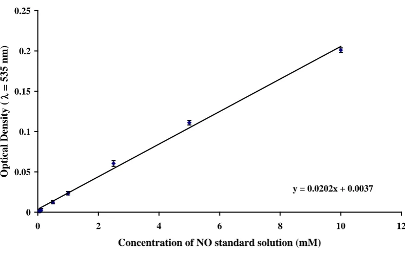
3.1 NO standard linear regression curve………...………… 91
3.2 Cholesterol mean concentrationbefore and after the active 75% fucoidan treatment……….. 96
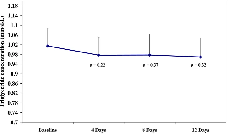
3.3 Triglyceride mean concentration before and after the active 75% fucoidan treatment………..……… 97
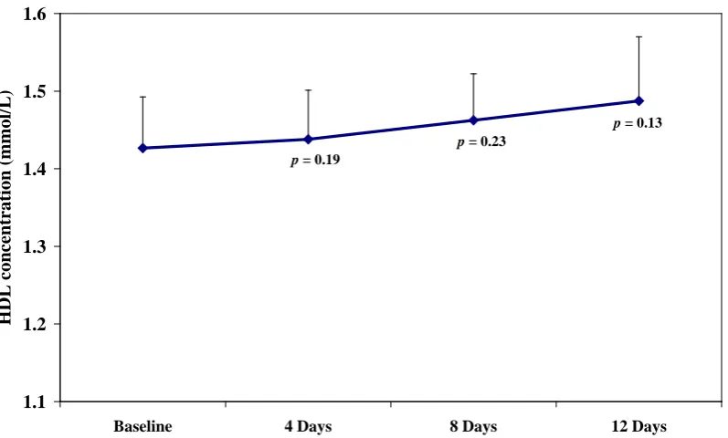
3.4 HDL mean concentration before and after the active 75% fucoidan treatment………..……… 98
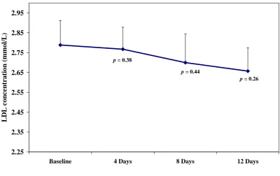
3.5 LDL mean concentration before and after the active 75% fucoidan treatment……….……… 99
3.6 Nitric oxide concentration before and after the active 75% fucoidan treatment………..……… 100
3.7 Insulin concentration before and after the active 75% fucoidan treatment. 101 4.1 Schematic diagram showing the competitive ELISA reactions inside one well……….. 134
4.2 Fucoidan standard linear regression curve prepared using DMB dye…… 136
4.3 Inhibition curves for heparin and fucoidan using 1B1 Ab……….. 138
4.4 Concentration of free fucoidan in plasma measured by 1B1……….. 140
4.6 Median concentration of fucoidan in urine measure by DMB method…... 142
5.1 aPTT measurements in volunteers treated with 3 g of 75% fucoidan tid for 12 days………... 157
5.2 Individual aPTT in volunteers treated with 75% fucoidan………. 157
6.1 SDF-1 standard linear regression curve……….. 173
6.2 IFN-γ standard linear regression curve………... 175
6.3 IL-12 standard linear regression curve……… 177
6.4 Total number of PB circulating CD34+ cells at baseline and after 4, 8 and 12 days of taking 75% fucoidan………... 182
6.5 Total number of PB CD34+/CXCR4+ cells at baseline and after 4, 8 and 12 days of taking 75% fucoidan………. 184
6.6 Flow cytometric histograms for one subject after ingesting 75% fucoidan showing CD34+ cells stained with CD34-FITC and CXCR4-PE………... 185
6.7 Percentage of total number of PB CD34+CXCR4+………. 186
6.8A Total number of CFU-GM per µL blood……… 187
6.8B Total number of CFU-GM per 105 blood cells………... 187
6.9A CD4+ lymphocyte counts before and after active 75% fucoidan treatment 188 6.9B CD4+CXCR4- and CD4+CXCR4+ lymphocyte counts before and after active 75% fucoidan treatment……… 189
6.9C Percentage of CD4+CXCR4- and CD4+CXCR4+ before and after active 75% fucoidan treatment……….. 189
6.10A CD8+ lymphocyte counts before and after active 75% fucoidan treatment 190 6.10B CD8+CXCR4- and CD8+CXCR4+ lymphocyte counts before and after active 75% fucoidan treatment……… 191
6.10C Percentage of CD8+CXCR4- and CD8+CXCR4+ before and after active 75% fucoidan treatment……….. 191
6.11 CD16+ myeloid cell count before and after active 75% fucoidan treatment……….. 192
6.12 CD19+ B-cell count before and after active 75% fucoidan treatment……. 193
6.13 SDF-1 plasma level before and after ingesting 3 g of 75% fucoidan for 12 days……… 194
6.14 IFN-γ plasma level before and after ingesting 3 g of 75% fucoidan for 12 days………. 194
7.1 Two bivariate dot-plots showing the KG1a cells (R1) with respect to beads (R2)………... 217
7.2 Growth curve for KG1a cells cultured without beads………. 218
7.3 Growth curve for KG1a cells cultures with beads……….. 218
7.4 Bivariate dot-plots showing the KG1a cells (R1) with respect to beads (R2). The cells were cultured with different concentrations of 75% fucoidan w/w for 5 days………...…………... 220
7.5 Bivariate dot-plots showing the KG1a cells (R1) with respect to beads (R2). The cells were cultured with different concentrations of N3 for 5 days………. 221
7.6 Effect of N3 on the growth of KG1a cells over 5 days………... 222
7.7 Effect of 75% GFSTM on the growth of KG1a cells over 5 days………… 222
7.8 Effect of different fucoidan extracts (75% GFSTM, N1, N2 and N3) on the growth of KG1a cells at three different concentrations………. 223
7.10 Fractional factorial design to investigate the interactions among growth
factors, DP-GFS-001 and CD34+ cells……… 225 7.11 Contrast analysis of the effect of various growth factor and GFSTM (high
= 500 μg/mL, low = 100 μg/mL) combinations on the growth of
different cell lineages………. 226 7.12 Effect of fucoidan (N3) on PB CD34+ cell expansion at 10 days………... 227 7.13 Effect of growth factors (0 versus 100 ng/mL) and fucoidan on the
percentage of CD34+ cells that are CXCR4+……….. 228 7.14 Effect of growth factors (100 versus 1000 ng/mL) and fucoidan on the
percentage of CD34+ cells that are CXCR4+………..….…... 229 7.15 Effect of growth factors (100 versus 1000 ng/mL) and fucoidan on the
percentage output of CD34+ cells………... 230 7.16 PB cell expansion when cells were cultured with either 75% GFSTM or
DP-GFS-001 at a concentration of 0.335 mg/mL over a period of 5 days. 231 7.17 Bivariate dot-plots showing the PB cells (R1) with respect to beads (R2)
and CB cells (R3) in respect to PI. The cells were cultured with either 75% GFSTM
232 or DP-GFS-001 at a concentration of 0.335 mg/mL over a
period of 5 days………... 7.18 CB cell expansion when cells were cultured with either 75% GFSTM or
DP-GFS-001 at a concentration of 0.335 mg/mL over a period of 5 days. 233 7.19 Bivariate dot-plots showing the CB cells (R1) with respect to beads (R2)
and CB cells (R3) in respect to PI. The cells were cultured with either 75% GFSTM or DP-GFS-001 at a concentration of 0.335 mg/mL over a
period of 5 days………... 234 7.20 Bivariate dot-plots showing the PB cells (R1) with respect to beads (R2).
The cells were cultured with either 75% GFSTM or DP-GFS-001 at a concentration of 0.335 mg/mL over a period of 5 days and stained with
anti CD34, CD38 and CFSE………... 236 7.21 Effect of the different fucoidan fractions on cord blood CD34+ cell
expansion……….... 237
7.22 Bivariate dot-plots showing the CB cells (R1) with respect to beads (R2). The cells were cultured with either 75% GFSTM or DP-GFS-001 at a concentration of 0.335 mg/mL over a period of 5 days and stained with anti CD34, CD38 and CFSE………... 240 7.23 Effect of fucoidan on the appearance of cell cultures. All contained
T
ABLEO
FA
BBREVIATIONSThe following abbreviations have been used throughout this thesis:
°C Degree Celsius
µg Microgram
µL Microlitres
1B1 A new novel MoAb (IgM isotype)
Ab Antibody
AGAL Australian Government Analytical Laboratories
ALB Albumin
ALP Alkaline phosphatase ALT Alanine aminotransferase AML Acute myelogenous leukaemia AP Alkaline phosphatase
Baso Basophil
BFU-E Erythrocyte burst forming unit
BM Bone marrow
BSA Bovine serum albumin BUN/Urea Blood urea nitrogen
CB Cord blood
CD Cluster of differentiation antigen CFCs Colony forming cells
CFSE 5-(and-6)-carboxyfluorescein diacetate, succinimidyl ester CFU Colony forming unit
CFU-GM Colony forming unit-granulocyte-macrophage Chol Cholesterol
Cl- Chloride
CO2 Carbon dioxide
Creat Creatinine
CTCE-0021 SDF-1 peptide agonist
CY Cyclophosphamide
DMB 1,9-dimethylmethylene blue DMSO Dimethyl sulfoxide
D-PBS Dulbecco’s phosphate buffered saline ECM Extracellular matrix
EDTA Disodium ethylenediamine tetra-acetic acid ELISA Enzyme-linked immunosorbent assay
Eos Eosinophil
ESR Erythrocyte sedimentation rate FACS Fluorescent activated cell sorting FBC Full blood count
FBS, FCS Foetal bovine serum, foetal calf serum FDA Food and Drug Administration
Flt-3 FMS-like tyrosine kinase-3 FSC Forward scatter
g Gram
G-CSF Granulocyte colony stimulating factor
GF Growth factor
GFSTM Galactofucan sulphate GGT Gamma-glutamyl transferase
GM-CSF Granulocyte-macrophage colony stimulating factor GvHD Graft versus host disease
h Hour
Hb Haemoglobin
HCO3 Bicarbonate
Hct Haematocrit
HDL High density lipoprotein cholesterol HIV Human immunodeficiency virus HLA Human leukocyte antigen
HOXB4 Homeobox B4 (a transcription factor) HPC Haematopoietic progenitor cell(s) HRP Horse-radish peroxidase
HSC Haemopoietic stem cell(s)
HSCT Haemopoietic stem cell transplantation HSPC Haemopoietic stem and progenitor cell(s) HSV Herpes simplex virus
HUCB Human umbilical cord blood i.p Intraperitoneal
i.v Intravenous
ICAM Intercellular cell adhesion molecule IFN-γ Interferon gamma
IL Interleukin
IMDM Iscove’s modified Dulbecco’s medium IU International unit
K+ Potassium
kD Kilodalton
L Litre
LDH Lactate dehydrogenase LDL Low density lipoprotein
LFA-1 Lymphocyte function-associated antigen-1 LTC-IC Long-term culture-initiating cell
LTR Long term repopulating
Lymph Lymphocyte
MACS Magnetic activated cell sorting MCH Mean cell haemoglobin
MCV Mean cell volume
mg Milligram
MGDF Megakaryocyte growth and development factor MHC Major histocompatibility complex
min Minute
mL Millilitre
mM Millimolar
MM Multiple myeloma mm3 Cubic millimetre
mmol Millimole
MMP-9 Matrix metalloproteinase-9
MNC Mononuclear cell
MoAb Monoclonal antibody
Mono Monocyte
MPV Mean platelet volume MSC Mesenchymal stem cells MSE Mean standard error
Na+ Sodium
N-Cad N-cadherin
Neut Neutrophil
NHL Non-Hodgkin’s lymphoma
NK Natural killer
nm Nanometre
NMR Nuclear magnetic resonance
NO Nitric oxide
NOD-SCID Non-obese diabetic-severe combined immunodeficiency mice NOS Nitric oxide synthase
NSW New South Wales
OPN Osteopontin
p Probability
PB Peripheral Blood
PBMC Peripheral blood mononuclear cells PBS Phosphate buffered saline
PBSC Peripheral blood stem cells PBS-T PBS Tween-20 0.1% (v/v) PDW Platelet distribution width
PI Propidium iodide
Plat Platelets
PMN Polymorphonuclear cells PNP Pooled normal plasma PoAb Polyclonal antibody PPP Platelet poor plasma
PPR Parathyroid hormone/ Parathyroid hormone- related protein receptor PTH Parathyroid hormone
PTHrP Parathyroid hormone-related protein RBC Red blood cell(s)
RCC Red cell count
RDW Red cell distribution width
rhGH Recombinant human growth hormone RHH Royal Hobart Hospital
rhPTH Recombinant human parathyroid hormone rhTPO Recombinant human thrombopoietin rHuSCF Recombinant human stem cell factor
s.c Subcutaneous
SDF-1 Stromal derived factor-1
sec Second
SEM Standard error of the mean
SSC Side scatter
TAS Tasmania
TBIL Total bilirubin
Tid “tie in die”, three times daily TNF Tumour necrosis factor
TP Total protein
TPO Thrombopoietin
Trig Triglyceride
U Unit
UNSW University of New South Wales USA United States of America UTAS University of Tasmania
VCAM Vascular cell adhesion molecule VEGF Vascular endothelial growth factor
VIC Victoria
VLA Very late antigen WBC White blood cell WCC White cell count
I
NTRODUCTIONThis thesis concerns the uptake and clinical effects of ingested fucoidan on blood parameters, and the in vitro and in vivo effects of fucoidan on haemopoietic stem cells. Fucoidan is a naturally occurring fucose rich sulphated polysaccharide which is found in brown algae, sea urchins and sea cucumbers with the former being the main source. “Fucoidan” is a general term and encompasses a number of different preparations. The type of fucoidan, its sulphations, molecular weight and conformation of sugar residues varies with the species of seaweed (Berteau & Mulloy, 2003) and the extraction procedure. However, fucoidans consist of long branched chains of carbohydrates and include a substantial amount of fucose.
Fucoidan has a long history of use in experimental biology as a selectin blocker, and has a plethora of biological effects such as anticoagulant (Mourao & Pereira, 1999), anti-tumour (Maruyama et al., 2003), anti-viral (Furusawa & Furusawa, 1989), anti complementary activity (Blondin et al., 1994) and many others. More recently, fucoidan has been shown to induce a marked and prolonged stem cell release from the bone marrow in animal models (Sweeney et al., 2000; Frenette & Weiss, 2000).
The use of whole seaweeds (brown algae) in the diet or as “medicine” seems to correlate with some of these activities. In countries where seaweed is a normal everyday part of the diet such as Japan and Korea, rates of cancer and HIV prevalence are lower (Shibata et al., 2000). It has been proposed that high levels of seaweed ingestion, especially of brown algae (Undaria pinnatifida) may contribute to that low level of cancer and low HIV incidence (Cooper et al., 2002).
Whilst fucoidans have known biological effects in vivo when ingested, there is little data on the amount of uptake. Indeed, it is a common assumption that this very high MW substance is not changed or absorbed in the intestine and no studies to date have reported on the detection of fucoidan in plasma after oral doses. Thus, a part of this thesis concerns a method for identifying uptake and measuring it especially after establishing the safety of treating human subjects with large quantities of fucoidan. Few options were available taking into consideration that the method had to be quantitative and reproducible and able to be used in routine settings. Two methods in this study were successful in measuring the level of fucoidan in solutions, an antibody based method using ELISA and a colorimetric method using a dye.
The known effects of fucoidan on haemopoiesis have been induced by intravenous dosing. In this thesis, it was hypothesised that oral fucoidan has an effect on this system. Haemopoiesis is the process of formation of blood cellular components from haemopoietic and progenitor stem cells (HPC). These cells are the most undifferentiated precursor cell type in the haemopoietic system and are defined on the basis of their functional and phenotypic properties. They are pluripotent, capable of self-renewal and capable of giving rise to long-term haemopoietic reconstitution. Once they lose pluripotency they become lineage-committed with only limited potential for self-renewal (Berenson et al., 1988 & 1991; Charbord, 1994).
The majority of HPC reside in the bone marrow (BM) with a small number continually escaping into the circulation then homing back into the BM in a process called “cell trafficking”. Stromal cells in the BM constitutively express and secrete stromal cell derived factor (SDF-1), which is a highly conserved chemokine, strongly basic, and binds to heparin. It acts as a chemo-attractant for CXCR4+ cells, so playing a role in regulating stem cell trafficking.
cell mobilisation, allows the collection of HSC via apheresis for both autologous and allogeneic transplantation (Cashen et al., 2004).
Although the HSC normally reside in the BM, in recent years we have learned how to enhance trafficking of HSC into the PB in simple and safe ways. These HSC are capable of homing to the BM cavity and regenerating a full array of haemopoietic cell lineages in a timely fashion after ablative and non-myeloablative conditioning. This process mimics enhancement of the physiological release of HSC from the BM reservoir in response to stress signals during injury and inflammation. In the early 1980s the first autologous stem cell transplants using mobilised peripheral blood stem cell (PBSC) collected by apheresis were documented (Kessinger et al., 1986; Juttner
et al., 1989). Initially, the mobilisation protocols used chemotherapy alone; however, since the discovery and clinical development of human granulocyte-colony-stimulating factor (G-CSF) (Welte et al., 1985), cytokine mobilisation has become the standard of care. Currently, the unique cytokines approved by the United States Food and Drug Administration (FDA) for autologous and allogeneic stem cell mobilisation are G-CSF and granulocyte-macrophage colony-stimulating factor (GM-CSF). PBSC mobilisation and collection have been optimized in different clinical trials. Nevertheless, 14% of patients receiving standard mobilisation for the purpose of autologous donation and 4% of allogeneic donors still fail to mobilise (Moncada et al., 2003). The pursuit of an enhanced understanding of HSC biology, the processes involved in HSC microenvironmental interactions, the ligands and receptors involved in HSC homing and mobilisation, with an emphasis on fucoidan induced HSC mobilisation, form the basis of this work.
It was found that fucoidan can increase the leucocytes and HPC in mice and monkeys (Frenette & Weiss, 2000). Its ingestion can lead to an increase in SDF-1 plasma level but a decrease in the BM. SDF-1 acts as chemoattractant for mature leucocytes and HPC which carry its receptor CXCR4 (Sweeney et al., 2002). Intravenous injection in rats of LMW fucoidan significantly increased the SDF-1 level in plasma (Luyt et al., 2003). Clinical trials have not demonstrated a definitive effect on the level of plasma SDF-1 in patients mobilised with G-CSF. Would ingesting fucoidan have the same effect on the SDF-1 plasma level?
Fucoidan is known to bind the lectin domain of P-selectin on platelets (Shibata et al., 2003). All fucoidan fractions of different molecular weights and different sulphate contents induced irreversible platelet aggregation in a dose-dependent manner. The low molecular weight fucoidan FF7/3 combines potent anticoagulant and fibrinolytic properties with only minor platelet activating effects (Durig et al., 1997).
Fucoidan extracts have been shown to have in vitro activity against herpes simplex virus (HSV). The extracts appear to inhibit HSV by blocking virus attachment and entry into the host cell (Cooper et al., 2002).
In animal models, ingestion of fucoidan has inhibitory effects on tumours, which appear to be associated with a rise in IFN-γ, IL-12, and stimulation of innate immunity (Maruyama et al., 2003; Mavier et al., 2004; Funahashi et al., 2001). In vitro
treatment of BM mononuclear cells (MNC) with IFN-γ can up-regulate the expression of CXCR4 on granulocyte precursors and monocytes (Funahashi et al., 2001). On the other hand, nitric oxide synthase (NOS-2) (iNOS) is rapidly induced by IFN-γ and IL -12 to produce nitric oxide (NO); a recently identified biological signal molecule that plays an important role in vascular regulation, immune responses, and neural signal transduction (Liao et al., 1999; Huang et al., 2001; Chesler & Reiss, 2002). Therefore ingestion of fucoidan in humans may have a similar effect on IFN-γ and IL-12 to subsequently regulate the expression of CXCR4 and the production of NO.
cancer therapies. Large-scale production and administration of expanded mobilised peripheral blood stem cells has been shown to abrogate postmyeloablative cytopenia (Boiron et al., 2006). In the future it may be possible to manufacture myeloid progenitors using HPC from alternate sources such as cord blood. Stromal layers, which are the current method for long-term maintenance or expansion of blood stem cells, are unsuitable for clinical trials. There would be considerable clinical interest in a synthetic matrix that was suitable for blood stem cell maintenance or expansion. Fucoidan preparations may provide an easily manufactured proteoglycan component of an artificial BM matrix, since it displays sulphate sugars that interact with heparin binding proteins, integrins, and selectins. The present studies were designed to directly examine the characterization of fucoidan effects on in vitro human CD34+ cells proliferation and differentiation and to determine the interaction between fucoidan and cytokines in the expansion system.
Objectives and Aims:
In this study the safety and possible effects of ingesting fucoidan on human volunteers will be examined. This work will define the effects of ingestion of fucoidan on the haemopoietic systems and will study the interaction of fucoidan with different haemopoietic cytokines in vivo and in vitro. It will also study the kinetics of the mobilisation effect of HSC and will seek possible detection methods for the presence of fucoidan in plasma.
The specific aims of this study are:
1. To establish the safety and maximum tolerated dose of Undaria pinnatifida -derived fucoidan extracts in volunteers. The pre-treatment evaluation includes volunteers’ history, physical examination and routine laboratory studies. 2. To study and report the clinical, biological and pathological changes after oral
doses of fucoidan extracts by examining volunteers’ general health, physical examination and routine laboratory studies.
3. To study the effect of ingesting fucoidan on the lipid profile.
4. To develop a method suitable for routine laboratory settings that can measure the level of fucoidan in blood circulation after oral administration.
6. To study the immune-system by examining any changes in the WBC and their subsets including B-cells and T-cells.
7. To examine the direct effect of oral fucoidan on the level of pro-inflammatory cytokines such as IFN-γ and IL-12.
8. To define the possible effects of fucoidan on HPC and HSC in BM and PB by studying the PB CD34+ properties, receptors, trafficking and cytokines.
9. To study the direct effect of fucoidan and G-CSF on SDF-1 level in PB. 10.To examine the effects and refine the characterization of different fucoidan
C
HAPTERO
NEL
ITERATURER
EVIEWChapter contents:
1.1 Haemopoietic stem and progenitor cells (HSPC) [8]
1.1.1 Historical background [8] 1.1.2 Definition of HSC and HSPC [9]
1.1.3 Characterisation of HSC in BM and microenvironment [12]
1.1.4 The bone marrow microenvironment [14]
1.1.4.1 Stromal cells [15]
1.1.4.2 The extracellular matrix [15]
1.1.5 Mobilisation of HSPC [15]
1.1.5.1 Different mobilisation mechanisms [17]
1.1.5.1.1 Chemotherapy [17]
1.1.5.1.2Cytokines such as G-CSF [18]
1.1.5.1.3 Chemotherapy plus cytokines [22]
1.1.5.1.4 AMD-3100 [22]
1.1.5.1.5 CTCE-0021 (SDF-1 peptide agonist) [24]
1.1.5.1.6 Stem cell factor (SCF) [24]
1.1.5.1.7 CXCL2 (Gro-β)[25]
1.1.5.1.8 Interleukin-8 (IL-8) [25]
1.1.5.1.9 Recombinant human growth hormone (rhGH) [26]
1.1.5.1.10 Recombinant human parathyroid hormone (rhPTH) [26]
1.1.5.1.11 Pegfilgrastim(pegylated G-CSF) [27]
1.1.5.1.12 Thrombopoietin (TPO/MGDF) [27]
1.1.5.2 The role of neutrophils and proteases in mobilisation [28]
1.1.6 Homing of HSPC [31]
1.1.6.1 Extravasation of HSPC through BM sinuses [34]
1.1.6.2 Migration of HSPC through the BM stroma [35]
1.1.6.3 Lodgement of HSPC into specific HSC niches [37]
1.1.7 In vitro expansion of HSPC [39]
1.1.8 Engraftment and repopulation [41]
1.2 Haemopoietic cytokines and their receptors [42]
1.2.1 SDF-1 (CXCL12) [42]
1.2.2 CXCR4 [43]
1.2.3 SDF-1 and CXCR4 interaction and disruption [44]
1.2.4 CXCR4 and tumour [46]
1.2.5 Interleukin-12 (IL-12) [46]
1.2.6 Interferon-gamma (IFN-γ)[48]
1.2.7 Nitric oxide (NO) [51]
1.3 Seaweed (Algae) [52]
1.3.1 Undaria pinnatifida[52]
1.3.2 Undaria fucoidan [55]
1.3.3 Structural comparison of different sulphated polysaccharides [57]
1.3.3.1 Fucoidan structure [57]
1.3.3.2 Heparan sulphate [57]
1.3.3.3 Heparin [58]
1.3.3.4 Hyaluronic acid [59]
1.3.4 Physiological properties of fucoidan [60]
1.3.5 Urinary sulphated glycosaminoglycan [63]
1.3.6 Fucoidan antiviral activity [64]
1.3.7 Fucoidan anti-tumour activity [65]
1.3.8 Fucoidan anticoagulation effect [65]
1.4 Factorial experimental design and analysis [68]
1.1 Haemopoietic stem and progenitor cells (HSPC) 1.1.1 Historical background
In 1909, a Russian biologist, Alexander Maximow, claimed that a small number of
cells circulate in the PB within the lymphocyte population that might be capable of
reacquiring pluri-potentiality and he called these cells "gemeinsame stamzellen"
(Maximow, 1909). The first clinical investigation in the field of HSCT occurred in the
1940’s, when experiments using mice showed that protection of the spleen by lead
shielding allowed animals to survive otherwise lethal total body irradiation (Jacobsen
et al., 1949).
Until the 1950’s, few attempts were made to confirm this concept, until Ford and his
colleagues established through a series of murine experiments that cellular
repopulation is the mechanism by which spleen and other tissues bring about their
therapeutic effect rather than through “humoral factors” (chemical factors) (Ford et al.,
1956).
After that, and for the first time, a French group in Paris reported long-term survival
of an adult patient with acute lymphoblastic leukaemia who received BM transplants
from several relatives after being subjected to whole-body irradiation and
methyl-nitro-imidazolyl-mercaptopurine administration (Mathé et al., 1963).
Until this point, it had been known that i.v injection of BM or spleen cells into lethally
irradiated animals could lead to animal recovery. This recovery had been shown to be
the result of repopulation of the damaged haemopoietic tissues by stem cells from the
donor. At the same time circulating stem cells were under investigation but the PB as
a source of stem cells was still considered inadequate to permanently reconstitute
haemopoiesis (Micklem et al., 1975).
Decades later, a glycoprotein (CD34) that is present on colony-forming cells and
myeloblasts but is lost at the level of the promyelocyte was detected on the surface of
immature haemopoietic cells (Krauss et al., 1996). Culture assays were able to
document the number of haemopoietic progenitor cells in any preparation. At different
levels of maturation they were called Colony Forming Units (CFUs). The PB of
at a much lower level than in human BM (Jansen et al., 2005). Also, it was
documented in a dog model that HSC obtained from the PB could permanently
reconstitute irradiated animals, just like BM could (Calvo et al., 1976). These
discoveries, plus the use of haemopoietic cytokines such as G-CSF and combined with
the development of other techniques such as cell culture assays and the quantification
of stem cells has led to a complete paradigm shift in the world of stem cell
transplantation.
1.1.2 Definition of HSC and HPC
Stem cells are defined as cells capable of unlimited self-renewal and with the ability to
give rise to multiple tissue types (Thomson et al., 1998). Both of these parameters are
subject to wide interpretation and depend to some degree on whether the stem cell is
present in situ (in its normal environment) or in an experimental setting [Figure 1.1].
There are three possibilities when stem cells divide: the first is self-renewal that is
when stem cells divide and generate new stem cells; the second is to differentiate into
mature blood cells; the third is to be destroyed through apoptosis. Some genes that are
[image:35.595.249.395.443.566.2]involved in these processes have been identified (Sorrentino, 2004).
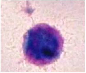
Figure 1.1: Haemopoietic stem cell. Stained with May-Grunwald-Giemsa at magnification of x1000
A haematopoietic stem and progenitor cell (HSPC) is a generic term to include both
haematopoietic progenitor cells (HPC) and haematopoietic stem cells (HSC). This is
because many properties are shared by these two cell populations. HSC and HPC are
very similar, but the later term is used to describe cells which are more immature or
self-renewal and differentiation, although these properties may be limited. For
instance, both HSC and HPC share properties such as CAM expression, KIT
expression, and trafficking in the blood and spleen.
HSC and HPC are found in BM and give rise to all the types of both the myeloid and
lymphoid lineages. They are used in clinical transplantation protocols to treat a wide
variety of diseases. The ability to increase the number of HSC or HPC either in vivo or
in vitro or change their phenotypic properties would provide new treatment options.
However there are fundamental differences between the two. By definition, HSC are
the only ones able to reconstitute the whole haematopoietic and immune systems for
the life-time of the recipient following transplant. This functional definition is
equivalent to what is also call long-term constitutive cells. Short-term
re-constitutive cells reconstitute for 1 month and then crash. As they do not self-renew
beyond this, they are not considered as HSC but multi-potent HPC. There are also all
the possible HPCs including multi-potent, myeloid (such as CMP, GMP, MEP,
colony-forming cells) and lymphoid progenitors (such as CLP).
The main functional properties of HSC as mentioned before are multi-potency and
self-renewal. Multi-potency means an individual HSC have the ability to give rise to
any of the end-stage blood cell types. During differentiation, daughter cells derived
from HSC undertake a series of commitment decisions, retaining differentiation
potential for some lineages while losing others. Intermediate cells become
progressively more restricted in their lineage potential, until eventually
lineage-committed end stage cells are generated. Self-renewal of HSC means some kinds of
stem cells are thought to undertake asymmetric cell division, generating one daughter
cell that remains a stem cell and one daughter cell that differentiates. For HSC,
however, whether asymmetric cell division occurs during self-renewal is not known
with certainty. It is instead possible that haematopoiesis occurs via symmetrical
divisions, that sometimes give rise to two daughter HSC, and that at other times give
rise to progeny that are committed to differentiate. The balance between self-renewal
versus differentiation would therefore be regulated by the control of these two kinds
It is known that a small number of HSC can expand to generate a very large number
of progeny HSC. This phenomenon is used i
HSC reconstitute the hematopoietic system. This indicates that at least during BM
transplant, symmetrical cell divisions that give two progeny HSC must occur, as
expansion in HSC numbers seen during BM transplant cannot occur in any other way.
Stem cell self-renewal is thought to occur in the stem cell niche in the BM, and it is
reasonable to assume that key signals present in this niche will be important in
self-renewal. There is much interest in the environmental and molecular requirements for
HSC self-renewal, as understanding the ability of HSC to replenish themselves will
eventually allow the generation of expanded populations of HSC ex vivo that can be
used therapeutically.
There are different types of stem cells. Embryonic stem (ES) cells, derived from the
inner cell mass of mammalian embryos, have unlimited self-renewal properties and
give rise to all embryonic tissue types in vitro, although unlimited self-renewal is not a
property of cells of the inner cell mass in situ, where they differentiate into various
tissues of the body and the ES cell phenotype is lost. These cells represent the ultimate
in stem cells because of their abilities to be both self-renewing and multi-potent.
Adult stem cells, of which HSC are the best studied, give rise to a wide range of
progenitor and mature cells within the confines of the haemopoietic system, and have
self-renewal properties for the life of the organism. Umbilical cord blood has also
been established as a clinical source of HSC.
HSC are relatively rare and are difficult to identify by morphology alone; they are thus
frequently characterised by functional assays. Several methods exist to enrich HSPC,
including fluorescence-activated cell sorting (FACS), immunomagnetic separation,
and density-gradient centrifugation (Thomas et al., 1999). The major cellular antigens
1.1.3 Characterisation of HSC in BM and microenvironment
BM has been traditionally envisioned as a ‘home’ of HSC (Ratajczak et al., 2004b).
HSC in BM are supported by an extracellular matrix (ECM) rich in fibronectin,
collagens, and various proteoglycans (Nervi et al., 2006) and cell-cell interactions
with non-haemopoietic cells, which include osteoblasts, fibroblastic BM stromal cells
and endothelial cells. These interactions form what is called the microenvironment or
“niche” for HSC that is responsible for their localization to specific anatomical
regions in the BM (Sorrentino, 2004). These niches play an important role in
regulating HSC trafficking as well as in self-renewal, proliferation, and differentiation.
The BM endothelium is the first anchoring site for homing cells, presenting adhesion
molecules and stimulating chemokines. Human and mouse BM share common
structures, but species dependent differences can also be observed. The small blood
vessels in both human and murine BM, the sinusoids in which trans-endothelial
migration is thought to take place, are composed of specialized cell structures that
regulate cell trafficking (Lapidot et al., 2005). Different niches have been described
by researchers such as “endosteal niche” where HSC are physically associated with
osteoblasts at the endosteum of the BM (Petit et al., 2002) and endothelial niches
where HSC in the BM are closely associated with sinusoidal endothelial cells (Kiel et
[image:38.595.137.505.103.315.2]al., 2005).
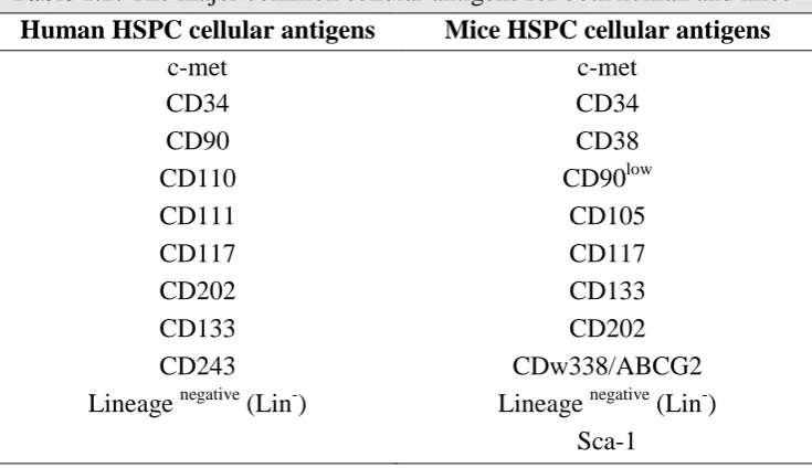
Table 1.1: The major common cellular antigens for both human and mice
Human HSPC cellular antigens Mice HSPC cellular antigens
c-met c-met
CD34 CD34
CD90 CD38
CD110 CD90low
CD111 CD105
CD117 CD117
CD202 CD133
CD133 CD202
CD243 CDw338/ABCG2