Ecology and Epidemiology
Development of Nested Polymerase Chain Reaction Detection
of
Mycosphaerella
spp. and Its Application to the Study
of Leaf Disease in
Eucalyptus
Plantations
M. Glen, A. H. Smith, S. R. H. Langrell, and C. L. Mohammed
First, second, and fourth authors: Forest Biosecurity and Protection, Ensis, Private Bag 12, Hobart, Tasmania, Australia, 7001; second and fourth authors: CRC for Sustainable Production Forestry, University of Tasmania, Private Bag 12, Hobart, Tasmania, Australia, 7001; and third author: CSIRO Forestry and Forest Products, Private Bag 5, Wembley, Western Australia, 6913.
Current address of S. R. H. Langrell: European Commission, Directorate General Joint Research Centre, Institute for Health and Consumer Protection, I-21020 Ispra, Italy.
Accepted for publication 9 August 2006.
ABSTRACT
Glen, M., Smith, A. H., Langrell, S. R. H., and Mohammed, C. L. 2007. Development of nested polymerase chain reaction detection of Mycosphaerella spp. and its application to the study of leaf disease in Eucalyptus plantations. Phytopathology 97:132-144.
Mycosphaerella leaf disease (MLD) is a serious disease of two of the major eucalypt species grown in temperate regions worldwide, Eucalyp-tus globulus and E. nitens. More than 30 species of Mycosphaerella have been reported on eucalypts worldwide. Accurate, rapid, and early dis-crimination of Mycosphaerella spp. causing crown damage to E. globulus and E. nitens will assist the development of sustainable management strategies. This study describes the development, and incorporation in a nested polymerase chain reaction (PCR) approach, of specific primers for the detection and identification of Mycosphaerella spp. commonly re-ported from leaf lesions of E. globulus and E. nitens in Australia. Primer design was assisted by sequence alignment and phylogenetic analysis of 165 nonredundant sequences from the nuclear ribosomal DNA internal transcribed spacer regions of Mycosphaerella and related species. Phylo-genetic analysis revealed very high sequence similarity for two taxon groups, Mycosphaerella grandis and M. parva, and M. vespa, M.
ambi-phylla, and M. molleriana, and primers were designed to differentiate each of the two groups. Three other species, M. cryptica, M. nubilosa, and M. tasmaniensis, were distinct and distinguished by species-specific primers. In double-blind trials, the detection test accurately and rapidly identified Mycosphaerella spp. in cultures and discriminated against other pathogens that co-occur in or on Eucalyptus leaves, thereby verifying its reliability. The detection test has an internal amplification control in the first-round PCR with fungal-specific primers to raise confidence in test results, particularly to highlight negative results due to PCR inhibition. When applied to DNA extracted from leaf or stem samples either as multiple or single lesions, it detected and identified up to five Mycosphaerella spp. or taxon groups in both positively identified and in young (putative) MLD lesions. The samples were 20 mm2 or larger in
surface area and were collected while undertaking disease rating assess-ments in an experimental investigation of Eucalyptus plantations and regrowth forest. Using nested PCR detection, Mycosphaerella spp. were positively identified in 2 days, 1 to 5 months earlier than by classical methods, demonstrating the potential application of this detection test to the early discrimination of MLD components in ecological, epi-demiological, and genetic investigations.
Eucalyptus spp. are a major global hardwood pulp crop.
Mycosphaerella spp. cause severe leaf diseases of temperate
Eucalyptus plantations in wetter areas of Australia and serious disease in New Zealand, South Africa, Spain, and some regions of Chile (19). Many species appear to have spread to other temperate regions from Australia, and species that also occur in the Australian subtropics appear to have spread to Indonesia, Vietnam, China, and South America (21,58,62). Indigenous temperate Australian forests have mixed eucalypt species where Mycosphaerella leaf disease (MLD) generally is of minor significance, except in coppice regrowth in certain Western Australian forests (1,14). In southern Australia, MLD in plantations appears to be caused principally by Mycosphaerella nubilosa and M. cryptica, although
M. vespa, M. tasmaniensis, M. parva, and M. grandis occur fre-quently. M. marksii and other species are encountered less often (13,49,51,52,60). The most damaging are reported to be M. nubi-losa and M. cryptica (10–12,14,25,59–62), which cause the most severe leaf disease, resulting in significant crown damage of
E. globulus, E. nitens, and their hybrids (30,39), the most widely
grown temperate plantation species. Crown damage in E. globulus
in Australia has been reported to range from 10% leaf necrosis to complete defoliation and tree death in locations strongly condu-cive to disease development (22,23,50,52). MLD has restricted the use of commercially desirable eucalypt species in Australia, New Zealand, and South Africa (28).
Currently, MLD is detected visually by lesion characteristics that also are used in crown damage assessment of plantations (71). Many foliar diseases that have much lower impact also produce symptoms similar to MLD and are difficult to distinguish without experience or training. MLD identification may be con-firmed using conventional methods. Conventional methods for positive identification of these and other leaf pathogens involve waiting until lesions produce “typical” reproductive structures or forms, isolating them, and identification of putative pathogens using taxonomic criteria. A challenge for those evaluating
Mycosphaerella spp. affecting eucalypts is that reproductive struc-tures may or may not form in leaves or culstruc-tures and, often, the most readily available identifying characteristics, such as germi-nating ascospores, are also taxonomically ambiguous (10,15,49, 51,59–62). In addition, pseudothecia do not form in axenic cultures of most species and are found only in older lesions or, for a few species, on inoculated aseptic leaves in culture (59,60). Identification is further confounded by the fact that more than one
Corresponding author: M. Glen; E-mail address: Morag.Glen@csiro.au
DOI: 10.1094 / PHYTO-97-2-0132
Mycosphaerella spp. may be present in a single lesion (43,62). Other limitations of conventional methods include low isolation fre-quencies of all or even most Mycosphaerella spp. from lesions. De-tailed sampling does not always deliver all of the Mycosphaerella
spp. present in each lesion. Hence, crown damage assessment based on lesions is not necessarily attributable to a particular
Mycosphaerella spp.
Molecular detection of plant pathogens is used in many agri-cultural spheres to assist decision support systems. These include immunological and DNA and polymerase chain reaction (PCR)-based methods. Immunological detection of Mycosphaerella bras-sicicola from spore traps has been developed to rationalize pesti-cide applications within crop protection programs (42). However, pathogen discrimination is still dependent on spore production, which may occur at a late stage in development in the disease cycle and by which stage prophylactic intervention may not be a management option. Furthermore, fungicide use may be finan-cially or environmentally prohibitive in tree plantations. Detection at an earlier stage in the fungal life-cycle would be preferable and may permit development of silvicultural options to prevent dis-ease or reduce disdis-ease severity (9,11,52). PCR-based methods exploit genetic sequence differences to provide early, rapid, specific, and sensitive detection and identification of pathogens of concern regardless of the presence of reproductive structures. PCR amplification using species-specific primers has been widely used for disease diagnosis in humans, other animals, and plants (2,67). Specific PCR tests have been developed for M. gramini-cola (Septoria tritici) in wheat (27) and to distinguish M. fijiensis
and M. musicola from banana leaves (40). PCR detection may target specific genes or anonymous products, such as microsatel-lites, random amplified polymorphic DNA, or amplified fragment length polymorphism fragments (35,67,68,72). In order to provide simultaneous discrimination for several Mycosphaerella spp. of eucalypts with high reproducibility and reliability, a well-characterized DNA region which is likely to be highly stable must be targeted. There are more than 30 species of Mycosphaerella on
Eucalyptus spp., and related anamorphs increase this number to over 50 (5,6,15,17,18,49). In wheat, the M. graminicola-specific PCR amplifies a fragment of the β-tubulin gene (27). However, many more species occur in eucalypts than on wheat, and few β -tubulin sequences are currently available for eucalypt species. The specific primers for M. fijiensis and M. musicola (banana pathogens) were designed to sequence divergence within internal transcribed spacer (ITS)1 (40). In addition, molecular phylo-genetic studies in the families Mycosphaerellaceae on Myrtaceae have been based mainly on ITS1 and ITS2 sequences (19,20, 33,62,64,70). These studies have provided reliable characters for the discrimination of species for which morphological characters such as spore dimensions are variable within species and overlap among species (62), leading to taxonomic confusion and frequent misidentifications. The value of ITS sequence data is attested to by the numerous Mycosphaerella sequences available on public databases. Multiple sequences are available for species causing serious diseases of industrial crops, allowing comparison of geo-graphically diverse isolates from different host species. In addi-tion to providing interspecific discriminaaddi-tion, the ribosomal RNA genes, including the ITS regions, are multicopy, assisting in sensitive detection from plant material. Sensitivity is essential as lesions may have mixtures of many fungal species in addition to several Mycosphaerella spp. A PCR test based on ITS sequences exists for some Mycosphaerella spp. on eucalypts; however, the published test requires a restriction digestion after PCR, with discrimination based on fragment size (43), adding considerably to the processing and analysis time. Analysis of restriction fragments also may be confounded when multiple species are present, as is often the case in MLD.
Therefore, the primary aim of this investigation was to develop PCR-based systems for direct detection and identification of several
Mycosphaerella spp. from lesions of all ages in eucalypt species as a research tool aimed at refining current knowledge of epidemiology, ecology, and host resistance and to provide the potential for certification of disease-free germ plasm. Although the tests developed here were primarily for use on Eucalyptus spp. leaves and stems, they also were intended to be applicable to a wider range of woody species that are potential hosts for
Mycosphaerella spp. For example, other members of the family Myrtaceae, members of other Myrtales families, and species of the family Proteaceae are actual or potential hosts that co-occur naturally throughout the landscape in Australia with Eucalyptus
spp. Plantations are grown in close proximity to natural vegetation harboring Mycosphaerella spp. in Australia, South Africa, most probably South America, which shares a Gondwanan origin, and Indonesia. In addition to detecting Mycosphaerella spp. directly from leaves, a detection system also could be used to verify whether samples collected or isolated during earlier surveys belong to the species or taxon group in which they had been identified according to the taxonomic criteria at the time.
PCR, although fast, sensitive, and capable of being automated, is not without problems. These include enzyme inhibition by substances present in the template DNA, which may cause false negative results. Plant material is particularly notorious for PCR inhibitor compounds, many of which often are co-precipitated during DNA extraction (45). Strategies to overcome PCR inhi-bition and increase confidence in negative results include using a range of DNA template dilutions (40), spiking of negative samples (40), buffer additions during the DNA extraction process (4,34,63) magnetic DNA capture (4,45,63), post-extraction puri-fication (44), and the use of alternative polymerases (3). In the interests of speed, economy, and wide applicability of the tests, it is desirable to avoid repetitious assays or lengthy and involved DNA purification techniques. A rapid, simple DNA purification method based on binding of DNA to silica in the presence of chaotropic salts (31) avoids the use of toxic and volatile reagents and has been used extensively in our laboratory for obtaining amplifiable DNA from a wide range of material. Nested PCR also has been used to amplify DNA containing inhibitors from plant material (53,75). Because none of these strategies for overcoming PCR inhibition were completely successful, detection of PCR inhibition is important to raise confidence in negative results. This can be achieved by the use of internal amplification controls (IACs). Amplification of endogenous IACs using a different primer pair has been shown to be unreliable in detecting PCR inhibition (37); therefore, we constructed an exogenous IAC that can be added at a controlled concentration to each sample in the first round of a nested PCR. Specific primers were used in the second-round PCR to detect the presence of five of the most commonly reported
Mycosphaerella spp. in southern Australian plantations. The results of a survey of Mycosphaerella spp. in an E. globulus plantation in northern Tasmania to establish a baseline of the species present prior to the setting up of experimental trials also are presented here. The increased coverage, spatial resolution, and accuracy provided by using the detection test as a survey tool for Mycosphaerella spp. are discussed.
MATERIALS AND METHODS
Fungal material. Cultures (Table 1) were maintained on 2% malt extract agar. Nonviable cultures up to 10 years old also were used for DNA extraction. Standard reference cultures for each of the target species were nominated based on the most reliably identified material available. These were M. cryptica (AA/1/3/10), morphologically identified by Milgate (51), ITS sequenced here, sequence consistent with sequences in GenBank for nine other
sequenced (51), sequence identical with that of AC165, neo- type material from A. J. Carnegie; M. nubilosa (Z/1/1/11), morphologically identified by Milgate (51), ITS sequenced, and consistent with 13 other sequences in GenBank; M. tasmaniensis
(STE-U1555), an ex-type culture from Tasmania (21); and
[image:3.612.53.564.120.649.2]M. vespa (BS/3/2/1), morphologically identified by Milgate in collaboration with A. J. Carnegie, and ITS sequenced (51). These cultures have been deposited in the Department of Agriculture Western Australia Plant Pathogen Collection (WDCM77).
TABLE 1. Fungal cultures used to test species-specific primers
Species Codea Host Origin
Botryosphaeria sp. WA36b Eucalyptus wandoo Western Australia
Cryptosporiopsis eucalypti I00108ab E. camaldulensis Queensland
Cylindrocladium retaudii I00150b E. urophylla Queensland
Mycosphaerella africana CBS680.95c,d E. viminalis South Africa
M. cryptica AA/1/3/10e E. globulus Tasmania
NZ301He E. nitens New Zealand
E7444af E. pilularis New South Wales
I00205b E. globulus Victoria
I00207b E. globulus Victoria
I00208b E. globulus Victoria
I00210b E. globulus Victoria
I00214b E. globulus Victoria
I00216b E. globulus Victoria
I00217b E. globulus Victoria
I00225b E. obliqua Victoria
I00226b E. obliqua Victoria
I00234b E. globulus Victoria
I00235b E. cypellocarpa New South Wales
I00236b E. cypellocarpa New South Wales
M. grandis AC165g,h E. saligna Victoria
I00206b E. globulus Victoria
I00219b E. globulus Victoria
S/1/2/3e E. globulus/nitens Tasmania
Q/1/1/1e E. nitens Tasmania
H/1/3/1e E. nitens Tasmania
M. nubilosa AC106g,h E. globulus Victoria
L/1/2 multisporee Not recorded Victoria
StMarys/2/4e E. globulus Tasmania
StMarys/2/8e E. globulus Tasmania
Z/1/1/11e E. globulus Tasmania
E1 multisporee E. globulus Tasmania
Z/1/1/13e E. globulus Tasmania
S/2/1/1e E. globulus Tasmania
M. parkii CBS 387.92c,d E. grandis Brazil
M. parva AC83g,h E. globulus Victoria
AC85g,h E. globulus Victoria
AC89g,h E. globulus Victoria
CBS110503c E. globulus Western Australia
M. pini DT3b Pinus radiata New South Wales
M. suberosa CBS 436.92c E. dunnii Brazil
Mycosphaerella sp. 1 (received as M. parkii) CBS 208.94c E. grandis Indonesia
CBS 209.94c E. grandis Indonesia
Mycosphaerella sp. 2(received as M. parkii) CBS516.93c E. globulus Brazil
M. tasmaniensis I555d,e,h Not recorded Tasmania
BS10/2e,h E. nitens Tasmania
S/3/2/7e,h E. globulus/nitens Tasmania
M. vespa A/1/3e E. globulus Tasmania
B/3/2/1e E. globulus Tasmania
A/1/7e E. globulus Tasmania
B/3/2/4e E. globulus Tasmania
BrunyIs/1/1e E. globulus Tasmania
Phaeophleospora eucalypti NZFS85C/1i E. nitens New Zealand
NZFS85C/3i E. nitens New Zealand
NZFS85C/23i E. nitens New Zealand
Quambalaria cyanescens IMI178848j E. pauciflora New South Wales
Q. eucalypti S8k E. globulus (bark) Uruguay
Q. pitereka E7440f Corymbia maculata Western Australia
a Superscripts identify culture source.
b CSIRO FFP Canberra.
c Centraal Bureau voor Schimmelcultures..
d Type strain.
e University of Tasmania.
f CSIRO FFP Perth.
g Angus Carnegie, LaTrobe University.
h DNA was extracted from an old, nongrowing culture.
i New Zealand Forest Research.
Sampling of leaf material from plantations and forest re-growth sites. A survey of 2-year-old E. globulus was conducted at Smithton in northwestern Tasmania on 8 August 2003 (S40°55′21′′, E144°59′39′′). The plantation of approximately 21 ha was divided into six sections and 10 leaf samples were taken from separate trees in each section. Juvenile leaves were sampled from branches at a height of 1.2 m from trees that were up to 4 m tall. These trees were selected using systematic sampling starting from the first two trees, moving over a row and sampling the next two trees, and so on, until the required number of samples was taken. Leaves were taken from the fourth to fifth leaf pair on each branch to minimize variation in leaf age, physiological development, height in the canopy, and age of the lesions. Lesions from lateral branch stems also were sampled from seven randomly selected E. globulus trees in the same trial.
Additional samples for confirmation that the test was applicable to other host species were collected at four different sites in north and northeastern of Tasmania. In all, 16 E. nitens samples were taken from plantations at Weldborough (S41°13′14′′, E147°51′60′′), St. Helens (S41°20′41′′, E148°3′55′′), and Scotts-dale (S41°15′34′′, E147°25′25′′). Leaf material was sampled from branches at a height of 1.2 m and sampled from the fourth to fifth pair on each branch from trees ≈5 to 6 m tall, selected using a systematic sampling regime as described above. A collection of four samples was made from natural forest regeneration of E. regnans at a roadside site near Bicheno (S41°48′19′′, E148°14′53′′). Leaves were taken from the fourth pair on branches. Trees were ≈1.5 m tall and ≈12 months old.
Lesions were excised with a scalpel, using a fresh blade for each sample to avoid DNA cross-contamination. Each sample was 1 cm2 and consisted ofup to 15 lesions per leaf, pooled. The stem
samples from E. globulus were obtained by scraping the surface of stem lesions with a scalpel to produce shavings and approxi-mately 80 mm2 of stem surface area was taken.
DNA extraction, PCR, and sequencing. Plant material and fungal mycelium were ground in 1.5-ml microfuge tubes with a pellet pestle mixer (in liquid nitrogen for leaf and stem samples), mixed with extraction buffer (65), and incubated for 1 h at 65°C. Tubes were centrifuged at 14,000 rpm for 15 min and the super-natant removed. Purification was achieved by binding to a silica matrix in the presence of NaI (31). PCR was carried out in an Applied Biosystems (Foster City, CA) GeneAmp PCR System 2700 thermocycler. Reactions of 25 µl contained 0.55 U of TTH+ Polymerase (Fisher Biotec, Subiaco, Western Australia) in 1× polymerization buffer (Fisher Biotec), 2.0 mM MgCl2, bovine
serum albumen at 0.2 mg/ml, 0.2 mM each dNTP, and 0.25 µM each primer. For sequencing, the entire ITS1, 5.8S, and ITS2 regions were amplified using primers ITS1-F (29) and ITS4 (76) and the following thermocycler program: 94°C for 3 min; 35 cycles of 94°C for 30 s, 55°C for 30 s, and 72°C for 30 s; fol-lowed by an extension step of 72°C for 7 min. PCR products were electrophoresed on a 1% agarose (Fisher Biotec) gel at 4 V/cm for 1 h. Before sequencing, PCR products were purified with an UltraClean PCR Clean-up DNA purification kit (MoBio Labora-tories, Inc., West Carlsbad, CA). Sequences were determined with an ABI Prism Dye Terminator Cycle Sequencing kit on a Bio-Rad 373 Sequencer (Hercules, CA) with stretch upgrade. Sequences were obtained in both directions and assembled with Lasergene software (DNAStar, Madison, WI).
Phylogenetic analysis and primer design. Sequences generated in this project are listed in Table 2. Further sequences were retrieved from GenBank and EMBL. Mycosphaerella and anamorph species included in the analysis were selected on the basis of (i) occurrence on members of Myrtaceae or (ii) high sequence similarity to a species from Myrtaceae. All available sequence variants for each species were included with the exception of two related anamorph groups (Cercospora and
Trimmatostroma), with sequence similarity of <85% to any of the
target species and which previously have been shown to form distinct clusters in phylogenetic analyses (20,33). Hortaea werneckii was included on the basis of high sequence similarity, but only one ITS sequence was selected from the 17 available at the time. Alignments were performed with CLUSTALW (73) and adjusted manually. Phylogenetic analyses were executed with DNAPars and DNAML of the PHYLIP package (26) on the Australian National Genome Information Service (ANGIS, Sydney, NSW, Australia).Initial analysis was by DNAPars, with
Phaeoramularia hachijoensis (AY251086) set as the outgroup, jumbling 10 times, and all other options as default. The set of most parsimonious trees were subjected to maximum likelihood analysis by DNAML with Phaeoramularia hachijoensis
(AY251086) set as the outgroup, global rearrangements on, and all other options as default. The sequence alignment and phylogenetic tree were deposited in TreeBase (accession number TBA). Primers were designed following the general concepts outlined by Dieffenbach (24), with two additional aims. To permit future multiplexing of the five specific PCRs, primer sites were selected to produce amplicons of different size for each species and primer lengths were modified to result in the same annealing temperature for all primers. BLAST searches of GenBank and EMBL also were carried out using the taxon-specific primer sequences.
Primer specificity testing. Primers were tested for specificity on a broad range of template DNA, including pure cultures of
Mycosphaerella spp., other eucalypt leaf pathogens (Table 1), and clean eucalyptus leaves. To obtain optimum specificity and efficiency of amplification, several different annealing tempera-tures were tested. Primer lengths were adjusted by 1 or 2 bases until all primer pairs amplified the target species or group at the same annealing temperature, with no amplification from other species.
Construction of an IAC for first-round PCR. A plasmid containing primer-binding sites for the primers ITS1-F (29), ITS5, and ITS4 (76) was constructed by ligation of a histone gene fragment amplified from Phaeophleospora eucalypti DNA using composite primers (Table 3). The forward primer sequence is a composite of ITS1-F, ITS5, and H3-1a (32), with H3-1a at the 3′ end, and the reverse a composite of ITS4 and H3-1b (32), with H3-1b at the 3′ end. A product of ≈1,100 bp was amplified using conditions for the H3-1a/H3-1b primers (32). This product was purified with an UltraClean PCR Clean-up DNA purification kit (MoBio Laboratories, Inc.) and transformed into Escherichia coli
using a pGEM-T cloning kit (Promega Corp. Madison, WI). Plasmid DNA was extracted using an Ultraclean plasmid extrac-tion kit (MoBio Laboratories, Inc.).
[image:4.612.316.568.621.750.2]Nested PCR. All reactions were set up in UV-irradiated laminar flow hoods with positive (DNA from pure culture of target species) and negative (DNA from pure cultures of nontarget species and no added template) controls included in each set of reactions. Aerosol barrier tips were used routinely and gloves
TABLE 2. Sequences generated in this project for primer design and species verification
Species Accession numbers
Mycosphaerella africana AY626981
M. cryptica AY667576, AY626989, DQ471928
M. grandis AY626986
M. nubilosa AY667577, DQ471926
M. parkii AY626979(type isolate)
M. parva AY626980
Mycosphaerella sp. 1 AY66982,AY626983
Mycosphaerella sp. 2 AY626984
M. suberosa AY626985
M. tasmaniensis AY667578 (type isolate)
M. vespa DQ471925, DQ471927
were always worn and changed frequently to minimize cross-contamination. First-round PCR used primers ITS1-F and ITS4 as outlined above, with the addition of 25 fg of IAC in each 25-µl reaction. Failure of first-round amplification (as occurred in a few leaf samples) demonstrated the need to repeat the first-round PCR using a more dilute sample. First-round product was diluted 1/10 and a 5-µl aliquot was used as template in a 25-µl nested PCR reaction with species-specific primers. Concentrations were 0.25 µM for each primer, 2 mM MgCl2, and 0.2 mM each dNTP
in 1× polymerization buffer (Fisher Biotec), with 0.55 U of TTH+ polymerase (Fisher Biotec) per 25-µl reaction.
Cloning and sequencing of PCR products from multiple fungal species in leaf samples. Independent confirmation of PCR test results by isolation of corresponding species from the same leaf samples was not possible because not all samples had pseudo-thecia and not all samples with pseudopseudo-thecia had mature spores. Therefore, confirmation was sought by amplification, cloning, and sequencing of fungal PCR products from selected leaf samples. Fungal ITS products were amplified with ascomycete-specific primer combination ITS1-F (29) and ITS4-A (46) using the same reagent concentrations and thermocycler program as given above for ITS1-F/ITS4. Products were purified with an UltraClean PCR Clean-up DNA purification kit (MoBio Labora-tories, Inc.) and cloned using a pGEM-T cloning kit (Promega Corp.). These primers had not been used previously in the laboratory and amplified a larger fragment than any primers used previously, so that the possibility of chance contamination with PCR product was eliminated. Individual clones were tested with the species-specific primers, and a representative of each species from each leaf sample was sequenced using primers ITS1-F and ITS4 as described previously.
Plantation survey. Every set of PCR reactions included five samples containing DNA from cultures of each of the five species or taxa groups and one sample with no template DNA. The four nontarget species in each of the species-specific tests were treated as additional negative controls. The test was repeated on ≈30% of samples to demonstrate the consistency of the analysis. Although the ITS primers for M. vespa also are able to detect M. ambi-phylla and M. molleriana, these species previously have not been recorded in Tasmania, whereas M. vespa has been recorded over the majority of the state (51). For simplicity, the positive results for this group are referred to as M. vespa. The possibility of M. molleriana and M. ambiphylla occurring in Tasmania has not been eliminated.
RESULTS
Phylogenetic analysis and primer design. A phylogenetic analysis of ITS sequences was carried out after sequence align-ment to accommodate the following points: (i) intraspecific varia-tion in ITS sequences of some Mycosphaerella spp., (ii) a degree
of taxonomic uncertainty for some species, and (iii) quality control for sequences obtained from public databases (the accuracy and reliability of which was beyond our control). Phylogenetic analysis showed species with ITS sequences most similar to the target species to facilitate the selection of interspecific divergent regions most suitable for species-specific primer design. Phylo-genetic analysis also assisted in the detection of misidentified species from public sequence databases or culture collections.
In all, 165 nonredundant ITS sequences from 95 Mycosphaerella
and related anamorph species that were obtained from GenBank or produced here were included in the phylogenetic analysis (Fig. 1). All available sequences of Mycosphaerella spp. that occur on
Eucalyptus spp. were included in the analysis, as were several species from other hosts. Species known from eucalypts for which ITS sequences were not available include M. delegatensis, M. endophytica, M. gracilis, M. longibasalis,and M. swartii. Se-quences AY045519 (M. suttoniae)and AY626984 (Mycosphaerella
sp.) had segments of 157 and 181 bp, respectively, removed from ITS1 after the initial alignment revealed that these were most probably insertions. The insertions were at different sites and did not appear to be homologous. Analysis by DNAPARS produced 428 equally most parsimonious trees of 3,170 steps. DNAML analysis of this set produced an ML tree with an Ln likelihood = –13,649.6.
For each of the five target species (M. cryptica, M. nubilosa, M. vespa, M. grandis, and M. tasmaniensis), variable regions were visually selected from the alignment of 95 Mycosphaerella
spp. Particular attention was paid to discriminating species that had high overall sequence similarity and that clustered closely in the ML tree. The variable regions anchored the 3′ end of species-specific primers and primer length was varied at the 5′ end to give similar annealing temperatures. Species-specific primers were designed for M. cryptica, M. nubilosa, and M. tasmaniensis. High interspecific sequence similarity combined with intraspecific variation prevented the design of species-specific primers for
M. vespa and M. grandis. Therefore, primers were designed for two taxa groups: one primer pair for M. vespa, M. ambiphylla, and M. molleriana, and one primer pair for M. grandis and
M. parva. Regions were chosen to give a different product size for each species with a view to future multiplexing of the tests. Primer sequences were checked for self-annealing and BLAST search results were checked to ensure that only the target species matched both primers. Primer sequences and expected product sizes are given in Table 3.
[image:5.612.45.569.614.752.2]Arrows on branches indicate groups that have an exact match to primer sequences (Fig. 1). Each of these branches is supported by 99% confidence limits. This includes all available sequences for each target taxon, with two exceptions: AF173307 was very dif-ferent from all other M. tasmaniensis sequences, grouping instead with Mycovellosiella eucalypti and Phaeoramularia saururi, and AY534227 grouped with the other M. vespa sequences but dif-
TABLE 3. Species-specific primers designed from Mycosphaerella spp. internal transcribed spacer (ITS) sequences and composite primers used to construct an
internal amplification control for ITS1-F/ITS4 and ITS5/ITS4 PCR
Species
Primer
name Primer sequence
Product size (bp)
Mycosphaerella cryptica McrypF 5′ CATCTTTGCGTCTGAGTGATAACG
McrypR 5′ GGGGGTTGACGGCGCGAC 331
M. grandis/parva MgpF 5′ CCCATTGTATTCCGACCTCTTG
MgpR 5′ CGCTTAGAGACAGTTGGCTCAG 359
M. nubilosa MnubF 5′ CAACCCCATGTTTTCCCACCACG
MnubR 5′ CGCCAGACCGGTCCCCGTC 395
M. tasmaniensis MtasF 5′ GTCACGCGGCCGACCGC
MtasR 5′ CATTAGGGCACGCGGGCTG 298
M. vespa/ambiphylla/molleriana MvamF 5′ GCATCTCTGCGTCTGAGTCAC
MvamR 5′ GCTCGGCCGGAGACTTCG 264
Phaeophleospora eucalypti ITSPCF 5′ CTTGGTCATTTAGAGGAAGTAAAAGTCGT AACAAGGACTAAGCAGACCGCCCGCCAGG
Fig. 1. Maximum likelihood tree produced by phylogenetic analysis of 165 internal transcribed spacer sequences from Mycosphaerellaand related anamorph
species, Ln likelihood = –13,649.6. The outgroup is Phaeoramularia hachijoensis and the scale bar represents expected nucleotide variation of 10%. Clades that
are supported with 99% confidence are denoted by an asterisk above the branch. Arrows on branches represent the groups that have 100% similarity to the
taxon-specific primer sequences as follows: G, Mycosphaerella grandis/parva group; V, M. vespa/ambiphylla/molleriana group; N, M. nubilosa; C, M. cryptica; and T,
M. tasmaniensis. Abbreviations for genera are: M., Mycosphaerella and the anamorph genera C., Cladosporium; Mv., Mycovellosiella; Pa., Passalora; Php.,
Phaeophleospora; Phr., Phaeoramularia; Ps., Pseudocercospora; and R., Readeriella. The anamorph genera are those given in the GenBank accessions, although many of them recently have been recombined (5,6,15), appropriate formal name changes have not been made by the sequence submitters. In addition, some
sequences listed in GenBank as belonging to M. juvenis and M. ellipsoideaare included as belonging to those species, though the isolates that were sequenced
probably represent different taxa (P. W. Crous, perssonal communication). To improve readability of the tree, one branch, distant from the target taxa, has been
pruned. Group A represents two Mycosphaerella spp., M. fori and M. musicola, and 14 Pseudocercospora spp.: P. basiramifera, P. cordiana, P. cruenta,
fered by 2.3% and had two mismatches to the forward primer for
Mycosphaerella vespa/ambiphylla/molleriana.
By contrast, M. ambiphylla and M. molleriana sequences grouped closely with the remaining M. vespa sequences with 99% confidence and had a 100% match to the M. vespa primers. Percent difference among this group was only 0.2 to 1.0%. In addition, sequences for M. grandis and M. parva were very similar to each other (99.2%) and form a single clade with high support. Furthermore M. bellula, derived from a Proteaceae family host, and other unnamed isolates from eucalypts also fall within this clade and all have a 100% match to the
M. grandis/parva primers.
IAC for first-round PCR. Plasmid p35.12 was selected for use as an IAC because it had an insert of the correct size (≈1,100 bp) that was amplifiable with the ITS1-F and ITS4 primers. A PCR product was visible when 2.5 fg of IAC was the only DNA tem-plate added to a 25-µl reaction (Fig. 2); however, addition of leaf or fungal DNA required a higher concentration of IAC (Fig. 3). Therefore 25 fg was added to each 25-µl PCR, which equates to 6 × 10⁴ copies per reaction. Successful first-round amplification
with ITS1-F and ITS4 primers verified that the template DNA from plant material or fungal cultures was amplifiable, even if fungal DNA was at too low a concentration to be detected after one round of PCR. This increased confidence that subsequent negative results for taxon-specific primers were not false nega-tives caused by PCR inhibition. If fungal DNA was present at a much higher concentration, amplification of IAC was prevented by competition. Thus, a visible product from either the IAC or fungal DNA or, in some cases, both, indicated successful first-round PCR.
Specificity, sensitivity, and reliability testing of primer sets.
Species-specific primers (0.25 µM) were used in the second-round amplification of a nested PCR after first-second-round ampli-fication with ITS1-F and ITS4. After specificity testing of all primer pairs at several annealing temperatures, the optimum thermocycler program to run the species-specific primers
simul-taneously was 94°C for 3 min; 20 cycles of 94°C for 30 s, 62°C for 30 s, and 72°C for 30 s; followed by 72°C for 10 min.
Each individual primer pair, designed to detect Mycosphaerella
spp. or closely related species groups from plantation Eucalyptus
spp., were highly specific for their respective targets (Fig. 4B to F). The specific primer pairs verified the taxa of 3 to 15 isolates of each of the target species from culture collections (Table 4). These tests were done in a series of experiments with replication of ran-domly chosen standard isolates and had identical results, showing the reliability of the test. In double-blind trials, the primers un-equivocally discriminated among the five species or taxon groups and did not detect cultures of nontarget species, including possible co-occurring fungal pathogens, verifying their reliability (Table 4; Fig. 4B to F). With nested PCR, each primer pair was sensitive enough to detect pathogen DNA when present at concentrations of 10 to 100 fg per 25-µl reaction (Fig. 5). Amplification at these low levels of template DNA was not reliable in repeated trials and this may be attributed to stochastic events involving target sequences at such low concentrations. Amplification from 10 fg of M. grandis
DNA with no amplification from 100 fg (Fig. 5B and C) supports this hypothesis. The addition of plant DNA to the first-round PCR, equivalent to that in a commonly used dilution of the average-sized leaf sample, appeared to reduce final sensitivity by a factor of ≈10 (Fig. 5B). Addition of IAC appeared to have the reverse effect, though this also may have been a result of stochastic effects.
In 2 days in a double-blind trial, one person using the detection test successfully differentiated the appropriate species (M. cryp-tica, M. grandis/parva, M. nubilosa, and Mycosphaerella sp. nov.) (Table 1; data not shown) from 30 cultures from disease surveys in Eucalyptus plantations that had been identified at the time of isolation according to the taxonomic criteria available at that time. The PCR detection test results were verified by subsequent se-quencing of the complete ITS regions of these isolates (Table 2; data not shown).
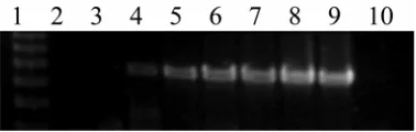
Testing of primers on plant material. The primers success-fully detected Mycosphaerella spp. directly from infected leaves and stems (Fig. 6). At least one, and up to four, Mycosphaerella
spp. were detected from each leaf or stem sample. The robustness of the detection systems to test for Mycosphaerella spp. in a wide range of tissue types was further indicated by amplification of specific products from dead, dried, herbarium collections. The nested PCR reactions enabled detection and discrimination of the five Mycosphaerella taxa in 30 samples with lesions at any developmental stage by one person within 2 days.
Verification of infection with multiple Mycosphaerella spp. by cloning of PCR products. Isolation of Mycosphaerella spp. from leaf lesions does not produce cultures of nonsporulating species; therefore, infection by multiple Mycosphaerella spp. was verified by cloning PCR products from selected leaf samples, then testing individual clones by species-specific PCR and sequencing. A comparison of species detected by taxon-specific nested PCR with those detected by cloning and sequencing (Table 5) shows that, although fewer species were detected by the latter method, the presence of multiple species in individual leaves or even individual lesions was confirmed. One leaf sample that had given a negative result for all the taxon-specific nested PCRs produced a clone containing an M. grandis sequence (Table 5, sample 3). Other species of possible leaf pathogens also were detected by the cloning method (data not shown). No clones containing M. vespa
[image:7.612.77.266.435.495.2]or M. tasmaniensis sequences were detected, even though five and two samples, respectively, had tested positive to these species by nested PCR. To verify that the nested PCR product was amplified from these two species and not from an untested fungus present in the leaf sample, the taxon-specific amplicons were sequenced. The sequences were 99 to 100% identical to published sequences from M. vespa and M. tasmaniensis, though single nucleotide polymorphisms occurred among the M. vespa sequences from dif-ferent geographic areas (Table 5).
Fig. 2. Evaluation of sensitivity of amplification of the internal amplification control plasmid (IAC) using primers ITS1-F/ITS4. Lane 1 contains a 200-bp ladder showing fragments of 600 to 1,600 bp and lane 10 is no template
control. Template concentrations are lane 2, 10–18 g/µl or 60 copies per 25-µl
reaction; lane 3, 10–17 g/µl or 600 copies; lane 4, 10–16 g/µl or 6,000 copies;
lane 5, 1 fg/µl or 6 × 10⁴ copies; lane 6, 10 fg/µl or 6 × 10⁵copies; lane 7,
100 fg/µl or 6 × 10⁶ copies; lane 8, 1 pg/µl or 6 × 10⁷ copies; and lane 9, 10 pg
or 6 × 10⁸ copies.
[image:7.612.60.282.599.700.2]Plantation survey. Most leaves contained more than one lesion and, therefore, most 1-cm2 samples consisted of subsamples from
more than one lesion. No relationship was observed between the number of lesions sampled per leaf and the number of species detected. Leaves from both E. nitens and E. globulus with only one lesion contained two to three Mycosphaerella spp. (Fig. 6).
The Mycosphaerella spp. most frequently detected on
E. globulus were M. cryptica, M. nubilosa, and M. grandis/parva
[image:8.612.115.502.141.487.2](Table 6). All E. nitens, E. globulus, and E. regnans leaves from the plantation survey contained multiple Mycosphaerella spp., with three or more species occurring on most leaves (Table 7). The analysis for 30% of samples was repeated independently and produced consistent results. All leaf and stem lesions were analyzed at an early developmental stage. E. globulus leaves con-tained M. cryptica, M. grandis/parva, and either M. vespa, M. tasmaniensis, or M. nubilosa (Table 7), including several
TABLE 4. Results of taxon-specific nested polymerase chain reaction detection tests for the five Mycosphaerella taxaa
Species (no. of isolates tested) C G N T V
Botryosphaeria sp. (1) – – – – –
Cryptosporiopsis eucalypti (1) – – – – –
Cylindrocladium retaudii (1) – – – – –
Mycosphaerella africana (1) – – – – –
M. cryptica (15) + – – – –
M. grandis (7) – + – – –
M. nubilosa (8) – – + – –
M. parkii (1) – – – – –
M. parva (4) – + – – –
M. pini (1) – – – – –
Mycosphaerella sp. 1(3) – – – – –
Mycosphaerella suberosa (1) – – – – –
M. tasmaniensis (3) – – – + –
M. vespa (5) – – – – +
Phaeophleospora eucalypti (3) – – – – –
Quambalaria cyanescens (1) – – – – –
Q. eucalypti (2) – – – – –
Q. pitereka (1) – – – – –
a Columns C, G, N, T, and V represent results for taxon-specific tests for M. cryptica, the M. grandis/parva group, M. nubilosa, M. tasmaniensis, and the
[image:8.612.46.565.548.731.2]M. vespa/ambiphylla/molleriana group, respectively.
leaves with a single lesion. All stem samples contained
M. grandis/parva, with two testing positive for only M. grandis/ parva. Five stem samples contained M. tasmaniensis and one had
M. cryptica.
M. grandis/parva occurred on every leaf tested; however, it always occurred with either M. cryptica or M. nubilosa and often with M. tasmaniensis, M. vespa, or both. M. nubilosa was de-tected on one E. nitens sample. M. tasmaniensis was detected in 35% of E. globulus samples and 77% of E. nitens samples but was the only Mycosphaerella sp. absent from our samples of regrowth E. regnans. M. vespa occurred in 62% of E. globulus, 23% of E. nitens, and in samples from regrowth E. regnans.
The absence of false negatives due to PCR inhibition was demonstrated by amplification in first-round PCR, either of IAC or fungal ITS. Four samples that initially were negative for all species and the first-round amplification, gave positive results upon additional dilution of the DNA template (i.e., from 10- to 100-fold) for first-round PCR. Every positive was confirmed by lack of amplification in all of the negative controls. Samples from the various treatments and controls were processed randomly, and there was no evidence of cross contamination.
DISCUSSION
Five nested PCR detection tests were developed and their specificity verified against classically identified cultures of Myco-sphaerella spp. and other genera pathogenic on Eucalyptus spp. The test identified cultures which had been given only a tentative identification during disease surveys and whose species relation-ship was confirmed by subsequent ITS sequence analysis. The sensitivity of all the nested PCR tests, at 10 to 100 fg using DNA from pure cultures, with or without the addition of plant DNA (Fig. 5), approximates to an ascospore or hyphal cell with two to three nuclei, assuming a genome size of 10 to 100 Mbp. This is an important attribute, potentially enabling the detection tests to discriminate a few Mycosphaerella cells in leaf or stem samples or to identify cultures nondestructively sampled 7 to 14 days after they are initiated from germinated ascospores. The specificity of the primers is high under the stringent conditions of the tests because they have not amplified DNA from a wide range of related organisms. The probability of 18 to 24 nucleotide primers
amplifying the DNA of an unrelated organism is ≈4–40 (equivalent
to the probability of random occurrence of a 40-bp sequence, assuming that ITS sequences develop over a long period of time through a random process of unconstrained mutation and subse-quent fixation), which is <10–24.
[image:9.612.326.558.133.325.2]The detection system distinguished among pathogens in culture and produced accurate, rapid diagnosis at an early stage of infec-tion by five Mycosphaerella taxon groups, which is impossible
Fig. 5. Sensitivity of detection from cultures with and without internal amplification control (IAC) and plant DNA in the first-round amplification. Polymerase chain reaction (PCR) products from second-round amplification with specific primers are shown. Lane 1 contains a 200-bp DNA ladder showing fragments from
200 to 1,200 bp. A, Pure fungal DNA as template for round 1 PCR; B, equal volume of Eucalyptus globulus (lanes 2 to 16) or E. grandis (lanes 17 to 26) DNA,
from a sample size and dilution equivalent to that used in testing diseased leaves; C, the IAC in round 1; and D, both plant DNA as in B and IAC as in C. Isolates
details are: Mycosphaerella cryptica AA/1/3/10 amplified with McrypF/McrypR, M. grandis S/1/2/3 amplified with MgpF/MgpR, M.nubilosaSt Marys/2/8
[image:9.612.101.522.489.690.2]amplified with MnubF/MnubR, M. tasmaniensis BS/10/2 amplified with MtasF/MtasR, and M. vespa A/1/7 amplified with MvamF/MvamR.
Fig. 6. Detection of Mycosphaerella spp. from leaf lesions. Polymerase chain
reaction products from A, first-round amplification with primers ITS1-F/ITS4
and Bto F, second-round amplification with specific primers. B, McrypF/
McrypR; C, MgpF/MgpR; D, MnubF/MnubR; E, MtasF/MtasR; and F,
MvamF/MvamR. Lane 1, Promega 1-kb ladder showing fragments of 1,000
(A, C, and D only), 750, 500, and 250 bp; lane 2, E7417 Eucalyptus globulus,
all lesions on leaf; lane 3, E7418 E. nitens, all lesions on leaf; lane 4, E7445,
E. regnans, all lesions on leaf; lane 5, E. nitens, random sample, no disease
evident; lanes 6 to 10, E. nitens, single leaf lesions at a range of
using current methodology (52,62). Data in this article also raise concerns about the usefulness of lesion appearance as a diagnostic tool for identifying leaf pathogens. Different necrosis symptoms occur on Phaseolus vulgaris when co-inoculated with both com-mon bacterial blight (Xanthomonas campestris pv. phaseoli) and rust (Uromyces appendiculatus var. appendiculatus) than those observed with single inoculations (78). Even within eucalypts there is considerable host variation with necrosis symptoms for the same Mycosphaerella spp. (8), and M. cryptica and M. nubi-losa lesions often coalesce (59). Therefore, the identification of the full complement of Mycosphaerella spp. using lesion appear-ance, shape, and position of pseudothecia may not be accurate due to the presence of several species in lesions once identified as having only one species.
The first-round amplification with general fungal-specific primers increases sensitivity and avoids time-consuming proce-dures such as incubation (48). A high degree of species specificity is not essential at this stage, though preferential amplification of fungal DNA improves sensitivity in the nested PCR. Incorpora-tion of an internal amplificaIncorpora-tion control provides addiIncorpora-tional con-fidence that negative results are not due to PCR inhibition by some component of the DNA extract, a common problem with plant, particularly eucalypt, DNA (7). The first-round PCR is more critical for enzyme inhibition, because plant components will be considerably diluted by the second round of PCR. The larger size of the IAC product was planned to ensure the IAC amplified less efficiently than the fungal DNA target.
The inability to obtain cloned PCR products of M. tasmaniensis
and M. vespa from leaf samples that had tested positive for these species is puzzling. M. grandis also was lacking in cloned products from five of the samples. This discrepancy may have been caused by preferential amplification of other fungal species with the primers used, though amplification of these three species with ITS1-F/ITS4A was verified from pure fungal DNA. All possible precautions were taken to prevent cross-contamination
with PCR product. The variation among the M. vespa sequences (Table 5), corresponding with geographic location of the leaf samples, is evidence against cross-contamination. A more likely explanation is that these three species were present in much smaller amounts and testing of a larger number of clones may have been necessary to locate clones containing DNA from these underrepresented species. Development of species-specific real-time PCR may provide further information.
The phylogenetic analysis revealed discrepancies among iso-lates within species indicative of possible misidentifications. Three isolates received as M. parkii (Table 1) were shown to be two new species quite distinct from M. parkii (Fig. 1), although they had produced a Stenella anamorph in culture (P. W. Crous,
personal communication). Several GenBank accessions also ap-peared to come from misnamed isolates (AF173298, AF173299, AF173307, AF468879, AY251086, AY725545, and SAR244260) (Fig. 1). These difficulties are compounded when inaccurately identified cultures are used as reference strains; therefore, culture verification is vital for any studies involving molecular identi-fication.
Phylogenetic analysis showed the close relationships for
M. grandis and M. parva, and M. vespa, M. ambiphylla, and
M. molleriana, which are all species from Eucalyptus. The high degree of interspecific ITS sequence similarity and lack of alternative well-characterized genomic loci preclude differentia-tion of each currently recognized species in these groups. Addi-tional studies based on collections from many hosts and geo-graphic regions are needed to confirm that these species are distinct taxa and not merely subpopulations of a single species (22). The ecological niche occupied or pathogenicity factors are secondary taxonomic characters for Mycosphaerella spp. from eucalypts (62). They have been used as part of the rationale for discriminating between M. grandis and M. parva (12), with
[image:10.612.45.567.449.571.2]M. grandis regarded as pathogenic and M. parva as endophytic, though some workers regard these as a single species (18). The
TABLE 5. Recombinant clones and sequence accession numbers of fungal ribosomal DNA internal transcribed spacer polymerase chain reaction (PCR) products amplified from leaf lesionsa
Clones
Leaf sample No. of lesions Nested PCRb M. cryptica M. nubilosa M. grandis Other Accession no.c
1 10 CGNV 18 5 … 9 DQ465671
2 1 C 4 … … 4 …
3 1 … … … 1 13 …
4 1 G … … 7 21 …
5 1 CG 31 … … 1 …
6 12 GNV … 8 … 8 DQ465671
7 1 CGTV 24 … 2 3 DQ465672, DQ465674
8 1 GNTV … 1 … 15 DQ465672, DQ465675
9 2 CGNV 2 … … 9 DQ465673
10 6 C 1 … … 13 …
a A variable number of clones from each leaf sample was tested by nested PCR to determine the number of clones from each Mycosphaerella sp. At least one
representative of each species from each leaf sample was sequenced to confirm species designation, including one or more clones that were negative for all five
Mycosphaerella spp. tested.
b Results for nested PCR: positive for C = Mycosphaerella cryptica, G = M. grandis/parva, N = M. nubilosa, T = M. tasmaniensis, and V = M. vespa.
c No clones containing M. vespa or M. tasmaniensis sequences were recovered; therefore, taxon-specific amplicons for these species were sequenced directly to
confirm that the product came from the target species. Because the M. vespasequences obtained from samples 1 and 6, and samples 7 and 8, were identical, only
one GenBank submission was made for each of these sequences.
TABLE 6. Frequency of detection for Mycosphaerella spp. on leaves of Eucalyptus globulus, E. nitens, and E. regnans and stems of E. globulus
Frequency (%)
Tree species Sample type, number Mycosphaerella cryptica M. grandis M. nubilosa M. tasmaniensis M. vespa
E. globulus Leaf, 60 92 100 83 35 62
E. nitens Leaf, 13 85 100 8 77 23
E. regnansa Leaf, 4 Y Y Y N Y
E. globulus Stem, 7 14 100 0 71 0
[image:10.612.42.567.672.739.2]ubiquity of M. grandis/parva in plantation samples reinforces the need for taxonomic clarification. Additional unnamed isolates and another species from a non-Myrtaceae host (M. bellula) also have high ITS sequence similarity to M. grandis and M. parva (Fig. 1). Whether this is a single, variable species with a broad host range or many species with highly conserved ITS sequences is unclear. The anamorphs of M. grandis and M. parva are unknown and there is uncertainty regarding their taxonomic differentiation (15). There is need for a simplified, workable taxonomy (20); however, in the interim, analysis of Mycosphaerella spp. on eucalypts and other woody hosts must accommodate an evolving taxonomic framework.
There is potential to increase the range of tests by developing further primers for more taxa, though additional groups, such as
M. aurantia and M. africana (Fig. 1), also may be difficult to distinguish using ITS sequences. The failure of most Myco-sphaerella spp. from Eucalyptus spp. to produce teleomorphs in culture complicates taxonomic investigations (62). Comparative analysis involving cross inoculations under a range of conditions onto various hosts may indicate variation in teleomorph charac-teristics of individual isolates. These investigations also could confirm anamorph relationships to teleomorphs, especially for isolates that do not form anamorphs in culture. If further taxo-nomic studies confirm the validity of currently recognized species that are not differentiated by ITS sequences, another genomic region may be able to provide greater differentiation among those species.
Increasingly, β-tubulin, elongation factor, actin, calmodulin, histone, and DNA-dependent RNA polymerase II genes are being used in phylogenetic studies of fungi. Phylogenetic analysis based on multiple genes was able to differentiate several species previ-ously all identified as Botryosphaeria dothidea (69). Analysis of elongation factor 1-α and β-tubulin genes supported the synonymy of M. juvenis with M. nubilosa (18). Any region that provides phylogenetic discrimination is a suitable candidate for species-specific detection. For example, the calmodulin gene was in-cluded in a multiple gene phylogeny that provided greater phylo-genetic discrimination than ITS sequences for Fusarium spp. in the Gibberella fujikuroi species complex (57). Subsequently, the calmodulin gene was exploited successfully for species-specific detection of two toxigenic Fusarium spp. in asparagus plants (54). As yet, none of these regions have been examined in a large number of Mycosphaerella spp.
The occurrence of highly similar ITS sequences in isolates of distinct species from different host genera raises questions about past evolutionary relationships or recent ones involving new host relationships. This is highlighted by the high similarity among
M. aurantia, M. africana (Eucalyptus spp. hosts), M. confusa
(Rubus spp.), and M. pini (Pinus spp.). However, the group is distinct from other species infecting Eucalyptus spp. and M. dear-nessii, another species infecting Pinus, is very different from
M. pini. In addition, there is significant support for a group con-taining M. intermedia, M marksii (both from eucalypts), and
M. musae (from banana) (Fig. 1). In contrast, three poplar-infect-ing species, M. populi, M. populicola, and M. populorum, form a distinct group. A multiple gene molecular phylogenetic study on carefully isolated and identified isolates of a broad cross-section
of Mycosphaerella spp., including those from members of Myrtaceae and other hosts, could be valuable in elucidating evolutionary relationships, possible host jumps, and some of the current taxonomic quandaries.
The Eucalyptus plantation industry is relatively new to Australia and the study of Mycosphaerella spp. in Australia has only recently received attention. Native Australian eucalypt forests mainly consist of mixtures of species of uneven age, in which MLD is usually a minor disease. The establishment of increasing areas of even-aged monocultures provides ideal conditions for epidemics. Crous et al. (19) have suggested that certain Myco-sphaerella spp. found on eucalypts in plantations in South Africa, South America, and Europe probably originated in Australasia although, as yet, not all have been found in Australia. The possibility that many species currently known only from overseas are yet to emerge on eucalypt plantations in Australia implies that more serious disease may yet occur.
The capacity to detect Mycosphaerella spp. or taxon groups in samples from plantations and natural forests and to provide detailed and spatially explicit information on the species present within and between trees in a plantation clearly was demon-strated. In our case, this information provided baseline infor-mation prior to establishing a rotational length trial for the long-term monitoring of disease impact and the efficacy of different control options. Using standard techniques based on lesion attributes, ascospore germination, and cultural characteristics, rapid determination of the Mycosphaerella spp. associated with the large number of lesions sampled in this study would be impractical. Considering that all the stems and many of the leaves analyzed in the present study had lesions without reproductive structures, the use of classical techniques to rapidly identify species also would be impossible. Spore release from mature fruiting bodies is highly variable, with clusters of spores released by only one or two sporulating species per lesion. Therefore, in the past, Mycosphaerella spp. damage usually has been attributed to these one or two sporulating species (11,14,25,38,47,50,51,61). Although the species detected at the trial site and elsewhere in Tasmania support the findings of a seasonally and geographically more comprehensive survey (51), the detection frequency for all species was much higher in this study, presumably due to the sensitivity and specificity of the molecular detection methods, the ability of the test to detect species in tissue without reproductive structures and to test large numbers of samples. It is evident from this survey that, in Tasmania, where E. globulus is indigenous in native forests and E. nitens, which is indigenous to mainland Australia, has been grown in plantations for over 30 years, species compositions are far more complex than original surveys in eucalypt plantations using classical techniques have suggested.
There is substantial evidence from this study to indicate that the co-occurrence of several pathogenic and saprophytic species in a
[image:11.612.43.567.671.740.2]Mycosphaerella leaf lesion is typical for Eucalyptus spp. in Tasmania. Several Mycosphaerella spp. previously have been identified on the same lesions on Myrtaceous and Proteaceous hosts (16). The co-occurrence of several Mycosphaerella spp. in a small leaf volume raises the issue of species–species interaction and their roles as primary or secondary pathogens or saprophytes. In other systems, the co-occurrence of pathogenic species is
TABLE 7. Coexistence of multiple Mycosphaerella spp. in single leaves of Eucalyptus nitens, E. regnans, and E. globulus
Percentage of samples with the corresponding number of Mycosphaerella spp.
Tree species Sample type, number 1 2 3 4 5
E. nitens Leaf, 13 0 22 67 11 0
E. globulus Leaf, 60 0 10 37 33 20
E. regnansa Leaf, 4 N Y Y N N
E. globulus Stem, 7 29 57 14 0 0