This is a repository copy of Epidemiology and health related quality of life in
hypoparathyroidism in Norway..
White Rose Research Online URL for this paper:
http://eprints.whiterose.ac.uk/100026/
Version: Accepted Version
Article:
Astor, M.C., Løvås, K., Debowska, A. et al. (12 more authors) (2016) Epidemiology and
health related quality of life in hypoparathyroidism in Norway. Journal of Clinical
Endocrinology and Metabolism , 101 (8). pp. 3045-3053. ISSN 0021-972X
https://doi.org/10.1210/jc.2016-1477
eprints@whiterose.ac.uk https://eprints.whiterose.ac.uk/ Reuse
Unless indicated otherwise, fulltext items are protected by copyright with all rights reserved. The copyright exception in section 29 of the Copyright, Designs and Patents Act 1988 allows the making of a single copy solely for the purpose of non-commercial research or private study within the limits of fair dealing. The publisher or other rights-holder may allow further reproduction and re-use of this version - refer to the White Rose Research Online record for this item. Where records identify the publisher as the copyright holder, users can verify any specific terms of use on the publisher’s website.
Takedown
If you consider content in White Rose Research Online to be in breach of UK law, please notify us by
1
Epidemiology and health related quality of life in hypoparathyroidism in NorwayMarianne C Astor1,2, Kristian Løvås1,2, Aleksandra Debowska3, Erik F Eriksen4
Johan A Evang5, Christian Fossum6, Kristian J Fougner7, Synnøve E Holte8, Kari Lima9,11, Ragnar B Moe10, Anne Grethe Myhre11, E. Helen Kemp12,Bjørn G Nedrebø13, Johan Svartberg 14, 15, Eystein S Husebye1,2
1
Department of Clinical Science, University of Bergen, Bergen, Norway, 2Department ofMedicine, Haukeland University Hospital, Bergen, Norway, 3Department of Medicine, Vestfold Hospital,
Norway. 4Dept of Endocrinology, Morbid Obesity and Preventive Medicine, Oslo University Hospital, Norway, 5Section of Specialized Endocrinology, Oslo University Hospital, Rikshospitalet, Norway,
6
Department of medicine, Innlandet Hospital, Gjøvik, Norway, 7Department of Endocrinology, St. Olavs Hospital, Trondheim University Hospital, Trondheim, Norway, 8Department of Medicine, Sørlandet Hospital, Arendal, Norway,9Department of medicine, Akershus University Hospital, University of Oslo, Oslo, Norway, 10Department of medicine, Østfold Hospital, Fredrikstad, Norway
11
Department of Pediatrics, Rikshospitalet, Oslo University Hospital, Oslo, Norway, 12Department of Oncology and - Metabolism, University of Sheffield, Sheffield, UK, 13Department of Medicine, Haugesund Hospital, Haugesund, Norway, 14Division of Internal Medicine, University Hospital of North Norway, Tromsø, Norway, 15Institute of Clinical Medicine, UiT The Arctic University of Norway, Tromsø, Norway,
Abbreviated Title: Hypoparathyroidism in Norway
Key Terms: Hypoparathyroidism, quality of life, post-surgical HP, non-surgical HP, epidemiology Word count: 3636
Number of figures and tables: 5
Corresponding author:
Marianne Catharina Astor, MD.
Department of Clinical Science.
University of Bergen at Haukeland University Hospital, Norway.
Phone: +47 559 73077
Fax + 47 559 75890.
E-mail: marianne.astor@helse-bergen.no
2
AbstractObjective The epidemiology of hypoparathyroidism (HP) is essentially unknown. We aimed to
determine prevalence, etiologies, health related quality of life (HRQoL) and treatment of HP.
Methods Patients with HP and 22q11 deletion syndrome (DiGeorge syndrome) were identified in
electronic hospital registries. All identified patients were invited to participate in a survey. HRQoL
was determined by Short Form 36 (SF-36) and Hospital Anxiety and Depression scale (HADS). Assay
of autoantibodies and genes were performed for classification of etiology.
Results We identified 522 patients (511 alive) and calculated the overall prevalence to 102 per million
divided among post-surgical HP (64 per million) and non-surgical HP (38 per million). The latter
comprised pseudo-HP (22%), autosomal dominant hypocalcemia (16%), autoimmune polyendocrine
syndrome type 1 (13%), DiGeorge/22q11 deletion syndrome (12%), idiopathic HP (34%), and others,
3%. Among the 283 respondents (median age 53 years (range 9-89), 75% females), seven formerly
classified as idiopathic were reclassified after genetic and immunological analyses, whereas 26 (30%
of non-surgical HP) remained idiopathic. Most were treated with vitamin D (94%) and calcium (70%),
and 10 received parathyroid hormone. HP patients scored significantly worse than the normative
population on SF-36 and HADS; patients with post-surgical scored worse than those with non-surgical
HP, especially on physical health and depression.
Conclusions We found higher prevalence of non-surgical HP in Norway than reported elsewhere.
Genetic testing and autoimmunity screening of non-surgical HP seems warranted as HP is often a
component of a clinical syndrome. Further research is necessary to unravel the causes of idiopathic HP
3
IntroductionPrimary hypoparathyroidism (HP) is caused by a group of heterogeneous diseases in which
hypocalcemia and hyperphosphatemia occur as a result of insufficient parathyroid hormone (PTH)
secretion or receptor dysfunction in target organs. The most common etiologies among adults are
surgical damage to the parathyroid glands in the course of treatment for thyroid or parathyroid-disease.
Non-surgical HP can be either autoimmune or genetic (Table 1), but in many cases the cause remains
unknown, referred to as idiopathic HP.
Epidemiological studies on HP are sparse and mostly cover certain subgroups. In Denmark, the
prevalence of post-surgical HP was 220 per million inhabitants, non-surgical HP 23 per million, and
pseudohypoparathyroidism (PTH resistance; pseudo-HP) 11 per million (1-3) totaling 254 per million.
An estimate among insured people from the US revealed that about 77 000 have chronic HP of all
causes (4), which translates into an approximate prevalence of 250 per million. In Japan, the
prevalence numbers of idiopathic HP and pseudo-HP were 7 and 3 per million, respectively (5). The
prevalences in other countries have not been reported.
Patients with pseudo-HP and vitamin D resistance have elevated PTH, in contrast to classical HP. The
clinical picture in pseudo-HP and vitamin D resistance is equal to other forms of HP and is therefore
included as a subgroup of HP. An activating mutation in the calcium sensing receptor (CaSR) is
probably the most common genetic cause of HP, called autosomal dominant hypocalcemia (ADH) (6).
Many are asymptomatic or exhibit mild symptoms that can go undiagnosed.
Autoimmune HP is mainly seen as part of autoimmune polyendocrine syndrome type 1 (APS-1), in
which it is present in about 80% (7, 8). About half of the APS-1 patients with HP have autoantibodies
against NACHT leucine-rich-repeat protein 5 (NALP5), an intracellular protein with unknown
function highly expressed in parathyroid tissue (9). Autoantibodies against interferon omega (IFN- )
can be detected in nearly all APS-1 patients regardless of organ involvement (10). Autoantibodies that
4
of various syndromes, most commonly the 22q11 deletion syndrome (DiGeorge syndrome). In thissyndrome about 60% have serum calcium levels below the reference range (13, 14), and most have
PTH-levels below or in the low reference range (15) due to underdeveloped parathyroid glands. Only a
minority requires treatment for chronic hypocalcemia, and only 7% of those with DiGeorge syndrome
were diagnosed based on hypocalcemia and HP in a Norwegian national survey (16).
The conventional treatment of HP is calcium and active vitamin D supplementation to alleviate
symptoms of hypocalcemia. To ensure normal level of 25-hydroxyvitamin D (25(OH)D) many
patients also need supplementation with calciferol, as treatment with calcitriol or alphacalcidiol does
not affect the 25(OH)D status. Calciferol is probably important for several cellular processes, as
hydroxylation to active vitamin D also occurs inside the cell. The well-known neuromuscular
problems accompanying vitamin D insufficiency, together with proposed association of vitamin D
insufficiency to a number of different conditions like cancer and diabetes mellitus is a reasonable
argument to ensure adequate vitamin D status also in HP patients (17).
To minimize the ensuing hypercalcuria and hyperphosphatemia, serum calcium should be kept in the
low normal range or slightly below. Undertreatment can lead to complications like convulsions and
arrhythmias, and overtreatment to tissue calcification with risk of kidney failure (18). PTH
replacement therapy is not approved in Europe, but is advocated as a treatment option for patients who
are difficult to manage on conventional therapy (17, 19).
Given the scarcity of epidemiological data and the unique possibility to obtain nation-wide data in
Norway, we aimed to establish the epidemiology, etiology, quality of life and to map current treatment
modalities in a nation-wide survey of HP.
Material and Methods
5
We aimed to identify all living patients with HP in Norway, who had been registered in an electronichospital registry, as we assumed that the vast majority of the patients would have been admitted to
specialist care at least at the time of diagnosis. The health care system in Norway consists of four
Regional health authorities (RHA) that own the health trusts, altogether 19 somatic health trusts,
responsible for the hospitals in each region (varying from 1 to 6 hospitals in each trust). Invitations to
participate in the study were sent to all but two health trusts with altogether 5 hospitals which were
considered too small and also lacked endocrinology departments. The research department in two of
the health trusts declined participation (seven hospitals), and one health trust (four hospitals) and three
single hospitals did not respond to our request. Thus, we searched the in-patient and out-patient
registries at departments of medicine, surgery and pediatrics in 35 of altogether 54 hospitals, including
all the tertiary and the majority of the secondary endocrine centers. Altogether 80% of the Norwegian
population was covered. In addition, the survey was advertised through the Norwegian HP patient
association.
The inclusion period was from October 2010 till September 2013. The search criteria were the
International Classification of Diseases version 10 (ICD10) codes E20.0-9 (HP), E21.4 (Other
specified disorders of parathyroid gland), E89.2 (post-surgical HP), and D82.1 (DiGeorge syndrome).
In two of the university hospitals the search also included codes E83.5 (disorders of calcium
metabolism), R29.0 (tetany), P71.0-9 (transitory neonatal disorders of calcium and magnesium
metabolism) in addition to ICD 9 codes 252.1, -8, -9 (disorders of the parathyroid gland), 275.40, -41
and -49 (disorders of calcium metabolism), 781.7 (tetany), 775.4 (hypocalcemia and hypomagnesemia
in the newborn), 279.11 (DiGeorge syndrome).
Medical records were reviewed and the diagnosis of HP was verified by an endocrinologist in each
case. The diagnostic criteria were one of the following: 1) serum calcium below reference range with
simultaneously low or inappropriately normal PTH, 2) serum calcium below reference range with
simultaneously high PTH and normal renal function (pseudo-HP), 3) criterion one plus need of
6
Patients who fulfilled the inclusion criteria were invited to participate in the study and to complete aquestionnaire including time of diagnosis and symptoms, treatment, and cause of the disease (if
known), the Short Form 36 (SF-36) and Hospital Anxiety and Depression scale (HADS). Blood and
urine samples were collected. Non-respondents received a second invitation, and eventually a phone
call to ask for willingness to participate. All the participants or their guardians gave written informed
consent. The regional committee for medical and health research ethics of Western Norway approved
the study, as well as separate approval at each participating hospital trust’s research department.
Blood and urine analyses
Serum was analyzed for total calcium, albumin, phosphate, magnesium, creatinine, thyroid-stimulating
hormone (TSH) and free thyroxine (FT4). Absolute estimated GFR (eGFR) was calculated based on
measured creatinine and calculated body surface according to the formula: Calculated eGFR
(Modification of Diet in Renal disease (MDRD) formula) x (0.20247 x height (m) 0.725 x weight (kg)
0.425
)/1.73, where the MDRD formula is 175 x (s-Creatinine/88.4)-1.154 x (age)-0.203 x 0.742 (if female).
Albumin corrected calcium was calculated from the formula: serum calcium (mmol/L) + 0.02 x (40 –
measured serum-albumin (g/L)). In spot urine creatinine and calcium per mmol creatinine was
assayed. Assays of autoantibodies against NALP5 and interferon omega (IFN- ) were performed
using radioligand binding assay (20). Calcium-sensing receptor (CaSR) antibodies were tested using
immunoprecipitation (21).
Sequencing of genes was carried out by Sanger sequencing. The MLPA (Multiplex
Ligation-dependent Probe Amplification) technique was used for analysis of large deletions/duplications. DNA
was purified from blood using QIASymphony SP Midi Kit. GATA3 was sequenced to identify one
patient with the syndrome of hypoparathyroidism, sensorineural deafness and renal disease (HDR),
and AIRE was sequenced in one patient with NALP5 autoantibodies.
7
SF-36 is a 36 item quality of life questionnaire with response alternative scores 1-6 for each item. Ascoring algorithm transforms the raw score to a score from 0-100 were a high score indicates better
HRQoL. Eight scales are calculated: perception of physical functioning (PF), role limitations due to
physical problems (RP), bodily pain (BP), general health (GH), vitality (VT), social functioning (SF),
role limitations due to emotional problems (RE), and mental health (MH). Missing data were replaced
by the mean scores of the completed items in the same scale if at least half of the items in the actual
scale were answered. HADS is a 14 items questionnaire, seven for anxiety and seven for depression.
Scores are 0-3 for each item, and lower scores are favorable. If a single item from a subscale was
missing, the data was replaced by using the mean of the remaining six items. If several items were
missing the subscale was discarded. Norwegian normative data are available for both SF-36 (22) and
HADS (from the Health Study of Nord-Trøndelag 1995-97, HUNT II) (23).
Statistics
Norway’s population in 2012 (4 985 870 inhabitants) was used to calculate the prevalence (Statistics
Norway) (24). Two sample t-test and the Mann-Whitney U test were used for continuous data that
were normally and not normally distributed, respectively. Data are presented as median, unless
specified. A significance level at 0.05 was chosen for all tests. Pearson’s was calculated for bivariate
correlations.
Results
Patient identification and epidemiology
The initial search in two hospital registries using extended search criteria yielded over 2000 hits, but
only 132 were verified as HP. For subsequent searches, all ICD9 codes and three ICD10 codes (E83.5,
R29 and P71.0-9), were omitted. Even the narrowed search criteria revealed a coding practice that
could not alone be trusted to identify patients. The erroneous coding was mostly attributed to
hypocalcaemia of other causes, such as critical illness, malignancy, renal failure and transient HP after
8
Altogether 522 patients with HP were identified, of whom 511 were alive at the end of the registrationperiod yielding an overall prevalence of 102 per million. Post-surgical HP comprised 321 individuals
(64 per million) and non-surgical 190 individuals (38 per million). There were large regional
variations in post-surgical HP prevalence (Table 2), which accounts for most of the variation in overall
HP prevalence. Among non-surgical HP patients (n=193) most were idiopathic (n=67, 35%).
Forty-one (21%) had pseudo-HP, while 85 had genetic or autoimmune HP, of which ADH (n=31, 16%),
APS-1 (n=25, 13%), and DiGeorge syndrome (n=23, 12%) were most common. Four had HDR, one
vitamin D-dependent rickets type 1 and one Stormorken’s syndrome (Table 3). In our study, 8% of the
identified patients with DiGeorge syndrome had permanent treatment for HP of more than one year
duration. Ninety percent of the patients were identified through search of hospital registries, whereas
10% were identified from other sources, in particular the patient organization.
National survey
Two hundred and eighty three (55%) agreed to participate (median age 53 years (range 9-89); 75%
females). The sex and age distribution of the identified patients and respondents were similar, but
surgical HP was slightly more common among the respondents (Table 3). Patients with
post-surgical HP, ADH and APS-1 had a response rate at about 60%, whereas the response rates for
patients with idiopathic HP, pseudo-HP and DiGeorge syndrome were 35-40%.
Etiology
Positive IFN- autoantibodies were found in 16 patients, of whom 15 had APS-1 (100%). One had
post-surgical HP and had previously been operated on for malignant thymoma and was diagnosed with
myasthenia gravis. NALP5 autoantibodies were detected in 11 patients, of whom seven had known
APS-1 (median titer 822, range 787-1555, cut-off 65). One patient with high titer (1020) was
diagnosed with idiopathic HP at age 22; all other tested antibodies were initially negative, including
9
and c.967_979del) consistent with APS-1 (8), and a new serum sample taken 2 years after the first wasnow clearly positive for interferon autoantibodies. Three patients with positive NALP5 autoantibodies
and no evidence of APS-1 had low titers (indices 66-161). One patient had positive CaSR
autoantibodies, although only a slightly elevated index (2.69, cut-off value 2.26). This patient was
later diagnosed with DiGeorge syndrome. Seven patients (21%) formerly classified as idiopathic HP
were reclassified after genetic testing. Four had activating mutations in CaSR (ADH), one had
DiGeorge syndrome, one had the HDR syndrome and one APS-1 (see above).
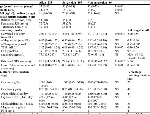
Treatment and follow-up
Calcium supplementation was used by 198 (70%) and active vitamin D formulations by 237 (84%)
(Table 4). About half (n=136, 48%) used either ergocalciferol (39%) or cholecalciferol (61%) of
whom 102 (75%) in combination with active formulations of vitamin D. Eleven used ergocalciferol as
the only vitamin D supplementation. Ten patients were treated with subcutaneous PTH injections or
delivery by a pump. There was no significant difference in types of medication used by post-surgical
and non-surgical patients, except treatment with PTH; nine of them were post-surgical. Median
albumin corrected serum calcium was below the reference range (2.08 mmol/L, reference range
2.20-2.55), whereas the median urine calcium value was slightly above the reference range (0.51
mmol/mmol creatinine, reference range 0.04-0.50) (Table 4). Post-surgical HP had significantly higher
albumin corrected serum calcium than non-surgical HP patients (P=0.005), whereas serum magnesium
and phosphate were similar. Eighteen percent had kidney failure (eGFR <60 ml/min). The median
eGFR was 80.8 (14.6-215.7) ml/min. Patients with post-surgical HP had significantly higher calcium
excretion (P=0.01) and lower eGFR (P=0.002) than non-surgical HP patients.
The non-surgical were younger than the post-surgical HP patients both at the time of diagnosis
(median 18 vs 40 years) and at the time of the study (median 45 vs 56 years). Overall, the median age
at diagnosis was 36 years (range 0-81). Most (70%) were diagnosed with HP within the first six
months from presentation of hypocalcemic symptoms, but 17% were diagnosed between two and five
10
with non-surgical HP were diagnosed late; 33% between two and five years and 18% more than fiveyears after symptom debut, as opposed to post-surgical HP (corresponding numbers were 11 and 6%,
respectively).
Most of the patients (64%) were diagnosed by an internist, endocrinologist or pediatrician, but 15%
were diagnosed by a general practitioner and 21% by others. A higher percentage of the post-surgical
patients were diagnosed by others (23%), primarily a surgeon, but also 14% of non-surgical patients
were diagnosed by non-internists, mostly neurologists.
The majority (82%) had their serum calcium levels assessed every six months or more frequently. A
higher percentage of patients in the surgical group than the non-surgical group reported that urine
calcium never had been measured (66% vs 43% respectively).
Quality of life and working ability
The SF-36 and HADS scores are given in Table 5 as mean ± SD compared with respective Norwegian
normative data (22, 23). HP patients had significantly lower SF-36 score than normative population in
all eight dimensions, and also lower than Norwegian Addison patients in six of eight dimension (PF,
RP, BP, VT, SF, MH) and Norwegian congenital adrenal hyperplasia patients in five of eight
dimensions (PF, VT, SF, RE, MH) (25, 26) (data not shown). Female patients scored worse than male
patients for PF (p=0.03) and VT (p=0.03), whereas patients with post-surgical HP scored worse than
non-surgical for PF (p=0.01), RP (p=0.001), BP (p=0.01) and VT (p=0.02).
HP patients displayed significantly higher symptom score for anxiety, depression and total HADS
score than normative Norwegian population. The post-surgical group scored worse on depression than
non-surgical (p=0.04). Thirty-eight percent had anxiety scores ≥ 8, and 26% had depression score ≥ 8,
11
No correlation between SF-36 or HADS scores (overall and subgroups) were found, neither withcorrected calcium levels nor serum magnesium levels. However, there was a positive correlation
between GH and serum magnesium (Pearson’s 0.3; p=0.03) in non-surgical patients.
Working ability
Forty percent had permanent or temporary social security benefits (SSB) (Table 4). Among the general
population in Norway aged 18 to 66 years the proportion of permanent SSB is about 10% and
temporary SSB about 4% (24).
Discussion
We found an overall prevalence of HP in Norway less than half the prevalence recently established in
Denmark (1-3) and USA (4). This difference mainly reflects fewer with post-surgical HP in our study.
Despite the lower overall prevalence in Norway, the prevalence of non-surgical HP was higher than in
Denmark (2). Non-surgical HP was most common in Western Norway, where ADH in a few large
families (27, 28) and APS-1 accounted for the difference. These differences could be genuine or due to
underdiagnosing in other regions. ADH, for instance, can present with only mild hypocalcemia and be
easily missed. Higher prevalence of idiopathic and pseudo-HP were found in the Norwegian cohort
than in studies in Japan (5), but the prevalence of pseudo-HP in Norway was similar to that recently
found in Denmark (3).
Not surprisingly, IFN- autoantibodies were detected in all the APS-1 patients, but also one
post-surgical patient had a high titer, which was ascribed to thymoma-associated myasthenia gravis, in
which IFN- autoantibodies have been found in 60% of the patients (29). In concordance with earlier
studies (9), NALP5 autoantibodies were detected in 50% of the patients with previously known
APS-1. One patient with a high titer of NALP5 autoantibodies, who had been diagnosed with idiopathic HP
39 years ago and in recent years also treated for autoimmune hypothyroidism, initially tested negative
for IFN- autoantibodies, but sequencing of AIRE eventually confirmed APS-1. A new sample taken
12
Despite thorough testing for underlying causes of idiopathic HP, about one third of non-surgicalpatients still have unknown cause. Concealed in this remarkable high fraction of idiopathic HP must
be hitherto unidentified forms of HP. The medical history and clinical vigilance can to some extent
guide the clinician to the underlying cause of HP, but in many cases the cause is far from obvious.
According to our results it seems reasonable to test for ADH, APS-1 and DiGeorge syndrome in cases
without findings indicating a specific diagnosis. Antibodies against IFN- and NALP5 are excellent
markers of APS-1 (9, 30, 31). General testing for antibodies against the CaSR among patients with
idiopathic HP does not seem justified based on our results.
We believe that systematic search for ADH among patients with idiopathic HP is important, since
these should receive treatment with calcium and vitamin D only if the disease is symptomatic. The
treatment itself can increase hypercalcuria and the risk of renal calcifications and renal failure more
than other forms of HP (6). Symptomatic patients should be treated, but only to alleviate symptoms,
not to restore normocalcemia, as low dosages of calcitriol results in less frequent renal calcifications
(6). Diagnosis of APS-1, DiGeorge or other syndromes is also of great importance, since other
components of these disorders needs to be diagnosed and treated early to avoid untimely morbidity
and mortality. Most of the patients in the Norwegian HP population received conventional calcium
and active vitamin D supplementation, which was associated with a high proportion of kidney failure,
indicating need for improvement of the therapy.
Our study corroborates earlier studies showing reduced HRQoL among HP patients (32-34), especially
among patients with post-surgical HP. One plausible explanation could be a higher proportion of
absolute PTH depletion or related to the cause of surgery (for instance Graves’ disease or thyroid
cancer). The cause of the overall reduced HRQoL is not obvious, as there is no correlation to low or
high calcium levels. Receptors for PTH are found in several tissues, including the central nervous
system and probably the adrenal cortex (35), and lack of PTH in tissues not related to calcium
homeostasis or bone metabolism is a plausible explanation for the reduced HRQoL. If so, PTH
13
in HRQoL with PTH therapy (32, 34, 36). Another study found no such improvement (33), but manypatients became hypercalcemic, which could be an explanation. It is reasonable to assume that the
uncertainty about effectiveness of PTH treatment is due to dose or delivery, which so far has not been
able to restore physiological calcium homeostasis properly. Our study revealed a higher percentage of
patients with clinical significant anxiety and depression compared to other disease groups in Norway
which have been studied using HADS (37, 38). Although not directly comparable, our results are in
concordance with the result from one study among 25 post-surgical patients (39).
The large sample size and the study design as a national study without major selection bias is the
greatest strength of this study. The added inclusion criterion with need of permanent treatment for HP
for more than one year for patients with post-surgical HP and HP due to DiGeorge syndrome ensured
that only patients with permanent HP were included. A limitation is that even though the overall
sample size is large, it constitutes a very heterogeneous group. The response rate of 55% in the patient
survey should ideally been higher, but the basic characteristics of the respondents and identified
patients were not significantly different; we therefore believe that this group is representative.
Furthermore, the response among the patients who comprise the largest subgroups of the cohort
(post-surgical, APS-1, ADH) were higher than for the patients within the smaller subgroups.
In conclusion, the prevalence of genetic, autoimmune and idiopathic HP in Norway is higher than
reported elsewhere, whereas the prevalence of post-surgical HP is lower than expected. Systematic
assessment of the underlying cause of HP is important to tailor the treatment, especially for patients
with ADH, APS-1, DiGeorge syndrome, and HDR. Still, a large proportion of the patients have
unknown cause despite systematic investigation. Despite conventional calcium, magnesium, and
vitamin D supplementation complications such as kidney failure and reduced HRQoL is common,
indicating need for improvement of the therapy.
14
The study was supported by The Regional Health Authorities in Western Norway and the NorwegianMinistry of Health.
Acknowledgements
We are very grateful to the participating patients for their cooperation. Mrs. Elisabeth Halvorsen and
15
References1. Underbjerg L, Sikjaer T, Mosekilde L, Rejnmark L. Cardiovascular and renal
complications to postsurgical hypoparathyroidism: a Danish nationwide controlled historic follow-up study. J Bone Miner Res. 2013;28:2277-2285
2. Underbjerg L, Sikjaer T, Mosekilde L, Rejnmark L. The Epidemiology of Nonsurgical
Hypoparathyroidism in Denmark: A Nationwide Case Finding Study. J Bone Miner Res. 2015;30:1738-1744
3. Underbjerg L, Sikjaer T, Mosekilde L, Rejnmark L. Pseudohypoparathyroidism -
epidemiology, mortality and risk of complications. Clin Endocrinol (Oxf). 2015;
4. Powers J, Joy K, Ruscio A, Lagast H. Prevalence and incidence of hypoparathyroidism in
the United States using a large claims database. J Bone Miner Res. 2013;28:2570-2576 5. Nakamura Y, Matsumoto T, Tamakoshi A, Kawamura T, Seino Y, Kasuga M,
Yanagawa H, Ohno Y. Prevalence of idiopathic hypoparathyroidism and
pseudohypoparathyroidism in Japan. Journal of epidemiology / Japan Epidemiological Association. 2000;10:29-33
6. Raue F, Pichl J, Dorr HG, Schnabel D, Heidemann P, Hammersen G, Jaursch-Hancke C, Santen R, Schofl C, Wabitsch M, Haag C, Schulze E, Frank-Raue K. Activating
mutations in the calcium-sensing receptor: genetic and clinical spectrum in 25 patients with autosomal dominant hypocalcaemia - a German survey. Clin Endocrinol (Oxf). 2011;75:760-765
7. Orlova EM, Bukina AM, Kuznetsova ES, Kareva MA, Zakharova EU, Peterkova VA, Dedov, II. Autoimmune polyglandular syndrome type 1 in Russian patients: clinical variants
and autoimmune regulator mutations. Hormone research in paediatrics. 2010;73:449-457 8. Wolff AS, Erichsen MM, Meager A, Magitta NF, Myhre AG, Bollerslev J, Fougner KJ,
Lima K, Knappskog PM, Husebye ES. Autoimmune polyendocrine syndrome type 1 in
Norway: phenotypic variation, autoantibodies, and novel mutations in the autoimmune regulator gene. J Clin Endocrinol Metab. 2007;92:595-603
9. Alimohammadi M, Bjorklund P, Hallgren A, Pontynen N, Szinnai G, Shikama N, Keller MP, Ekwall O, Kinkel SA, Husebye ES, Gustafsson J, Rorsman F, Peltonen L, Betterle C, Perheentupa J, Akerstrom G, Westin G, Scott HS, Hollander GA, Kampe O.
Autoimmune polyendocrine syndrome type 1 and NALP5, a parathyroid autoantigen. The New England journal of medicine. 2008;358:1018-1028
10. Meager A, Visvalingam K, Peterson P, Moll K, Murumagi A, Krohn K, Eskelin P, Perheentupa J, Husebye E, Kadota Y, Willcox N. Anti-interferon autoantibodies in
autoimmune polyendocrinopathy syndrome type 1. PLoS medicine. 2006;3:e289
11. Blizzard RM, Chee D, Davis W. The incidence of parathyroid and other antibodies in the
sera of patients with idiopathic hypoparathyroidism. Clinical and experimental immunology. 1966;1:119-128
12. Kifor O, McElduff A, LeBoff MS, Moore FD, Jr., Butters R, Gao P, Cantor TL, Kifor I, Brown EM. Activating antibodies to the calcium-sensing receptor in two patients with
autoimmune hypoparathyroidism. J Clin Endocrinol Metab. 2004;89:548-556 13. Kobrynski LJ, Sullivan KE. Velocardiofacial syndrome, DiGeorge syndrome: the
chromosome 22q11.2 deletion syndromes. Lancet. 2007;370:1443-1452
14. Oskarsdottir S, Persson C, Eriksson BO, Fasth A. Presenting phenotype in 100 children
with the 22q11 deletion syndrome. Eur J Pediatr. 2005;164:146-153
15. Lima K, Abrahamsen TG, Wolff AB, Husebye E, Alimohammadi M, Kampe O, Folling I. Hypoparathyroidism and autoimmunity in the 22q11.2 deletion syndrome. Eur J
Endocrinol. 2011;165:345-352
16. Lima K, Folling I, Eiklid KL, Natvig S, Abrahamsen TG. Age-dependent clinical problems
16
17. Bollerslev J, Rejnmark L, Marcocci C, Shoback DM, Sitges-Serra A, van Biesen W,Dekkers OM, European Society of E. European Society of Endocrinology Clinical
Guideline: Treatment of chronic hypoparathyroidism in adults. Eur J Endocrinol. 2015;173:G1-20
18. Mitchell DM, Regan S, Cooley MR, Lauter KB, Vrla MC, Becker CB, Burnett-Bowie SA, Mannstadt M. Long-term follow-up of patients with hypoparathyroidism. J Clin
Endocrinol Metab. 2012;97:4507-4514
19. Bilezikian JP, Khan A, Potts JT, Jr., Brandi ML, Clarke BL, Shoback D, Juppner H, D'Amour P, Fox J, Rejnmark L, Mosekilde L, Rubin MR, Dempster D, Gafni R, Collins MT, Sliney J, Sanders J. Hypoparathyroidism in the adult: epidemiology, diagnosis,
pathophysiology, target-organ involvement, treatment, and challenges for future research. J Bone Miner Res. 2011;26:2317-2337
20. Oftedal BE, Wolff AS, Bratland E, Kampe O, Perheentupa J, Myhre AG, Meager A, Purushothaman R, Ten S, Husebye ES. Radioimmunoassay for autoantibodies against
interferon omega; its use in the diagnosis of autoimmune polyendocrine syndrome type I. Clinical immunology. 2008;129:163-169
21. Kemp EH, Habibullah M, Kluger N, Ranki A, Sandhu HK, Krohn KJ, Weetman AP.
Prevalence and clinical associations of calcium-sensing receptor and NALP5 autoantibodies in Finnish APECED patients. J Clin Endocrinol Metab. 2014;99:1064-1071
22. Loge JH, Kaasa S. Short form 36 (SF-36) health survey: normative data from the general
Norwegian population. Scand J Soc Med. 1998;26:250-258
23. NTNU HUNT Research Centre HUNT databank. In.
https://hunt-db.medisin.ntnu.no/hunt-db
24. Statistics Norway 2015 Population statistics. In. http://www.ssb.no/en/forside
25. Lovas K, Loge JH, Husebye ES. Subjective health status in Norwegian patients with
Addison's disease. Clin Endocrinol (Oxf). 2002;56:581-588
26. Nermoen I, Husebye ES, Svartberg J, Lovas K. Subjective health status in men and women
with congenital adrenal hyperplasia: a population-based survey in Norway. Eur J Endocrinol. 2010;163:453-459
27. Lovlie R, Eiken HG, Sorheim JI, Boman H. The Ca(2+)-sensing receptor gene (PCAR1)
mutation T151M in isolated autosomal dominant hypoparathyroidism. Human genetics. 1996;98:129-133
28. Sorheim JI, Husebye ES, Nedrebo BG, Svarstad E, Lind J, Boman H, Lovas K.
Phenotypic variation in a large family with autosomal dominant hypocalcaemia. Hormone research in paediatrics. 2010;74:399-405
29. Meager A, Wadhwa M, Dilger P, Bird C, Thorpe R, Newsom-Davis J, Willcox N.
Anti-cytokine autoantibodies in autoimmunity: preponderance of neutralizing autoantibodies against interferon-alpha, interferon-omega and interleukin-12 in patients with thymoma and/or myasthenia gravis. Clinical and experimental immunology. 2003;132:128-136
30. Tomar N, Kaushal E, Das M, Gupta N, Betterle C, Goswami R. Prevalence and
significance of NALP5 autoantibodies in patients with idiopathic hypoparathyroidism. J Clin Endocrinol Metab. 2012;97:1219-1226
31. Cervato S, Morlin L, Albergoni MP, Masiero S, Greggio N, Meossi C, Chen S, del Pilar Larosa M, Furmaniak J, Rees Smith B, Alimohammadi M, Kampe O, Valenzise M, Betterle C. AIRE gene mutations and autoantibodies to interferon omega in patients with
chronic hypoparathyroidism without APECED. Clin Endocrinol (Oxf). 2010;73:630-636 32. Cusano NE, Rubin MR, McMahon DJ, Irani D, Tulley A, Sliney J, Jr., Bilezikian JP. The
effect of PTH(1-84) on quality of life in hypoparathyroidism. J Clin Endocrinol Metab. 2013;98:2356-2361
33. Sikjaer T, Rolighed L, Hess A, Fuglsang-Frederiksen A, Mosekilde L, Rejnmark L.
Effects of PTH(1-84) therapy on muscle function and quality of life in hypoparathyroidism: results from a randomized controlled trial. Osteoporos Int. 2014;25:1717-1726
17
Hypoparathyroidism: A Prospective, Open-Label Investigation of Efficacy and Quality of Life. J Clin Endocrinol Metab. 2015;100:3590-359735. Mazzocchi G, Aragona F, Malendowicz LK, Nussdorfer GG. PTH and PTH-related
peptide enhance steroid secretion from human adrenocortical cells. American journal of physiology. Endocrinology and metabolism. 2001;280:E209-213
36. Cusano NE, Rubin MR, McMahon DJ, Irani D, Anderson L, Levy E, Bilezikian JP.
PTH(1-84) is associated with improved quality of life in hypoparathyroidism through 5 years of therapy. J Clin Endocrinol Metab. 2014;99:3694-3699
37. Engum A, Bjoro T, Mykletun A, Dahl AA. An association between depression, anxiety and
thyroid function--a clinical fact or an artefact? Acta Psychiatr Scand. 2002;106:27-34 38. Felde G, Bjelland I, Hunskaar S. Anxiety and depression associated with incontinence in
middle-aged women: a large Norwegian cross-sectional study. International urogynecology journal. 2012;23:299-306
39. Arlt W, Fremerey C, Callies F, Reincke M, Schneider P, Timmermann W, Allolio B.
Well-being, mood and calcium homeostasis in patients with hypoparathyroidism receiving standard treatment with calcium and vitamin D. Eur J Endocrinol. 2002;146:215-222 40. Hannan FM, Nesbit MA, Zhang C, Cranston T, Curley AJ, Harding B, Fratter C, Rust
N, Christie PT, Turner JJ, Lemos MC, Bowl MR, Bouillon R, Brain C, Bridges N, Burren C, Connell JM, Jung H, Marks E, McCredie D, Mughal Z, Rodda C, Tollefsen S, Brown EM, Yang JJ, Thakker RV. Identification of 70 calcium-sensing receptor mutations
in hyper- and hypo-calcaemic patients: evidence for clustering of extracellular domain mutations at calcium-binding sites. Hum Mol Genet. 2012;21:2768-2778
41. Mannstadt M, Bertrand G, Muresan M, Weryha G, Leheup B, Pulusani SR, Grandchamp B, Juppner H, Silve C. Dominant-negative GCMB mutations cause an
autosomal dominant form of hypoparathyroidism. J Clin Endocrinol Metab. 2008;93:3568-3576
42. Upadhyay J, Steenkamp DW, Milunsky JM. The syndrome of hypoparathyroidism,
deafness, and renal anomalies. Endocrine practice : official journal of the American College of Endocrinology and the American Association of Clinical Endocrinologists. 2013;19:1035-1042
43. Parvari R, Hershkovitz E, Grossman N, Gorodischer R, Loeys B, Zecic A, Mortier G, Gregory S, Sharony R, Kambouris M, Sakati N, Meyer BF, Al Aqeel AI, Al Humaidan AK, Al Zanhrani F, Al Swaid A, Al Othman J, Diaz GA, Weiner R, Khan KT, Gordon R, Gelb BD, Consortium HRARK-CS. Mutation of TBCE causes
hypoparathyroidism-retardation-dysmorphism and autosomal recessive Kenny-Caffey syndrome. Nat Genet. 2002;32:448-452
44. Unger S, Gorna MW, Le Bechec A, Do Vale-Pereira S, Bedeschi MF, Geiberger S, Grigelioniene G, Horemuzova E, Lalatta F, Lausch E, Magnani C, Nampoothiri S, Nishimura G, Petrella D, Rojas-Ringeling F, Utsunomiya A, Zabel B, Pradervand S, Harshman K, Campos-Xavier B, Bonafe L, Furga G, Stevenson B, Superti-Furga A. FAM111A mutations result in hypoparathyroidism and impaired skeletal
development. Am J Hum Genet. 2013;92:990-995
45. Albaramki J, Akl K, Al-Muhtaseb A, Al-Shboul M, Mahmoud T, El-Khateeb M, Hamamy H. Sanjad Sakati syndrome: a case series from Jordan. Eastern Mediterranean
health journal = La revue de sante de la Mediterranee orientale = al-Majallah al-sihhiyah li-sharq al-mutawassit. 2012;18:527-531
46. Misceo D, Holmgren A, Louch WE, Holme PA, Mizobuchi M, Morales RJ, De Paula AM, Stray-Pedersen A, Lyle R, Dalhus B, Christensen G, Stormorken H, Tjonnfjord GE, Frengen E. A dominant STIM1 mutation causes Stormorken syndrome. Human
mutation. 2014;35:556-564
47. Lemos MC, Thakker RV. GNAS mutations in Pseudohypoparathyroidism type 1a and
18
Table 1. Causes of hypoparathyroidism
Cause Gene (when indicated) Reference
Postsurgical and/or following radioactive iodine thyroid ablation Autoimmune
Isolated
Component of APS-1 AIRE/21q22.3 (7, 8)
Genetic
Isolated
Activating CaSR-mutations CaSR/3q21.1 (6, 40)
PTH- mutations PTH/11p15
GCMB-mutations GCMB/6p24.2 (41)
X-linked recessive SOX3/Xq26-27
As part of syndromes
DiGeorge (22q11.2-deletion syndrome) TBX1/22q11 (13, 14)
HDR-syndrome GATA3/10p13-14 (42)
Hypoparathyroidism-retardation-dysmorphism syndrom (Sanjad-Sakati syndrome) and Kenny-Caffey syndrome
TBCE/1q42.3, FAM111A/11q12.1*
(43-45)
Mitochondrial associated (Kearns-Sayre and others)
Stormorken’s syndrome STIM1/11p15.4 (46)
Target organ resistance
Pseudohypopara type 1 and 2 GNAS,STX/20q13.3(type) (47)
Blomstrand chondrodysplasia PTHR1/3p22-p21.1
Hypomagnesemia TRPM6/9q21.13**
Vitamin D dependent rickets VDR/12q13.11 (type 2a)
Idiopathic Miscellaneous
Infiltrative disorders (Hemochromatosis, Thalassemia, Wilsons disease, metastasis)
*Kenny-Caffey syndrome type 2.
**Hypomagnesemia due to TRPM6 mutations is typically accompanied by secondary hypocalcemia, but severe hypomagnesemia of any cause can give target organ resistance.
19
Table 2. Prevalence and cause of HP in the health regionsamong living patients (n=511)
All RHA South-Eastern
RHA
Western RHA
Central RHA
Northern RHA
Inhabitants 4 985 870 2 785 259 1 041 886 687 968 470 757 HP n (prev/100000) 511 (10.2) 256 (9.2) 117 (11.2) 72 (10.5) 66 (14.0) Post-surgical n (prev/100 000) 321 (6.4) 183 (6.6) 47 (4.5) 51 (7.4) 40 (8.5) Non-surgical n (prev/100 000) 190 (3.8) 73 (2.6) 70 (6.7) 21 (3.1) 26 (5.5) Idiopathic 64 (1.3) 31 (1.1) 14 (1.3) 8 (1.2) 11 (2.3)
APS-1 25 (0.5) 9 (0.3) 9 (0.9) 3 (0.4) 4 (0.8)
ADH 31 (0.6) 4 (0.1) 26 (2.5) 1 (0.1) 0
HDR 4 (0.08) 1 (0.04) 3 (0.3) 0 0
DiGeorge 23 (0.5) 8 (0.3) 7 (0.7) 5 (0.7) 3 (0.6) PseudoHP 41 (0.82) 19 (0.7) 10 (1.0) 4 (0.6) 8 (1.7)
Miscellaneous 2 1 1 0 0
20
Table 3 Patients identified and survey respondents, n (%)
Identified HP n=522 Respondents n=283
Age, years (range) 51 (4-91) 53 (9-89)
Female 381 (73%) 212 (75%)
Post-surgical 329 (63%) 197 (70%)
Non-surgical 193 (37%) 86 (30%)
Idiopathic 67 26
APS-1 25 15
ADH 31 18
HDR 4 1
DiGeorge 23 8
Pseudo-HP 41 16
Other causes 2* 2*
APS-1: Autoimmune polyendocrine syndrome type 1, ADH: Autosomal dominant hypocalcemia, HDR:Hypoparathyroidism, deafness and renal syndrome.
21
Table 4 Basal characteristics, laboratory results and treatment among respondents (median- range)
All, n=283 Surgical, n=197 Non-surgical, n=86
Age (years), median (range) 53 (9-89) 56 (20-89) 45 (9-72) P<0.001
Female n (%) 212 (75) 161 (82) 51 (59) P<0.001
BMI (kg/m2), median (range) 25 (14-60) 25 (15-60) 25 (14-47) NS
Social security benefits (SSB)
Retirement pension, n (%) 51 (18) 46 (23) 5 (6)
Permanent SSB, n (%) 72 (25) 53 (27) 19 (22)
Temporary SSB, n (%) 42 (15) 33 (17) 9 (11)
Laboratory results Ref.range/cut off
Corrected s-calcium (mmol/L)
2.08 (1.47-2.84) 2.09 (1.61-2.69) 2.01 (1.47-2.84) P=0.005 2.20-2.55
s-Magnesium (mmol/L) 0.83 (0.64-1.22) 0.83 (0.64-1.22) 0.82 (0.65-1.10) NS 0.71-0.94 s-Phosphorus (mmol/L) 1.29 (0.76-2.55) 1.29 (0.77-2.55) 1.33 (0.76-2.27) NS 0.75-1.50* TSH (mIE/L) 1.22 (0.01-14.20) 0.69 (0.01-14.20) 1.87 (0.01-6.86) P=0.05 0.40-4.50 FT4 (pmol/L) 19.3 (9.3-39.6) 20.7 (11.0-39.6) 16.4 (9.3-24.2) NS 9.5-22.0 Creatinine (mol/L) 75.0 (33-247) 76.0 (34-247) 73.0 (33-168) NS Male: 60-105
Female: 45-90 Estimated GFR (ml/min) 80.8 (14.6-215.7) 78.6 (14.6-181.5.) 93.8 (30.4-215.7) P=0.002 > 90
Urine Calcium (mmol/mmol creatinine)
0.51 (0.02-2.29) 0.53 (0.02-1.91) 0.43 (0.02-2.29) P=0.01 0.04-0.50
Treatment, dose median (range)
Percentage
receiving treatment (all)
Calcium mg/day 1000 (167-10000)
1000 (167-10000) 1000 (250-4000) NS 70
Calcitriol µg/day 0.75 (0.13-4.00) 0.75 ((0.13-4.00) 0.61 (0.25-2.00) NS 40 Alphacalcidiol µg/day 1.50 (0.25-6.00) 1.50 (0.25-6.00) 1.50 (0.40-5.00) NS 44 Ergocalciferol (D2) U/day 4286 (1286-210
000)
4286 (1286-150 000)
4286 (2143-210 000)
NS 19
Cholecalciferol (D3) U/day 800 (200-8000) 800 (200-8000) 800 (400-2400) NS 29 Magnesium mg/day 300 (120-1200) 300 (120-1200) 300 (120-1200) NS 34
PTH use, n 10 9 1 4
22
Table 5. SF-36 and HADS score, mean (±SD) of the HP population and subgroups compared to Norwegian normative data
SF-36 HADS
n PF RP BP GH VT SF RE MH n Anxiety Depression Total
HADS-score Overall
HP 283 74.2 (24.6) 44.9 (43.8) 58.1 (26.9) 50.7 (27.2) 42.2 (22.9) 68.5 (27.3) 65.1 (42.5) 70.5 (19.5) 283 6.5 (4.4) 4.8 (4.1) 11.4 (7.7)
Normative 2311 87.2 (18.7) 77.9 (35.8) 75.1 (26.0) 76.8 (22.0) 60.0 (20.8) 85.5 (22.2) 81.6 (32.4) 78.8 (16.5) 58784 4.2 (3.3) 3.4 (3.0) 7.5 (5.5)
Females
HP 212 72.4 (25.1) 43.1 (43.6) 56.4 (26.5) 49.3 (27.3) 40.4 (23.0) 67.6 (26.9) 66.3 (42.2) 70.0 (19.1) 212 6.7 (4.3) 4.8 (4.0) 11.5 (7.6)
Normative 1184 84.8 (20.8) 75.4 (37.7) 73.0 (26.6) 76.3 (22.5) 56.9 (21.2) 83.7 (23.1) 79.1 (34.6) 77.6 (17.0)
Males
HP 71 79.6 (22.0) 50.4 (44.3) 63.0 (27.5) 54.8 (26.6) 47.2 (22.2) 71.2 (28.5) 61.3 (43.4) 72.3 (20.6) 71 5.9 (4.5) 5.0 (4.7) 10.9 (8.2)
Normative 1085 89.8 (15.5) 80.5 (33.6) 77.2 (25.0) 77.4 (21.3) 63.2 (19.9) 87.6 (20.9) 84.5 (29.7) 80.0 (15.8)
HP Subgroups
Surgical 197 72.2 (24.4) 39.2 (43.1) 55.3 (26.0) 48.7 (27.1) 40.0 (22.6) 67.4 (27.4) 63.9 (42.8) 70.2 (19.0) 197 6.6 (4.3) 5.2 (4.0) 11.8 (7.7)
Non-surgical 86 78.7 (24.5) 58.6 (42.6) 64.5 (27.9) 55.4 (27.0) 47.1 (23.0) 71.0 (26.9) 67.9 (41.8) 71.1 (20.9) 86 6.4 (4.4) 4.1 (4.3) 10.9 (8.2)