1988
Thymic structure and function in aging dogs
Belinda Lawler GoffIowa State University
Follow this and additional works at:https://lib.dr.iastate.edu/rtd
Part of theAllergy and Immunology Commons,Immunology and Infectious Disease Commons, and theMedical Immunology Commons
This Dissertation is brought to you for free and open access by the Iowa State University Capstones, Theses and Dissertations at Iowa State University Digital Repository. It has been accepted for inclusion in Retrospective Theses and Dissertations by an authorized administrator of Iowa State University Digital Repository. For more information, please [email protected].
Recommended Citation
Goff, Belinda Lawler, "Thymic structure and function in aging dogs " (1988).Retrospective Theses and Dissertations. 11182.
lëSi
JV'"' 'L'(' ;,„ s
ÎW
' ; A',
'4'f y^./ -''iLL/LSlfjl
11 r
l''i
mvM , \i I "i ^
n
5*
' i'S:,I:M I
'f'
'.4 ('/' V, /'"Y
The most advanced technology has been used to photograph and reproduce this manuscript from the microfilm master. UMI films the text directly from the original or copy submitted. Thus, some thesis and dissertation copies are in typewriter face, while others may be from any type of computer printer.
The quality of this reproduction is dependent upon the quality of the copy submitted. Broken or indistinct print, colored or poor quality illustrations and photographs, print bleedthrough, substandard margins, and improper alignment can adversely affect reproduction.
In the unlikely event that the author did not send UMI a complete manuscript and there are missing pages, these will be noted. Also, if unauthorized copyright material had to be removed, a note will indicate the deletion.
Oversize materials (e.g., maps, drawings, charts) are reproduced by sectioning the original, beginning at the upper left-hand corner and continuing from left to right in photographed in one exposure and is included in reduced form at the back of the book.
Photographs included in the original manuscript have been reproduced xerographically in this copy. Higher quality 6" x 9" black and white photographic prints are available for any photographs or illustrations appearing in this copy for an additional charge. Contact UMI directly to order.
University Microfilms International A Bell & Howell Information Company 300 North Zeeb Road, Ann Arbor, fvtl 48106-1346 USA
Thymic structure and Ainction in aging dogs
GofF, Belinda Sue Lawler, Ph.D.
Iowa State University, 1988
in aging dogs
ty
Belinda lawler Goff
A Dissertation Submitted to the
Graduate Faculty in Partial Fulfillment of the Requirements for the Degree of
DOCTOR OF mUCGOPHY
Interdepartmental program: Immunobiology Major: Immunobiology
i^jproved:
[e of Major Work
Chair, y^iaiMfetoiology Program
For''t@e(;6#SBuate College
Iowa State University Ames, Iowa
1988
Signature was redacted for privacy.
Signature was redacted for privacy.
TABLE OF COMmnS
Page
GENERAL DflRODUCTICN 1
EXPLANATION OF IHE DISSERTATION FORMAT 3
SECTION I: THÏMJinOFHIC AGENTS 4
imnoDucnoN 6
PnUHARÏ - THÏMUS IKEERACnONS 10
THYROID - THYMUS INIERACTIONS 17
GONAD - THYMUS INTERACTIONS 20
ADRENAL - THYMUS mCERACTIONS 24
SUMMARY 28
SECTION II: GRCWIH HOMCNE TREATMENT STIMULATES 30 THYMUUN PRODUCTION IN AGED DOGS
INTRODUCTION 32
MATERIALS AND METHODS 35
RESULTS 38
DISCUSSION 48
SECTION III: THE EFFECT OF ORAL CL£NIDINE ON GROWIH 52 HORMONE RELEASE, THYMIC STRUCTURE AND
IMMUNE FUNCTECN IN AGING DOGS
INTRODUCTION 54
MATERIALS AND METHODS 56
RESULTS 62
DISCUSSIΠ80
SECTION IV: THYMIC CYSTS IN AGING DOGS: LIGHT AND 84 ELECTRON MICRDSODFIC ANATCMY,
HISTOCHEMISTRY, AND DMJNOCYTOCHEMISTRY
INTRODUCTION 86
MATERIALS AND METHODS 89
RESULTS 92
DISCUSSION 111
SUMMARY AND CCNCUUSIONS 117
LITERATURE CITED 119
GENERAL TmSCOXTIŒ
Advancing age is associated with involution of the thymus, decreased thymic endocrine function, and depressed imnune function (DelafUente, 1985; Sdxuurman et al., 1985). Increased incidence of certain 'diseases of aging' are edso noted (Schultz, 1984; Monroe and Roth, 1986). An agent
or agents that would stimulate the immune system in the aged might also be associated with better health as assessed clinically.
Various hormones have been observed to enhance thymic morphology and immune function. A review of the literature regarding various
thymotrephic agents is presented in Section I. One such thymotrcphic agent is growth hormone (GH). Hie results of a stu^ on the effects of bovine GH on thymic structure and endocrine function in aging dogs is presented in Section II.
Since exogenous GH may be recognized as a foreign protein ty dogs, long term administration may lead to the production of antibodies against it, negating its beneficial effects. Previous results in our laboratory indicate that oral clonidine HCl stimulates the release of endogenous GH in aging dogs. However, dedly administration was associated with a loss
of the GH response to clonidine. The effect of intermittent
administration of oral clonidine on GH release, thymic structure and immune function in aging dogs is presented in Section III.
direc± evldenoe of the pcesenoe of at least t(«o thymic homones within the
EXPLANATICN OF THE DISSERimCN POBMAT
An alternative format was used in this dissertation. There are four sections eifter the General Introduction; each of the sections is an
individual ]papeac. Ihe first section is a review of the literature vAiich will be submitted for publication, the second section has been published in Clinical and E)^)er±anent5d Innunology, the third section will be
IHÏMJLnOFHZC ASENTS
Belinda lawler Goff
D^jartment of Veterinary Microbiology and Preventive Medicine, Iowa
nmmjcncN
The thymus gland is an organ located in the anterior mediastinum, and it plays an integral role in developnent and maintenance of cellular
imunity (Symners, 1966; Weksler, 1982). The thymus gland from a young animal is characterized histologically by an abundance of lymphocytes,
e^iecially in the cortical region, a distinct corticcnedullaiy junction, and the absence of adipose tissue either in the thin interlobular s^ïtae or in the parenchyma (Figure la). Age-related involution of the thymus, vAiich begins at about the time of puberty, is associated with massive loss of lymphocytes, especially from the cortex (Figure lb). Biis lymphocyte loss leads to an indistinct corticoroedullary junction and a decrease in
the cortex to medullary ratio, mterlcbular sqxtae become wider as thymic lobules reduce in size and separate &an each other. The amount of
adipose tissue progressively Increases within the sqitae and parenchyma. In some species involution is acocnpanied by the e^iparent hypertrophy of the thymiic epithelium, with some formiing cysts of a secretory nature (Goff, 1988). Concurrent with tbymiic morphological involution is a
decline in thymiic endocrine function, as assessed by the waning secretion
of various thymiic hormones (Goldstein et eQ.., 1974; Bach et al., 1975; Lewis et éd., 1978).
In general, the normal function of the immune system declines with age, with T-cell mediated immunity being e^seciedly affected (Hirdkawa and Makinodan, 1975; Weksler et al., 1978; Delafuente, 1985). Althou^
a, Ihymus from a dog a^^woodmately 4 months old. Note the distinct oortioonedullary junction (j), thin interlobular septae (s), and the absence of adipose tissue. The cortex (c) to medullary (m) ratio is 1:1 or greater and there are numerous lymphocytes present in the cortex.
[image:16.614.126.517.317.485.2]organ atrophy, generalized jmnunological deficiencies, and a progressive wasting dimpmmm leading to early death (Mstcalf, 1965; Miller, 1965; Taylor, 1965). The effects of adult thymectomy are delayed since an adequate pool of competent cells remain. However, eventually a striking dqAetion of cells in the secondary lymphoid organs and a defective innune response to newly enoountered antigens beormes e^pparent (Metcalf, 1965; Miller, 1965; Taylor, 1965). Thymic atrophy may also occur precociously
in the face of physiologies^. imbeOanoe, stress, or disease.
Ibe changes in thymic morphology and in some parameters of immune function that occur with age and in precocious involution are reversible. Several factors have been observed to regenerate or enhance thymus
EnXJTDffiY - THYMUS BnERACTIONS
The thymotrophic effect of the pituitary hormones growth hormone (GH)
and prolactin (ERL) has been frequently reported. Ihere is some sequence homology between these two adenohypopbyseal homcnes, and pituitary
derived preparations of one may be contaminated to some extent with the other. Still, a preponderance of the evidence (as follows) indicates that
GH has a more important role in affecting thymic morphology than does PRL. Kelley et al. (1986, 1987) have observed thymic regeneration and enhancement of some parameters of isnune function in aging female
Wistar-Rxtth rats implanted with Œg pituitary adenomas. The GH3 tumor secretes both GH and IKL at a ratio of approximately 70:30; GH3 implanted rats have a 20 to 100 times greater GH concentration and a 10 times greater PRL concentration in their plasma (Boodkfor et al., 1985; Kelley et al.,
1987). a\ro months after GH3 tumor implantation, thymic morphology similar to 3 month old controls was observed in 18 month old rats, lAile 24 month old rats showed only partial thymic regeneration (Kelley et al., 1986).
Similarly, the ^lenic lymphocyte reqxanse to the phytonitogens
phytohemagglutinin (HA) and Ooncanavalin A (Oon A) was restored to values comparable to 3 month old controls in GH3 implanted 18 mcnth old rats (Kelley et al., 1986). m GH3 implanted 24 mcnth old rats the mdtogenic
response was significantly increased as compared to age matched controls, but remedned about 90% less than the reqionse observed in the young
moderately hi^ier proportion of lymphocytes with the Thy-1.1 and % (helper) phenotypes as detected by flow microfluor±netry (KsUey et ed., 1986). It was concluded that GH and/or ERL from the GH3 pituitary
adenomas augmented thymic size and reconstituted mitogenic re^xxises in aged rats (Kelley et eil., 1986).
m attenpting to sort out the GH from the ERL effect on tl^mic
morphology and jjmune function Davila et ed. (1987) administered ovine GH (oGH) to 26 month old, female Fischer 344 (F344) rats, twice daily for five weeks. The oGH treatment was associated with enhancement of
aplenocyte proliferation to mitogens and natural killer cell (NK) activity at lew target to effector ratios. Thymic morphology at necropey and IL-2 synthesis were not significantly different from age-matched,
saline-injected controls; hcwever, thymic morphology in the control rats did not shew the expected signs of age-associated atrophy that would negate any thpnotrophic effect of GH vdien structural comparisons were made. It was concluded that VBL, in addition to GH (as from GH3 cells), may be
necessary for enhanced thymic structure and seme parameters of immune
function in aging rats; undefined factors secreted by GH3 pituitary cells, the discontinuous (daily) injection regimen and the stress associated with it may have been confounding factors éiffecting the results of these
studies. To overcome some of these problems, an experimental design in vMch groups of rats received either GH, HRL, both GH and ERL, GH3 tumors, or sedine all within the framework of a single ea^ieriment could be
attempted.
administered bovine GH (bGH) to five dogs beWeen the ages of
apppoadinately 2.5 - 5 years, with five additional dogs serving as controls and receiving bovine serum albumin (BSA). The regimen of bGH injection in this study was 0.1 isg/kg dally for five doses, then every other day for five doses, then every third day for four doses for a total of 14 doses in
27 days. Ihoracotamy 10 days to three weeks before bGH treatment was begun revealed moderate to severely atrophied thymic morphology in all dogs. At necropsy, all five bQl treated dogs had regenerated thymic morphology. Ocntrol dogs, treated with BSA, had not regenerated as consistently or as markedly as bGH treated dogs although two of the five controls did have regenerated thymus glands. The toted peripheral vAilte
blood cell count, lymphocyte count, and serum concentration of thymosin alpha 1 did not change with bGH treatment; the response of peripheral lymphocytes to mitogens did not change with treatment, thou^i results were difficult to interpret due to assay variability and variation in re^xxise between dogs. Considering the great variability in lymphocyte ftmctlcai and the possible confounding factors of urikncun age and medical history, the surgical biopey procedure and acocapanying anesthesia, Mcairoe et al.,
(1987) could only conclude that bGH treatment contributed to thymic regeneration in these aging dogs, but may not have been the only factor
involved.
m a similar, but more controlled, stuc^ using retired breeder,
female beagle dogs of kncun age and medlced history, Goff et al. (1987) found enhanced thymic morphology and function eifter bGH treatment.
Pre-txeatxnent thymic biopsies were not performed. Stereologiced.
assessment of thymic morphology revealed regeneration in four of five bGH-treated middle-aged dogs and in one of five BSA-^bGH-treated controls at
necrop^; no inproviement in thymus morphology was observed in any of the old-aged dogs in this study. More frequent administration of bGH to a group of old-aged dogs was not associated with improvement of thymic
morphology. However, all bGH-treated dogs, regardless of age or the state of thymus morphology at necrop^, had significantly elevated plasma
concentrations of thymulin as compared to the BSA-^treated, age-matched
controls; the elevated thymulin concentrations were comparable to those of 4 month old dogs. It was concluded that exogenous GH may be useful for restoration of thymus structure and some immune functions in aging individuals (Goff et al., 1987). It is important to note that
immunoenhanoement (as measured by thymus endocrine function) did not require regeneration of thymus morphology.
Some of the earliest e)q)eriments indicating the relationship between
pituitary hormones, thymus structure and innune function were performed on mutant mice. The Snell-Bagg and Ames pituitary dwarf mouse strains have a shortened life ^lan and are deficient in GH, FRL, thyroid stimulating
hormone (TSH), and adrenocarticotrophic hormone (ACIH), and develop a wasting syndrome (Fabris et e&l., 1971; Monroe and Both, 1986). Concurrent with hypopituitary function is a deficient immune system, eq^ecially
jjDÇiroved euitilxx^ re^xxise to th^nus-dependent antigens, and enhanced tbymlin production (van Buul-Offers and Van den Ecande, 1981; RLerpaoli
et al., 1969; Baroni et al., 1969). If the dwarf mice were thynectonized before CH an^/or thyroxine ther%y the effects of immune system
restoration, prolonged life ^)an, and reversal of the wasting disease did not occur (Fabris et al., 1971; Fahris et al,, 1972; Sorkin et al., 1972). These results tphold the relationship between pituitary function, tliymus structure and immune function and implicate their malfunction eis a cause
of the inmunodeficiency and shortened life q>an in dwarf mice.
Innunodeficient dwarfism in an inbred colony of 7/8 Weimaraner^l/S German Shepherd dogs was associated with a wasting syndrome at weaning, severe th^c hypoplasia, decreased peripheral lymphocyte re^xanse to HA, and significant depression of GH release in reqxxise to clonidine (Roth et al., 1980). Treatment with bGH resulted in clinical improvement and
regeneration of thymic morphology as ccnpared to pretreatment biopsies, but was not associated with a significant increase in lymphocyte
blastogenesls (vAlch was hi^ily variable) or thymosin alpha 1
conoentrations in plasma (Both et al., 1984). The results of studies in dwarf dogs and mice indicate the relationship between pituitary
hypofunction and T-oell dependent inmunodeficlencies and edso demonstrate the role of GH in improving seme of these deficits, including thymic morphology.
Prolonged nursing or intraperitoneal injections of mouse milk in
Ouquesncy, 1971). Hie imamoemhanoing agent (s) in milk are not known, but mey include GH and ERL (Duguesncy, 1971).
The presenoe of receptors for GH on the membranes of thymocytes has been rqxxrted (Talwar et al., 1974; Pandian et al., 1975). Hânjan and Talwar (1975) studied the io vitro effect of (31 on surface charge and electrcphoretic mobility of thymocytes from aged rats. The percentage of GH-sensitive thyoncytes decreased from 65% to 19% by 1 year of age, and no effect was detectable in 1 1/2 year old rats; those thyonocytes still
responding to GH in the older rats studied did so to the same extent as young rat thymocytes (Hanjan and Talwar, 1975). It was concluded that it is not the density and quantitative disposition of receptors for GH that
diminish with age, rather the number of cells reacting with the hormone decreases (Hanjan and Talwar, 1975).
Hypophysectomy (the surgical removed of the pituitary gland) results in thymic atrcphy resembling that in age-involution or the naturally occurring dwarf mutants. Oomsa et al. (1979) hypophysectomized male
Sptague-Dawley rats at 35 days of age, then injected them with GH daily for 15 days. Rats treated with GH had thymus morphology e^^arently
restored to normal in contrast to saline-injected controls at necropsy (Oomsa et ed., 1979). Nagy and Berczi (1978) observed restoration of the suppressed inrame response in hypophysectomized rats sifter ERL
administration.
Another experimental approach in the stuc^ of pituitary-immune
interactions is the use of anti-typophysis or anti-GH antiserum. Thymic atrophy and a wasting syndrome resulted vdien rabbit anti-mouse hypophysis
a WEUsting syndrome (Pieccpaoli and Sorkin, 1967). Similar results were observed vAien anti-bGH serum was administered to young mice but the effect could be blocked by simultaneous GH treatment (Pierpaoll and Sorkin,
1968). Ihe effect of Πin blocking the development of the wasting syndrome is e^parently via its action on the thymus since its effect was negated in neonatally thymectonized mice (Eabrls et al., 1970).
Recipinxally, thymectomy has been associated with changes in
THXBDID - CKMDS INlERAlCTIGNS
Tliyroid hormones have been observed to hacve a thymotrophic action vAien administered to various thyroid deficient animals, and may synergize with other hormones (especially GH) to this end.
Few studies have directly addressed the question of thyroid hormones
as thymotrophic factors in aging animals. Fabris et al., (1982a) observed that the plasma concentrations of triiodothyronine (T3) and thyroxine (T4) in Balb/c mice remained relatively constant from 3 to 12 months of age; beyond this age T4 concentrations progressively decreased lAiile T3
concentrations increased until 20 months of age and declined thereafter. Treatment of aging, male Balb/c mice with Irthyroocine reversed the age-dependent decline of various parameters of immne function including: plague-forming-cell cz%)aci^ (after janunization with sheq) red blood cells [siRBC's] ), splenooyte response to Wk, and plasma concentration of thymulin (Fabris et al., 1982a). No significant ailarganent of lymphoid organs was noted, edthouc^ the data were not presented (Fabris et al., 1982a). Similar enhancement of thymulin concentration and lymphocyte
response to BHA was observed in aging Balb/c mice that received a neonatal thymus graft (Fabris et al., 1982b). Fran these results it was concluded
that thyroxine acts at the level of the tt^mas to exert effects on immune function in aging mice (Fabris et ed., 1982b).
is also associated with increased plasma concentrations of thynulin (Fabris et al., 1986).
Pituitary dwarf mice, as diflaiBaed in the previous section, have imnme function deficits associated with decreased concentrations of pituitary hormones including thyroid stimulating hormone (ISH), GH, FSL, and AdH, and have shortened life spans (Pierpaoli et al., 1969).
Administration of GH alone or with thyroxine was observed to improve
thymic morphology in these mice (Van Buul-Offers and Van den Brande, 1981; Pierpaoli et al., 1969).
Hie sex-linked dwarf chicken has normal plasma GH concaitrations and near normal thyroid activity, but peripheral conversion of to Tg is decreased resulting in a functional hypothyroid condition (Scanes et al.,
1982; Marsh et al., 1984). Thyroxine administration significantly
stimilated thymus growth in dwarf chickens, while GH and thyroxine acted aynergistically resulting in an even greater thymotrophic effect (Marsh et al., 1984). Interestingly, GH and GH-thyroodne had a bursotrcphic effect in dwarf chickens; GH, but not thyroxine or GH-thyroodne, was associated with an increased antibocfy reqxmse to sRBC's (Marsh et al., 1984).
Ihyroidectonized or prqaylthiouracil-treated rats have thymic
morphology similar to that associated with age-involution, as well as decreased plasma concentrations of thymulin (Oomsa et al., 1979; Savino et al., 1984). Thymic morphology was reconstituted by thyroxine
administration in thyroidectcmized rats (Ocnsa et al., 1979).
chicten, and a variety of wild birds such as mallards, robins, and house ^anxws (Glide, 1984). In frogs and squirrels, thymic involution was observed to begin before hibernation with subsequent thymic hypertrophy occurring after the hibernation period during the return to active life (Ply^cz and Bigaj, 1983; Shivatcheva and Han^ioloff, 1987). m wild
GONAD - THUfIS imERACEIGNS
The interacticns between the gonadal steroids and the innune systan have been reviewed xeoently (Grossman, 1985). In general, the gonadal steroids are inhibitory to cell mediated isnune re£^3onses. Castration
before the time of puberty delayed the onset of thymic involution,
producing thymic hypertrophy instead (Castro, 1974). In rats, ceistxation eifter puberty, and even much later, was associated with thymic
regeneration and enhanced isnune response to antigen (Greenstein et al., 1987).
Ctiemiced castration by the administration of a potent analogue of the
hypothalamic luteinizing hormone releasing hormone (IHRH) resulted in reversible iidiibition of testicular steroidogenesis and ^jematogenesis (Idnde et al., 1981) and was associated with thymic regeneration in old male rats (Greenstein et al., 1987). The mechanism of action of the IHRH analogue was hypothesized to be via desensitization (negative feedback on the pituitary, resulting in decreased IH releeise) althou^ a direct action on the thymus was not ruled out (Greenstein et al., 1987).
Seasonal fluctuation in the morphology of the avian thymus was
attributed in part (along with thyroid function) to spasnnal regression of
the testes and androgen production (Click, 1984).
In addition to the thymotrophic effect, castration in males and
females of many qpecies has also been associated with an increased rate of
blood œil counts (Greenstein et al., 1987). These results indicate that imune function as well as thymic morphology is affected by gcneulectcny.
Ihe thymotrophic effect of castration is counteracted by the
administration of gonadal steroids (Ocnsa et al., 1979; Fitzpatrick and Greenstein, 1987). In orchidectouized rats treated with estradiol or testosterone, the thymus glands weighed 50% less than controls and were
histologically involuted; total white blood cell counts were also decreased, reflecting primarily lymphocyte loss (Fitzpatrick and
Greenstein, 1987). Progesterone treatment was associated with decreased thymic weight but did not eiffect total white blood cell counts
(Fitzpatrick and Greenstein, 1987).
Pregnancy in mice was associated with thymic involution, egpecictlly
of the steroid sensitive cortical lymphocytes; the steroid resistant medullary lymphocytes were not affected (Grossman, 1985).
Steroid receptors for estrogen, androgen and progesterone hawe been identified and characterized in thymic tissue (Grossman, 1985). Most authors have rqxarted that the gonadal steroid receptors are in the epithelial reticular cells of the thymus and not in the thymocytes themselves; however, Wo authors have reported the presence of androgen
and estrogen receptors in T-lynphocytes (Grossman, 1985), thou^ the following experiments tend to refute these results.
Grossman et al. (1982) and Grossman and Roselle (1983) performed a series of eo^jeriments regarding the mode of action of estradiol on the cell mediated isnune reqxxise. When added to the culture medium of a
as cxnpared to serum fran normal oontrols. Serum frcn rats that had been thymec±cniized or castrated and thymnotcmlzed was associated with
significantly Icwer responses to the mitogens, as ccmpared to that from castrated rats. Physiological concentrations of estradiol or testosterone added to the medium containing normal rat serum had no effect on the
re^wnse to mitogens, as compared to normal rat serum alone. However,
serum that was obtained from castrated rats that had been treated io vivo with physiological concentrations of estradiol significantly decreased the mitogenic response as compared to castrate serum. It was concluded that the release of a thymic serum factor was inhibited jo vivo in the presence of estradiol, and was stimulated in the absence of estradiol (serum ftom castrate male). These results indicate the need for consideration of hormone effects not only on lynphocytes themselves, but edso on the thymic q)ithelial cells that affect their differentiation and function.
The action of estrogens and androgens is primarily noted in
suppression of cell mediated immunity, but they may affect different cell types. It was hypothesized that estrogen enhances the antiboc^ reqxmse due to its inhibitory effects on Tg (stppressor) cells (discussed later), vAiile androgens may eiffect the antibocty ire^mse throu^ their metabolic
conversion to estrogens (Grossman, 1985). The net stimulatory or
Inhibitory effect on the iitnune response may be dependent on the ratio of
estrogens to androgens (Grossman, 1985).
Subpopulations of mouse lymphocytes may vary in their ability to be modified by sex steroids. For example, estradiol treatment in mice decreased the percentage and absolute nunkers of cells identified as
cells identified as nature thynccytes and % (helper) precursors (Novotny
et al., 1983). Diese results are consistent with the observation of increased frequency of autoimnune disease in females, including systemic
AHŒNAL - IHÏMUS INlERACriGNS
Ihe influence of the pituitary-adrenal axis en the immune system has reoently been reviewed (Berczi, 1986a). Ihere is a ocnplex relationship between the adrenal oorticosteroids and thymus morphology and immune function. In oomparing the results of e^qierimenks addressing adrenetl-immune interactions one must consider: the dosage of gluoooortiooids administered (physiological versus pharmacological) ; vAiether a steroid sensitive (mouse, rat, hamster, and rabbit) or steroid resistant (guinea
pig, monkey, human) i^jecies was used; and, whether cortisone sensitive or resistant cell populations were being assayed. Hie effect of other
stressors, such as handling, must also be taken into consideration since they can cause glucocorticoid release, thereby confounding the results, ïhus, the vast literature on this topic does contedn some e^iparent
contradictions. In general, adrenal hypofUnction or adrenaLLectcny results in thymic hypertrophy Wiile adrenal hyperfUnction, exogenous adrenal
corticosteroids, and 'stress' are associated with thymic atrophy and
immunosLgpression (Berczi, 1986a).
Ihymic hypertrophy was observed following adrenalectomy; normal (non-hypertrophied) thymic morphology returned in adrenalectonized rats
receiving implants of corticosterone and aldosterone vAiile
deso^corticosterone treatment was associated with some degree of
hyperplasia (Gonsa et al., 1979). Adrened corticosteroid treatments
results Indicate seme variability of effect of various adrenal hormones on thymic mocphology.
adrencxxartiootrupic hormone (AdH) was associated with increased adenine intake in the vAole thymus, and increased oxygen oonsunption and thymidine uptake by thymic epithelial cells (Ocnsa et al., 1979). These results indicate that AdH itself can enhance thymic function. This
contradicts the previous observations that AdH antagonized the effects of GH and FRL on the immune system (review: Berczi, 1986a).
Fitzpatridk et al. (1985) reported that the presence of the adrenals did not prevent regeneration of thymic morphology in old rats that had been orchidectomized. It was concluded that the adrenal cortex does not have as important a physiological role as the testes in age-related atrophy of the thymus (Fitzpatzick et al., 1985).
Ihe effect of exogenous glucocorticoids on thymic morphology in rats and humans has been observed to occur quickly, within 48 hours (Weaver, 1955; HLoodworth et al., 1975). Tyan (1979) has observed that
susceptibility to the effects of hydrocortisone on thymus atroply in mice is associated with the major histocompatibility (MHC) antigens; mice having H-2^ MHC antigens were more sensitive to hydrocortisone-induced thymic atrophy than mice of other MHC types.
Cortisone sensitive thymocytes are located primarily in the thymic cortex whereas medullary thymocytes are more cortisone resistant
(Weiseman, 1973), Wiich accounts for the massive loss of predominantly corticed lymphocytes in the stress-atrophied thymus. This loss of
gluœoortioolds is lost vAien medullary thymocytes are tested jj) vitro (Weisaman and Levy, 1975; Berczi, 1986a). It has been hypothesized that thymic humoral factor, produced fay the thymus, confers cortisone
resistance to the thymocytes (Trainin et ed., 1974) ; pharmacologiced doses of glucocorticoids have a detrimental effect on thymic epithelial cell secxetion (Berczi, 1986a).
Ihe organs of the immune system, including the thymus, are innervated by the autonomic nervous system; lymphocytes and other cells in these
organs are thus eoqxased to the catecholamine, norepin^shrine (Felten et ed., 1985). Ihe major circulatory catecholamine, ^inephrine, is produced
almost exclusively fay the adrenal medulla; norepinqArine is released by sympathetic nerve endings and to a lesser extent by the adrenal medulla (Berczi, 1986a). Catecholamine synthesis is regulated by nerve
stimulation and by ACIH mediation of glucocorticoid synthesis; glucocorticoids control the rate limiting enzyme of catecholamdne
synthesis, tyrosine hydrooylase (Berczi, 1986a). Catecholamine synthesis increases greatly in re^xaise to stress (Berczi, 1986a). Epinephrine was
observed to have a synergistic effect with corticotropin releasing factor (a hypothalamic honnone) in stimulating the release of AŒH from the
pituitary (labrie et al., 1984). Catecholamines had an inhibitory effect on the release of other pituitary hoooones, such as ERL and GH (Huang and McCann, 1983; Labrie et al., 1984). Ihrouc^ these honnone interactions, and through specific recqxtors identified on peripheral lymphocytes and
other cells of the immune system, the catecholamines have been observed to hawe a variety of mostly siçpressive effects on immune function (Berczi,
satwŒd
mvolukion of the thymus gland is a nommai oocucrenoe with advancing age and is also observed pceoociously in certain strains of animals, or due to stress or other physiological imbalanoes. In some :^)ecies thymic
atrophy and subsequent hypertrophy is a aenamnl occurrence. Oiymic involution, and associated abnormalities in immune function, may be reversed by the thymotrophic factors. An understanding of these
thymotrophic factors is the first step toward the eventual goal of being able to enhance thymus-related a^ects of imune functions.
As (iismflfied in this review the thymotrophic factors include the pituitary hormones GH and HRL, thyroid hormones, gonadectony (reflecting decreased gonadal steroids), and adrenalectomy (reflecting decreased glucocorticoids). These factors han/e in ummuii being part of, or
affecting hormones ;Aich are a part of, the endocrine system. Rirther, those factors associated with thymic hypertrophy (GH, FRL, thyroxine) are all classified as peptide hormones, vdiereas those associated with thymic atrophy (gonadal steroids, glucocorticoids) are classified as steroid
hormones. Receptors for all of these hormones have been demonstrated in thymic tissue. These observations demonstrate the inportance of the
interrelationship between the nervous, endocrine, and imune systems. An evolution in the stu^ of neuroendoorine-immme interactions is apparent. Early r^x>rts were concerned simply with the anatomical description of thymic hypertrophy. More recent studies are focusing on the function of the regenerated thymus and immune system since it cannot
Hie investigation of the actions of thymotrophlc factors is ocnplex because thymus structure and function are ocnpleK. Ihere are many
different cell populations and subpopulations to be discerned, and a given hormone may affect a certain subpopulation of cells and not another. With further elucidation of the mechanisms of action of the various
SECnCN II: GROMIH HORMONE IXŒASDlENr STIHUIAIES IHXMOUN EBODUCnON IN
GBOWIH HGRCME TREAIMENT SmVLfOES CKMOUN HRDDUCnON IN AGED DOGS
Bpilinria lawler Goff^, Janes A. Roth^,
Lawrence H. Azp^, and Genevieve S. Inoefy^
^D^artnenb of Veterinary Microbiology, College of Veterinary
Medicine, Iowa State University, Ames, lA 50011.
^D^artment of Veterinary Pathology, College of Veterinary Medicine, Iowa State Uhiversity, Ames, lA 50011.
Manorial Sloan-Kettering Cancer Center, New Y%k, NY 10021.
INTRODUCTION
Morphological involuticn and functional decline of the thymus normally occurs with advancing age (DelafUente, 1985; Schuuzman et al., 1985). Histologically, the age-involuted thymus is characterized
^imarily by a decreased cortex:medullary ratio, reflecting low lynphocyte numbers and fatty infiltration. Oell-mediated immune mechanisms, vdiich depend on the thymus as a source of differentiated effector cells, also
decline with exivancing age. Die pattern of release of sane of the adenohypophyseal hormones such as growth hormone (GH) is affected in
aging, with an overall decrease in production with advancing age (Sonntag et ad., 1983). Ihe thymus gland has an endocrine component, the thymic ^ithelieLL cells, vftiich produce several well-characterized pqxtides (Monroe and Both, 1986). The general function of these peptides is to
induce differentiation of pre-T lympho^tes and to modulate the function
of more mature T lymphocytes (Incefy, 1983). One such peptide is thymulin, the zinc-conjugated form of facteur thymigue serigue (FIS).
Plasma thymulin concentration has been shewn to be significantly lower in aged versus young mice and humans (Fabrls and Mocchegiani, 1985; Bach et al., 1975; Iwata et al., 1981).
The concurrent age-associated waning of hypophyseed. and thymic hormone concentrations is significant in the li^xt of reports that the
nervous and immune systems are functionally and structurally linked. The thymus is probably not the primary site of the age-associated functional
thymulim production (Bach and Beaurain, 1979; Fabris and Hoochegiani, 1985). A hypothalamo4%)ophyseal^tAym^ circuit, including feedback control of thymulin production, has been proposed (HSdl and Goldstein, 1983; Savino et al., 1983). Results of e)q)eriments testing the
developnented and functional effects of thymectomy on the pituitary, or hvpo{3hysectony on the thymus, support such a hypothesis (Fabris and
Piantanelli, 1982; Pierpaoli and Sockin, 1967).
We previously reported the results of treatment of inounodef icient dwarf pigpies with bovine growth hormone (bGH) (Roth et al., 1984; Roth and Goff, 1985). Ihese ptçpies, from a cdory of inbred Weimaraners maintained at Iowa State Uhiversity, had subnormal thymic development
associated with a wasting syndrome that was usually fatal if untreated. Growth hormone release in re^xxise to clonidine administration was also low in these ptgpies. Clonidine provocation of GH release is used because GH is normally released in a pulsatile manner making random sampling
meaningless. Qiers^y with bGH resulted in clinical improvement of the wasting syndrome and enhanced thymic development. Since diminished thymic morphology and function and a decreased provocative GH response are
characteristic of normal aging, it was hypothesized that bGH treatment of old dogs would improve their thymic morphology and function. Preliminary studies in a diverse groqp of aged dogs indicated that bOS treatment
tended to improve thymic morphology (Monroe et al., 1987). ïhe purpose of this stu(^ was twofold: (1) to determine thynulin concentrations in
plasma from dogs of various ages, (2) to evaluate the effect of bGH
would observe a decline in plasma concentration of thynulin with advancing
MMERIAIS AND MEIH0D6
13295
Thirty-three pure^ared female beagles of known age and medical
history served as subjects for the present stu^. Dogs were purchased frcn laboratory Research Enterprises, Inc., KEdamazoo, MI. All but the 'young' age group were retired breeders.
TllVnHlin oonoentrations Jj} doos various aaes
Subjects Dogs in this ejqperdment were of three age groups:
'young' (five dogs, 4 months old), 'middle-aged' (10 dogs, 33 to 55 months old), and 'old' (18 dogs, 63 to 83 months old).
Assay Blood for the thymulin assay was collected into EDEA by jugular venipuncture between 0830 and 0930 hours. The blood collection
tubes were immediately placed in an ice bath to preserve thymulin activity. The tubes were centrifuged at 4° C (750g) ; the plasma was removed and stored frozen, at -70°C. Thymulin determinations were
performed in the laboratory of Dr. 6. S. Incefy according to the rosette inhibition assay of Dardenne & Bach (1975) with minor modifications as described by Iwata et 2lL. (1981). The thymulin assay is based on the
observation that rosette-forming cells in the i^leens of thymectcndzed
Data were ocRverted to the log2 of the reciprocal of the titer for comparison.
Aggay validation Immmoadsorption using a monoclonal antibody to FES (MArFTS) (Ohga et al., 1982) was performed in duplicate on six canine plasma samples oontadning hi^ levels of thpulin activity as a validation step to ensure that thynulin was the active ccnponent in the bioassay. Briefly, plasma filtrates were incubated for 2 hours at 4°C with MA-MS
cross-linked to cyanogen bromide activated Sepharose 4B beads. After incubation, the suspensions were centrifUged and the activity of the sipematant evaluated by the rosette-inhibition assay. Using this
procedure, thymulin is adsorbed by the anti-FDS imnunoeorbant, lAereas other factors that may be active in the rosette-inhibition assay,
particularly T lymphocyte derived allogeneic factor, renedn unads<%bed in the supernatant. In addition, human growth hormone was analyzed at
various concentrations in the bioassay because the exogenous hormone mic^t be present in the plasma samples and could, theoretically, be active in the assay. HUman GH was used in place of bGH, lAich was not available at the time of validation.
SB theracv
Subjects In order to evaluate the effects of GH therepy on thymic function, the middle-aged and old dogs were assigned to three treatment
grops blocked fay age (group A ['middle-aged']: 10 dogs, 33-55 months old; group B [ 'old-aged' ] : 10 dogs, 63-83 months old; group C [ 'old-aged' ] : ei^ dogs, 66-82 months old).
(BSA) as a oontrol. Pituitary derived bGH was purchased fccn Dr. A. F.
Parlcw, Research and Education Institute, Torrance, CA. Ihe BSA was purchased from Sigma Chemical Gb., St. Louis, MO. Dogs in grotps A
('middle-aged') and B ('old-aged') received 14 doses of either bGH or BSA at 0.1 mg/kg boc^ weight given subcutaneously over 30 days as previously described (Roth et al., 1984). In preliminary studies, 'old' dogs showed no change in thymic morphology with this GH regimen, so dogs in groip C
('old-aged') received daily subcutaneous injections of 0.1 mg^Oog of either bGH or BSA for 38 days to determine the effects of noce frequent GH
administration. Necropsies were performed immediately after sodium pentobarbital euthanasia; tissue samples were collected and fixed as guidcly as possible. Paraffin embedded sections (Sum) were stained with hematoxylin and eosin.
Stereoloav In middle-aged dogs, vAiere histoonrphologiced assessment indicated differences between groups, two randomly selected regions on each of three slides from each dog were used for stereological evaluation of thymic morphology. Ihe volume fraction of the various
thymic ccnpartments was determined by point-counting using a systematic lattice (Weibel, 1979; Elias et al., 1971). Ihe cortex to medullary ratio was determined for each dog and compared between grotçs. Additionally, subjective histonocphologiced evaluation of the thymic tissue was
performed and was r^licated fay a diplomate of the American OoUege of
RESUIOS
TlwmxTIn aosadaabisQS io
agsâ âsas
Assay validation In each plasma sanple in which innunoadscnrption with MAl-FTS was perfomned, the thpoulin titer was decreased by at least 4-fold, indicating that the original activity was due to thynulin. HUman growth hocnone did not shew activity in the assay.
omiaeHtrations at various aaes An age-associated decline in the plasma concentration of thynulin was detected (Figure 1). Ihe
tbymulin concentration decreased from the young to the middle-aged groups (P<0.01, Student's t-test), with no further discernible decrease with advancing age.
gï treatment of aoed doos
Thymic marcholoav Four of the five middle-aged dogs treated with bGH had improved thymic morphology \Aien compared to age matched
BSA-treated dogs. Only one of the five BSAr^BSA-treated, middle-aged dogs had
thymic morphology similar to the majority of the bGH-treated dogs. As illustrated in Figure 2, the corticomeduUary junctions of bGH treated
dogs were more distinct and the lobule size was greater, reflecting an increase in the number of lymphocytes present. Stereological examination of thymus glands from the bGH-treated middle-aged dogs, as compared to the BSAr'treated dogs, revealed: (a) an increase in the volume fraction of cortex but no change in medulla, and (b) an increase in the cortex to
ro w
i
•I •
II •
: II»
Nbte the marked increase in oellularity and the distinct
from middle-aged dogs. CEX = cortex; MED = medulla; EAT = adipose tissue; C/M ratio = cortex to medullary ratio. CYST refers to regions of columnar to pseudostratified, ciliated columnar epithelium lined cysts, vAich are normally found in canine age-involuted thymus glands. * P=0.07, level of
BSA ^ GH
0 . 2
-CrX MED FAT CYST
bGH or BEA treated dogs in either of the old age grotge (B and C) (data not shown).
ocaimaiLraticna As demonstrated in Figure 4, bGH-treated dogs had significantly greater plasma ttymulim oonoentrations than BSA-treated csntrols regardless of age (P<0.01, Student's t-test). Most of the BSk treated dogs showed either no change or a decline in plasma
6
4
-•—• GH treated
•—• BSA treated
Group A
Pre
Post
Group B
Pre
Post
Treatment
Group C
DISGUSSIGN
As eo^eoted, plasma thysulin oonoentxations decreased with age in dogs (Figure 1). Similar declines have been reported in rodents and humans, vAiere an initial decrease is followed by relatively oomstant, but Icwer, thymulin oonoentrations with advancing age (Fabris and Mocdiegiani, 1985; Bach et al., 1975; Iwata et al., 1981).
Improved thymic morphology was observed in four of the five bGH treated middle-aged dogs, confirming preliminary studies (Monroe et al., 1987). Ihese changes were obvious at the gross and li^it microscopic levels (Figure 2). Stereological evaluation of the tissue sections reflected these changes (Figure 3), especially with regard to the cortex to medullary ratio (P=0.07). That these obvious changes, «Aien evaluated by point-counting to determine volume fractions of various thymic
components, were not hi^ily statistically significant indicates the propwtional growth of the different tissue ccnponents in the thymus.
Such proportioned, mrarphological changes mask the actual changes in
particular components of the organ vAien per-unit-volume analysis is used. To demonstrate the true change in the tissue components the measured volume fractions should be multiplied by the overall tissue volumes to indicate the absolute volumes (Loud and Anversa, 1984). However, the
nature of the thymus, particularly the involuted thymus, precludes accurate measurement of the tissue volume, as other investigators have also realized (Khmel'nitskii et al., 1986). Measurements of thymic volume
cxxitributlng to the total tissue volume, and then be lost to varying extents during tissue collection, handling, and routine processing before stereological measurements can be made. One of the bGH-treated dogs did not respond with greatly improved morphology, and one of the five
BSA-treated dogs did hove a thymus with better than eoqœcted morphology.
Ihere is a normal variation in the morphology of the thymus in individuals of the same age (Steimann et al., 1985) ; factors that can cause changes in thymic morphology include stress, viral infections, and other hormones in addition to GH.
In contrast to the middle-aged dogs, there was no detectable
histcmorphological change in the thymus glands of the 'old' dogs, vAiich
could be interpreted as a loss of the ability to respond to bGH in advanced age. However, a change (or a lack of change) in thymic morphology does not prove increased or decreased thymic function;
immunological or endocrine function must also be assessed. Ihe present results indicate that bGH treatment did stimulate the endocrine function
of the thymus, as measured by its thymulin production. As demonstrated in
Figure 4, even the oldest dogs studied (v^ch had no change in thymic morphology) had consistent increases in plasma thymulin concentrations. More frequent bGH injections (Grotg) C) did not result in any further increase in thymulin titers (Figure 4).
Enhancement of thymus aidocrine function after bGH treatment is
significant, considering the imnaunostimulatory effects of thymulin. Ihere are several conditions in animals and humans in vdiich a decline in
thymulin concentration has been documented. These include jjmune-related
dircnic graft-^versus-host disease), certain nutritional deficiencies
(protein-calorie malnutrition, zinc or pyrLdoodne deficiency, and advanced anorexia nervosa), and some miscellaneous conditions (hypothyroidism, some asthmatic children and most Down's syndrome patients) (jSeod^ et al., 1985; Atkinson et al., 1982; Chandra, 1980; Made et al., 1985; Garaci et al., 1978; Franoeschi et al., 1981; liœCy et éd., 1986). llxymulin treatment in vivo, or in vitro treatment of lymphocytes, have yielded some positive results in conditions such as riieumatoid arthritis, systemic liçus
erythematosus, and some types of jununodeficiencies in children (Faure et éd., 1984; Bene et al., 1982). Ihymalin treatment in aged individuals resulted in partial correction of age-associated immune deficiencies including lymphocyte-mediated cytotoxicity and interleukin 2 production (Bach, 1977; Schulof, 1985; Zatz and Goldstein, 1985). One problem with
exogenous thymulin treatment is that the half-life of thynulin in the blood is short (less than 15 min. ), peihapB due to proteolysis, binding to
a carrier protein or seme other clearance mechanism (Hadden and Keskiner Herriam, 1985; Savlno et ed., 1983). Treatment with GH may enhance immune function in aged individuals by causing a sustained elevation of plasma thymulin concentration and perhe^ will stimulate increases in other thymic hormone concentrations as well.
It has been hypothesized that the decline of thymulin concentration with age is largely dependent on eige-associated endocrinological
imbalances. It may therefore be more effective to treat with a hormone
hormone^ thynulin. Okher pituitary hoomes, and hooaones under pituitary cxntrol, also affect thpiic morphology and thymic endocrine function. Prolactin and growth hormane-secrsting GH3 pituitary adenoma cells inplanted into aged rats improve thymic structure and T cell function (Kelley et ed., 1986). Hie thyroid hormone, thyroxine, ^diidi is under the direct influence of the pituitary hormone thyroid stimulating hormone
(TSH), has been shown to increase thynulin production in aged animals edthou^ ijqarovanent in thymic morphology was not r^xsrted, and an
increase in thymulin was not observed in very old animals (Fabris and Mocchegiani, 1985; Fabris et al., 1982b; Fabris et al., 1982a). Human hyper- and hypothyroid patients have thymulin conoentrations that are higgler and Icmer, respectively, than in nomml patients; a significant correlation was found bebraen thynulin and T3 and T4 concentrations (Fabris et al., 1986). However, thyroxine can €iffect both prolactin and
growth hormone production (Feake et al., 1973; Martial et al., 1977). Rather than acting singly, it seems likely that these and other hormones
interact in an as yet undefined manner to produce their effects on the immune system.
We have demonstrated that OH treatment not only improves thymic morphology in middle-aged dogs, but also thymic function as evidenced by
increases in thymulin levels even in the oldest dogs studied. Ihe results suggest that exogenous GH may be useful for restoration of sane immune
SECnCN III: THE EFFECT OF ORAL CLONIDINE ON GRCWIH HORMONE RELEASE,
THE EFFECT OF ORAL CD3NID1NE ON (90N1H HORMONE RELEASE,
THaac smrciuRE and HffiNE functicn in aging dogs
Belinda lawler Goff^, James A. Roth^, and Lawrence H. Aip^
^D^)artinent of Veterinary Microbiology, College of Veterinary
Medicine, Icwa State Uiiversity, Ames, lA 50011.
^D^artment of Veterinary Pathology, College of Veterinary Medicine,
INTODCUCnCN
Hob structure and function of the thymus change with age, beginning
at about the time of puberty (DelafUente, 1985; Schuurman et éd., 1985).
Thymic involution in the dog is characterized by a decrease in the cortex to medullary ratio (reflecting a decrease in the lymphocyte population), and increases in adipose tissue and the incidence of thymic cysts. Oell-mediated immune function, vAiich depends on the thymus as a source of
differentiated effector cells, also declines with age (Weksler et al., 1978; DeKcuyff @t al., 1980; Oowan et sd., 1981; Schultz, 1984). Thymic
endocrine function, as assessed by the secretion of various thymic hormones, is decreased in aged versus young animals, including humans (Oostercm and Kater, 1982; Hirokawa et éd., 1982; Fabris and Mcxxhegiani, 1985). Since the secretion of growth hocnane (GH) from the
e»denchypophysis also diminishes with age (Scnntag et al., 1983)
investigations of the relationship between GH, thymus function, and aging have been undertaken. A hypothalamo4iypophyseal-tbymus circuit, including
feedback control of thynulin (a thymic hormone) production, has been
proposed (Hall and Goldstein, 1983; Savino et al., 1983). This hypothesis is siçported by the results of es^sriments testing the developnentcd and functional effects of thymectcny on the pituitary or of hypophysectcny on the thynus (Pierpaoli and Sorkin, 1967; Fabris and Piantanelli, 1982).
We have previously reported the thymotroghic and innunoenhancing
effects of bovine QI (bGH) treatment in isnunodeficient dwarf Weimaraner piçpies and in aging female beagle dogs (Roth et aX., 1984; Roth and Goff,
midclle aged (33 - 55 months) but not in old aged (63 - 83 months) dogs (Goff et al., 1987). However, thymus endocrine function, as measured by
tl^mulin secreticn, was enhanced in every bGH treated dog, regardless of age or of the bGH effect on thymic morphology (Goff et al., 1987).
The long term administration of exogenous GH has two draA&adks: bGH
must be administered by injection, and the subject is likely to develop antibodies to the foreign protein, vAiicii would block its effect. It is more desirable to cause the release of endogenous GH vAiich would more closely e%«oodmate the true physiological state of a younger subject, m an effort to induce endogenous GH secretion in aging dogs, Morrison (1987) assessed various secretagogues of GH, including arginine,
ornithine, and clonidine hydrochloride. Clonidine is an alpha 2
adrenergic agonist and is used in the provocative testing of GH secretion in humans (Hunt et al., 1986). Of the secretagogues tested, clonidine was the most reliable stimulant of GH secretion, and an optimal dosage for oral administration was determined (Morrison, 1987). However, the dosage regimen (100 ugyTog, twice daily in the food) resulted in an apparent desensitization, or down regulation, of the pituitary GH re^xsnse to
clonidine administration; GH secretion and immune function were ootparable in ejqjerimental and control groigas by day 30 of the stuc^. Ihe purpose of this study was to evaluate the effect of intermittent clonidine
administration on GH release, thymic morphology and various parameters of
VKSmiMB AND MEIIKXS
subiec±a aid fiafl2ficifflsÉal daiao
El^xteen female beagles, 47 - 67 months old, served as subjects for this stuc^. All dogs were retired breeders, of known medical history, purchased from Laboratory Research Enterprises (Kalamazoo, MI). Hie dogs ware fed Hill's Science Diet maintenance formula (Hill's Bet Products,
Inc., Tcpeska, K5) at 700 calories per day, vMch is an e^ipropriate
medntenanoe ration for caged dogs of this age and wei^it range (Lewis et
al., 1987).
Ihe dogs were randomly assigned to one of three groups blodked by age and wei^t. Dogs in grotp 1 served as controls by receiving empty gelatin capsules. Groip 2 dogs were given clonidine every second day (total of 15 doses). Dogs in groip 3 received clonidine every third day (total of 10 doses). Clonidine HCl tablets (American Iherspeutics, Inc., Bohemia, NY)
were placed within gelatin capsules and were administered orally at epproodmately 0900 hours at a dosage of 100 ug/kg.
The dogs were euthanatized on day 30 of the stuc^ period ky sodium pentobaztital injection; thymic tissue was collected as quicikly as possible and fixed in 10% neutral buffered formalin.
ftpffTffnm*' s£ qgwffi twnmg
Blood collection Blood to be assayed for growth hormone content was collected at the beginning (day 1), middle (day 15 or 16), and end (dey 28 or 29) of the study period. Sample collection began between 0830
collected into EQIA, put immediately into crushed ice, centrifiiged at 4*C, and stored at -70" C until assayed.
6c anins qrcwth homonB Plasma GH content was determined by a double antibody radioimnunoassay ^lecific for canine growth hormone. Pituitary derived, purified canine GH (cGH) for
iodination and standards, monkey anti-cGH gamma globulin, and procedural details for the assay were acquired from Dr. A. F. Barlow (Pituitary Hormones and Antisera Center, Harbor-UCEA Medical Center, Torrance, CA). Briefly, cGH was solubilized and then immediately icdinated to low
spécifia activity using I(XX)-BEADG (Pierce Chemical Co., Rockford, IL).
lodinated cGH was collected eifter being s^arated over a S^Aadex G-lOO column.
For the radioimnmoassay, 0.05 M PBS, cold standard or unknown plasma saznple, iodinated cGH and monkey anti-cGH gamma globulin (1:31,250) were mixed together and incubated in tubes for 18 - 24 hours at room
teuperature. Goat anti-monkey gamma globulin (1:12), normal monkey serum (1:40) (both from Antibodies Inc., Davis, CA) and polyethylene glycol were added to each sample tube. After an incubation of two hours at room
temperature, the tubes were centrifiiged at 1500 x g at 4*C. "Rie
sigiematant was removed and the radioactivity in the pellet was determined using a gamma counter.
s£ aWs TOCrtwloqy
slides that were ooded so that the identity of the subject and grov^ were unknown to the observer. Subjective analysis of the degree of age
involution was based en several parameters known to change progressively with increasing age, as shewn in Table 1. An age-involuted thymus was defined as having an increase in the amount of fatty infiltration,
discontinuity of thymic lobules, presence and extent of thymic cysts and a
decreased cortex to medullary ratio, the thymus of each subject was then assigned a score from one to five; a score of 1 was assigned to a thymus with essentially no signs of age involution, whereas a score of 5
represented a thymus with advanced age involution. Ihe subjective
histcmorphological evaluation of the thymic tissue was performed and then was replicated by a diplomate of the American College of Veterinary
Pathologists (L.H.A.).
TkmatQlogy seA ainial
Hematologic and clinical chemistry procedures were performed ly the Clinical Pathology Section of the OoUege of Veterinary Medicine, Iowa State Uhiversity (Ames, lA) on serum samples collected âxm each dog at
the beginning, middle, and end of the esqierimental period. The parameters measured included total and differential white blood cell count, packed cell volume, total serum protein, blood urea nitrogen (BUN), serum
alkaline phosphatase, serum glutamic-pyruvic transaminase, serum albumin and blood glucose.
Ae@9@@ment SS primary antibody response
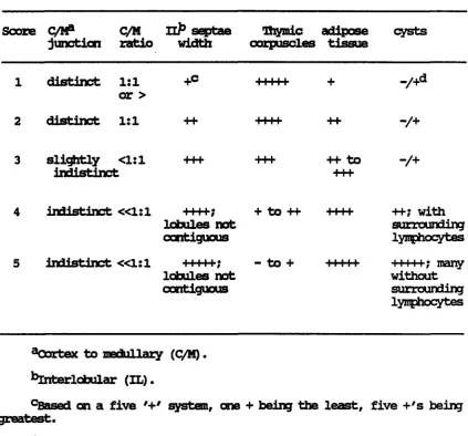
Table 1. Explanation of thymic morphology scaring system
Score C/RP Q/M Hp septae Dipdc adipose cysts junction ratio width corpuscles tissue
distinct 1:1 or >
distinct 1:1
+c 11111
-/+
3 sli^xtly <1:1 +++ indistinct
++ to -/+
indistinct «1:1 +++f; lobules not contiguous
+ to ++ I I I I
indistinct «1:1 +++++; - to + lobules not contiguous 11111 •H-; with surrounding lymphocytes
M i l l ; m a n y without surrounding lymphocytes
^Oortex to medullary (C/M).
^^Interlobular (XL).
°Based on a five system, one + being the least, five +'s being greatest.
[image:68.607.88.512.117.511.2]iraaDunizatlon and weekly thereafter for the duration of the e«perjjnent. Brucella abortus Hie serum antibody titer to abortus was
determined using the standard tube agglutination procedure, standard lUbe
Test Antigen was obtained from the Nationed Veterinary Services laboratory (Ames, lA). Hie titer was eo^xressed as the log2 of the inverse of the titer.
Tetanus toxoid The tetanus taxoid titer was determined using an enzyme-linked immunosorbent assay (EUSA) procedure. Tetanus tonmid was bound to a 96-well microtiter plate (Innulon 2, Dynatech Labs, Alexandria,
VA) and serial 2-fold dilutions of serum, beginning with a 1:10 dilution, were made in the wells. A perooddase-conjugated goat anti-canine IgG (heavy and li#it chain specific) was used to detect the presence of bound
antiboc^.
SŒMnsgisfcyUD qvantttatiçn
Serum samples to be assayed for immunoglobulin content were collected from each dog at the beginning and at the end of the e:q)erimental period. Ihe total concentration of serum IgG, igM, and IgA was measured using radial ismunodiffUsion kits (lOMT ImnunoBiologicals, Lisle, IL) specific for the canine immunoglobulins.
Lvnchocvte response tg mitogens
Venous blood was collected from each dog into acid citrate dextrose (ACD) once per week (beginning one week prior to the start of clonldlne
administration). %e blood samples were collected at e^proximately 0900 hours, prior to feeding. Lymphocyte blastogenesis in response to
MO) purified lymphocytes as previously described (Roth et al., 1984). Briefly, mitogen-stimilated lymphocytes were incubated for 48 hours before the addition of [^]-tfapddine. After 16-18 hours additional incubation, the cultures were harvested and prepared for liquid scintillation
RESUIinS
SOKa) tJfiOBDS
Four of the six dogs in each treatment grocp re^onded to clonidine with GH release, although this xespaneB was inconsistent. On day 1, only
three of Qie 12 dogs receiving clonidine reqwnded fay secreting GH (Figure la). Peak release of GH occurred 60 or 90 minutes after clonidine
edministration. On day 15, five of the 12 dogs receiving clonidine
ra^wnded by releasing GH (Figure lb) ; by day 30, clonidine administration resulted in GH release from seven dogs (Figure le). Those dogs responding to clonidine on day 1 continued to respond by GH release throu^iout the study period. There was no statistically significant difference between grotps in the amount of GH released eoaseç/t 60 minutes after clonidine
administration on day 30, far group 2. Control dogs, which had received amply gelatin cs^sules, did not re^xmd with GH secretion.
ThYmi? mgmhplpqy
The morpholow scores assigned to each thymus sample are given in Table 2 along with referenoes to photogrE^iis showing the lic^t microscopic
e^pearance of the glands (Figures 2 - 4).
The mean score for thymic morphology in groip 2 dogs was less than
A
[=1 0 MINUTES •• 30 MINUTES BSa 60 MINUTES m 90 MINUTESCONTROLS GROUP 2
jiu GROUP 3
B
CONTROLS GROUP 2 GROUP 3
c
âjt _cu
G)
Table 2. Ifaymic morphology scores by grotp, with Figure references
aoLp Animal Score 1® Score 2 Mean Score Figure
1 1223 5 4 4.5 2a
1226 3.5 4 3.75 2b
1231 2 2 2 2c
1232 3.5 3 3.25 2d
1235 4.5 4 4.25 2e
1237 4 4 4 2f
mean = 3.63
2 1222 2 2 2 3a
1224 3.5 3 3.25 3b
1227 2.5 3 2.75 3c
1228 2.5 3 2.75 3d
1230 4 4 4 3e
1234 2 2.5 2.25 3f
mean = 2.83
3 1220 2.5 2 2.25 4a
1221 5 4.5 4.75 4b
1225 5 4 4.5 4c
1229 4 4 4 4d
1233 2 2.5 2.25 4e
1236 4.5 4 4.25 4f
mean = 3.67
[image:74.610.91.485.107.494.2]Hanatwlgqy and çTInlçal (AimlPtTy
Values for the various clinical chemistry parameters, complete blood counts, and differentials did not vary from normal during the study period
(individual data not shewn) except in one animal. A group 2 dog had conoentraticns of serum alkaline phoephatase and serum glutamic pyruvic transaminase (SGFT) vAiich were greatly elevated at day 15. By day 30 her alkaline pho^ihatase concentration had returned to normal; the SGFT
cmcentration was greatly decreased but still was slightly elevated above normal.
While all clinical chemistry results were within normal ranges significant differences between groups were detected. As ccnpared to controls, significantly lower mean concentrations of blood urea nitrogen
(BUN) were detected in grotp 2 dogs at days 15 and 30, and in group 3 dogs at day 15 (Figure 5a). Mean blood glucose concentrations in group 2 and 3 dogs were significantly Icwer than in control dogs at day 30 (Figure 5b). Ihere were no significant differences detected between groups in conplete blood counts or differentials.
PrWry response
Brucella abortus The mean log2 titer of anti-B& abortus antibody for each grotp at each date tested is given in Table 3. Dogs in groups 2
20-1
C=1 CONTROLS m GROUP 2
GROUP 3
E 15. O o 0» E Z ZD 00
1 0
-DAY 1 DAY 15
**
DAY 30
"O
150-E
o 1254
CZ] CONTROLS on GROUP 2 BSa GROUP 3
0» 100-UJ
8
i
a o3
m 75 50-Table 3. Mean log2 of the inverse of the titer of anti-Bi. abortus antibody by groip
Day Grctp 1 Gaxp 2 Geaap 3
8® 0 + 0 0 + 0 0 ± 0
15 8.82 ± 0.43b 9.49 ± 0.17 8.99 ± 0.56
22 8.66 ± 0.42 9.82 ± 0.34* 9.82 + 0.34* 29 8.49 ± 0.48 8.99 ± 0.21 9.32 ± 0.45
^Dogs were ianmunized to heat-killed strain 19 of & abortus on day 8.
^faan ± standard error of the mean.
[image:84.610.94.439.89.390.2]Tetanus toMold The mean log2 titer of anti-tetanus toxoid
antibo(^ for each groqp at each date tested is given in Table 4. Ihere were no significant differences between grotgs in their primary antiboi^ responses to tetanus toxoid.
jBaamsglsiaiLiD quantitation
The mean serum concentrations of IgG, igM, and IgA, as measured ly radial immmodiffusion, are presented in Table 5. Ihere were no
significant differences detected bebœen groups in the serum concentration of the imunoglobulin classes tested.
lyrPBtWgyte XI^SSISS ïa mitogens
Table 4. Mean log2 of the inverse of the titer of anti-tetanus toxoid antibody by grotp
Day Grotg) 1 Grotp 2 Gscup 3
sa 0 + 0 0 + 0 0 + 0
15 0 + 0 0 + 0 0 + 0
22 8.49 ± l.Olb 8.32 ± 0.52 8.32 + 1.03 29 9.16 + 0.79 8.66 + 0.76 9.16 ± 0.65
^Dogs were immunized to tetanus toxoid on day 8.
[image:86.609.89.430.87.290.2]Table 5. Mean serum œnoentraticn of IgG, Iç|Mr and IgA on days 1 and 29, as measured by radial imunodiffusion
Grotqp Day IgG igM IgA
1 1 2458.3 ± 232.5® 95.0 ±_ 7.3 18.2 ± 2.9 2 3233.3 ± 518.8 96.2 ± 9.9 17.0 ± 2.9 2 1 2100.0 ± 227.7 130.2 ±17.1 26.7 ± 8.8 2 2766.7 ± 391.3 120.2 ± 18.5 19.3 ± 4.1 3 1 2616.7 ± 332.1 125.2 ± 16.1 26.0 ± 7.8 2 2500.0 ± 335.7 135.7 ± 28.3 20.2 ± 3.8
[image:87.607.85.479.94.308.2]