Abstract
This study explores whether self-reported depth of hypnosis and hypnotic suggestibility are associated with individual differences in neuroanatomy and/or levels of functional connectivity. Twenty-nine people varying in suggestibility were recruited and underwent structural, and after a hypnotic induction, functional magnetic resonance imaging at rest. We used voxel-based
1. Introduction
Hypnosis can be used as an adjunct treatment for pain (Montgomery et al., 2002), depression (Alladin and Alibhai, 2007), weight loss (Kirsch et al., 1995; Kirsch, 1996), irritable bowel syndrome (Whitehead, 2006; Wilson et al., 2006), and it can also be used to study psychological phenomena (Szechtman et al., 1998; Barnier, 2002; Egner et al., 2005; O'Connor et al., 2008). It is not effective for everyone, however, and certain individuals appear to respond favourably to hypnosis and hypnotic suggestions and others are unaffected (Kirsch and Braffman, 2001).
Some studies suggest that hypnosis enables participants to successfully respond to certain task suggestions such as those aimed at altering cognition or perception (Faymonville et al., 2000; Kosslyn et al., 2000; Raz et al., 2002; Raz, 2005). Others suggest that successful performance predominantly relies on abilities/skills already possessed by the participants, which can be accessed with or without hypnosis (Raz et al., 2006; Raz, 2007; Raz et al., 2007; McGeown et al., 2012). Indeed, the difference between the number of suggestions that high suggestible participants respond to with or without hypnosis is small (Braffman and Kirsch, 1999; Kirsch and Braffman, 2001) and abilities thought possible only following a hypnotic suggestion can be achieved without hypnosis (e.g. colour hallucination (Mazzoni et al., 2009), Stroop effect reduction (Raz et al., 2006; Raz, 2007)). Low suggestible people on the other hand do not seem to be capable of demonstrating these
abilities whether a hypnotic induction is attempted or not. Such findings suggest that, regardless of whether hypnosis is induced, highly suggestible people differ from low suggestible people on certain behavioural capabilities. In this study we investigate whether individual variations in brain structure or function exist that might explain differences in response to hypnotic suggestions and/or self-reported depth of hypnosis.
1.1.Structural neuroimaging
interest approach which focused on volumetric corpus callosum measurements only, provided evidence that high suggestible (who could eliminate pain) compared to low suggestible people, had a larger rostrum. They suggested that this characteristic might facilitate transfer of information providing benefit for attention, monitoring and inhibitory abilities. An alternative and potentially more informative approach is to use voxel-based morphometry (VBM), which does not restrict analyses to a priori regions of interest only or use manual measurements (it is an automated whole-brain technique). VBM was used by Hoeft et al., (2012) and in the current study. Hoeft et al. found no volumetric differences between high and low suggestible participants applying their principal statistical threshold (combined height threshold p<0.01, extent threshold p<0.01 FWE corrected), but when adopting a less conservative threshold (uncorrected height threshold of p<0.001), between group differences were detected in parietal, temporal and cerebellar regions. The
direction of the differences was not specified, nor was whether the regional differences occurred in grey or white matter specifically.
We sought to extend this literature, and using VBM, examined whether self-reported levels of depth of hypnosis and differences in hypnotic suggestibility were associated with regional volumetric variations in grey and white matter.
outside of the hypnosis literature. Since hypnosis and certain forms of meditation appear to share features, such as absorption and the disengagement of attention from distracting stimuli (Holroyd, 2003; Cardeña, 2005; Lutz et al., 2008; Demertzi et al., 2011) and given that structural neuroimaging studies on meditation have shown volumetric adaptations within the cingulate (Grant et al., 2010; Holzel et al., 2011) and insular cortex (Lazar et al., 2005; Holzel et al., 2008; Luders et al., 2012), these brain structures might also be linked to hypnotic response. Visual cortical areas may be additional candidates for close investigation, given evidence of higher levels of activity in these regions during hypnosis (Maquet et al., 1999; Rainville et al., 1999) and reports of spontaneous imagery during hypnosis (e.g. Maquet et al., 1999; Cardeña, 2005).
1.2 Functional neuroimaging
Intrinsically connected networks (ICNs), identified through fMRI analysis include, but are not restricted to, the default mode network (DMN; Raichle et al., 2001; Greicius et al., 2003), the Salience Network (Critchley et al., 2004; Critchley, 2005; Seeley et al., 2007) and the Executive Control Network (Seeley et al., 2007). Hypnosis has been found to alter brain activity and connectivity within these networks (McGeown et al., 2009; Demertzi et al., 2011; Deeley et al., 2012).
Using independent components analysis (ICA), Hoeft et al., (2012) found that high suggestible compared to low suggestible people, in the absence of a hypnotic induction, had greater
also reported in the ‘extrinsic system’ (which processes sensory information and overlaps with the bilateral executive control network) and interpreted as a reflection of dissociation from the environment.
Using a block design rather than a data-driven approach, McGeown et al., (2009) reported decreased activity within the DMN in high suggestible participants during rest after a hypnotic induction. These changes fitted with Oakley and colleagues’ hypothesis (2008; Oakley and Halligan, 2009) that DMN activity may be altered in hypnosis. Deeley et al., (2012) also showed decreased activity during hypnosis within the DMN (along with increases within bilateral regions of the frontal cortex) in medium to high suggestible people, during passive visual stimulation. In the hypnosis condition and presumably in correspondence with the neural changes, participants reported more absorption and relaxation, and less distractibility, analytical thought and cluttering of the mind.
In the current study we used ICA to examine whether reports of varying depths of hypnosis are associated with variation in connectivity within ICNs. In line with previous neuroimaging research (McGeown et al., 2009; Deeley et al., 2012), we expected that deeper levels of hypnosis would be associated with lower levels of functional connectivity within the DMN.
We also predicted alterations in the salience and executive networks, given descriptions of attentional focus and absorption in hypnosis (e.g. Demertzi et al., 2011; Deeley et al., 2012), evidence from PET studies showing activity modulation within the ACC during hypnosis (Rainville et al., 2002), findings of decreased connectivity in the ‘extrinsic system’ during hypnosis (Demertzi et al., 2011), and the findings of Hoeft et al., (2012) who observed differences in connectivity within these networks in high and low suggestible people (in the absence of hypnosis). Specifically, we anticipated that lateral cortical connectivity would decrease with greater depths of hypnosis, in correspondence with a reduction in processing of the external environment as suggested by Demertzi et al., (2011), and that connectivity in the dorsal ACC would increase, due to increased levels of absorption and inhibition of information processing of the external environment, as
and accompanying activation in the visual cortex (Maquet et al., 1999; Rainville et al., 1999) we also expected connectivity to increase within visual (medial/ventral) networks.
2. Method 2.1. Participants
One hundred and fifty-six non-psychology university students were screened for hypnotic suggestibility with Comey and Kirsch’s (1999) modification of the Carleton University Responsiveness to Suggestion Scale (CURSS; Spanos et al., 1983). The scale ranges from 0-7, and consists of
ideomotor suggestions, motor challenges and cognitive/perceptual alterations. A score of one is assigned for a response to each suggestion. Participants scoring between 0-2 are classified as low in suggestibility, 3-4 medium and 5-7 high. The CURSS was used because 1) it has extended hypnotic induction and introductory instructions that are similar to the Stanford Hypnotic Suggestibility Scale, Form C (SHSS:C; Weitzenhoffer and Hilgard, 1962), 2) It is straightforward to administer to large groups, 3) It contains a number of very difficult suggestions thus ensuring that high-scoring
individuals are indeed very highly suggestible. The CURSS shows adequate internal consistency and is highly correlated (r = .65) with the SHSS:C (Spanos et al., 1983), and the two scales yield ‘good levels of agreement’ in identifying highly responsive individuals (Perry et al., 1992, p. 482). Specifically, 75% of participants who score in the high range on the CURSS also score in the high range on the SHSS:C. All 13 participants high in suggestibility were invited for scanning, but only 7 could participate for reasons which included safety and personal issues. All 13 participants low in suggestibility were scanned. Thirteen people with medium scores, randomly selected, were invited for scanning and 9 participated. See Table 1 for demographics. Two participants were left-handed, and the sample contained 17 males and 12 females.
Approval was granted by the local ethics committee in Venice, Italy, where scanning took place. Participants were informed that the purpose of the study was to investigate whether
differences exist between the brains of those people who can be hypnotized and those who cannot. Written informed consent was obtained for all participants in accordance with the Declaration of Helsinki. Participants were told that while in the scanner, they should lie as still as possible with their eyes closed and listen to the instructions via the headphones. A high-resolution structural brain scan would then be acquired. The participants were informed that a hypnotic induction would then be played through headphones. An abbreviated version of the induction described by Kirsch, Lynn, and Rhue (1993) was used. Participants were invited to close their eyes, relax, imagine being in a pleasant place, and to go deeper and deeper into hypnosis. After the induction participants the resting state scan was acquired (eyes closed). After a script to terminate hypnosis, participants rated on a 10 point scale how deeply hypnotized they were during the session (with “0” corresponding to “not at all hypnotized” and “10” relating to “very deeply hypnotized”) and how relaxed they were (with “0” relating to “not at all relaxed” and “10” corresponding to “very deeply relaxed”). Responses were recorded in the scanner with fibre optic response devices placed at the left and right hand.
2.3. Structural MRI scanning: acquisition and analysis
Three dimensional T1-weighted MRI images were acquired on a 1.5 T Philips system with a Turbo Field Echo sequence using a SENSE headcoil. Voxel dimensions were 1.1 x 1.1 x 0.6 mm. Number of slices was 280, TR 7.4 msec, TE 3.4 msec, flip angle 8°, and total duration 4 minutes 41 seconds. A number of preprocessing steps were taken to isolate the grey and white matter from the 3D T1-weighted structural scans. Initially the T1 images were manually aligned to approximate Montreal Neurological Institute (MNI) space. The VBM8 Toolbox
segmentation algorithm (which is based on the adaptive Maximum A Posterior technique), the tissue probability maps available within SPM8 as inputs and an inhomogeneity intensity correction. For normalization, the Diffeomorphic Anatomical Registration Through Exponentiated Lie (DARTEL) algebra toolbox (Ashburner, 2007) was used to align diffeomorphically the participants' brain images to the IXI-database template (http://www.brain-development.org), which is in MNI space. The non-linear deformations were used to preserve the tissue volumes (Davatzikos et al., 2001), however the linear transforms were not. This meant that adjustments for brain size, alignment and orientation were made, but these did not affect the volume quantifications locally. Smoothing was carried out with an 8 mm full width at half maximum (FWHM) Gaussian kernel to compensate for any residual variability after spatial normalization.
A regression model was used to investigate the presence of linear correlations between grey matter volume and self-reported depth of hypnosis. Additional models assessed relaxation and hypnotic suggestibility. An absolute threshold of 0.15 was used. A similar analytic approach was used with white matter segments. Due to the potential influence of age, sex (Pell et al., 2008; Barnes et al., 2010) and years of education (Gonul et al., 2009) on VBM analyses, these were included as covariates. The x, y, z coordinates of significant correlations were first converted into Talairach coordinates using the Matlab function mni2tal
(http://imaging.mrc-cbu.cam.ac.uk/downloads/MNI2tal/mni2tal.m) and then labelled using the Talairach Daemon Client (http://www.talairach.org/). A combined thresholding approach similar to that described in Poline et al., (1997) and Hoeft et al. (2012) was used, with height threshold p<0.01 and cluster extent p<0.05 FWE. For regions considered to be of importance and identified a priori, a height threshold p<0.01 and cluster extent p<0.05 uncorrected was also used for directed search. To control for the known issues with making cluster inferences on smoothed grey and white matter images, non-stationarity correction was enabled within SPM8.
Two hundred and ten sets of 20 contiguous 5 mm thick axial T2* images were acquired (TR = 2000
ms, TE = 50 ms, flip angle = 90°, voxel size = 3.28 x 3.28 mm in plane resolution). Field of view was 230 x 230 x 100. Ten dummy scans enabled the scanner to reach equilibrium and these were removed prior to pre-processing (leaving 200 scans per participant).
SPM8 was used for the pre-processing and statistical analyses. All volumes from each subject were re-aligned to their mean and re-sliced using 4th Degree B-Spline interpolation methods to adjust for residual motion related signal changes. The images were then slice time corrected using the 10th image as the reference slice (ascending acquisition). The 3D anatomical T1-weighted image was coregistered to the mean image produced during realignment, segmented (and normalized to MNI stereotactic space using non-linear estimation of parameters) and the normalization parameters applied to the re-aligned functional images. The normalised fMRI data were then smoothed with a 12 mm FWHM isotropic Gaussian kernel. GroupICATv3.0a (GIFT v2.0a)
(http://mialab.mrn.org/software/gift) was used for ICA. The toolbox adopts a group approach, in which the data from each individual is first concatenated across time. The GIFT dimensionality estimation tool indicated that the data could be decomposed into 16 components (mean). Principal components analysis was used for data reduction and ICA was carried out (Infomax algorithm). As a final step, the toolbox was used to back re-construct subject specific images (group ICA; GICA). The components were transformed from arbitrary units to z-scores. One-sample t-tests on the
component images, thresholded at p<0.05 FWE voxel-level identified the various networks. These were saved as binary masks (Figure 2), and used to constrain the regression analyses to the networks of interest. Regression was used to assess associations between the functional connectivity of the different components, and ratings of hypnotic depth, relaxation and hypnotic suggestibility. Combined thresholding was used, height threshold p<0.01, cluster extent p<0.05 FDR correction.
3. Results
3.1.1. Depth of Hypnosis and GM Volume
Participants reporting greater depths of hypnosis had significantly larger GM volume in brain regions which included the ACC, and the medial and superior frontal gyri, bilaterally (see Table 3a, and Figure 1a+b). To examine whether associations were present within the additional regions identified a priori (the remainder of the ACC, prefrontal cortex, insular cortex, and the visual association areas), the less conservative threshold (p < 0.01 height, p < 0.05, uncorrected cluster-level) was applied. Uncorrected clusters should be viewed with some degree of caution given the risk of false positives inherent with such thresholds, but were reported given the evidence based hypotheses and their potential theoretical significance. The more liberal threshold identified further significant correlations in the right dorsal ACC, the left superior frontal gyrus and medial frontal gyrus, in addition to the left insula. No associations were detected within the visual association cortex. Furthermore, no brain regions were identified with lower GM volume in relation to increased hypnotic depth.
3.1.2. Degree of Relaxation and GM Volume
No significant relationships between the ratings of relaxation during hypnosis and regional GM volume were identified.
3.1.3. Hypnotic Suggestibility and GM Volume
Higher suggestibility was associated with greater GM volume in the left middle occipital and middle and superior temporal gyrus (see Table 3b and Figure 1c). Application of the p < 0.01 height, p < 0.05 uncorrected cluster-level threshold revealed further correlations within the left insula and the right inferior parietal lobule. No brain areas were identified in which higher levels of suggestibility were associated with smaller GM volumes.
Depth of hypnosis rating, hypnotic suggestibility and relaxation rating were also entered with GM volume into a single regression model with age, education and sex as additional covariates. Using this alternative model specification, the correlations between hypnotic depth and the ACC and insula were maintained, as were those between hypnotic suggestibility and the left occipito-temporal and insular cortex (using the same significance thresholding).
3.1.5. WM Volume Analyses
Hypnotic depth ratings did not correlate (positively or negatively) with WM volume at the specified significance threshold, nor did relaxation ratings or suggestibility. To assess potential relationships between the corpus callosum and suggestibility in greater detail, and attempt replication of the findings of Horton et al., (2004), a mask of the corpus callosum (with “1” entered in the dilate operator) was created using the WFU pick atlas tool (From the template under “TD Brodmann areas +”; Maldjian et al., 2003; Maldjian et al., 2004) and a very liberal level of statistical threshold was set at p < 0.05, uncorrected voxel-level. No significant correlations were detected within the rostrum.
3.1.6. Global correlations of GM and WM
For information on global brain volume see Table 1. Suggestibility correlated negatively with total GM and total WM (Table 2). A likely explanation for that finding is that slightly more males than females were found within the lower range of suggestibility in our sample (low 3F:10M, med 6F:3M, high 3F:4M). Partial correlations were also computed, while controlling for total intracranial volume (summing GM, WM and CSF). In this analysis CURSS scores no longer correlated significantly with the global GM, WM or CSF measures. Of further note, in relation to the morphometric analyses described above, overall brain size was adjusted in the preprocessing stage, prior to the statistical analyses, and sex included as a covariate in the models, with age and education.
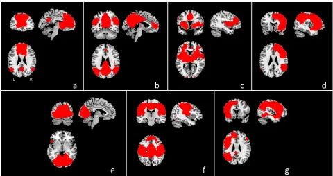
Figure 2 illustrates networks that were identified through the ICA analyses on the resting state scans.
3.2.1. Correlation of hypnotic depth ratings with resting state networks
Hypnotic depth correlated negatively with anterior DMN connectivity within the left medial and superior frontal gyri and the ACC (BA 32; See Table 4, Figure 3). No significant positive correlations were observed, nor were any correlations (positive or negative) detected with connectivity within the posterior DMN, salience network, executive control networks (right and left), or visual network. As each of these networks and/or the brain structures underlying the networks have been
implicated in a previous intrinsic connectivity studies (Demertzi et al., 2011) and/or functional neuroimaging studies (Maquet et al., 1999; Rainville et al., 1999; Rainville et al., 2002) of hypnosis and suggestibility, an uncorrected cluster level was also applied. One cluster within the salience network (the ACC) correlated positively with hypnotic depth, whereas another cluster (the left insula) correlated negatively. A cluster within the right executive network (dorsal ACC) correlated positively, whereas another (right dorsolateral prefrontal cortex; DLPFC) correlated negatively. In the left executive network, three clusters in the left DLPFC, superior temporal and medial frontal cortex showed negative correlations. Within the visual network a positive correlation was observed in the left cuneus.
3.2.2. Correlation of relaxation ratings with resting state networks
There were no significant correlations at the specified threshold (positive or negative) between with the relaxation ratings and connectivity in the DMN, salience or executive networks.
3.2.3. Correlation of suggestibility scores with resting state networks
4. Discussion
4.1 Structural analyses
4.1.1. Depth of hypnosis – structural analyses
Depth of hypnosis correlated positively with GM volume in the frontal cortex, which included the ACC (BA 32). An uncorrected cluster-threshold revealed further positive correlations within the ACC (BA 24) and the insula. This combination of brain regions is noteworthy as a recent fMRI study by McGeown and colleagues (2009) showed that hypnosis during rest was characterized by decreased activity in the anterior DMN. The functional analysis in the current study similarly showed decreased connectivity within this network in relation to self-reported depth of hypnosis. The observation that larger GM volume in the anterior DMN is associated with reports of increased hypnotic depth might explain the ability for people to effectively modulate activity within that network e.g. to reduce spontaneous thought.
Functional neuroimaging studies have highlighted that activity in the ACC occurs during hypnosis (Maquet et al., 1999; Rainville et al., 1999; Rainville et al., 2002). It might be speculated that greater development of the ACC provides participants with the ability to not only engage in hypnosis, but to reach more intense states of hypnosis. Interestingly, an analysis from the neuroimaging study by Rainville et al (2002) revealed that activity in the ACC (BA 32) increased as participants reported greater absorption during hypnosis (while statistically covarying for relaxation). Furthermore, a recent morphometric study examining meditators found greater cortical thickness in the ACC in association with increased subjective reports of absorption (Grant et al., 2013). Although not measuring absorption as a distinct concept in our study, the positive correlation between grey matter volume in the ACC and depth of hypnosis might have captured this facet of hypnotic
The ACC is involved in a variety of functions however, and the areas identified in the current structural analysis fell both within the rostral-ventral (affective) and dorsal (cognitive) divisions as defined using the Bush et al. (2000) partitions. In relation to affectivity, a number of studies provide evidence for an association between emotion and hypnosis (e.g. Cardeña, 2005; Pekala and Kumar, 2007; Cardeña et al., 2008). Dorsal regions, on the other hand, tend to be involved more in
executive control and attentional functions (e.g. Pardo et al., 1990; Botvinick et al., 1999; MacDonald et al., 2000; Milham et al., 2001; Lutcke and Frahm, 2008). The association with ACC volume in the current study may reflect the capacity for absorption, dissociation and spontaneous changes in affectivity characteristic of deep hypnosis (Cardeña, 2005; Pekala and Kumar, 2007). The insula was also associated with hypnotic depth. The insular cortex operates in combination with the ACC (Craig, 2009; Medford and Critchley, 2010; Menon and Uddin, 2010), and like the ACC, given its diverse number of functions such as integrating sensory, affective and cognitive information (Medford and Critchley, 2010) may play a key role in facilitating hypnotic phenomena.
Previous research has drawn attention to visual areas which are active during hypnosis (Maquet et al., 1999; Rainville et al., 1999; Rainville et al., 2002). In the current study no significant associations between GM volume in visual brain areas and reports of depth of hypnosis were detected however. In summary, a more developed circuit of brain regions, including the ACC, dorsolateral and
ventromedial PFC, and insula appears to lead to a more effective use of attentional, executive and affective functions which in turn facilitate the depth with which hypnosis is experienced.
4.1.2. Level of suggestibility – structural analyses
The auditory association cortex appears to play an important role in both hallucination and mental imagery of sounds (Zatorre et al., 1996; Lennox et al., 2000; Kraemer et al., 2005; Jardri et al., 2007) as does the visual association cortex for hallucination and mental imagery in that modality
(D'Esposito et al., 1997; Ffytche et al., 1998). To summarize, the auditory and visual association cortices can be seen to relate to the occurrence of alterations in perception, and it might be that greater structural development of these neural structures facilitates the generation of complex perceptual experiences which is then reflected by higher suggestibility scores.
An association between suggestibility and GM volume was also detected in the left insula. In addition to the processing/functions mentioned above, the insula contains a motor map (e.g. Fink et al., 1997), and is often activated during movement and when motor representations are called upon (e.g. Fink et al., 1997; Cunnington et al., 2002; Mutschler et al., 2007). It is involved with awareness of body movements and/or feelings of agency (Farrer and Frith, 2002; Farrer et al., 2003). The left insular cortex was the only brain area that was related to both hypnotic depth and suggestibility. Its roles in processing and integrating diverse types of information (and its association with the ACC) might help to explain the propensity for highly suggestible participants to experience both suggestions and hypnotic phenomena. In summary, the brain regions that were associated with suggestibility mostly support a range of perceptual and motor functions and may be involved in modulating introspective awareness.
Hoeft et al., (2012) reported differences between high and low suggestible people in the parietal, temporal and cerebellar regions at an uncorrected level of significance. It seems due to that
and/or reliance on different brain networks. Further research is required to explore the reliability of neuroanatomical differences in relation to hypnotic suggestibility.
No significant relationships were observed between regional white matter volume and either suggestibility or depth of hypnosis. Even when using a corpus callosum mask and a liberal threshold, still no relationship was observed between the volume of the rostrum and suggestibility. Possible reasons for the lack of replication of the findings of Horton et al. (2004) include differences in measurement techniques and the two stage selection procedure adopted by Horton et al., in which high suggestible participants were first selected for the study and only those who could modulate pain were then scanned. We selected only on the basis of hypnotic suggestibility, not on any further distinguishing characteristics.
4.2. Functional connectivity analyses
Deeper levels of hypnosis were associated with decreased connectivity within the anterior DMN. This finding fits closely with the results of McGeown et al., (2009), who found decreased BOLD activity in the anterior DMN in high suggestible participants during hypnosis. The decreased functional connectivity/activity in the anterior part of the network may reflect a reduction in spontaneous mental activity and self-awareness, outcomes of hypnosis that have been reported in studies of its phenomenology (Tart, 1970; Cardeña, 2005).
information. Conversely, greater functional connectivity in the dorsal regions of the ACC, may reflect executive and attentional processes, and, as mentioned above, absorption.
A discrepancy between the results of the intrinsic connectivity study by Demertzi et al. (2011) and our study arises in terms of the direction of modulation within the anterior DMN during hypnosis. The activity undertaken during scanning might explain the difference. In the current study, after the induction participants merely had to rest (with no instructions provided) for the subsequent period of scanning. We observed a decrease in connectivity for deeper levels of hypnosis which we suggest may be due to decreased spontaneous thought or a similar process. Demertzi et al., had participants revive autobiographical memories during the induction and the subsequent scanning period. That task might have increased anterior DMN connectivity, as autobiographical retrieval leads to activation of the DMN (Spreng and Grady, 2010; Ino et al., 2011). Examination of figure three (p. 316) in the article by Demertzi et al. also suggests that connectivity decreased in the anterior DMN during hypnosis compared to the rest condition (although functional connectivity in these conditions was not directly compared). In line with the inverse correlation between connectivity in lateral brain regions and hypnotic depth found in our study, Demertzi et al., (2011) observed decreases in the extrinsic system during hypnosis. The authors postulate that these changes are associated with higher levels of dissociation (which was self-reported by participants).
In summary, self-rated deeper levels of hypnosis were associated with less anterior DMN
connectivity. Changes in the other networks were consistent with the phenomenology of hypnosis e.g. higher levels of absorption, greater dissociation, and increased mental imagery.
4.3. Limitations, future directions and conclusion
induction) (see Mazzoni et al., 2013). Further reports of the phenomenology are also necessary and while self-rated measures of hypnotic depth, like all self-report measures, may be subject to
response biases, they remain the most common measure of hypnosis and self-report is used extensively in neuroimaging studies relating to hypnosis.
Suggestibility in the current study was only assessed after a hypnotic induction. Due to this, we may have identified brain areas involved in determining the degree of hypnotisability of a person, but those findings are likely to be confounded with the association non-hypnotic suggestibility (waking suggestibility) has with grey matter. Future research could take measurements of suggestibility with and without hypnosis to assess which brain regions are associated with hypnotisability per se (see Kirsch et al., 2011) and which are associated with responsiveness to suggestion.
In relation to clinical practice, if the networks associated with suggestibility and hypnotic response can be further defined, appropriate interventions (e.g. with pharmacological agents or stimulation devices) may enhance the desired response.
In conclusion, the findings of this study indicate that individual differences in hypnotic suggestibility and the feeling of being deeply hypnotized correlate with regional variance in specific brain areas associated with spontaneous thought, attention and executive control, sensory integration and interoception, and also visual perception. Less anterior DMN functional connectivity was also related to experiences of deeper levels of hypnosis.
Acknowledgment
We thank Irving Kirsch for his contribution and comments on an earlier draft of this article and two anonymous reviewers for their helpful suggestions.
References
Ashburner, J., 2007. A fast diffeomorphic image registration algorithm. NeuroImage 38, 95-113. Barnes, J., Ridgway, G.R., Bartlett, J., Henley, S.M., Lehmann, M., Hobbs, N., Clarkson, M.J.,
MacManus, D.G., Ourselin, S., Fox, N.C., 2010. Head size, age and gender adjustment in MRI studies: a necessary nuisance? NeuroImage 53, 1244-1255.
Barnier, A.J., 2002. Posthypnotic amnesia for autobiographical episodes: a laboratory model of functional amnesia? Psychological Science 13, 232-237.
Botvinick, M., Nystrom, L.E., Fissell, K., Carter, C.S., Cohen, J.D., 1999. Conflict monitoring versus selection-for-action in anterior cingulate cortex. Nature 402, 179-181.
Braffman, W., Kirsch, I., 1999. Imaginative suggestibility and hypnotizability: an empirical analysis. Journal of Personality and Social Psychology 77, 578-587.
Bush, G., Luu, P., Posner, M.I., 2000. Cognitive and emotional influences in anterior cingulate cortex. Trends in Cognitive Sciences 4, 215-222.
Cardeña, E., 2005. The phenomenology of deep hypnosis: quiescent and physically active. International Journal of Clinical and Experimental Hypnosis 53, 37-59.
Cardeña, E., Terhune, D., Loof, A., Buratti, S., 2008. Hypnotic Experience is Related to Emotional Contagion. International Journal of Clinical and Experimental Hypnosis 57, 33-46.
Craig, A.D., 2009. How do you feel--now? The anterior insula and human awareness. Nature Reviews. Neuroscience 10, 59-70.
Critchley, H.D., 2005. Neural mechanisms of autonomic, affective, and cognitive integration. The Journal of Comparative Neurology 493, 154-166.
Critchley, H.D., Wiens, S., Rotshtein, P., Ohman, A., Dolan, R.J., 2004. Neural systems supporting interoceptive awareness. Nature Neuroscience 7, 189-195.
Cunnington, R., Windischberger, C., Deecke, L., Moser, E., 2002. The preparation and execution of self-initiated and externally-triggered movement: a study of event-related fMRI. Neuroimage 15, 373-385.
D'Esposito, M., Detre, J.A., Aguirre, G.K., Stallcup, M., Alsop, D.C., Tippet, L.J., Farah, M.J., 1997. A functional MRI study of mental image generation. Neuropsychologia 35, 725-730.
Davatzikos, C., Genc, A., Xu, D., Resnick, S.M., 2001. Voxel-based morphometry using the RAVENS maps: methods and validation using simulated longitudinal atrophy. Neuroimage 14, 1361-1369.
Demertzi, A., Soddu, A., Faymonville, M.E., Bahri, M.A., Gosseries, O., Vanhaudenhuyse, A., Phillips, C., Maquet, P., Noirhomme, Q., Luxen, A., Laureys, S., 2011. Hypnotic modulation of resting state fMRI default mode and extrinsic network connectivity. Progress in Brain Research 193, 309-322.
Draganski, B., Gaser, C., Busch, V., Schuierer, G., Bogdahn, U., May, A., 2004. Neuroplasticity: changes in grey matter induced by training. Nature 427, 311-312.
Draganski, B., Gaser, C., Kempermann, G., Kuhn, H.G., Winkler, J., Buchel, C., May, A., 2006.
Temporal and spatial dynamics of brain structure changes during extensive learning. The Journal of Neuroscience 26, 6314-6317.
Egner, T., Jamieson, G., Gruzelier, J., 2005. Hypnosis decouples cognitive control from conflict monitoring processes of the frontal lobe. Neuroimage 27, 969-978.
Farrer, C., Franck, N., Georgieff, N., Frith, C.D., Decety, J., Jeannerod, A., 2003. Modulating the experience of agency: a positron emission tomography study. Neuroimage 18, 324-333.
Farrer, C., Frith, C.D., 2002. Experiencing oneself vs another person as being the cause of an action: The neural correlates of the experience of agency. Neuroimage 15, 596-603.
Faymonville, M.E., Laureys, S., Degueldre, C., DelFiore, G., Luxen, A., Franck, G., Lamy, M., Maquet, P., 2000. Neural mechanisms of antinociceptive effects of hypnosis. Anesthesiology 92, 1257-1267.
Fink, G.R., Frackowiak, R.S., Pietrzyk, U., Passingham, R.E., 1997. Multiple nonprimary motor areas in the human cortex. Journal of Neurophysiology 77, 2164-2174.
Gonul, A.S., Demirel, O., Kitis, O., Eker, C., Eker, O.D., Ozan, E., 2009. The Effects of the Duration of Formal Education on Adult Brain: A Voxel-Based Morphometry - (Diffeomorphic Anatomical Registration Using Exponentiated Lie Algebra) DARTEL Study. Bulletin of Clinical
Psychopharmacology 19, 221-226.
Grant, J.A., Courtemanche, J., Duerden, E.G., Duncan, G.H., Rainville, P., 2010. Cortical thickness and pain sensitivity in zen meditators. Emotion 10, 43-53.
Grant, J.A., Duerden, E.G., Courtemanche, J., Cherkasova, M., Duncan, G.H., Rainville, P., 2013. Cortical thickness, mental absorption and meditative practice: possible implications for disorders of attention. Biological Psychology 92, 275-281.
Greicius, M.D., Krasnow, B., Reiss, A.L., Menon, V., 2003. Functional connectivity in the resting brain: a network analysis of the default mode hypothesis. Proceedings of the National Academy of Sciences of the United States of America 100, 253-258.
Hoeft, F., Gabrieli, J.D., Whitfield-Gabrieli, S., Haas, B.W., Bammer, R., Menon, V., Spiegel, D., 2012. Functional brain basis of hypnotizability. Archives of General Psychiatry 69, 1064-1072.
Holzel, B.K., Carmody, J., Vangel, M., Congleton, C., Yerramsetti, S.M., Gard, T., Lazar, S.W., 2011. Mindfulness practice leads to increases in regional brain gray matter density. Psychiatry Research 191, 36-43.
Holzel, B.K., Ott, U., Gard, T., Hempel, H., Weygandt, M., Morgen, K., Vaitl, D., 2008. Investigation of mindfulness meditation practitioners with voxel-based morphometry. Social Cognitive and Affective Neuroscience 3, 55-61.
Horton, J.E., Crawford, H.J., Harrington, G., Downs, J.H., 3rd, 2004. Increased anterior corpus callosum size associated positively with hypnotizability and the ability to control pain. Brain 127, 1741-1747.
Ino, T., Nakai, R., Azuma, T., Kimura, T., Fukuyama, H., 2011. Brain activation during autobiographical memory retrieval with special reference to default mode network. The Open Neuroimaging Journal 5, 14-23.
Jardri, R., Pins, D., Delmaire, C., Goeb, J.L., Thomas, P., 2007. Activation of bilateral auditory cortex during verbal hallucinations in a child with schizophrenia. Molecular psychiatry 12, 319.
Kirsch, I., 1996. Hypnotic enhancement of cognitive-behavioral weight loss treatments--another meta-reanalysis. Journal of Consulting and Clinical Psychology 64, 517-519.
Kirsch, I., Cardena, E., Derbyshire, S., Dienes, Z., Heap, M., Kallio, S., Mazzoni, G., Naish, P., Oakley, D., Potter, C., Walters, V., Whalley, M., 2011. Definitions of hypnosis and hypnotizability and their relation to suggestion and suggestibility: A consensus statement. Contemporary Hypnosis and Integrative Therapy 28, 107-115.
Kirsch, I., Lynn, S.J., Rhue, J.W., 1993. Introduction to clinical hypnosis, in: Rhue, J.W., Lynn, S.J., Kirsch, I. (Eds.), Handbook of Clinical Hypnosis. American Psychological Association, Washington DC, pp. 3-22.
Kirsch, I., Montgomery, G.H., Sapirstein, G., 1995. Hypnosis as an Adjunct to Cognitive-Behavioral Psychotherapy: A meta-analysis. Journal of Consulting and Clinical Psychology 63, 214-220.
Kosslyn, S.M., Thompson, W.L., Costantini-Ferrando, M.F., Alpert, N.M., Spiegel, D., 2000. Hypnotic visual illusion alters color processing in the brain. American Journal of Psychiatry 157, 1279-1284.
Kraemer, D.J., Macrae, C.N., Green, A.E., Kelley, W.M., 2005. Musical imagery: sound of silence activates auditory cortex. Nature 434, 158.
Lazar, S.W., Kerr, C.E., Wasserman, R.H., Gray, J.R., Greve, D.N., Treadway, M.T., McGarvey, M., Quinn, B.T., Dusek, J.A., Benson, H., Rauch, S.L., Moore, C.I., Fischl, B., 2005. Meditation experience is associated with increased cortical thickness. Neuroreport 16, 1893-1897.
Luders, E., Phillips, O.R., Clark, K., Kurth, F., Toga, A.W., Narr, K.L., 2012. Bridging the hemispheres in meditation: thicker callosal regions and enhanced fractional anisotropy (FA) in long-term
practitioners. NeuroImage 61, 181-187.
Lutcke, H., Frahm, J., 2008. Lateralized anterior cingulate function during error processing and conflict monitoring as revealed by high-resolution fMRI. Cerebral Cortex 18, 508-515.
Lutz, A., Slagter, H.A., Dunne, J.D., Davidson, R.J., 2008. Attention regulation and monitoring in meditation. Trends in Cognitive Sciences 12, 163-169.
MacDonald, A.W., Cohen, J.D., Stenger, V.A., Carter, C.S., 2000. Dissociating the role of the dorsolateral prefrontal and anterior cingulate cortex in cognitive control. Science 288, 1835-1838.
Maguire, E.A., Gadian, D.G., Johnsrude, I.S., Good, C.D., Ashburner, J., Frackowiak, R.S., Frith, C.D., 2000. Navigation-related structural change in the hippocampi of taxi drivers. Philosophical
Transactions of the Royal Society of London Series B-Biological Sciences 97, 4398-4403.
Maldjian, J.A., Laurienti, P.J., Burdette, J.H., 2004. Precentral gyrus discrepancy in electronic versions of the Talairach atlas. NeuroImage 21, 450-455.
Maldjian, J.A., Laurienti, P.J., Kraft, R.A., Burdette, J.H., 2003. An automated method for
neuroanatomic and cytoarchitectonic atlas-based interrogation of fMRI data sets. NeuroImage 19, 1233-1239.
Mazzoni, G., Rotriquenz, E., Carvalho, C., Vannucci, M., Roberts, K., Kirsch, I., 2009. Suggested visual hallucinations in and out of hypnosis. Consciousness and Cognition 18, 494-499.
Mazzoni, G., Venneri, A., McGeown, W.J., Kirsch, I., 2013. Neuroimaging resolution of the altered state hypothesis. Cortex 49, 400-410.
McGeown, W.J., Mazzoni, G., Venneri, A., Kirsch, I., 2009. Hypnotic induction decreases anterior default mode activity. Consciousness and Cognition 18, 848-855.
McGeown, W.J., Venneri, A., Kirsch, I., Nocetti, L., Roberts, K., Foan, L., Mazzoni, G., 2012. Suggested visual hallucination without hypnosis enhances activity in visual areas of the brain. Consciousness and Cognition 21, 100-116.
Medford, N., Critchley, H.D., 2010. Conjoint activity of anterior insular and anterior cingulate cortex: awareness and response. Brain Structure & Function 214, 535-549.
Menon, V., Uddin, L.Q., 2010. Saliency, switching, attention and control: a network model of insula function. Brain Structure & Function 214, 655-667.
Milham, M.P., Banich, M.T., Webb, A., Barad, V., Cohen, N.J., Wszalek, T., Kramer, A.F., 2001. The relative involvement of anterior cingulate and prefrontal cortex in attentional control depends on nature of conflict. Brain Research. Cognitive Brain Research 12, 467-473.
Mutschler, I., Schulze-Bonhage, A., Glauche, V., Demandt, E., Speck, O., Ball, T., 2007. A rapid sound-action association effect in human insular cortex. Public Library of Science. One 2, e259.
O'Connor, A.R., Barnier, A.J., Cox, R.E., 2008. Deja vu in the laboratory: a behavioral and experiential comparison of posthypnotic amnesia and posthypnotic familiarity. International Journal of Clinical and Experimental Hypnosis 56, 425-450.
Oakley, D.A., 2008. Hypnosis, trance and suggestion: Evidence from neuroimaging, in: Nash, M.R., Barnier, A. (Eds.), Oxford handbook of hypnosis. Oxford University Press, Oxford, pp. 365-392.
Oakley, D.A., Halligan, P.W., 2009. Hypnotic suggestion and cognitive neuroscience. Trends in Cognitive Sciences 13, 264-270.
Pardo, J.V., Pardo, P.J., Janer, K.W., Raichle, M.E., 1990. The anterior cingulate cortex mediates processing selection in the Stroop attentional conflict paradigm. Proceedings of the National Academy of Sciences of the United States of America 87, 256-259.
Pekala, R.J., Kumar, V.K., 2007. An empirical-phenomenological approach to quantifying
consciousness and states of consciousness: with particular reference to understanding the nature of hypnosis, in: Jamieson, G.A. (Ed.), Hypnosis and Conscious States: The Cognitive Neuroscience Perspective. Oxford University Press, New York, pp. 167-194.
Perry, C., Nadon, R., Button, J., 1992. The measurement of hypnotic ability, in: Fromm, E., Nash, M.R. (Eds.), Contemporary Hypnosis Research. Guilford Press, New York, pp. 459-490.
Poline, J.B., Worsley, K.J., Evans, A.C., Friston, K.J., 1997. Combining spatial extent and peak intensity to test for activations in functional imaging. NeuroImage 5, 83-96.
Raichle, M.E., MacLeod, A.M., Snyder, A.Z., Powers, W.J., Gusnard, D.A., Shulman, G.L., 2001. A default mode of brain function. Proceedings of the National Academy of Sciences of the United States of America 98, 676-682.
Rainville, P., Hofbauer, R.K., Bushnell, M.C., Duncan, G.H., Price, D.D., 2002. Hypnosis modulates activity in brain structures involved in the regulation of consciousness. Journal of Cognitive Neuroscience 14, 887-901.
Rainville, P., Hofbauer, R.K., Paus, T., Duncan, G.H., Bushnell, M.C., Price, D.D., 1999. Cerebral mechanisms of hypnotic induction and suggestion. Journal of Cognitive Neuroscience 11, 110-125.
Raz, A., 2005. Attention and hypnosis: neural substrates and genetic associations of two converging processes. International Journal of Clinical and Experimental Hypnosis 53, 237-258.
Raz, A., 2007. Suggestibility and hypnotizability: mind the gap. American Journal of Clinical Hypnosis 49, 205-210.
Raz, A., Kirsch, I., Pollard, J., Nitkin-Kaner, Y., 2006. Suggestion reduces the stroop effect. Psychological Science 17, 91-95.
Raz, A., Moreno-Iniguez, M., Martin, L., Zhu, H., 2007. Suggestion overrides the Stroop effect in highly hypnotizable individuals. Consciousness and Cognition 16, 331-338.
Raz, A., Shapiro, T., Fan, J., Posner, M.I., 2002. Hypnotic suggestion and the modulation of Stroop interference. Archives of General Psychiatry 59, 1155-1161.
Seeley, W.W., Menon, V., Schatzberg, A.F., Keller, J., Glover, G.H., Kenna, H., Reiss, A.L., Greicius, M.D., 2007. Dissociable intrinsic connectivity networks for salience processing and executive control. The Journal of Neuroscience 27, 2349-2356.
Spanos, N.P., Radtke, H.L., Hodgins, D.C., Bertrand, L.D., Stam, H.J., Dubreuil, D.L., 1983. The Carleton University Responsiveness to Suggestion Scale: Stability, reliability, and relationships with expectancy and "hypnotic experiences.". Psychological Reports 53, 555-563.
Spreng, R.N., Grady, C.L., 2010. Patterns of brain activity supporting autobiographical memory, prospection, and theory of mind, and their relationship to the default mode network. Journal of Cognitive Neuroscience 22, 1112-1123.
Tart, C.T., 1970. Transpersonal potentialities of deep hypnosis. Journal of Transpersonal Psychology 2, 27-40.
Weitzenhoffer, A.M., Hilgard, E.R., 1962. Stanford Hypnotic Susceptibility Scale: Form C. Consulting Psychologists Press, Palo Alto, CA.
Whitehead, W.E., 2006. Hypnosis for irritable bowel syndrome: the empirical evidence of therapeutic effects. International Journal of Clinical and Experimental Hypnosis 54, 7-20.
Wilson, S., Maddison, T., Roberts, L., Greenfield, S., Singh, S., Birmingham, I.B.S.R.G., 2006. Systematic review: the effectiveness of hypnotherapy in the management of irritable bowel syndrome. Alimentary Pharmacology & Therapeutics 24, 769-780.
Figure legends:
Figure 1: a) Regions in which grey matter volume was greater in relation to reports of deeper levels
of hypnosis (height threshold p<0.01 and cluster extent p<0.05 FWE – yellow; height threshold p<0.01
and cluster extent p<0.05 uncorrected – red). b) Plot demonstrating the positive correlation between
GM in the left medial frontal gyrus [Talairach co-ordinates -6, 58, 1] and self-reported hypnotic
depth. c) Regions in which grey matter volume was positively associated with responsiveness on the
CURSS (height threshold p<0.01 and cluster extent p<0.05 FWE – yellow; height threshold p<0.01 and
Figure 2: ICNs of interest identified from the ICA analysis - a) anterior DMN, b) posterior DMN, c)
salience network, d) right executive control network, e) visual network, f) sensorimotor network, g)
left executive control network. These images were used as masks to constrain the regression models
to the networks of interest.
Figure 3: Regions in which functional connectivity in the anterior default mode network correlated
[image:32.595.72.330.470.618.2]Table 1. Demographics and information on suggestibility, hypnosis ratings and relaxation ratings for the sample.
Mean SD Range Skewness Kurtosis
Age 28.52 9.16 21-56 1.58 2.11
Education 17.17 2.49 14-23 1.21 0.38
Hypnotic suggestibility
2.83 2.14 0-6 0.08 -1.20
Depth of Hypnosis ratings
4.59 3.73 0-10 0.06 -1.55
Depth of Relaxation ratings
6.86 2.84 0-10 -1.03 0.56
Total grey matter
667.18 50.01 599.26-798.06 0.76 -0.36
Total white matter
526.03 57.69 434.05-652.67 0.69 -0.15
Table 2. Correlation matrix for suggestibility scores, hypnosis ratings, relaxation ratings, covariates (age and education [sex not included in this diagram]), total intracranial volumes, grey matter volumes, white matter volumes and cerebrospinal fluid (CSF).
Suggestibili ty score Hypnoti c depth rating Relaxatio n rating
Age Educatio n Total Intracrani al Volume Grey matter White matter CS F Suggestibili ty score 1 Hypnotic depth rating
0.524** 1
Relaxation rating
0.378* 0.660** *
1
Age -0.447* -0.464* -0.364 1
Education -0.463* -0.388* -0.476 0.658* *
1
Total Intracranial Volume
-0.528** -0.278 -0.325 0.267 0.092 1
Grey matter
-0.380* -0.094 -0.280 -0.027 -0.021 0.877*** 1
White matter
-0.507** -0.285 -0.234 0.426* 0.114 0.919*** 0.661** *
1
CSF -0.512** -0.445* -0.407* 0.305 0.208 0.740*** 0.515** 0.609** *
1
*** p<0.001, ** p<0.01, * p<0.05
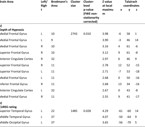
Table 3. Areas of significant (positive) correlation between grey matter volume and a) the depth of hypnosis reported by the participants, b) the suggestibility level of the participants as measured on the CURSS.
Brain Area Left/
Righ t Brodmann’s Area Cluster size Cluster-level p-value (FWE non-stationarity corrected) Z value at local maximu m Talairach coordinates x y z
a)
Depth of Hypnosis
Medial Frontal Gyrus L 10 2743 0.010 3.98 -6 58 1
Medial Frontal Gyrus L 9 3.90 -3 46 14
Medial Frontal Gyrus R 10 3.16 4 61 -6
Superior Frontal Gyrus R 10 3.12 9 61 -8
Anterior Cingulate Cortex R 32 2.97 6 46 9
Superior Frontal Gyrus R 11 2.78 12 52 -15
Superior Frontal Gyrus L 11 2.71 -7 53 -18
Medial Frontal Gyrus L 11 2.68 0 50 -16
Inferior Frontal Gyrus R 11 2.68 13 34 -16
Anterior Cingulate Cortex L 32 2.67 0 43 -8
Medial Frontal Gyrus R 11 2.55 9 41 -17
b)
CURSS rating
Superior Temporal Gyrus L 22 1485 0.028 4.29 -61 -60 14
Middle Temporal Gyrus L 37 4.07 -50 -64 9
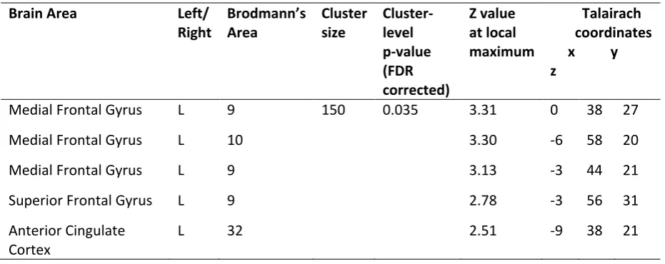
Table 4. Areas of significant (negative) correlation between reported depth of hypnosis and functional connectivity within the anterior default mode network (see Figure 2a).
Brain Area Left/
Right
Brodmann’s Area
Cluster size
Cluster-level p-value (FDR corrected)
Z value at local maximum
Talairach coordinates x y z
Medial Frontal Gyrus L 9 150 0.035 3.31 0 38 27
Medial Frontal Gyrus L 10 3.30 -6 58 20
Medial Frontal Gyrus L 9 3.13 -3 44 21
Superior Frontal Gyrus L 9 2.78 -3 56 31
Anterior Cingulate Cortex

![Table 2. Correlation matrix for suggestibility scores, hypnosis ratings, relaxation ratings, covariates (age and education [sex not included in this diagram]), total intracranial volumes, grey matter volumes, white matter volumes and cerebrospinal fluid (C](https://thumb-us.123doks.com/thumbv2/123dok_us/1602205.113064/34.595.66.536.132.591/correlation-suggestibility-relaxation-covariates-education-included-intracranial-cerebrospinal.webp)