Biomolecular Chemistry
PAPER
Cite this:DOI: 10.1039/c9ob00123a
Received 16th January 2019, Accepted 27th February 2019
DOI: 10.1039/c9ob00123a
rsc.li/obc
Control of conformation in
α
-helix mimicking
aromatic oligoamide foldamers through
interactions between adjacent side-chains
†
Irene Arrata,
a,bClaire M. Grison,
a,bHeather M. Coubrough,
a,bPanchami Prabhakaran,
a,bMarc A. Little,
aDarren C. Tomlinson,
b,cMichael E. Webb
a,band Andrew J. Wilson
*
a,bThe design, synthesis and structural characterization of non-natural oligomers that adopt well-defined conformations, so called foldamers, is a key objective in developing biomimetic 3D functional architec-tures. For the aromatic oligoamide foldamer family, use of interactions between side-chains to control conformation is underexplored. The current manuscript addresses this objective through the design, syn-thesis and conformational analyses of model dimers derived from 3-O-alkylatedpara-aminobenzoic acid monomers. The O-alkyl groups on these foldamers are capable of adoptingsyn- oranti-conformers through rotation around the Ar–CO/NH axes. In thesyn-conformation this allows the foldamer to act as a topographical mimic of theα-helix whereby theO-alkyl groups mimic the spatial orientation of theiand
i+ 4 side-chains from theα-helix. Using molecular modelling and 2D NMR analyses, this work illustrates that covalent links and hydrogen-bonding interactions between side-chains can bias the conformation in favour of the α-helix mimicking syn-conformer, offering insight that may be more widely applied to control secondary structure in foldamers.
Introduction
The design and synthesis of foldamers that adopt well-defined conformations represents the foundation for construction of non-natural folded structures with well-defined secondary, ter-tiary and quaternary structures1–6 possessing biomimetic and emergent functions.7–11 The aromatic oligoamide foldamer family12–25 has received considerable attention due to their predictable and well-defined folding behaviour. Control over conformation in the aromatic oligoamide family may be achieved by harnessing judiciously positioned hydrogen-bonding functionality along a planar backbone with well-defined steric constraints.12–14 Elegant studies have estab-lished the role of interactions between peripheral side chains in regulating the conformation of β-peptides,26–28 however
such interactions in the aromatic oligoamide family and other abiotic oligomers are less explored.29,30 This is surprising given the role of interactions between adjacent side-chains in controlling natural peptide conformation31–34and in control-ling conformation in self-assembled conjugated materials.35 Our group previously reported a series of aromatic oligoamide scaffolds designed to mimic the α-helix and inhibit α-helix mediated protein–protein interactions.36–39For the 2- and 3-O -alkylated scaffolds, these oligomers adopt an extended confor-mation determined by the trans configuration of the amide bond and in which rotation around the Ar–CO/NH axes is restricted by S(6) or S(5) intramolecular hydrogen bonding respectively (Fig. 1a for an illustration of the 3-O-alkylated scaffolds).41,42 Given the free rotation around the remaining amide–aryl bond in these oligomers, we observed no prefer-ence forsyn- oranti-conformers in helix mimetics derivatized with simple alkyl alkoxy groups.41,42Helix mimicry (Fig. 1b) of aniandi+ 4 cluster of side-chains (and thei+ 7 position for trimeric oligoamides) occurs where the oligomers adopt the
syn-conformation. Herein we demonstrate that covalent links or hydrogen-bonding interactions between suitably functiona-lized side-chains bias the conformation in favour of the syn -conformer so as to further stabilize the helix mimicking conformer in a manner that mirrors stabilisation of helical conformation throughitoi+ 4 salt bridges (Fig. 1c).31,32,43 †Electronic supplementary information (ESI) available: Synthetic procedures
and characterization, additional studies on covalently constrained helix mimetics. CCDC 1889089. For ESI and crystallographic data in CIF or other elec-tronic format see DOI: 10.1039/c9ob00123a
aSchool of Chemistry, University of Leeds, Woodhouse Lane, Leeds LS2 9JT, UK.
E-mail: [email protected]
bAstbury Centre For Structural Molecular Biology, University of Leeds,
Woodhouse Lane, Leeds LS2 9JT, UK
cSchool of Molecular and Cellular Biology, Faculty of Biological Sciences,
University of Leeds, Woodhouse Lane, Leeds, LS2 9JT, UK
Open Access Article. Published on 02 April 2019. Downloaded on 4/10/2019 1:54:03 PM.
This article is licensed under a
Creative Commons Attribution 3.0 Unported Licence.
View Article Online
Results and discussion
Using established solid-supported synthesis methodologies developed by our group,44,45model foldamer dimers1–3were prepared (Fig. 1a, see ESI† for syntheses of novel monomers and solid-phase assembly procedure), together with trimer4. Dimer1was synthesized bearing acid and amine terminated side-chains with the hypothesis that these would promote a
syn-conformer through hydrogen-bonding; evidence that such interactions between adjacent side-chains might control con-formation in helix mimetics was obtained from studies in which a constraint or“staple”30,46,47between adjacent mono-mers was introduced by olefin metathesis (discussed below for trimer 4). Dimer 2 was synthesized bearing two acid side-chains as a control predicted to disfavour thesyn-conformation due to repulsion of the carboxylic acids whilst dimer3was syn-thesised bearing two aliphatic side-chains and predicted to adopt a mixture ofsyn- andanti-conformers.
To provide evidence supporting the potential to exploit interactions between peripheral side-chains for control of fol-damer conformation, we first characterized trimer 4by X-ray and 2D NMR (Fig. 2). The molecules pack in the crystal lattice with C–H⋯πinteractions between the alkyl side chain and the
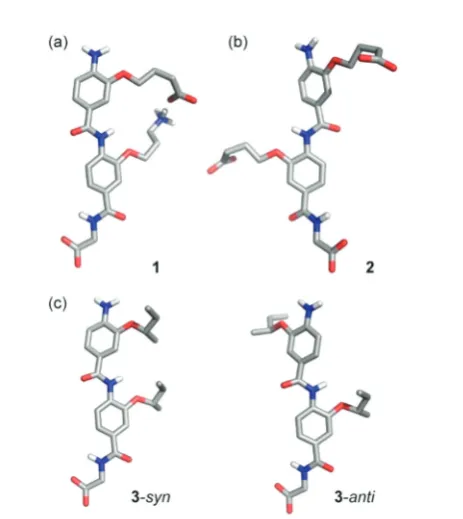
Fig. 1 Controlling conformation through side-chain side-chain inter-actions in 3-O-alkylated aromatic oligoamides: (a) model dimers 1–3
used in this study illustratingsyn-/anti-conformers; (b) model of dimer1
superimposed on an α-helix illustrating ideal matching between the
O-alkyl groups and theα-carbons in theiandi+ 4 positions (green spheres); (c) segment of an α-helix bearing a stabilising salt bridge between a Glu and Lys residue at theiandi+ 4 positions in the helix (excised fromCrystal Structure of the Mosquito-Larvicidal Toxin Cry4Ba
PDB ID: 1 W99).40
Fig. 2 Analyses of covalently constrained trimeric aromatic oligoamide foldamer4(a) single crystal structure analyses highlights the restricted nature of rotation around the Ar2–CO axis enforced by the covalent link and theanti-relationship of the groups either side of the Ar2–CO axis (b)
1
H–1H NOESY spectrum of compound4demonstrating propensity for a covalent link to restrict rotation in helix mimetics: (7–9 ppm, 500 MHz, CDCl3, 40 mM).
Fig. 3 Molecular modelling structures illustrating potential confor-mational landscape of helix mimetics: (a) dominantsyn-conformer of1
highlighting side-chain carboxylate and side-chain ammonium comple-mentarity; (b) dominantanti-conformer of 2; (c) mixture of syn- and
anti-conformers observed for3.
Open Access Article. Published on 02 April 2019. Downloaded on 4/10/2019 1:54:03 PM.
This article is licensed under a
[image:2.595.61.275.50.312.2] [image:2.595.334.521.51.316.2] [image:2.595.316.548.403.663.2]benzyl group (not shown). This extended conformation is stabilized through five membered N–H⋯O intramolecular H-bonds (dNH1⋯O = 2.15 Å,dNH1⋯O: 2.17 Å, Fig. 2a). There is clear evidence that rotation around the Ar2–CO axis by the hydrocarbon “staple”axis is restricted; the alkoxy groups are constrained to have a syn relationship, whereas unrestricted rotation around the Ar1–CO axes is evidenced by the anti -relationship between the benzyloxy and central alkoxy groups. The 2D NMR data (Fig. 2b) further reinforce these conclusions
–whilst a 2NH to 2-H2 NOE is observed, a correlation between 2-NH and 2-H6 is absent indicative of thesyn-conformation. In contrast correlations are observed from 1-NH to 1-C2H and 1-NH to 1-C6H indicating free rotation around the Ar1–CO axis and a mixture ofsyn- andanti-conformers.
To characterize the conformational preferences of these dimers, molecular modelling was performed. A Monte Carlo Multiple Minima (MCMM) molecular mechanics conformational search was carried out using MacroModel (Schrödinger) and the Merck Molecular Force Field (MMFF) force field in implicit water and with fixed ionisation state on the aromatic oligoamide
side-chains (Fig. 3). ForO-alkylated dimers, the conformations obtained depended on the composition of the side-chains. For the dimer 1 bearing complementary terminal carboxylate and ammonium groups in the side-chains, thesyn-conformation was preferred (100 of 100 low energy conformers, Fig. 3a), stabilised by side-chain to side-chain H-bonding. For the dimer2bearing two terminal carboxylates on the side-chains, the anti -confor-mation was predominant (100 of 100 low energy conformers Fig. 3b); and finally, for the dimer3bearing two alkylsBu groups as side-chains, a mixture of syn- and anti-conformation was observed (86 : 14syn:antiratio for the 100 lowest energy confor-mers, Fig. 3c). The potential for pH and therefore protonation state to affect conformational preference was further explored by modelling the dimers in different states (see ESI, Fig. ESI16–18†). These indicate that dimer1prefer thesyn- conformation in all protonation states other than where the carboxylic acid is depro-tonated resulting in repulsion of the amine. Dimer 2 on the other hand prefers an anti-conformation when both carboxylic acids are deprotonated, but thesyn- conformation is enabled by carboxylic acid dimerization.
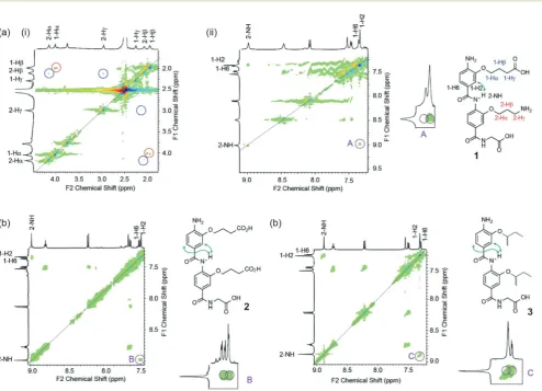
Fig. 4 NMR studies demonstrating propensity for side-chains to controlsyn-/anti-conformational preference in helix mimetics: (a)1H–1H NOESY
spectrum of compound1(i) 2–4.5 ppm and (ii) 7–9 ppm (500 MHz, DMSO-d6, 40 mM) together with expansion highlighting key diagnostic NOE (b) 1H–1H NOESY spectrum of compound2(7–9 ppm, 500 MHz, DMSO-d
6, 40 mM), together with expansion highlighting key diagnostic NOEs; (c) 1H–1H NOESY spectrum of compound3(7–9 ppm, 500 MHz, DMSO-d
6, 40 mM), together with expansion highlighting key diagnostic NOEs.
Open Access Article. Published on 02 April 2019. Downloaded on 4/10/2019 1:54:03 PM.
This article is licensed under a
[image:3.595.54.549.315.672.2]Using1H–1H NOESY NMR (Fig. 4), we could establish that the capability for dimer 1to form a hydrogen-bond between the two side-chains promoted the syn-conformation (in con-trast to mimetics bearing only acidic or aliphatic side-chains). A NOE between 1-alkoxy or 2-alkoxy side-chain resonances was not observed, however this was not surprising to us as our models indicated that the distance between these protons (e.g.
5.2 Å between 1-Hγand 2-Hγ) would likely result in low or no correlations. A correlation between 2-NH and 1-H2 however was indicative of thesyn-conformer being populated whilst the absence of a correlation between 2-NH and 1-H6 imply the absence of theanti-conformer. Furthermore, the data match those observed for the covalently constrained foldamer4(see Fig. 2b). The syn-conformation more accurately mimics the i
andi+ 4 residues in anα-helix and replicates the side-chain to
side-chain stabilization of helices observed in nature
(Fig. 1c).31,32In contrast correlations from 2-NH to 1-H2 and 1-H6 were evident for both compounds2and3indicating the presence of bothsyn- andanti-conformers. In the case of2, comparison to the protonation state dependent modelling data implies a mixture of protonation states in the experi-mental sample. Thus the modelling and experiexperi-mental data indicate hydrogen-bonding between adjacent side chains can control conformation in aromatic oligoamides, and imply that pH could be used to regulate this. Such pH switchable folda-mers will be explored in future work.
Conclusions
In summary we report side-chain mediated conformational control in model aromatic oligoamide helix mimetics. Such control might be exploited in the context of bifacial helix mimetics48to more effectively pre-organise side-chains on the opposing face for recognition of proteins and consequent inhi-bition ofα-helix mediated PPIs.38Similarly, the ability to more accurately mimic the conformational control exhibited by peptides might be exploited in future efforts to construct tertiary49,50 and quaternary structures from this class of foldamer.
Experimental section
General considerations
Unless stated otherwise, solvents and reagents were used as received from major suppliers without prior purification. Anhydrous acetonitrile, chloroform, dichloromethane were obtained from the in-house solvent purification system from Innovative Technology Inc. PureSolv®. Anhydrous
dimethyl-sulfoxide was obtained from major chemical suppliers equipped with a SureSeal or equivalent. Non-anhydrous sol-vents were of HPLC quality and provided by Fisher or Sigma-Aldrich. Water used for formation of aqueous solutions and quenching was deionised. Thin Layer Chromatography (TLC) was performed on Merck Kieselgel 60 F2540.25 mm precoated aluminium plates. Product spots were visualised under UV light (λmax = 254 nm) and/or staining with anisaldehyde. Purifications were performed with either silica gel 60 (0.043–0.063 mm VWR) using head bellows or by flash chrom-atography using an Isolera Four Biotage®. HPLC experiments were run on an Agilent 1290 Infinity Analytical Preparative
system. 1H NMR spectra were recorded on Bruker DPX 300
(300 MHz) or Avance 500 (500 MHz) spectrometers and refer-enced to residual non-deuterated solvent peaks.13C NMR were recorded on a Bruker Advance 500 (125 MHz) and referenced to the solvent peak. Chemical shifts (δ) are expressed in part per million ( ppm) and coupling constants are expressed in hertz (Hz). Spectral assignments were facilitated by COSY, HMQC and HMBC experiments when appropriate. HPLC LC-MS were recorded on a Bruker HCT ultra under the con-ditions of electrospray ionisation (ESI). HPLC separation was performed on an Agilent 1200 series instrument equipped with a Phenomenex C18 column (50 × 2 mm) using aceto-nitrile/water/1% formic acid as the eluent for positive ion spectra. HRMS were performed using a Bruker Maxis impact mass spectrometer, using ESI.
Dimer synthesis
Glycine loaded Wang resin (0.79 mmol g−1, 100–200 mesh; carrier: polystyrene, crosslinked with 1% DVB; 127 mg, 0.1 mmol) was swelled in anhydrous N-methyl-2-pyrrolidone (5 mL) 15 minutes prior to reaction. The appropriate mono-mers were dissolved in anhydrousN-methyl-2-pyrrolidone and pre-activated for coupling with Ghosez’s reagent (20% in chloroform) for 1 hour at 50 °C. The reactions were carried out on a CEM Liberty® automated microwave assisted peptide synthesizer, under microwave heating, following the con-ditions detailed in Table 1. Before each coupling, standard washing and deprotection (25% piperidine solution in
N-methyl-2-pyrrolidone) cycles were carried out on the synthe-sizer. The samples on resin were washed with dichloro-methane and diethyl ether. When required, cleavage off the resin was performed manually, using a 50% solution of TFA in dichloromethane (2 mL). TFA was removed by bubbling the solution with nitrogen and the solvent was evaporated under vacuum.
To simplify NMR assignment of the trimers, the following nomenclature is used. The monomers constituting the dimers/
Table 1 Conditions for SPPS forO-alkylated scaffold
Monomer (mmol) Ghosez (mmol) NMP (mL) Method Coupling time (min) Temperature (°C)
0.2 0.2 4 Single 30 50
Open Access Article. Published on 02 April 2019. Downloaded on 4/10/2019 1:54:03 PM.
This article is licensed under a
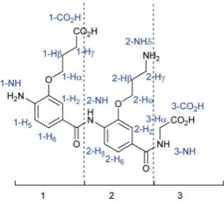
trimers are considered separately, numbered 1 to 2 or 3 start-ing from the N-terminus, and the glycine is numbered 3 or 4. All the monomers are numbered following the same standard system: the carbon bearing the carboxylic acid is C1 and the one bearing the amine is C4. The carbon attached to the nitro-gen is Cα, and the numbering of the aliphatic part of the chain continues with Cβ, etc. Numbering of protons corres-ponds to numbering of the carbons (Fig. 5). For clarity, the monomer number is added as a prefix to the proton number.
4-(2-Amino-5-((2-(3-aminopropoxy)-4-((carboxymethyl) carbamoyl)phenyl)carbamoyl)-phenoxy)butanoic acid 1
Using the general SPPS procedure with 4-((((9H-fluoren-9-yl) methoxy)carbonyl)amino)-3-(3-((tert -butoxycarbonyl)amino)pro-poxy)benzoic acid (107 mg, 0.2 mmol); 4-((((9H-fluoren-9-yl) methoxy)carbonyl)amino)-3-(4-(tert-butoxy)-4-oxobutoxy)benzoic acid (104 mg, 0.2 mmol). The residue was purified using semi-preparative HPLC (eluent: acetonitrile/water/formic acid: 0.5/9.5/0.1 to 9.5/0.5/0.1) and the product was collected as a pale cream powder (9.3 mg, 19%): δH (500 MHz, DMSO-d6) 9.01 (s, 1H, 2-NH), 8.48 (s, 1H, 3-NH), 8.11 (d, 1H,J= 8.3 Hz, 2-H5), 7.55 (s, 1H, 2-H2), 7.50 (d, 1H,J= 8.3 Hz, 2-H6), 7.38 (s, 1H, 1–H6), 7.36 (s, 1H, 1-H2), 6.71 (d, 1H, J= 8.1 Hz, 1-H5), 5.43 (s broad, 2H, NH2), 4.18 (t, 2H,J= 5.5 Hz, 2-Hα), 4.05 (t, 2H,J= 6.5 Hz, 1-Hα), 3.79 (d, 2H,J= 4.9 Hz, 3-Hα), 2.99 (t, 2H,
J= 6.6 Hz, 2-Hγ), 2.29 (t, 2H,J= 6.6 Hz, 1-Hγ), 2.08–2.15 (m, 2H, 2-Hβ), 1.96–2.01 (m, 2H, 1-Hβ); δC (125 MHz, DMSO-d6) 175.7, 171.8, 166.2, 165.2, 149.1, 145.0, 142.5, 131.1, 130.1, 130.0, 122.4, 121.6, 121.3, 120.2, 112.9, 111.2, 110.3, 68.0,
66.0, 42.2, 36.7, 27.7, 25.4. HRMS: Calcd [M + H]+
(C23H29N4O8)m/z= 489.1980, found [M + H]+m/z= 489.1978.
4-(2-(4-Amino-3-(3-carboxypropoxy)benzamido)-5-((carboxymethyl)carbamoyl)phenoxy)-butanoic acid 2
Using the general SPPS procedure with 4-((((9H-fluoren-9-yl) methoxy)carbonyl)amino)-3-(4-(tert-butoxy)-4-oxobutoxy)benzoic acid (104 mg, 0.2 mmol) for each cycle of coupling. The residue was purified using semi-preparative HPLC (eluent: acetonitrile/water/formic acid: 0.5/9.5/0.1 to 9.5/0.5/0.1) and the product was collected as a pale cream powder (4.6 mg,
9%);δH(500 MHz, DMSO-d6) 8.98 (s, 1H, 2-NH), 8.77 (t, 1H,J= 5.9 Hz, 3-NH), 8.14 (d, 1H,J = 8.4 Hz, 2-H5), 7.53 (d, 1H,J= 1.7 Hz, 2-H2), 7.50 (dd, 1H,J= 8.4, 1.7 Hz, 2-H6), 7.36 (dd, 1H,
J= 8.1, 1.9 Hz, 1-H6), 7.34 (d, 1H,J= 1.9 Hz, 1-H2), 6.68 (d, 1H,
J= 8.1 Hz, 1-H5), 4.14 (t, 2H,J= 6.2 Hz, 2-Hα), 4.04 (t, 2H,J= 6.2 Hz, 1-Hα), 3.92 (d, 2H,J= 5.8 Hz, 3-Hα), 2.44–2.47 (m, 4H, 1-Hγand 2-Hγ), 2.05 (quint., 2H,J= 6.7 Hz, 2-Hβ), 1.99 (quint, 2H, J = 6.7 Hz, 1-Hβ); δC (125 MHz, DMSO-d6) 174.9, 174.7, 171.9, 166.4, 165.1, 148.9, 145.0, 142.6, 131.3, 129.6, 122.0, 121.3, 121.1, 120.4, 112.7, 111.0, 110.7, 68.2, 67.5, 41.6, 30.9, 30.8, 24.8, 24.7; HRMS: Calcd [M + H]+ (C24H28N3O10) m/z= 518.1769, found [M + H]+m/z= 518.1775.
(4-(4-Amino-3-(sec-butoxy)benzamido)-3-(sec-butoxy)benzoyl) glycine 3
[image:5.595.88.247.49.191.2]Using the general SPPS procedure with 44-((((9H-fluoren-9-yl) methoxy)carbonyl)amino)-3-(sec-butoxy)benzoic acid (86 mg, 0.2 mmol) for each cycle of coupling. The residue was purified using semi-preparative HPLC (eluent: acetonitrile/water/formic acid: 0.5/9.5/0.1 to 9.5/0.5/0.1) and the product isolated as a dark yellow solid (10 mg, 22%);δH(500 MHz, DMSO-d6) 8.89 (s, 1H, 2-NH), 8.74 (t, 1H, 3-NH) 8.24 (d, 1H,J= 8.4 Hz, 2-H5), 7.56 (d, 1H,J= 1.7 Hz, 2-H2), 7.52 (dd, 1H,J= 8.4 Hz, 1.7 Hz, 2-H6), 7.34–7.31 (m, 1H, 1-H2), 7.30 (d, 1H,J = 1.9 Hz, 1-H6), 6.72 (d, 1H, J = 8.1 Hz, 1-H5), 5.41 (s, 2H, 1-NH2), 4.56 (sxt., 1H,J= 5.9 Hz, 2-Hα), 4.40 (sxt., 1H,J= 5.9 Hz, 1-Hα), 3.92 (d, 2H,J= 5.9 Hz, 3-Hα), 1.79–1.61 (m, 4H, 1-Hβ, 2-Hβ), 1.32 (dd, 3H,J= 6.1 Hz, 1.3 Hz, 2-CHα(CH3)), 1.28 (dd, 3H,J= 6.1 Hz, 2.3 Hz, 1-CHα(CH3)), 0.99–0.94 (m, 6H, 1-Hγ, 2-Hγ); δC (125 MHz, DMSO-d6) 171.4, 165.8, 164.4, 147.0, 143.5, 143.2, 131.7, 128.9, 121.1, 120.9, 119.9, 112.8, 112.2, 76.0, 75.3, 41.3, 40.4, 28.6, 28.5, 19.1, 19.0 18.9, 9.6, 9.4; HRMS: Calcd [M + H]+(C24H32N3O6) m/z= 458.2286, found [M + H]+m/z= 458.2299.
Trimer 4
Trimer4was prepared directly from cross-linked dimer using procedure (b) followed by coupling with using dichlorotriphe-nyl phosphorene in chloroform as previously described. Yield: 26%; mp: 237 °C; 1H (CDCl3, 500 MHz)δ: 8.66–8.63 (m, 3H, 1-NH, 2-H5, 3-H5), 8.49 (s, 1H, 2-NH), 7.95–7.93 (d,J= 8.3 Hz, 1H, 1-H5), 7.78 (s, 1H, 1-H2), 7.75–7.72 (m, 2H, 2-H6, 3-H6), 7.56 (s, 1H, 3-H2), 7.50–7.48 (m, 2H, Ph-H1), 7.43–7.40 (m, 3H, 1-H6, Ph-H2), 7.37–7.34 (m, 1H, Ph-H3), 7.17 (s, 1H, 2-H2), 5.34 (s, 2H, OCH2Ph), 4.33–4.30 (m, 2H, OCH2), 4.17–4.15 (m, 2H, OCH2), 3.91 (s, 3H, OCH3), 1.89–1.83 (m, 4H, CH2), 1.61–1.50 (m, 8H, CH2);13C (CDCl3, 125 MHz)δ: 166.8, 165.3, 163.1, 152.2, 146.9, 146.6, 142.1, 139.8, 135.0, 132.0, 131.2, 131.0, 128.8, 128.5, 127.2, 125.9, 125.2, 123.3, 122.7, 120.4, 118.7, 117.9, 115.0, 111.1, 107.4, 71.5, 69.3, 68.6, 52.1, 29.3, 26.8, 26.6, 26.4, 24.7, 22.1; IR (neat)ν(cm−1): 3424, 2934, 2864, 1722, 1689, 1667, 1594, 1515, 1345, 1262, 1204, 1124, 1108, 1006; HRMS: Calcd [M − H]− (C37H36N3O9) m/z = 666.2452. Found [M−H]−m/z= 666.2485. Elemental analysis calculated for C37H37N3O9: C, 66.56; H, 5.59; N, 6.29; Found: C, 65.85; N, 5.50; N, 6.00.
Fig. 5 Example of numbering for anO-alkylated dimer1.
Open Access Article. Published on 02 April 2019. Downloaded on 4/10/2019 1:54:03 PM.
This article is licensed under a
Procedure for modelling
Monte Carlo Multiple Minima (MCMM) molecular mechanics conformational search was carried out using MacroModel (Schrödinger) and the Merck Molecular Force Field (MMFF), with a cut-offof 1 Å in implicit water. The calculation used a maximum of 10 000 steps and maximum of 100 steps per rotational bond, 100 structures were saved within an energy window of 10 kJ mol−1. Angle and torsional constraints with a force field of 1000 were applied to all amine and amide func-tional groups to fix an sp2conformation and charges were set to account for the pKa of the relevant groups and anticipated protonation state.
Conformational analyses by1H NMR
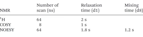
NMR experiments performed to determine the conformation of the dimers 1–3 and trimer 4 were acquired on a Bruker Avance 500, at 500 MHz and 26 °C ( parameters in Table 2).
Con
fl
icts of interest
There are no conflicts to declare.
Acknowledgements
This work was supported by the Leverhulme Trust [RPG-2013-065] and the EPSRC [EP/KO39292/1], for funding NMR facili-ties. Thanks to Silvia Rodriguez-Marin for preliminary syn-thetic efforts towards compound 3. Thanks to Christopher Pask for assistance with X-ray structure deposition (CCDC 1889089†).
Notes and references
1 W. S. Horne and S. H. Gellman,Acc. Chem. Res., 2008,41, 1399.
2 G. Guichard and I. Huc,Chem. Commun., 2011,47, 5933. 3 B. A. F. Le Bailly and J. Clayden,Chem. Commun., 2016,52,
4852.
4 A. Roy, P. Prabhakaran, P. K. Baruah and G. J. Sanjayan,
Chem. Commun., 2011,47, 11593.
5 I. Saraogi and A. D. Hamilton, Chem. Soc. Rev., 2009, 38, 1726.
6 Foldamers: Structure, Properties, and Applications, ed. S. Hecht and I. Huc, Wiley-VCH, Weinheim, 2007.
7 F. G. A. Lister, B. A. F. Le Bailly, S. J. Webb and J. Clayden,
Nat. Chem., 2017,9, 420.
8 P. K. Mandal, B. Baptiste, B. Langlois d’Estaintot, B. Kauffmann and I. Huc,ChemBioChem, 2016,17, 1911. 9 J. W. Checco, E. F. Lee, M. Evangelista, N. J. Sleebs,
K. Rogers, A. Pettikiriarachchi, N. J. Kershaw,
G. A. Eddinger, D. G. Belair, J. L. Wilson, C. H. Eller, R. T. Raines, W. L. Murphy, B. J. Smith, S. H. Gellman and W. D. Fairlie,J. Am. Chem. Soc., 2015,137, 11365.
10 S. Kwon, B. J. Kim, H.-K. Lim, K. Kang, S. H. Yoo, J. Gong, E. Yoon, J. Lee, I. S. Choi, H. Kim and H.-S. Lee, Nat. Commun., 2015,6, 8747.
11 M. Wolffs, N. Delsuc, D. Veldman, N. V. Anh,
R. M. Williams, S. C. J. Meskers, R. A. J. Janssen, I. Huc and A. P. H. J. Schenning, J. Am. Chem. Soc., 2009, 131, 4819.
12 D.-W. Zhang, X. Zhao, J.-L. Hou and Z.-T. Li, Chem. Rev., 2012,112, 5271.
13 B. Gong,Acc. Chem. Res., 2008,41, 1376. 14 I. Huc,Eur. J. Org. Chem., 2004, 17.
15 Y. Zhao, A. L. Connor, T. A. Sobiech and B. Gong,Org. Lett., 2018,20, 5486.
16 A. Lamouroux, L. Sebaoun, B. Wicher, B. Kauffmann,
Y. Ferrand, V. Maurizot and I. Huc,J. Am. Chem. Soc., 2017, 139, 14668.
17 S. Kumar, A. Henning-Knechtel, I. Chehade, M. Magzoub
and A. D. Hamilton, J. Am. Chem. Soc., 2017, 139,
17098.
18 R. Annala, A. Suhonen, H. Laakkonen, P. Permi and M. Nissinen,Chem.–Eur. J., 2017,23, 16671.
19 M. Kortelainen, A. Suhonen, A. Hamza, I. Pápai, E. Nauha, S. Yliniemelä-Sipari, M. Nissinen and P. M. Pihko,Chem.– Eur. J., 2015,21, 9493.
20 S. S. Kale, S. M. Kunjir, R. L. Gawade, V. G. Puranik, P. R. Rajamohanan and G. J. Sanjayan, Chem. Commun., 2014,50, 2886.
21 G. Priya, A. S. Kotmale, D. Chakravarty, V. G. Puranik, P. R. Rajamohanan and G. J. Sanjayan,Org. Biomol. Chem., 2015,13, 2087.
22 Z. Lockhart and P. C. Knipe,Angew. Chem., Int. Ed., 2018, 57, 8478.
23 J. Luccarelli, I. M. Jones, S. Thompson and A. D. Hamilton,
Org. Biomol. Chem., 2017,15, 9156.
24 P. C. Knipe, I. M. Jones, S. Thompson and A. D. Hamilton,
Org. Biomol. Chem., 2014,12, 9384.
25 N. Busschaert, S. Thompson and A. D. Hamilton, Chem.
Commun., 2017,53, 313.
26 R. P. Cheng and W. F. DeGrado, J. Am. Chem. Soc., 2001, 123, 5162.
27 P. I. Arvidsson, M. Rueping and D. Seebach, Chem.
Commun., 2001, 649.
28 L. M. Johnson, D. E. Mortenson, H. G. Yun, W. S. Horne, T. J. Ketas, M. Lu, J. P. Moore and S. H. Gellman, J. Am. Chem. Soc., 2012,134, 7317.
[image:6.595.43.292.66.131.2]29 M. Baskin, H. Zhu, Z.-W. Qu, J. H. Chill, S. Grimme and G. Maayan,Chem. Sci., 2019,10, 620.
Table 2 Parameters for 2D NMR experiments
NMR
Number of scan (ns)
Relaxation time (d1)
Mixing time (d8)
1H 64 2 s
COSY 8 1 s
NOESY 64 1.8 s 1.2 s
Open Access Article. Published on 02 April 2019. Downloaded on 4/10/2019 1:54:03 PM.
This article is licensed under a
30 L. Zheng, C. Yu, Y. Zhan, X. Deng, Y. Wang and H. Jiang,
Chem.–Eur. J., 2017,23, 5361.
31 J. M. Scholtz, H. Qian, V. H. Robbins and R. L. Baldwin,
Biochemistry, 1993,32, 9668.
32 S. Marqusee and R. L. Baldwin, Proc. Natl. Acad. Sci. U. S. A., 1987,84, 8898.
33 L. K. Tsou, C. D. Tatko and M. L. Waters,J. Am. Chem. Soc., 2002,124, 14917.
34 R. M. Hughes and M. L. Waters,J. Am. Chem. Soc., 2006, 128, 13586.
35 S. A. Sharber, R. N. Baral, F. Frausto, T. E. Haas, P. Müller and S. W. Thomas III, J. Am. Chem. Soc., 2017, 139, 5164.
36 G. M. Burslem, H. F. Kyle, A. L. Breeze, T. A. Edwards, A. Nelson, S. L. Warriner and A. J. Wilson,Chem. Commun., 2016,52, 5421.
37 A. Barnard, K. Long, H. L. Martin, J. A. Miles,
T. A. Edwards, D. C. Tomlinson, A. Macdonald and A. J. Wilson,Angew. Chem., Int. Ed., 2015,54, 2960.
38 V. Azzarito, J. A. Miles, J. Fisher, T. A. Edwards, S. L. Warriner and A. J. Wilson,Chem. Sci., 2015,6, 2434. 39 G. M. Burslem, H. F. Kyle, A. L. Breeze, T. A. Edwards,
A. Nelson, S. L. Warriner and A. J. Wilson,ChemBioChem, 2014,15, 1083.
40 P. Boonserm, P. Davis, D. J. Ellar and J. Li,J. Mol. Biol., 2005,348, 363.
41 P. Prabhakaran, V. Azzarito, T. Jacobs, M. J. Hardie, C. A. Kilner, T. A. Edwards, S. L. Warriner and A. J. Wilson,
Tetrahedron, 2012,68, 4485.
42 V. Azzarito, P. Prabhakaran, A. I. Bartlett, N. S. Murphy, M. J. Hardie, C. A. Kilner, T. A. Edwards, S. L. Warriner and A. J. Wilson,Org. Biomol. Chem., 2012,10, 6469.
43 P. E. Czabotar, E. F. Lee, G. V. Thompson, A. Z. Wardak, W. D. Fairlie and P. M. Colman,J. Biol. Chem., 2011,286, 7123.
44 N. S. Murphy, P. Prabhakaran, V. Azzarito, J. P. Plante, M. J. Hardie, C. A. Kilner, S. L. Warriner and A. J. Wilson,
Chem.–Eur. J., 2013,19, 5546.
45 K. Long, T. A. Edwards and A. J. Wilson, Bioorg. Med. Chem., 2013,21, 4034.
46 J. A. Miles, D. J. Yeo, P. Rowell, S. Rodriguez-Marin, C. M. Pask, S. L. Warriner, T. A. Edwards and A. J. Wilson,
Chem. Sci., 2016,7, 3694.
47 D. J. Yeo, S. L. Warriner and A. J. Wilson,Chem. Commun., 2013,49, 9131.
48 S. Rodriguez-Marin, N. S. Murphy, H. J. Shepherd and A. J. Wilson,RSC Adv., 2015,5, 104187.
49 M. K. P. Jayatunga, S. Thompson and A. D. Hamilton,
Bioorg. Med. Chem. Lett., 2014,24, 717.
50 O. V. Kulikov, S. Thompson, H. Xu, C. D. Incarvito, R. T. W. Scott, I. Saraogi, L. Nevola and A. D. Hamilton,
Eur. J. Org. Chem., 2013, 3433.
Open Access Article. Published on 02 April 2019. Downloaded on 4/10/2019 1:54:03 PM.
This article is licensed under a