THE FUNCTIONAL ORGANIZATION OF THE NEURONES OF THE CEREBELLAR CORTEX
by
Rodolfo R. Llina s
Thesis submitted for the Degree of Doctor of Philosophy in the Australian National University
Owing to the complex nature of the experimental procedures employed in these investigations, the experiments to be- reported here were carried out in collaboration with Professor Sir John C. Eccles and with Dr. Kasuo Sasaki. However, I was responsible for the initiation of the experiments on stellate and Golgi cell in-hibition as well as for the mossy fibre "axon reflex" and the
"climbing fibre reflex". The deafferented cerebellum preparation, developed for the studies of the antidromic field potentials of Purkinje cells and the field potentials generated by parallel fibre stimulation, is entirely my own work.
As a result of the investigations in which I have participated during the tenure of my scholarship at the Australian National University, the following communications and papers have appeared or are in the course of publication:
Eccles J.C. , R. Llinas and K. Sasaki: Excitation of cerebellar Purkinje cells by the climbing fibres. Nature (Lond.) 203; 245-246, 1964.
Eccles J.C. , R. Llina's and K. Sasaki: Golgi cells inhibitions in the cerebellar cortex. Nature (Lond.) 204; 1265-1266, 1964.
#
Eccles J.C. , R. Llinas and K. Sasaki: The excitatory synaptic
actions of climbing fibres on the Purkinje cells of the cerebellum.
J. Physiol (In press) 19 6 5.
Eccles J.C. , R. Llina's , K. Sasaki and P. Voorhoeve: Interaction experiments on the responses evoked in Purkinje cells by climbing
fibres. J. Physiol (In press) 1965.
.
,
Eccles J.C., R. Llinas and K. Sasaki: The actions of antidromic
impulses on, the cerebellar Purkinje cells. J. Physiol (In press) 1965.
•·
Eccles J.C. , R. Llinas and K. Sasaki: The inhibitory
interneu-rones within the cerebellar cortex. Exp. Brain Res. 1; 1-16, 19 6 5 .
,
Eccles J.C. , R. Llinas and K. Sasaki: Parallel fibre stimulations and the responses induced thereby in the Purkinje cells of the cere-bellum. Exp. Brain Res.
l;
17-39, 1965.I
Eccles J. C . , R. Llina s and K. Sasaki: The mossy fibre - granule
cell relay of the cerebellum and its inhibitory control by Golgi cells.
Exp. Brain Res.
l;
82-101, 1965..
.Eccles J.C., R. Llinas and K. Sasaki: Intracellularly recorded re-sponses of the cerebellar Purkinje cells. Exp. Brain Res. (In press), 1965.
,
Table of Contents Abbreviations
I GENERAL INTRODUCTION II METHODS
A) Surgical techniques
B) General experimental arrangements
C) Surgical technique for cerebellar deafferentatibn.-..
I I I A) THE CLIMBING FIBRE AFFERENT SYSTEM OF THE CEREBELIAR CORTEX.
a) Potential field evoked in the cerebellar cortex by electrical stimulation in the region of the inferior olive.
b) Extracellular recording of ·:.spike potentials c) Intracellular recording from Purkinje cells
d) The projection from the inferior olive to Purkinj e cells e) Latency of the unitary responses
f) Other modes of activation of climbing fibres
g) Effects of polarization of the Purkinj e cell on the excitatory postsynaptic potential generated by climbing fibre impulses
B) Interaction between responses evoked in Purkirije <tells by
climbing fibres and other Purkinje cell tnputs
a) Inhibitory action on the responses evoked in Purkinje cells by climbing fibres .
b) Extracellular recording from single Purkinje cells
c) Interactions recorded intracellularly from Purkinj e cells d) Double activation of climbing fibres
I .
C) DISCUSSION
IV A) THE FlJ..NCTIONAL ORGANIZATION OF THE CEREBELLAR GLOMERULUS a) Field potentials generated by mossy fibre-granule cell activation b) Mossy fibre activation and Golgi inhibition of single granule
cells
c) Field potentials evoked on the molecular layer by M. G. R. activation
d) Golgi cell inhibition of the Purkinj e cell EPSP evoked by activation of the ·mossy fibre~gr anule celJ relay (M. G . R.). e) Mossy fibre II
axon reflex 11
B) DISCUSSION
V A) THE PARALLEL FIBRE SYSTEM AND ITS FUNCTIONAL REIATIONS WITH THE NEURONES OF THE CEREBELLAR CORTEX
a) Field potentials generated by impulse propagation in parallel fibres
b) Conduction times along the beam of excited parallel fibres c) Experiments on the refractoriness following a parallel fibre
volley
d) The slow potential waves produced by a parallel fibre volley e) Potential waves evoked by a second parallel fibre volley at
various intervals
f) · Generation of impulses in Purkinj e cells by the parallel fibre volleys
B) Parallel fibre activation of the interneurones of the cerebe1la r
· cortex
C} DISCUSSION
a) Interpretation of the slow potential waves generated by a parallel fibre volley
. .
b) Interpretation of. slow potential waves reduced by a parallel fibre volley conditioned by a preceding volley
VI A) THE ORGANIZATION OF THE PURKINJE CELL INPUTS ACTIVATED
BY THE M·. G9 R.
a} Orthodromic activation of Purkinje cells recorded intra-:-cellularly
b) Excitatory and inhibitory postsynaptic potentials of Purkinj e cells
c) Effects of applied trans membrane currents on inhibitory postsynaptic potentials
d) The inhibitory synaptic noise of Purkinj e cells
· e) The action of Loe. stimulation in conditioning the intracellular responses . evoked by Loe. · or mossy fibre stimulation
B) DISCUSSION
a) Inhibitory synaptic noise
b) The EPSP and IPSP generated by a parallel fibre volley
c) Production of IPSP by a parallel fibre volley when conditioned by a preceding parallel volley
d) Comparison of inhibitory synaptic actions evoked by Loe _. and
J.
F. stimulationVI I A) THE ANTIDROMIC INVASION OF PURKINJE CELLS a) Antidromic responses of single Purkinje cells
b) Field potentials produced in the cerebellar cortex by juxta-fastigial stimulation
d) Inhibitory action of antidromic impulses on the rhythmic responses of Purkinje cell
B) DISCUSSION
a) Interpretation of the field potentials produced by antidromic invasion of Purkinj e cells
b) Inhibitory and excitatory synaptic action on antidromic impulse conduction
VIII CONCLUDING REMARKS Acknowledgements
Abbreviations Anatomy:
BC - basket cell CF - climbing fibre GL - granular layer Glo - glomerulus Goe - Golgi cell GrC - granule cell MF - mossy fibre
MGR - mossy fibre-granule cell relay PA - Purkinj e axon
PAC - Purkinj e axon collateral PC - Purkinj e cell
PF - parallel fibre PL - Purkinj e layer SC - stellate cell Electroph ys iology:
EPSP - excitatory post-synaptic potential IPSP - inhibitory post-synaptic potential Stimulating electrodes:
DR - deep radial nerve IO - inferior olive
JF - juxta-fastigial region LOC - local cortical
LCN - lateral cuneate nucleus TF - transfolial
Recording electrodes: ME - microelectrode
I GENERAL INTRODUCTION
One of the most challenging problems in developing an under-standing of the physiology of the nervous system is the one concern-ing the relations between structure and function at the neuronal level.
In studying the structure of the central nervous system one can-not but be fascinated by the geometrical organization of the neuro-nal elements of the cerebellar cortex. This organization is the more amazing as the regularity of the relations between the different el-ements is constant throughout the extent of the cortex.
From the electrophysiological point of view the cerebellum re-presents a unique opportunity to study functional organization of complex neuronal chains .
The history of the discovery of the histology of the cerebellar cortex saw its dawn in 1837, when
J.
E. Purkinje, the Czech profes-sor of physiology at Breslau described the existence of what he call-ed IIGangliosen Korperchen II
in the cortex of the cerebellum. These cells named after their discoverer, Purkinje cells, are the central el-ement of the histological organization of the cerebellar cortex. Purkinj e described these voluminous bodies as forming a clear-cut layer, one element in thickness, close to the surface of the cerebellar crust.
In 18 6 5 Deiters described the axonal prolongation of the Purkinj e cells, their collaterals being described by C. Golgi in 188 6, with
2
erals of these cells and the plexus formed by their secondary and
tertiary branches Golgi described also in 1886 the existence of the large
stellate cells of the granule layer, which are known today as Golgi cells,.
In collaboration with Furasi (Golgi and FurasL, 1886) he discovered the existence of the basket cells, but as in the case of the large stars of the granule layer, failed to recognize the mode of termination of their axons .
However, it was the arduous work of S. Ram6n y Cajal which
gave the histology of the cerebellar cortex its status as the best known structure of the central nervous system. This anatomist described the mossy fibres and the granule cells, as well as their synapse (1888).
In 1890, he discovered the mode of termination of the Golgi cell axon of which he writes in 1904, 11
• • • se concentran especialmente en los
islotes cerebelosos, en donde entran en intimo contacto con las
ar-borizaciones dendrlticas de los granos 11
• In 1888, he demonstrated the
presence of climbing fibres in birds and in 1890 in mammals, illus-trating their intimate relation with the dendrites of the Purkinj e cells. In the same 1888 paper on the molecular layer of the cerebellum of birds, Cajal gave the first description of the parallel fibres and their origin as bifurcation of the axons of the granule cells. He discussed
also in this paper the relat1onsh1p between the depth of a granule
cells and to the interneurones of the cerebellar cortex.
It is of interest to mention that although it was C. Golgi who
first :described the basket cells, Cajal discovered the basket arrange-ment of their axon terminal around the bodies of the Purkinje cells,
3
this being the first time an axon had been seen to enter in contact with another nervous element of the gray matter. On the basis of this sin-gle histological finding Cajal arrived at three conclusions (1888), a)
11 nervous currents" can propagate between two elements by means of
their contacts and thus, jump from one corpuscle to another, b) Axonic prolongation can be the place of emission of "nervous currents II
and the soma the place of reception. c) The initial part of the axon can
also be a place of reception. Finally the stellate cells of the
molec-ular layer were first described by Smirnov in 1887, who described several types of them. Based on his studies, Cajal proposed in his monograph·. 11
La textura del sistema nervioso del hombre y de los ver-tebra dos 11
19 0 4, two main systems of II
currents II
serve as an "avalanche · conduction system" to boost the input from this omnicellular pathway to the Purkinje cell. The histological pic-ture, at this level, almost reached completion after the works of the early anatomists. In recent years the introduction of new histological techniques such as the Nauta stain as well as the use of electron mi-croscopy for the study of the ultrastructure of nervous tissue, have allowed further developments in the understanding of the morphologi-cal organization of this cortex. The new findings on the subject will be discussed in the body of this thesis.
The study of the functional organization of the different neural elements of the cerebellar cortex can be said to have started with the pioneering experiments of Dow in 1949. This investigator demon-strated that local stimulation of the surface of the cerebellar cortex produced negative potentials which were conducted along the main axis of the folia of the cerebellum and which he related to the acti
-vation of parallel fibres. However, . the modern analysis of the func-tional characteristics of the neural elements of the cerebellar cortex begins with the microelectrode studies by Granit and Phillips (19 56 and 195 7) on the electrical activity of single Purkinje cells of the cat cerebellum. These authors were the first to have clear extracellular and intracellular records from Purkinj e cells , identified by their anti-dromic invasion from the white matter of the cerebellum. Similar re-sults were also obtained by Matthews, Phillips and Rushworth (19 5 7) ,
II METHODS
A) Surgical techniques
The experiments to be described in this thesis were performed in cats ranging in weight from 3 to 3. 5 kilograms. They were anesthe-tized with 30 mgm/kilo of sodium pentobarbital, administered intra-peritoneally.
The radial vein was then catheterized in order to continue the barbiturate anesthesia throughout the length of the experiments, as well as to administer other drugs. A tracheotomy was always per-formed and a tracheal canulae inserted into place to allow artificial ventilation of the animal.
The cat's head was then fixed on a stereotaxic frame, the pelvis being suspended by an iliac clamp. A single craniocaudal incision of the skin of the head was performed, it's borders being sewn to a
stain-less steel ring in order to create a pool for the Ringer's solution. The
temporal and neck muscles were disinserted and the cranium scraped of periosteal tis sue.
a large proportion of the posterior lobe folia. Recordings were re-stricted to the culmen and lobus simplex. Laterally, ·the craniotomy extended the whole width of the vermal region. This ample exposure was heeded in order to allow the selection of a recording site in the area of maximum response to the multiple inputs under study. The craniotomy was performed with bone nibblers under continuously running warm Ringer solution in order to prevent air embolism.
B) General~erimental arra129ftffient.s
Figs. 1 and 2 illustrate diagrammatically the general experimen-tal arrangements. Monopolar surface recordings were obtained with a spring mounted silver ball electrode (SR) that was insulated with
6
P. V. C . except for the very small area in contact with the cortex. The stimulating electrodes included a juxta-fastigial electrode (J .. F. ).
consisting of three independent bipolar concentric electrodes. A num-ber 30 hypodermic needle tubing was insulated with epoxy-resin up ·to
SOOu from the tip and an enamel insulated internal nichrome wire
No. 48 (S. W. G.) protruded about 700u from the end of the tube.- These three electrodes were cemented together so that their tips were 1. 5 mm apart, allowing in this manner a very localized stimulus at each of
ferred to as the inferior olive electrode (I. 0.). A very short flat-ended concentric electrode (2 mm in length and outside diameter
0. 8 mm) mounted on springy wire, was utilized to stimulate the sur-face of the cortex (LOC.), and of an adjacent folium, the transfolial stimulating electrode (T. F.). This surface electrodes allow a very localized region of stimulation. The superficial and deep radial nerves were stimulated with platinum electrodes under art oil pool.
Once the optimum responses were recorded with the surface electrode, the latter was removed to a nearby region of the same folium; and, after covering the exposed area of the cerebellum with an agar gel, a microelectrode for depth recording was lowered into the region of maximum response. Microelectrodes were filled with
4 mol. NaCl for the laminar field analysis (2 M!l. average D. C.
resistance) and with 3 mol KC 1 or 2 mol K citrate for the intracellular recordings (10
Mn
average D. C. resistance). In order to minimize7
the movement of the cerebellar cortex, a bilateral pneumothorax was performed after the animal had been paralyzed with II
Flaxedil II
and artificially ventilated. A bridge circuit (Araki and Otani, 19 55) with a
100
Mn
resistor facing the grid of the cathode follower was used to pass current through the impaled cell. An R . C. coupled amplifier with a time constant of 3 to 10 seconds was used for the potentialmem-brane potential was constantly monitored with a D. C. amplifier feeding a voltmeter.
In the initial stages of this work there were histological con-trols (haematoxylin - e osin) of the stimulating electrode placements in the inferior olive. Once the "typical II
olivary response was es-tablished, however, only occasional checks were deemed necessary, because the cerebellar responses evoked by the olivary stimulation were so characteristic.
C) Surgical technique for cerebellar deafferentation
In order to deafferent the cerebellar cortex a bilateral pedun-culotomy was performed and a minimum post-operative survival time
8
of eight days allowed before experimentation. The surgery was done under heavy "nembutal II
anaesthesia (45 mgms/kg} and the cerebellum was approached from its caudal end by removal of the squamous
plate of the occipital bone; the craniotomy was extended to the lamb-doidal ridge. In order to obtain an ample exposure of the floor of the fourth ventricle, the caudal region of the vermis of the posterior lobe, from the fissura praepyramidalis, to the velum medulare posterior, was removed by suction. The ventro ,-lateral part of both cerebellar
I'
9
direct vision, and thus to spare as much as possible the circulation to the cerebellum. Following the pedunculotomy the neck muscles, clavo-trapesius and occipito-scapularis of both sides, were approxi-mated in the midline and joined with the caudal edge of the temporal muscles, which had been previously severed at their origins, so that a protecting muscle sheath was created over the craniotomy. The skin was closed using metal clips.
All the cats operated upon underwent the typical decerebellate syndrome (Dow and Moruzzi, 19 58) with a "dynamic stage" lasting from two to three days during which extensor rigidity ahd opisthotonos was observed. Following this state the characteristic atonic period appeared. The animals showed intentional tremor with dysmetria and nystagmus. Nystagmus was present specially during psychical
ex-citation, e.g. when presented with food. All through the post-operative period, lasting up to 2 3 days, the cats were completely ataxic and unable to stand . A week after the operation, however, they were all able to support their weight, and even to climb the wire screen on the sides of their cages, thus showing that their flexor responses were not as
im-paired as their antigravity n1e c h.anism. The cats were fed thr ough an oesophageal canula during the first week, after which they began to eat unaided.
'
10
were used to study the degeneration pattern produced by the pedun-culotomy. These techniques demonstrated that for the most part the
degenerating fibres were restricted to the white matter of the cerebellum, leaving the actual cerebellar cortex intact. Also small portions of
cerebellar cortex were removed at the end of the experiment , before ,
the death of the animals, fixed in 4% osmic acid and imbeded in "Vestopal II
for electronmicroscopical (E. M.) observations which were kindly performed for us by Professor C .A. Fox and Dr. D. E. Hillman of the Departm_ent of Anatomy, Marquette University. The E. M.
studies de.monstrated degeneration of mossy fibres leaving the granule I'
cells intact. A striking observation previously described by Hamori (19 6 4); Fox (personal communication) and S zenta'gothai (personal communication) was the survival of the cerebellar glomeruli of the
I
' i
III-A
la
I
THE CLIMBING FIBRE AFFERENT SYSTEML OF THE CEREBELLAR CORTEX
The .climbing fibres of the cerebellar cortex were first described by Ramon y Cajal in his study of the moiecular · layer of the cerebel-lum of the pigeon, (1888) and were further investigated in mammals
'
,
by him (Ramon y Cajal and Illera, 1907; .Ramon y Cajal, 1911) · and
many other neurohistologists of his time. According to this anatomist the climbing fibres are neurotubules which traverse the granular layer of the cerebellar cortex and enter in contact with the dendrites of
11
Purkinje cells. Upon reaching the Purkinje layer the climbing fibre branches out in order to twine around each of the large primary and secondary dendrites of the Purkinje cell (Fig.
1 )
without entering in contact with the small spiny branchlets. This very extensive and in-timate contact which resembles the pattern of a climbing vine wasas-sumed by Ramo'n y Cajal to form a powerful synaptic junction on to the Purkinje cell. The study of the ultrastuctural details of this synapse has demonstrated these assumptions to be quite correct. Ultrastruc-ture studies have revealed very large synaptic contacts between the
enlarged portions of climbing fibres and the smooth branches of the
,
Purkinj e cells. (Szentagothai, (19 64a, 19 64b) and Fox; personal com-munication).
I
'
'
12
Szentagothai (1964a, 1964b). The study of the origin of the climbing fibres using the conventional degenerative methods has proved to be technically, extremely difficult. As a consequence, several differ-ent origins have been proposed for the climbing fibres. On the gen-eral grounds of the widespread distribution of the oli vo-cerebellar fibres Dow (1942) suggested that the climbing fibres originated in the inferior olive. All other cerebellar inputs being via mossy fibres, similar conclusions had been reached by other authors in the past, Brouwer and Coenen (1919, 1921) and Winkler (1923). However, it
,
was not until the study by Szentagothai and Rajkovits (1959), that a sound experimental basis was provided --for the oHvary or'i'g'in -6£. :the climbing fibres.
The present investigation. ·has utilized this discovery of Drs.
,
Szentagothai and Rajkovits, it being assumed that climbing fibres
projecting to the anterior lobe of the cerebellar vermis could be
selec-tively excited by an electrode located near the accessory olives (Jansen and Brodal, 19 54). Stimulation of the inferior olive has been found to
evoke electrical responses widely dispersed over the cerebellum (Dow , 1939; Combs, 1956; Jansen, 1957). The dominant potential was a
in-ferior olive.
a) Potential field evoked in the. cerebellar cortex by electrical stimulation in the region of the inferior olive
1 3
A series of potentials evoked by .a.stimulus applied through an electrode in the .region of the inferior olive and recorded by a micro-electrode in the contralateral vermal cortex at various depths below the surface are shown in Fig .. · 3 ,A, B, C. This field is composed at all depths of an initial neg·ative wave with a fairly steep rise and a slower decline on which small irregular spike-like potentials were superimposed. This negative wave declined to a small slow positive wave, which was relatively larger at deeper levels.
In about half of the experiments on potential profiles this· late positive wave was dominant at the deeper levels (cf. Fig. 3 B, C, D) there being on l y :"a s:r'na ll initial negativity below 2 SOu ·
I'
microelectrode which prodded the cerebellar cortex in depth. At the superficial levels the respective potentials differed in size, but not significantly in configuration and in time course with those recorded deeper, due to a large initial negative wave and a later positive wave, which became dominant with depths from 2 30u downwards. However,
at 330u and 430u the strongest stimulus ( B ) evoked a positive wave
. '
which being larger, suggested that it could be .prcodw.ced:.in part} qy·_ a spread of stimulus to structures adjacent to the inferior olive, such
as the reticulo-cerebellar fibres from the lateral reticular nucleus.
Otherwise the potential profiles of Figs. 3B- D , may be regarded as being produced by the activation of the olivo-cerebellar pathway.
The latency of the negative wave in the superficial levels of the cerebellar cortex is so brief (3. 9 to 6. 8 msec in 4 experiments) that it must be produced directly by the olivo-cerebellar pathwq.y, which has been shown by Szentagothai and Rajkovits (1959). to end
ex-clusively as climbing fibres . Hence the potential profiles of Fig. 3 can be regarded as being produced by the impulse of climbing fibres and by the synaptic and action potentials generated by them on the Purkinj e cells .
II
p 1,
greatest synaptic density. The decline in negative potential and even its reversal to a positive wave below 200u in B-D are attrib-utable to passive sources at the somatic and axonic level of the Purkinje cell with respect to the active sinks of the main dendrites. In Fig. 3, it can be seen that at more sµperficial levels there is, correspondingly, a larger latency of the initial negative wave, which is attributable to the conduction time of the climbing fibres. An ap-proximate conduction velocity of O .15 to O. 5 msec was calculated on this assumption for the fine terminal branches of these fibres (four experiments) .
The diphasic negative-positive potential produced by inferior olive stimulation at very superficial levels (Fig. 3 ) and actually
15
on the surface (Fig. 4E ) is in good agreement with the potentials recorded with gross surface recording (usually a negative-positive wave) which are found following such stimulation (Dow, 19 59; Jansen, 19 5 7). There are also subsidiary synaptic excitatory actions of climb-ing fibres on basket cells and on Golgi cells , findclimb-ings which are in accord with histological observations (Scheibel and Scheibel, 19 54;
,
Szentagothai, 1964); but these actions could make, due to their weak-ness, only a small contribution to the potential field, and would in
any case produce a much deeper negativity (about 400u) than the one actually observed. There is, however, the further possibility that
16
.
,
19641 and Golgi cells (Eccles, Llinas and Sasaki, 1964b) may contrib-ute to the positive potential fields at the deeper levels in Fig. 3.
Since the climbing fibres are slender and rather sparsely distrib-buted, it would be expected that the action currents of these fibres would give a negligible contribution to the observed potential field, particularly as their range of conduction velocities would result in a temporal dispersion over several milliseconds. Furthermore, they should theoretically produce a potential field having an initial posi-tivity and later negaposi-tivity, not the initial negaposi-tivity actually observed.
It seems therefore justifiable to :c.onaludeu that the_ pe>tentia1· profiles of Figs. jA-D are generated by the synaptic excitatory action of climb-ing fibre impulses on the dendrites of Purkinje cells, and that they are due in part to excitatory postsynaptic potentials, and in part to the impulses generated thereby. This provisional conclusion was corraborated by such data as selective extracellular recording from indi -vidual Purkinj e cells.
b) Extracellular recording of spike potentials
superim-·'
~
I 1,
17
posed on a low background potential (?j_g. 4B)_. In most cases there is an initial large diphasic spike potential (positive-negative) followed by a succession of spike potentials of the same polarity but of various smaller sizes. For example, in Fig. 4B there were four superimposed traces, two below threshold and two above. The two latter gave four
successive superimposed spikes.
Since the Purkinje cells axons represent the only efferent system of the cerebellar cortex, a stimulus applied to the j uxta-fa stigial re-gion (cf. Fig. 1) would be expected to excite the axons of Purkinj e
cells and of no other cells in the cerebellar cortex; (Granit and Phillips 1 1956). In Fig. 4C ,_ ' there is an initial spike potential (up-going
arrow) having a configuration closely resembling the initial spike evoked from the inferior olive. In view of its short latency it can be concluded that this spike was an antidromic invasion evoked in the same Purkinje cell. Each subsequent spike potential in Fig.
4
18
histological description of the climbing fibre synapse would agree with such a response.
Other examples of unitary responses of Purkinje cells are shown in Fig. 4D, (E is the simultaneous recording from the surface of the
cerebellar cortex) where the initial spike was followed by a relati vely negative wave with small superimposed spikes. In Fig. 4F, G , two spikes of another Purkinje cell were superimposed on a relatively large, slow positive wave. The whole assemblage in
f
and G represents an all-or-nothing response superimposed on a field potential which is well shown inF ..
In Fig. 4B, D, F, G, the unitary potential complex is superimposed upon a background potential generated by more distantPurkinj e cells . When the recording electrode is located in the cortex superficially the unitary potentials are characterized by spikes super-imposed on a large slow negative potential. On the other hand, when the electrode is relatively deep (300u in Fig. 4F-G ) , the unitary
potential is formed by spikes superimposed on a slow positive pot ent ia l.
c)
Intracellular recording _ from Purkinje cellsThe intrace llul a rly recorded responses in Purkinje cells to an in -ferior olive stimulus are in good agreement with the extracellular re-sponses. For example the latency of the first spike in Fig. SB indi -cates antidromic invasion from a juxta-fastigial stimulation and so
19
followed by a large long lasting potential on which several small spikes were superimposed. The whole spike complex evoked by
in-ferior olive stimulation usually had a duration of about S msec. Fig. SD gives another example of the initial spike and later EPSP and spike
complex evoked in a Purkinj e cell which was identified by antidromic invasion in response to a juxta-fastigial stimulus (Fig. SE ) . The later spike of the Purkinje cell in Fig. SE will be shown below to
be generated by activation of the mossy fibre-granule cell pathway, and thus of the system of parallel fibres which in turn have a multitude of excitatory synapses on, the Purkinj e cell dendrites.
In deteriorating Purkinj e cells inferior olive stimulation may evoke in an all-or-nothing manner complex depolarizing potentials as in Fig. 6 which could suggest repetitive synaptic action. However, the sequence of Fig. 6 A-F shows that these complexities diminish and virtually
disappear as the cell further deteriorates and thus it can be concluded that they arise not from delayed synaptic bombardments, but from local responses which disappear as the spike generating mechanism is pro-gressively depressed by the decreasing membrane potential.
20
having a latency of 9. 0 msec. With further increase in the stimulus strength (B) the latency was identical with the shorter value in A, and with further strengthening there was further reduction in latency, to
6. 0 msec in C and to 4. 4 msec in D. At the same time it will be noted that there was virtually no change in the latency of the potential
(lower traces) .simultaneously recorded from the surface of the folium within 1 mm of the recording microelectrode. The initial negative peak of the surface potential had a latency of about 5. 5. msec throughout, and its latency of onset was as brief as 2. 6 msec in D. It seems
- .
that the weaker stimuli to the inferior olive were exciting presynaptic pathways to the cell of origin ·of the climbing fibre, and that only
with the strongest stimuli (D) was this cell directly e x c i
!f
I
~
II
11
Ir
1:
Ii " ii 1 I,
I
,,
i
il
Ii
~I
1,
21
manner as in Fig. 7 as the stimulation is progressively increased .1) It must further be postulated that the repetitive synaptic peaks are attributable to the repetitive discharge of the olivary axon which provides the climbing fibre to the Purkinje cell under observation in Fig. 7 . Such a repetitive discharge at about 500/ sec in response to a single presynaptic volley is very frequently observed by direct stimulation of the inferior olive (cf. Figs. 7 and 12).
d) The projection from the inferior olive to Purkinj e cells
The pathway from the inferior olive to the cortex of the cerebellum decussates in the medulla and enters the contralateral restiform body
(Jansen & Brodal, 19 54) traversing the cerebellar white matter in such a way that the climbing fibres going to the vermis should pass close to the fastigial nucleus. This expectation is confirmed by the finding that juxta-fastigial (J. F.) stimulation frequently evokes re-sponses in Purkinje cells which are identical with those identified as being produced by stimulation of climbing fibres ( CF ) by means of the electrode in the inferior olive. Similarly Granit and Phillips (19 56) found that 11
fastigial II
stimulation often evoked large and prolonged de-polarizations of Purkinj e cells, which they named II
D II
potentials and which are now identifiable as climbing fibre responses.
[image:29.1048.26.747.38.974.2]22
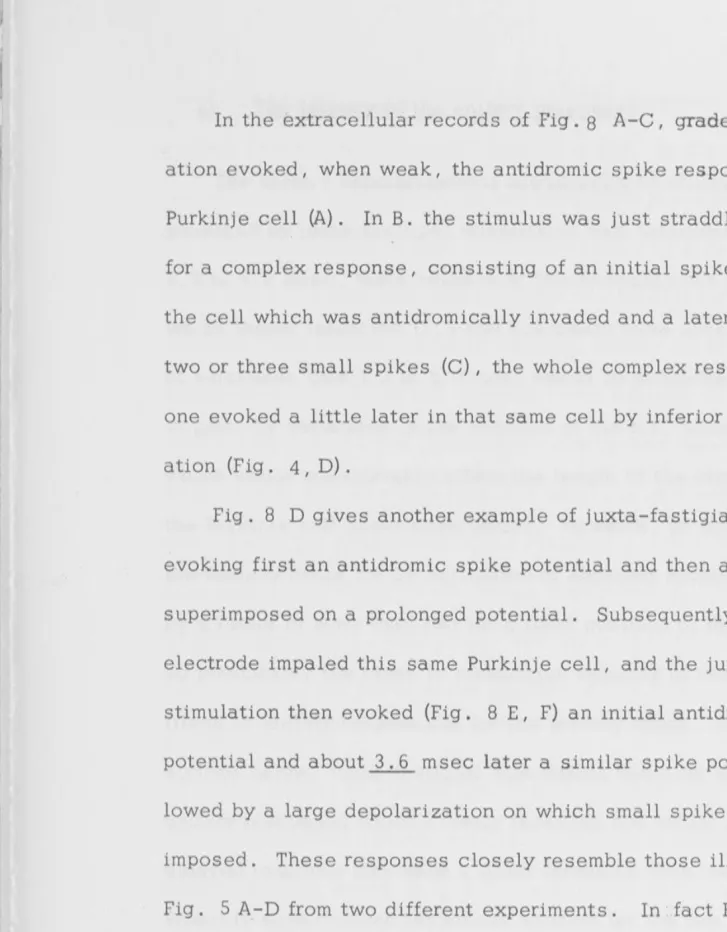
In the extracellular records of Fig. 8 A-C, graded
J.
F. stimul-ation evoked, when weak, the antidromic spike response of aPurkinje cell (A). In B. the stimulus was just straddling threshold for a complex response, consisting of an initial spike potential in the cell which was antidromically invaded and a later series of two or three small spikes (C), the whole complex resembling the one evoked a little later in that same cell by inferior olive stimul-ation (Fig. 4, D) .
[image:30.1048.27.755.39.972.2]I
J
'r
I:
Ir
1,
ll
,.
,,
I ,~
~I
II
e) The latency of the unitary responses
The unitary depolarizations attributable to climbing fibres are produced by juxta-fastigial stimulation with latencies ranging from
23
1. 2 to 5. 0 msec, there being only two examples out of almost 100 in which longer latencies (5. 5 and 6. 6 msec) were observed. This range of variations from 1 .. 2 to 5. 0 msec would be accounted for, at least in part, by variations in the location of the
J. F. stimulating electrode,
which would considerably affect the length of the climbing fibre tothe Purkinj e cell under observation. However, in most experiments the latency range for CF activation of adjacent Purkinje cells varied by a factor of more than two for a fixed position of the
J.
F. electrode, so presumably the range in conduction velocity of the excited climbing fibres is largely responsible for the latency range. As will bede-scribed below, juxta-fastigial stimulation can also set up delayed unitary responses which closely resemble the initial one and such a delayed response may have a lower threshold than the initial one. Probably such delayed responses account for the two exceptionally long latencies reported above.
When comparison w~s possible between the unitary responses evoked by inferior olive and juxta-fastigial stimulation, they were
stimul-I
•
J
:I
I:
,,
1,
It
r,
1:
,.
24
ation, and in the repetitive character often observed for the response evoked from the inferior olive (Figs. 7 & 12). In Fig. 5 B and C the
latencies were 4. 9 and 5. 0 msec, as ~gainst 6. 1 msec for A.
The differential latency of the two modes of stimulation (inferior olive and
J.
F.) has been measured for 79 Purkinje cells in which the complex depolarizing response could be evoked by both methods of stimulation. In 59 theJ.
F. response was between 1. 0 and 3. 6 msec briefer, while in 12 the differential latency was between 4. 5 and 7. 6 msec; and in 8 it was less than 1. 0 msec, approximating to zero in two. Since the actual conduction distance between the two sites ofstimulation was about 20 mm (range approximately 17 to 23 mm in dif-ferent experiments) , conduction velocities of 4. 7 to 2 3 msec can be calculated for the nerve fibres concerned in the latency differential of 1 . 0 to 3. 6 msec.
Wide ranges of stimulation strength through either the inferior olive or
J.
F. electrodes almost invariably failed to disclose the convergenc~ of two climbing fibres on to a sing le Purkinj e cell. But two out of more than 100 cells exhibited a clear superposition of two unitary responses as in Fig. 9 A-D. The first and second are displayed in thethreshold-straddling series of Fig. 9 A and C.
In the other example two quite different all-or-nothing EPSPs were evoked in a Purkinj e cell by the inferior olive (E, F) and
J.
F. stimul-ations (I, J}: With combined inferior olive andJ
.F. stimulations these two responses were summated at even the briefest stimulus intervals25
different climbing fibres which innervated the same Purkinj e cell.
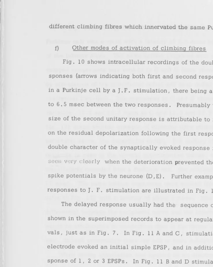
f) Other modes of activation of climbing fibres
Fig. 10 shows intracellular recordings of the double unitary
re-sponses (arrows indicating both first and second rere-sponses) often evoked in a Purkinje cell by a
J.
F. stimulation, there being an interval of 6. 0 to 6. 5 msec between the two response·s. Presumably the depressed size of the second unitary response is attributable to its superposition on the residual depolarization following the first response. Thedouble character of the synaptically evoked response in Fig. 10 is s2cn v ery c lec1rly when the deterioration prevented the generation of
spike potentials by the neurone (D, E). Further examples of such delayed responses to
J.
F. stimulation are illustrated in Fig. 11 .The delayed response usually had the sequence of EPSPs, which are shown in the superimposed records to appear at regularly spaced inter-vals, just as in Fig. 7. In Fig. 11 A and C, stimulation through one
J. F.
electrode evoked an initial simple EPSP, and in addition the later [image:33.1048.27.766.58.984.2].,
t
,,
I,\
"
In Fig. l '2A, B a typical unitary climbing-fibre response of a Purkinje cell was evoked with a latency of 3. 5 msec by stimulation
26
of the inferior olive. The same unitary response with superimposed delayed components was evoked in C and D (note slower sweep speeds) by stimulation of the superficial radial and deep radial nerves of the i.psilateral fore-limb. The responses in C and D showed some latency variation, 12. 2 and 13. 2 msec in C and 11. 5 and 12. 9 msec in D.
Fig. l 2F gives another example of climbing fibre stimulation from a peripheral nerve, the superficial radial. The unitary character of the response is seen in the several superimposed traces, which are ex-tracellularly recorded from a Purkinje cell, the minimal latency being
1 7 msec. The same unitary responses were evoked from the inferior olive i.n Fig . l 2E with a latency of about 5 msec. Most of the latency differential between the peripheral nerve and the inferior olive responses would be attributable to conduction time in the rather long neural path-way, \th·ough presumably there are one or more synaptic relays.
When strong stimulation was applied by an electrode on the surface of that folium into which the microelectrode had been inserted, unitary responses of the typical climbing fibre character were sometimes
[image:34.1048.24.738.8.982.2]I
I,I
I
Ii
VI
'
g) Effect of polarization of the Purkinje cell bn the excitatory postsynaptic potential generated by climbing fibre impulses.
27
Theoretically if the excitatory action of the climbing fibre synapse acts in the usual manner, that is, by creating temporarily a high ionic permeability (Eccles, 1964, pp. 51-53), it should be possible not only
to change the size of the EPSP by altering the membrane potential of the Purkin jE cell, but even to reverse the EPSP, as has been done with a relatively
few types of excitatory synapses (Coombs, Eccles and Fatt, 1965b; Burke and Ginsborg, 19 56; Nishi and Koketsu, 1960). In Fig. 13 the EPSP produced in a Purkinje cell by inferior olive stimulation (series C) is shown in A to have the typical unitary character of a climbing fibre synaptic action, and even to have sometimes the delayed additional re-sponses (B). In Fig. l 3C hyperpolarization of the Purkinje cell by a current applied through the recording electrode with the aid of a bridge, is seen to increase the EPSP, while a depolarization reduces and inverts
the EPSP so that the climbing fibre synapse evokes a large hyperpolar-izing potential. Fig. 13D and E illustrate in another cell a better exam-ple of these large EPSP changes produced by changes in membrane po-tential. They were observed in two successive series of current ap-plication to the same Purkin je cell.. E was recorded at lower
.,,
I
I
I
:
la
I
I
I . I
I
'
11
I
28
currents restored the impulse - generating property of the deteriorated cell and so increase d. th e :~~PS P that it \Vas abl e. to g e n .~ ru t e a s i'> J.k e
discharge, which is seen as an all-or-nothing event in the second low-est trace.
It has not been possible to determine the membrane potentials prod-uced by these applied currents; and, even if that had been possible, these measurements would obtain for the soma of the Purkinje cell and not for the region of the activated excitatory synapses on the dendrites. How-ever, the applied currents were recorded, as may be seen in one trace
-8
of Fig. l 3E for 3. 6 x 10 A . The plotted points in Fig. 14 from the
series partly illustrated in Fig. l 3E are seen to lie along a straight line, I
there being an approximately linear relationship between the applied currents and the EPSPs on either side of the reversal point that
oc-curred with a depolarizing current of 1. 2 3 x 1
o-
8 A. Such linear relation-ship has been observed in 3 of the 6 Purkinje cells in which this effect of current application on the EPSP has been successfully investigated.B) Interaction between responses evoked in Purkinje cells by the climbing fibres and other Purkinje cell inputs.
a) Inhibitory action on the responses evoked in Purkinje cells by the climbing fibres.
""l
I
I
1~1
I
I
Ii
1 1, I
ii(
29
of Purkinj e cells of the folium under _study, acting by the mediation both of basket cells (Andersen, Eccles and Voorhoeve, l 964~ and of the superficial stellate cells (Eccles, Llina1s and Sasaki, 1965 .b). Several procedures have been employed in investigating the influences of these postsynaptic inhibitions on the excitatory action of climbing fibre im-pulses on Purkinje cells.
The field l?otentials
As shown previously a single volley on the climbing fibre system (CF) produces a brief compound negative field potential with a max-imum a~ a depth of 100 to 200u, while at depths below 200u there is a later and slower positive wave (cf. Fig. l SA). Furthermore, it was shown that this potential profile can be satisfactorily explained by the distribution of the excitatory action of the climbing fibre synapses on the Purkinje cell dendrites. This explanation assumes that, in general accord with the usual histological descriptions, the climbing fibre
(CF) synapses on the Purkinje dendrites are most concentrated at a depth of 100 to 200u, with the consequence that both the EPSPs and the spike potentials generated thereby produce a maximum negative field poten-tial at this depth.
I
I
1,
'1 I
I, I,
I: ,,
I
u
I
11
·l
i
I30
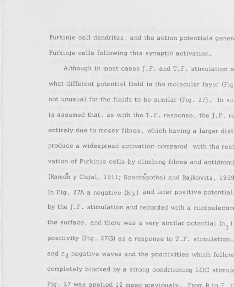
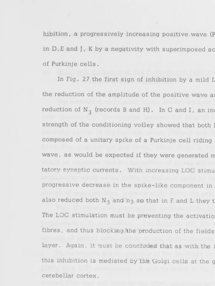
(Loe.). It was surprising to find on the other hand that the inhibi-tory action increased the initial negative potential at all depths, and that there was a reduction of the later positive wave, particularly at levels deeper than l SOu.
In Fig. l 5B these changes of the field potential were tested at the fixed interval of 19 msec, which is probably a little later than the maximum inhibitory action on the Purkinje cells (Andersen, Eccles and Voorhoeve, 19 64). In the series of Fig. C from another experiment the field potentials evoked by inferior olive stimulation were recorded at
l SOu depth and w'ere tested at various intervals after a conditioning Lbc. stimulation. The two control responses (CON) show the typir:;al
I
-initial sharply rising negativity followed by a declining phase broken by small spike-like potentials: When conditioned by the preceding Loe. stimulation at intervals from 5. 2 to 21 msec, the initial negative po-tential was increased in size (up to 160%) and had a much slower time course particularly in its declining phase. There was also a
notice-able decrease in the superimposed small spike potentials. The maximal effect was at 13 msec interval~ but it was still large at the longest test interval , 21 ms ec.
I
ij
j
I
'I
I
I
I
I
I
I:
Ii
I
increase. It is essential to examine the responses of individual Purkinje cells before attempting to give an account of these unex-pected observations .
Extracellular recording from single Purkinj e cells
31
In Fig. 16 the climbing fibre (CF) response evoked in an all-or-nothing manner in a single Purkinje cell (A, B) by inferior olive stim-ulation was depressed at a wide range of intervals after a
condition-ing Loe. stimulus. The i.ni.tial' spike response was unaffected, but the dim-inution in the subsequent partial spike complex was maximal at a test inter";" val of 4. 3 msec (D) and recovery was probably not completed even at the longest test interval of 33 msec (H). The briefest test interval (C) shows the frequent observation that at this short interval some facilitatory
influence increases the subsequent partial spike complex so that it consists of three large spikes.
I
'
II
Ii
I,
However, as in Fig. 16, the subsequent partial spike complex was depressed _, and had not fully recovered even at a test interval of 55 msec (H), by which time the antidromic invasion was restored.
32
Throughout Figs. 16 and 17 it will be observed that, in addition to this prolonged inhibitory action, the conditioning Loe. stimulation of the parallel fibres evoked itself a large initial spike and a variable sub-sequent partial spike complex. This is the expected : response gen-erated by the excitatory synaptic action which parallel fibre impulses exert directly on the dendrites of Purkinj e cells (Andersen, Eccles and Voorhoeve, 1964). I
c) Interactions recorded intracellularly from Purkinj e cells
In the intracellular recordings of Fig. 18 the Purkinje cell had already deteriorated, so that neither the conditioning Loe. stimulus nor
the testing inferior olive stimulus evoked spike discharges. However, there were slow fluctuations of membrane potential with a periodicity of about 90
mseo on which the responses of Fig. 18 were superimposed, as may be seen before the control response to the inferior olive stimulation in A. Furthermore, in G and H the periodicity of these fluctuations was
,,
i
I
I!
,,
,,
I}
l
'I
Jl1
"
11
II
II '
'
33
manner in D and E there was also a considerable lengthening of the superimposed EPSP, which occurred to a lesser degree in C and F. On the other hand, when superimposed on the depolarizing phase of the background rhythm, the EPSP was diminished and shortened (G), there being again recovery in size and time course during the devel-opment of the subsequent hyperpolarizing phase (H). Other examples of this influence of membrane potential on the CF responses are
il-lustrated in Fig .. 19. For the present it is sufficient to point out that the potentiation of the CF-evoked EPSP during a conditioning IPSP could explain the increased negative field potentials that CF stimulation produced in Fig. l SB, C, under similar conditions, if some assumptions to be mentioned later are granted.
In Fig. 19 (CON) the control CF response to juxta-fastigial (J .F .) stimulation was superimposed on the IPSP produced by parallel fibre stimulation. It is evident that these CF responses were affected by the background IPSP in the same way as in Fig. 18C, D, E; which is well illustrated in the potentials recorded from this _same cell at a
slower sweep speed (G-L).
When the height of the CF-EPSP is measured from control back-ground formed by the conditioning response, as in Fig. 19M it is im-mediately obvious that when the depolarization was superimposed on the initial phase of increasing IPSP, it was reduced in size (Fig. 19 B,
C)~ On the other hand it was potentiated when it was evoked later,
'
II
L,
'l
'I
J
l
34
These changes may be readily explained on the basis of the ionic con-ductance theories of excitatory and inhibitory postsynaptic potentials
(cf. Eccles, 1964).
The decrease at testing intervals from 3 to 12 msec is doubtless due to the shunting effect of the high membrane conductance during the incrementing phase of the IPSP (upper trace of Fig. 19M). At longer intervals this raised conductance will decline progressively, and the size of the CF-evoked EPSP becomes more influenced by the increased membrane potential. The prolonged falling phase of the
CF-EPSP under this condition will be considered later.
The IPSPs evoked by
J.
F. stimulation have the same action as those evoked from the Loe. stimulus, in conditioning the EPSPs produced by a CF impulse.d) Double activation of climbing fibres
Since, in Purkinj e cells with low membrane potential, climbing
fibres evoke only simple EPSPs uncomp1icated by spike potentials,· this situation provides conditions which are particularly appropriate for
,,
11
I ,
,,
I '
I·'',
I
I•
I
I
35
quite complete at the longest interval 410 msec (H). The full time course of the recovery process, except for the terminal phase, is exhibited in the curve of Fig. 20I. Similarly, in Fig. 21A-F with double stimulation of the inferior olive, the second CF-evoked EPSP was 70% of the first at 12 msec interval (A), and about 50% at the briefest interval for a second response, 1. 4 msec (D).
When I. O .. and
J.
F. stimulations evoke a CF response in a given Purkinje cell it is possible by the collision test diagramed in Fig. 21K to establish that the same CF fibre is responsible for both responses. For example, in Fig. 21 , utilizing theJ.
F. -I. 0. stimulus sequence the least interval for the second CF EPSP was 4. 7 msec (F-J), while it was only 1. 5 msec with the double I. 0. stimulus (D). This in-crease in the least interval by 3. 2 msec corresponds approximately to the latency differential for the respectiveJ.
F. -and I. O. -evoked EPSPs (4.3 - 1.9 msec,=
2.4 msec). As shown in Fig. 21 K, the former value is a measure of the time for antidromic propagation from theJ.
F. to the I . 0. sit es of stimulation of that climbing fibre and the latter is the time for the orthodromic propagation on the same fibre from the I. 0. to theJ.
F. site. This agreement would not be expected if an additional synapse were introduced on the I. 0. pathway, because the I. 0. stimulus would then act by exciting presynaptic fibres toI'
I
I
i
l
1
,,
,,
e) Repetitive activation of climbing fibres.
As with double stimulation, the simplest conditions for inves-tigation of repetitively activated CF synap·ses are provided by the intracellular recording of EPSPs from partially deteriorated Purkinj e
36
cells. In Fig. 22 A-E inferior olive stimulation at the indicated frequencies
evoked typical unitary CF EPSPs, which were, throughout, uncompli-cated by repetitive discharges from the inferior olive neurone. The second response of the repetitive series exhibited a depression of size matching those of Fig. 20, which is progressive for the first few im-pulses of the series. In the longer tetani of D and E the depression approached a steady level, which would be rather less than 40 % of the control in D and about 32 % in E. On cessation of these brief tetani there was no sign of the slowly declining depolarization which is characteristic of a lingering of transmitter action (Figs. 22,
J,
N, O; 2 3I) .The repetitive series of CF EPSPs in Fig. 22 F-J and K-0 were evoked in another Purkinje cell
b.Y
inferior olive andJ.
F. stimulation respective-ly. In the former series the first stimulus evoked a repetitive discharge of 4 to 5 CF impulses at about 500/ sec, resembling those previously illustrated. In the second series theJ.
F. stimulation directly evoked a single CF discharge, which was followed by a typical repetitive"reflex discharge II
1,
i
i
I I
! I
i
'
I
Ir
I'. 1,
37
500 /sec. In both series the subsequent s t imulations of the repetitive
series evoked only single CF responses which showed a depression of size which was more severe the higher the frequency. The relative
depres-sions of the amplitude at the steady level were less than in the first series (A-E) - about 70 to 80% at 70/ sec and 40 to 60% at 110/sec. In contrast with the first series there is clear evidence of a residual depolarizing action after cessation of the tetanus. This is particularly evident with the higher frequencies, 70 to 170/sec, where the depolar-ization persisted for as long as 25 msec after the summit of the last re-sponse.
Despite the depression of the EPSPs produced by repetitive stim-ulation, the CF synapses have an amazing ability to continue to produce repetitive spike discharges in Purkinje cells that are not injured by mi-croelectrode impalement. For example, Fig. 2 3A shows the complex re-sponse produced by
J.
F. stimulation, there being an initial antidromicI Ii
l
I
I
l'l
I
t
·, '
'
38
still well maintained for the 6 stimuli, but only the first stimulus evoked
the initial antidromic spike (a). Presumably the propagation down the Purkinje cell axon of the last spike discharge of each CF response pre-vented by refractoriness the next antidromic spike.
With the still more severe tetanus of Fig. 2 3F (16 stimuli at
180/sec) there was a rapid failure of large spikes, and only irregular small spike potentials could be evoked during the remainder of the tetanus. However, immediately after the tetanic stimulation was ar-rested there was a remarkable after-discharge of large spike potentials commencing at a frequency of about 3 50/ sec and declining to 18 0/ sec just before failing. The increase in spike size with slowing of frequency
may be entirely attributable to refractoriness. Comparable responses of Purkinje cells were observed by Granit and Phillips (19 56) following
tetanization by an electrode in the fastigial region, and likewise these may be attributable to the repetitive stimulation of climbing fibres.
[image:46.1048.17.731.78.983.2]I
1,
1,
11
I ,
I :
I
II
I
39
Finally in Fig. 2 3I intra cellular recording from another Purkinj e cell in this same experiment shows the large maintained depolarization during the tetanus (19 stimuli at 2 30/ sec), and the slow decline for almost 100 msec thereafter, though there was no associated generation of spike discharge as in F, but merely a recovery of the spontaneous discharge frequency. This build-up of residual depolarization was also noted in the more severe tetani of Fig. 22 I,
J,
M, N, 0, but was less well developed, presumably because this Purkinje cell was quite dete-riorated.It was assumed that in Fig. 23 repetitive activation of climbing fibres was responsible for all the subsequent after-discharge of the Purkinje cell due to the fact at lower frequencies the CF response was dominant (Fig. 2 3A-E). This characteristic type of response can also be produced by a brief tetanic stimulation of the inferior olive, and hence undoubtedly is due to climbing fibre activation.
C) DISCUSSION
I
II
:
I II I ,
Ii
1,
,,
40
all-or-nothing responses of Purkinje cells that were originally
observed by Granit and Phillips (19 5 6) . The physiological and histological evidence is further in agreement in showing that with rare exceptions (Fig .9) only one climbing fibre is distributed to each Purkinj e cell. Again, the
conclusion of Szentagothai and Rajkovits (1959) that the very extensive olivo-cerebellar tract is exclusively composed of climbing fibres is fully corroborated by the evidence that apparently pure climbing fibre
re-s ponre-s ere-s of Purkinj e cellre-s can be regularly evoked by re-stimuli applied to the inferior olivary nucleus.
Several illustrations have been given (Figs. 7, 12A, B; l 3B) of the frequent observation that stimulation of the inferior olive evokes a
brief sequence of all-or-nothing responses at about 2 msec intervals. These responses are identical with responses evoked by repetitive ac-tivation of a climbing fibre hence, presumably, they signal the repetitive discharge of impulses along a climbing fibre from its cell of origin in the inferior olive. The direct or short latency responses evoked by stimula -tion of the climbing fibre by the juxta-fastigial electrode (Figs. 1 OD, E, 11) have never exhibited this repetitive character; yet, as shown in Figs. 10 D ,E ; l lA-D, it was frequently observed with the reflex responses
following juxta-fastigial stimulation. With such reflex responses there must be synaptic excitation of the inferior olivary cells; so it can be concluded that, when similar responses are evoked by stimulation of the inferior olive, the stimulus is exciting presynaptic fibres in that nucleus.
.,
1,
.I
II I I
:
ll
II
i l
r
I
I,
41
recognized to evoke a brief efferent repetitive discharge by a single afferent volley, as indeed occurs with climbing fibre activation of Purkinje cells (Figs. 4, 5).
When juxta-fastigial stimulation leads to the "reflex II production of CF responses as in Figs. 10, 11, the latent period of these re-sponses is always sufficiently long for a reflex pathway through the inferior olive. There are two simple pathways by which juxta-fastigial stimulation could lead to the reflex discharge of impulses from the in-ferior olive. In one the axon collaterals of climbing fibres would ex-cite synaptically cells of the inferior olive. Fig. 11 B, D shows that this positive feedback pathway via an axon collateral would have to go at least in part to cells other than the cell of origin of that axon collateral. The alternative pathway would be antidromic transmission down mossy fibres and thence by axon collaterals to inferior olive cells. It has been recognized histologically that many fibres give
axon collaterals to the inferior olive as they pass by (Ramon y Cajal, 1909; Scheibel and Scheibel, 19 54; Scheibel, Scheibel and Vvalberg Brodal, 1956) and many of these fibres could well be cerebellar mossy fibres.
ac-'
,J
,, '
I
I:
I,
I,
I ,
II
Ii
I
I
11
•i
42
counted for by the postulate that the weakest stimuli excite fibres
that are pre synaptic to the cells of origin of the climbing fibres, while the stronger directly excite the discharge of impulses along the climb-ing fibres. In order to explain the additional latency steps, it is
necessary to postulate either serially arranged interneurones in the
inferior olive or, m.0re platrsibly the pathway via positive fe_edback .collat-erals which can go through several relays of inferior olive cells before reaching the cell of origin of the climbing fibre under observation.
Direct investigation of the inferior olive has shown the latter case to be correct, Ito (personal communication). Further investigation is re-quired before an explanation can be given of the very brief latency dif-ferential (less than 1. 0 msec) that has sometimes been observed for the CF responses evoked by I. 0. and
J.
F. stimulation respectively. A remarkable feature of the Purkinje cell responses to climbing fibre activation has been the relatively long duration (about 5 ms ec) of the repetitive spike discharges evoked by a single impulse (Fig. 4 A, B; SA-D; SB, C, E, F). In contrast, in the deeply deteriorated Purkinjetrans-I
"
I
Ii .I !I
r
I:
1
1,,
43
mitter action. In the case of intracellular records from Purkinj e cells in good condition, the depolarization produced by the synaptic ex-citation would have a longer duration; hence it seems possible to ac-count for the rather long duration of the repetitive discharge of im-pulses observed under such conditions (Figs. 5, 6, 8) or with extra-cellular leads (Figs. 4, l 2E, F).
In Figs. 4C, 8A-D, the spike potentials evoked by CF stimulation have a configuration (positive-negative diphasicity) that as usual
closely resembles the antidromic spike potential. However, this re-semblance merely shows that in both these cases the spike potential propagated into the region of the Purkinje cell under observation, which was the soma together with the large basal dendrites. It could be pos-tulated that the spike evoked by the CF impulse was generated in the axon and propagated into the soma-dendritic zone exactly as with the antidromic impulse set up by