Confocal microscopy using an InGaN violet laser
diode at 406nm
J. M. Girkin and A. I. Ferguson Institute of Photonics and Centre for Biophotonics,
University of Strathclyde, Glasgow G4 0NW, UK j.m.girkin@strath.ac.uk
D. L. Wokosin Centre for Biophotonics,
University of Strathclyde, Glasgow G4 0NR, UK
scopedoc@strath.ac.uk
A. M. Gurney
Centre for Biophotonics and Department of Physiology & Pharmacology, University of Strathclyde, Glasgow G4 0NR, UK
Abstract: We report on the application of a novel all-solid-state violet laser diode source to confocal microscopy. The source has the potential to replace argon ion lasers in a range of fluorescence based imaging systems. Improvements in system performance and image quality through the use of anamorphic prisms to modify the beam profile have been characterised. These modifications have permitted high quality, optically sectioned images to be obtained from laser diodes operating around 406nm. Living mammalian cells stained with a range of biologically significant fluorophores have been imaged. In addition, it has been shown that at this wavelength it is possible to image dyes that normally require excitation with UV argon laser lines.
OCIS codes: (180.1790) Confocal Microscopy; (170.3880) Medical and Biological imaging
References
1. J. B. Pawley (Ed.), “Handbook of Biological Confocal Microscopy,” 2nd Edition, Plenum (1995).
2. R. P. Haugland, “Handbook of Fluorescent Probes and Research Chemicals, 6th Edition,” Molecular Probes Inc.
3. S. Nakamura, M. Senoh, S. Nagahama, N. Iwasa T. Yamada, T. Matsushita Y. Sugimoto H. Kiyoku, “Optical gain and carrier lifetime of InGaN multi-quantum well structure laser diodes,” Appl. Phys Lett 69
1568-1572 (1996).
4. S. Nakamura “Developments in blue and UV optical semiconductor sources,” Proc. SPIE 3621-01 125-135 (1999).
5. J. Bewersdorf, S. W. Hell, “Picosecond pulsed two-photon imaging with repetition rates of 200 and 400 MHz,” Journal of Microscopy 191 28-38 (1998).
6. P. E. Hockerber, T. A. Skimina, V. E. Centonze, C. Lavin, S. Chu, S. Dadras, J. K. Reddy, J. G. White, “Activation of flavin-containing oxidases underlies light-induced production of H2O2 in mammalian cells,”
Proc. Natl. Sci. 96 6255-6260 (1999)
7. A. Miyawaki, J. Llopis, R. Heim, J. M. McCaffery, J. A. Adams, M. Ikura, R. Tsien, “Fluorescent indicators for Ca2+ based on green fluorescent protein and calmodulin,” Nature 388 882-887 (1997).
1. Introduction
argon ion based laser source to obtain the blue or ultraviolet light required to excite many of the more widely used flurophores. These sources have several disadvantages to the biological researcher including the cost, limited lifetime, heat generation, large size, noise and vibration from air-cooled systems and water cooling for UV sources. In particular the argon UV laser lines are not well matched for the excitation of many deep blue flurophores2. Other all solid state laser systems are now available for confocal imaging (diode pumped and frequency doubled lasers) but these are either expensive or demonstrate significant intensity fluctuations in addition to being centered mainly in the green. By contrast, the blue laser diode is small (around 5mm diameter, 5mm long) requires virtually no cooling, is significantly lower in cost, and, if required, can even be battery operated. Over the last two years, the performance of blue laser diodes has been improved with lifetimes rising from seconds of operation 3 to thousands of hours 4 at room temperature which compares favourably with argon ion gas lasers. The main thrust for the development of these devices has been for applications in optical storage with the aim of a low cost (few dollars), long lifetime device.
The diodes currently available for evaluation (Nichia Corporation, Ltd.) are restricted to a wavelength range from around 396 to 406nm, longer wavelengths being limited by growth defects in the devices. The 406nm wavelength region is, however, of interest for some biological applications in fluorescent microscopy, in particular some of the calcium ion indicators and stains for glutathione. In addition, many flurophores currently excited at 488 and 351nm have absorption wings around 400nm and hence have the potential for excitation with the new source. Of particular biological significance are green fluorescent protein (eGFP) and the variations now available.
2. Method
2.1 Optical Arrangement
A commercial evaluation laser diode (Nichia NLHV500) was mounted in a simple metallic heat sink. The device was not temperature controlled, but care was taken to ensure good thermal contact between the diode can and the heat sink in order to minimize wavelength and power drift caused by thermal fluctuations. The output of the laser diode was then collimated using a 4.5mm focal length, 0.45NA lens and the collimated beam was then directed into a confocal microscope scan head (Bio-Rad MRC1024) using the Bio-Rad standard T1 dichroic mirror on the input. This mirror has a reflectivity of around 90% at 406nm and two main transmission bands at 460-480nm and 510-545nm. The scan head was coupled to an inverted microscope (Zeiss Axiovert 100) through the side port, and a range of objective lenses (x40, 0.5NA and x63, 1.35NA oil) were used for the evaluations. The fluorescent light was descanned and then detected using the scan head photomultipliers (prism enhanced, S20 photocathode), with standard argon line dielectric filters being used to allow detection of the fluorescence.
In normal operation krypton/argon laser light was directed into the scan head through a collimated optical fibre coupling. For use with the new light source the fibre was removed and the laser diode light directed into the scan head without the use of optical fibres. Due to the mounting system employed on the scan head it was possible to reconnect the fibre without adjusting any of the other optical components, enabling direct comparisons of the imaging quality of the laser diode with that of the argon laser. In addition, a three times magnifying anamorphic prism pair could be placed in the laser diode beam. This prism pair was used to alter the beam profile to fill more efficiently the back aperture of the microscope objective.
these for further lenses. Measurements of the laser power were made throughout the system using a silicon photodiode power meter (Melles Griot 13PDC 001 with 13 PDH 001 head).
For the results imaging with monochlorobiamine labeled cells the laser source and scanning module (MRC 1024) was attached to a Nikon E600FN upright microscope and images obtained using a x60 1.0NA water dipping objective. In this system the optimal performance was obtained replacing the anamorphic prisms with a single plano-convex lens (750mm focal length, 1m from the scan head) to ensure that the back aperture of the objective was over filled. The diode laser beam in this system was combined with the output of the krypton/argon laser before entering the scan head using a 420DCLP mirror (Chroma Inc.) enabling direct comparisons between the sources.
2.2 Optical evaluation method
In order to assess the optical performance of the sources a number of techniques were employed concentrating on the 63x 1.35NA oil lens as the objective. The aim was primarily to make comparative measurements between the laser diode source and the argon ion laser. The axial resolution was measured using Coumarin 486 dye in benzyl alcohol on a cover slip. A line scanned z series of images was recorded and the derivative of the resulting image used to obtain a measure of the thickness of the axial slices5. Measurements were made with both the collimated laser diode and with the addition of the anamorphic beam shaping and these compared to measurements made using the 488nm argon laser line and theoretical calculations1. Theoretical approximations were made using the Abbe paraxial approximation which does not include the resolution enhancement possible with an infinitely small pinhole. This provides a realistic comparison for axial system resolution.
To obtain a measure of the lateral resolution 200nm diameter fluorescent beads were imaged and measured using the instrument software (Lasersharp). The results from 10 beads were then compared with those obtained using the fibre coupled argon laser after deconvolution had been applied to account for the non-point like nature of the beads.
2.3 Imaging biological samples
Smooth muscle cells were isolated from rat pulmonary artery and incubated for around one hour with fura2-AM (1µM) or for around 10 minutes with 1µM DAPI, 1µM DiBAC4 or
100nM tetramethylrhodamine ethyl ester (TMRE) at room temperature (all from Molecular Probes Europe BV, Leiden, Netherlands). The cells were imaged live without fixation. Additionally, smooth muscle cells were fixed, permeabilized and stained with FITC-phalloidin or FITC-labeled potassium-channel antibodies before imaging. Cos-7 cells were transfected with GFP (pEGFP-N1 Clontec Laboratories, Palo Alto CA) and imaged 24 hours later. Images were recorded with both the collimated laser diode and with the additional beam shaping optics in order to quantify the improvement through the more complex optics.
Samples of rat hepatocytes were imaged using the laser diode on the Nikon E600FN system to monitor the uptake of monochlorobiamine a probe indicating the presence of intracellular glutathione (Molecular Probes Europe BV, Leiden, Netherlands). This dye has a peak absorption at 395nm and so was ideally suited for use with the laser diode. The detection band on this system was from 440 to 565nm.
3 Results
3.1 Optical Measurements
was around 2.5mW and this power was reduced to around 2mW with the anamorphic beam shaping. This contrasts with the maximum of 5mW available from the standard air-cooled argon laser operating at 488nm originally supplied with the system. Without the anamorphic beam shaping the laser spot entering the scan head was measured to be around 2.1 by 0.5mm changing to 2.3 by 1.6mm with the prism pair.
The optical performance of the new source with the scanning system was then evaluated. The axial resolution was measured using the z-series line “scan” method. Results of the derivative of the line scan, for both the direct diode collimation and anamorphically shaped beam, are presented in figure 1. The differentiated slope of the fluorescent section gave a full width half maximum thickness of 1.5 (±0.2)µm for the collimated laser diode and 0.7 (±0.1)µm for the collimated and shaped beam using the 63x 1.35NA oil immersion objective. Previous measurements of this parameter with the fibre launched argon laser had given a thickness of 0.8 (±0.1)µm. Theoretical Abbe values (ignoring the effects of the pinhole size) for 406 and 488nm are 0.6µm and 0.72µm respectively and were calculated for comparative purposes.
Axial Profile at 406nm
0 10 20 30 40 50
0 2 4 6 8 10
Depth /µm
Signal /(arb)
Collimated Only
[image:4.612.153.473.255.448.2]Collimated and Shaped
Figure 1 Axial profile x63 1.35NA objective, laser diode with and without anamorphic beam shaping
The lateral resolution was measured using 200nm diameter beads. For the collimated only laser diode the lateral resolution was inferred (after deconvolution) at 0.5 (±0.05) µm, collimated at 0.24 (±0.02)µm and for 488nm at 0.25 (±0.02)µm. Full results are shown in table 1.
Table 1. Measured optical performance
Source Measured axial
resolution /µm
Calculated axial resolution /µm
Inferred lateral resolution /µm
Calculated lateral resolution /µm
406nm direct 1.5 (±0.2) 0.6 0.5 (±0.05) 0.18
406nm shaped 0.7 (±0.1) 0.6 0.24 (±0.02) 0.18
488nm fibre 0.8 (±0.1) 0.72 0.25 (±0.02) 0.22
and image contrast of the system. Figure 2 demonstrates a vascular smooth muscle cell stained with an FITC-conjugated potassium-channel antibody and imaged using the 63x 1.35NA oil objective. The total cell length was around 40µm. A significant improvement in the fluorescent image was produced by the shaped beam. The unshaped beam image (Fig 2a) has significantly less contrast and hence lower apparent resolution than the comparable image produced by the shaped beam (Fig 2b).
A number of other indicators were also successfully imaged with the laser diode. These included DiBAC4 a sensor for cell membrane potential and TMRE an indicator of
mitochondria. Both of these probes have excitation maxima above 490nm but appeared to have sufficient absorption at shorter wavelengths to enable imaging with excitation at 406nm, although the images were less bright than those obtained with the argon laser.
The ratiometric calcium ion indicator fura-2 has one excitation maximum at 380nm and this was well suited to excitation at 406nm. In addition DAPI (a DNA marker) loaded cells were imaged, although the normal peak excitation is at 350nm.
[image:5.612.124.491.244.442.2]2a
2b
2c
Figure 2 Live vascular smooth muscle cell stained with FITC imaged directly , 2a directly with laser diode, 2b laser diode with anamorphic beam shaping, 2c Cos-7 cell with eGFP transfection
A range of GFPs are available with excitation maxima ranging from 380nm to 513nm. The eGFP used in this work has an absorption peak at 498nm (Figure 2c). Nevertheless it was possible to achieve images using the laser diode that compared well with those obtained using an argon laser in the same system.
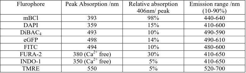
Table 2. Summary of flurophores imaged with 406nm Flurophore Peak Absorption /nm Relative absorption
406nm/ peak
Emission range /nm (10-90%)
mBCl 393 98% 440-640
DAPI 359 15% 410-600
DiBAC4 493 10% 490-590
eGFP 498 14% 490-610
FITC 494 10% 480-600
FURA-2 380 (Ca2+ free) 30% 410-650
INDO-1 350 (Ca2+ free) 5% 410-650
[image:5.612.111.502.551.668.2]The long-term stability of the source was demonstrated by imaging the uptake of monochlorobiamine in a time-stored series. The uptake rose and then reached a plateau as expected. The full range of flurophores successfully imaged is shown in Table 2.
4 Discussion
We have demonstrated the successful implementation of a violet InGaN laser diode for laser scanning confocal fluorescence imaging. Images were obtained by directly scanning the collimated output of the laser diode, but it has been demonstrated that additional beamshaping improves the resolution and contrast of the images. Without the shaping, one axis of the objective lens is significantly overfilled by the rectangular beam and hence the imaging power is not fully utilized.
The laser diode output is astigmatic with a divergence angle of 26° in one plane and 6° in the other. Using the three times anamorphic expansion these two are more closely matched, leading to an optimal fill of the objective lens and hence a tighter focus within the sample, resulting in improved axial resolution. Similar results could be achieved by directing the light into a single mode optical fibre, but in preliminary trials undertaken the laser output after the fibre was low (1mW). This figure could be increased, but the additional requirement for optical coupling into a fibre presents a more complex optical system than the lens and prism pair used in this work. These preliminary results indicate that system resolution can be improved through the use of 406nm excitation compared to 488nm, without resorting to UV excitation. Longitudinal chromatic aberration between 406nm and 488nm may also be limiting the potential resolution of the shorter wavelength source.
Although the 406nm source is not a good spectral match a wide range of flurophores can be excited through the use of the tails in the absorption spectrum, including those normally excited by UV lasers. In practical terms this means that a number of UV excited flurophores can be imaged without the need for special UV transmitting optics and UV lasers. With the blue/green excited flurophores the large difference between the exciting light at 406nm and the emitted light (typically greater than 500nm) means that highly efficient spectral filters can be used with very high transmissions for the emitted light and high rejection for the excitation source. A slightly higher power device would help in exciting many of the flurophores off their absorption peaks and it is anticipated that these will become available in the near future.
The use of 406nm light for excitation could have implications for cell viability when compared to the 488nm blue light6 used for many fluorophores or more significantly the 350nm UV used for dyes such as Indo-1. These effects were not examined in this work.
The excitation of eGFP has significance for biological applications, particularly with the emergence of fluorescent resonant energy transfer (FRET) techniques and the new cameleon variants 7. The short wavelength here specifically means that there should be better ratiometric imaging with lower background signals due to less acceptor excitation. In addition it is anticipated that the blue laser diode could be used in DNA sequencing instruments and as a source for Raman spectroscopy. The ease with which laser diodes can be modulated (in the case of InGaN laser diodes up to 1GHz) means that phase-sensitive detection is possible, which along with the confocal microscope will further improve the signal to noise of the system.
Acknowledgements