RESEARCH
YangZheng XiaoJi
exerts anti-tumour
growth effects by antagonising the effects
of HGF and its receptor, cMET, in human lung
cancer cells
Wen G. Jiang
1*, Lin Ye
1, Fiona Ruge
1, Sioned Owen
1, Tracey Martin
1, Ping‑Hui Sun
1, Andrew J. Sanders
1,
Jane Lane
1, Lucy Satherley
1, Hoi P. Weeks
1, Yong Gao
2, Cong Wei
2, Yiling Wu
2and Malcolm D. Mason
1Abstract
Background: Hepatocyte growth factor (HGF) is a cytokine that has a profound effect on cancer cells by stimulat‑ ing migration and invasion and acting as an angiogenic factor. In lung cancer, the factor also plays a pivotal role and is linked to a poor outcome in patients. In particular, HGF is known to work in combination with EGF on lung cancer cells. In the present study, we investigated the effect of a traditional Chinese medicine reported in cancer therapies, namely YangZheng XiaoJi (YZXJ) on lung cancer and on HGF mediated migration and invasion of lung cancer cells. Methods: Human lung cancer cells, SKMES1 and A549 were used in the study. An extract from the medicine was used. Cell migration was investigated using the EVOS and by ECIS. Cell–matrix adhesion and in vitro invasion were assessed. In vivo growth of lung cancer was tested using an in vivo xenograft tumour model and activation of the HGF receptor in lung tumours by an immunofluorescence method.
Results: Both lung cancer cells increased their migration in response to HGF and responded to YZXJ by reducing their speed of migration. YZXJ markedly reduced the migration and in vitro invasiveness induced by HGF. It worked synergistically with PHA665752 and SU11274, HGF receptor inhibitors on the lung cancer cells both on HGF recep‑ tor activation and on cell functions. A combination of HGF and EGF resulted in a greater increase in cell migration, which was similarly inhibited by YZXJ, and in combination with the HGF receptor and EGF receptor inhibitors. In vivo, YZXJ reduced the rate of tumour growth and potentiated the effects of PHA665752 on tumour growth. It was further revealed that YZXJ significantly reduced the degree of phosphorylation of the HGF receptor in lung tumours.
Conclusion: YZXJ has a significant role in reducing the migration, invasion and in vivo tumour growth of lung cancer and acts to inhibit the migratory and invasive effects induced by HGF and indeed by HGF/EGF. This effect is likely attributed to the inhibition of the HGF receptor activation. These results indicate that YZXJ has a therapeutic role in lung cancer and that combined strategy with methods to block HGF and EGF should be considered.
Keywords: Lung cancer, Tumour model, YangZheng XiaoJi, Hepatocyte growth factor, cMET, HGF, Cellular migration
© 2015 Jiang et al. This article is distributed under the terms of the Creative Commons Attribution 4.0 International License (http://creativecommons.org/licenses/by/4.0/), which permits unrestricted use, distribution, and reproduction in any medium, provided you give appropriate credit to the original author(s) and the source, provide a link to the Creative Commons license, and indicate if changes were made. The Creative Commons Public Domain Dedication waiver (http://creativecommons.org/ publicdomain/zero/1.0/) applies to the data made available in this article, unless otherwise stated.
Background
Lung cancer is the leading cancer worldwide and results in more death than any other cancer types. In the UK, the
incidence of lung cancer in males has steadily declined, but in contrast, the rate in females has steadily increased [1–3]. Worldwide, although the incidence rate has been declining in developed countries, since the later part of the last century, the incidence continues to rise in coun-tries in which smoking has peaked including China, Korea, and several countries in Africa and will con-tinue to do so for the next few decades [2, 4]. Treatment
Open Access
*Correspondence: jiangw@cf.ac.uk
1 Cardiff University‑Peking University Cancer Institute, Cardiff University
School of Medicine, Henry Wellcome Building, Heath Park, Cardiff CF14 4XN, UK
options for lung cancer remain limited, which is reflected in the poor survival, namely 5 year survival remains below 10 % and 10 year survival close to 5 %. Surgery, chemotherapy and radiotherapy remain the main treat-ment choice, although new targeted therapies are now available, namely anti-EGFR, etc. [3, 5, 6].
Recently, there have been early clinical reports that a Chinese medicinal formula, known as YangZheng XiaoJi (YZXJ), has a beneficial effect in patients with lung can-cer [7, 8]. In a number of small trials, YangZheng XiaoJi,
in combination with chemotherapy has been shown to increase the survival rate and at the same time, reduced the side effects. A similar beneficial effect has been reported in patients with primary hepatocellular car-cinoma [9]. Although it was initially proposed that the beneficial effects may be due to the improved immune function, such as the increase in NK cell functions, there have been recent reports to show that YangZheng XiaoJi
was able to directly inhibit angiogenesis and migration of cancer cells, including osteosarcoma cells, an effect attributable to the inhibition on the activation of focal adhesion kinase [10, 11].
Hepatocyte growth factor (HGF) is a cytokine that has strong effects on normal cells and cancer cells [12, 13]. In normal physiology, the cytokine is involved in tissue regeneration and organ repair, for example liver and lung regeneration. In cancer, however, the cytokine has been shown to have a profound effect on the migration, inva-sion and growth of cancer cells and has acted as a pow-erful angiogenic and lymphangiogenic factor [14, 15]. In the majority of solid tumour types, HGF and its receptor, cMET, have been found to be over-expressed in cancer cells and tumour tissues. It has been shown to be linked to disease progression, metastasis and long term clinical outcome of the patients [15–17].
In non-small cell lung cancer (NSCLC), HGF recep-tor protein over-expression has been frequently dem-onstrated [18, 19] and is shown to be associated with a poor clinical outcome of the patients. It has been shown that cMET protein expression is increased in NSCLC lung tumours with ALK gene rearrangement [20], and that gene amplification is uncommon in lung cancer. The amplified cMET protein expression may be the result of transcription factor ETS2 which was frequently down regulated in lung cancer [21].
In lung cancer, HGF has also been shown to inter-fere with EGF tyrosine kinase activation, which in turn results in induced resistance to EGFR inhibitor therapies [22]. Thus, combined use of MET tyrosine kinase inhibi-tor (TKI) and EGF TKI has been suggested to be a valid novel combination to overcome TGF TKI acquired resist-ance in lung cresist-ancer [23]. This was indeed shown in an in vitro study in which the cMET small inhibitor E7050
has the ability to circumvent resistance to the reversible, irreversible, and mutant-selective EGFR-TKIs induced by exogenous and/or endogenous HGF in EGFR mutant lung cancer cell lines, by blocking the Met/Gab1/PI3 K/ Akt pathway in vitro [24]. It is interesting to note that HGF-positive serum is a predictive factor for patients negative response to gefitinib therapy with advanced NSCLC who harbour wild-type EGFR [25, 26]. Serum HGF levels have been shown to be linked to disease pro-gression and overall survival, and interestingly even more so when EGFR status was considered [27].
cMET protein over-expression was seen in more than half of small cell lung cancer (SCLC) and patients with cMET phosphorylation in the SCLC tumours have a markedly poor overall survival (132 vs 287 days for those with cMET phosphorylated tumours and non-phospho-rylated tumours respectively, p < 0.001) [28]. Circulat-ing HGF (cHGF) was found to be a regular feature for patients with lung cancer and has been suggested to be a useful biomarker in choosing a HGF/cMET based tar-geted therapy [29, 30]. Lung cancer may attract neutro-phils to release their HGF storage and contribute to the local microenvironment for lung cancer progression [31].
Previous reports have demonstrated a profound effect of YangZheng XiaoJi on the migration of cancer cells and indeed on endothelial cells, suggesting that the medicine is an important regulator for cell motility and potential target for therapy [32, 33]. HGF is one of the most pow-erful motogens (motility stimulating cytokines) which, together with the HGF receptor, cMET, are aberrantly expressed in lung cancer. In the present study, we sought to investigate the effects of YangZheng XiaoJi on the migration, invasion and cell adhesion of lung cancer cells, in particular in the context of stimulation by HGF. In this context, the degree of the activation/phosphorylation of the HGF receptor was also investigated in order to deci-pher if and how YangZheng XiaoJi might affect the HGF/ cMET complex. The study also examined the effect of
YangZheng XiaoJi on the growth of lung tumours in vivo. Here, we report that YangZheng XiaoJi has an inhibitory effect on the functions and growth of lung cancer cells, in vitro and in vivo. YangZheng XiaoJi also exhibited an effect on HGF- and HGF/EGF-induced cellular migration on lung cancer cells and impacted on the phosphoryla-tion of the HGF receptor, cMET.
Methods
Cells and materials
Japan). Recombinant human EGF was from Sigma Aldrich (Poole, Dorset, England, UK). Small inhibitors to HGF receptor, SU11274 (IC50 for cMET 20 nM) and PHA665752 (IC50 for cMET 9 nM), and small inhibitor to the EGFR kinase, AG490 (IC50 for EGFR 2 µM) were obtained from Tocris Chemicals (Bristol, England, UK). Antibodies to human HGF, human cMET (SC-10), and phospho-cMET (sc-34088) were obtained from Santa Cruz Biotechnologies Inc., (Santa Cruz, CA, USA). Horse radish peroxidase (HRP) conjugated fluorescently tagged secondary antibodies were obtained from SigmaAldrich (Poole, Dorset, England, UK).
The herbal medicinal formula, YangZheng XiaoJi was obtained from Yiling Pharmaceuticals (Shijiazhuang, HeBei, China). The formula contained the following 16 ingredients: Panax ginseng C.A. Mey., Astragalus meme-branaceus (Fisch.) Bge.var. mongholicus (Bge.) Hsiao, Ligustrum lucidum Ait., Curcuma phaeocaulis Val., Gan-odema lucidum, Gynostemma pentaphylla (Thunb) Mak,
Atractylodes macrocephala Koidz, Scutellaria barbata D.Don, Oldenlandia diffusa (willd.) Roxb., Poria cocos, Duchesnea indica Focke, Solanum lyratum Thunb., Arte-misia scoparia (Bge.) Ki, Cynanchum paniculatum Kitag,
Eupolyphaga sinensis Walker, and Gallus domesticus Brisson. The chemical fingerprints of different batches were found to be highly consistent. Shown in Additional file 1 are fingerprints from 10 batches of YangZheng XiaoJi over a 2 year period (Additional file 1).
YangZheng XiaoJi extract, referred to as DME25, was prepared from YangZheng XiaoJi as previously reported using a DMSO based method [8, 10, 34]. Briefly, YangZ-heng XiaoJi formulated powder was added to pure DMSO solution (Sigma Aldrich). This was placed on a rotat-ing wheel (Labinco BV, Wolf Laboratory, York, England, UK) for 12 h in a cold room at 4 °C at 100 rpm [8, 10]. The preparation was centrifuged at 15,000×g, for 20 min at 4 °C. The supernatant was carefully removed and fil-tered using a disc filtration unit (pore size 0.20 μm, Sar-torius Stedim, SarSar-torius, Epson, Surrey, England, UK). The extract was diluted in a balanced salt solution (BSS) and standardised by quantifying the optical density of the preparation using a spectrophotometer at 450 nm wave-length (Biotek, Wolf Laboratory). A master preparation which gave 0.25 OD at 450 nm was stored as the master stock and so named as DME25 for the subsequent experi-ments. DME25 at dilutions below 1:40 did not show an effect on the growth of lung cancer cells. In the present study, we used the dilution range between 1:100 to 1:2000.
Electric cell‑substrate impedance sensing (ECIS) based cell adhesion and cell migration assays
This was modified from a method previously described [35, 36]. Briefly, 96-well W96E1 microarrays were used
on the ECIS Ztheta instrument (Applied Biophysics Ltd, Troy, New Jersey, USA). Lung cancer cells were added to the wells of the array, followed by immediate track-ing of cell adhesion over a range of frequencies, namely from 1000 to 64,000 Hz using automated modules. The adhesion was analysed using the mathematical modelling methods as previous described [37]. For cellular migra-tion, confluent lung cancer monolayers in the arrays were electrically wounded (2000 mA for 20 s each), after which the migration of the cells was immediately tracked, again over a range of frequencies. All the experiments were conducted in triplicate.
EVOS cellular migration assay (wounding assay)
Cellular migration assays were conducted on an EVOS system (EVOStm fl Digital Inverted Fluorescence Micro-scope, Fisher Scientific, Paisley, Scotland, UK). The human-interface free system integrated automated digi-tal time lapse recording, temperature and air control, automated sampling of same sample point over a speci-fied time and over large number of replicates (up to 96 wells).
and at a given point (38). The distance is shown as the mean number of pixels that cells had migrated from the 4 replicates. The migration is shown as a time course, and the comparison between the experimental settings was based on a fix time point.
In vitro invasion assay
Invasion assays were performed based on a previously published method [38]. Transwell invasion inserts with pore size at 8.0 µm were first coated with 50 µg Matrigel and allowed to air dry. The gel was then carefully rehy-drated. To each insert, 30,000 cancer cells were added together with the test agents or their combinations. After 72 h, Matrigel and non-invading cells were removed using a cotton swab. Invaded cells on the lower surface of the inserts were fixed with 4 % formaldehyde and then stained with 1 % crystal violet. The number of invaded cells were counted under a microscope.
SDS PAGE and western blot
Lung cancer cells at confluence were first sub-jected to serum hunger for 4 h in a serum free culture medium. The test agents including HGF (50 ng/ml), DME25 (1:1000), PHA665752 (10 nM), or their combi-nations were respectively added to the cells for a period of 45 min. Sodium orthovanadate (100 µM) with hydro-gen peroxide (0.1 %) was used as a positive control. Cells were collected using a cell scrapper. After centrifugation (1000 rpm for 5 min), media were removed from the cell pellets. Proteins from the cell pellets were extracted using a lysis buffer that contained 1.5 % Triton X100 as detergent, and protease inhibitors. Protein concentra-tions were standardised. Equal amount of proteins were loaded onto 8 % SDS PAGE. After separation, proteins were blotted onto nitrocellulose membrane, which were subsequently probed by specific antibodies to total MET, phospho-MET (p-MET) and GAPDH. The pro-tein blocking solution contained 1 % horse serum. After probing with horse-radish peroxidase conjugated anti-bodies, the protein signal was detected using chemilu-minescence methods (EZ ECL, Cellseco Ltd., Salisbury, Wiltshire, England, UK) and images were taken on a luminescent/fluorescent imager (Syngene, G: Box, Cam-bridge, England). The protein band signal was quantified using Image-J software. Shown here as pMET/cMET ratio.
In vivo tumour model
Athymic female nude mice, CD-1 of 4–6 weeks old were purchased from Charles River Laboratories (Mar-gate, Kent, England, UK). Due to the sensitive response to DME25 and the HGF receptor inhibitors, we chose A549 cell line for the in vivo tumour model. Tumour
cells, which were prepared at 5 × 10⁶/ml in a solution containing 3 mg/ml Matrigel, was injected subcutane-ously to give 0.5 million cells per injection [39, 40]. After one week when tumours became visible, mice were ran-domly divided into groups (n = 6 in each group). The size of tumours were measured using digital callipers. Treatments began one week after tumour cell inocula-tion and included the following: Control group (receiving control buffer), YangZheng XiaoJi extract via intraperi-toneal injection (IP) or by gavage (oral), cMET inhibi-tor (PHA665752), a combination of YangZheng XiaoJi
extract and cMET inhibitor. Treatments were given daily. The final concentration of YangZheng XiaoJi extract was 1 µl/g body weight and PHA665752 6.4 ng/gram body weight. Tumour size was measured weekly for 4 weeks. Mice were weighed and monitored daily. The procedure was reviewed and approved by the Cardiff University Joint Biological Ethics Committee and conducted under the UK Home Office Project License (PPL 30/2591). At the conclusion of the experiments, mice were terminated by euthanasia. Tumours were dissected and immediately stored at −80 °C for subsequent analysis. Tumour vol-umes were calculated by the following formula: tumour volume (mm3) = length × width × 0.54 [41].
Immunofluorescence staining
Frozen tumours were sectioned using a cryostat (Leica CM1900) at 7 µm thickness. After fixation using acetone/ methanol fixation buffer, the slides were rehydrated and blocked with a Tris buffer (25 mM, pH 8.4) that con-tained 10 % horse serum for 1 h. Primary antibodies (including anti-cMET, anti-pMET), diluted in the block-ing buffer was added to the respective slides which were kept in a humidified box for 1 h, and then thoroughly washed. FITC- or TRITC tagged secondary antibodies were then added to the respective slides together with DAPI for nuclear counter stain. After 1 h, the slides were thoroughly washed and mounted using FluroSave™ (Cal-biochem, Nottingham, England, UK). The slides were examined on an Olympus microscope and photographed using a Hamamatsu digital camera. The staining intensity was determined using Image-J. Briefly, the images were first converted to greyscale andthe staining intensity at the cell–cell junction areas where cMET is located, were calculated from Image-J, minus the reading from a back-ground control. Eight images were calculated from each staining.
Statistical analysis
Results
YangZheng XiaoJi had a profound influence on cell–matrix adhesion, cell migration and in vitro invasion
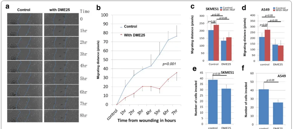
The effect of YangZheng XiaoJi was firstly tested on cel-lular migration. Using automatic cell tracking with the EVOS system, it was shown that YangZheng XiaoJi sig-nificantly reduced the migration speed of lung cancer cells (Fig. 1a, b). As expected, HGF increased the speed of migration of both cancer cells. It is interesting to observe that inclusion of YangZheng XiaoJi significantly reduced the pace of migration (Fig. 1c, d). Likewise, in vitro inva-siveness was also tested. As shown in Fig. 1e, f, YangZ-heng XiaoJi also significantly reduced invasiveness of both cells.
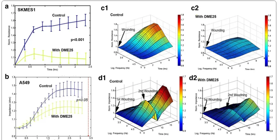
In the study, we also employed another automated method in tracking cell adhesion and migration, namely an ECIS based method. As shown in Fig. 2a, b,
YangZheng XiaoJi had some marked inhibitory effects on the adhesion of both cells. An electric wounding based method was applied to confluent lung cancer cells followed by tracking the migration of both cells. As shown in Fig. 2c, the migration of SKMES1 cells was markedly suppressed by YangZheng XiaoJi. A549 cells exhibited a similar response to YangZheng XiaoJi,
in that repeated wounding demonstrated a similar response (Fig. 2d).
YangZheng XiaoJi had a synergistic effect with cMET kinase inhibitors on HGF induced effects on lung cancer cells
To evaluate the potential interplay between YangZ-heng XiaoJi and HGF/cMET, the effects of combining
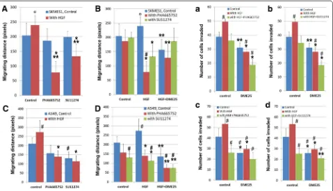
YangZheng XiaoJi and cMET kinase inhibitors, namely PHA665752 and SU11274, was tested. Using the EVOS method, it was found that YangZheng XiaoJi potenti-ated the effects of both inhibitors on cellular migration of both cells (Fig. 3A–D). Likewise, YangZheng XiaoJi
also increased the inhibitory effect of both inhibitors on in vitro invasiveness of both cells (Fig. 3A–D). Overall, A549 seems to be more sensitive than SKMES1 in the response to HGF, the cMET inhibitors and to DME25.
A similar observation was made when using the ECIS method in tracking cell adhesion (Fig. 4a–d) and cell migration (Fig. 4e–h) for both SKMES1 (a, b, e, f) and A549 (c, d, g, h). Figure 4 also demonstrates, in 3D imag-ing, the cell migration over a range of frequencies.
YangZheng XiaoJi had an inhibitory effect on the phosphorylation of the HGF receptor, cMET
The effect of YangZheng XiaoJi extract was further evalu-ated, directly or in combination with cMET small inhibi-tor PHA665752, on the action of the HGF recepinhibi-tor. In quiescent cells (4 h after serum hunger, only traceable
Fig. 1 Effects of YangZheng XiaoJi on cell migration, adhesion and invasion. a Cell images from A549 wounding model. Left control group in which cell monolayer was wounding and monitored continuously for 8 h. Right DME25 treated group, similarly wounded and monitored. DME25 dilution used was 1:1000. b Cellular migration of A549 as tracked by EVOS imaging system and quantified migration speed (in pixels). YangZheng XiaoJi had a significant inhibitory effect on the migration (p < 0.001). c, dYangZheng XiaoJi HGF‑induced cell migration (shown are at 6 h after wounding). HGF (shown at 50 ng/ml) had a significant stimulatory effects on cell migration of both A549 (d) (p < 0.05 vs control) and to some degree SKMES1 cells (c) (p = 0.08). YangZheng XiaoJi inhibited HGF‑induced cell migration (p < 0.05 vs control, p < 0.05 vs with HGF only). e, f Effects of YangZheng XiaoJi
[image:5.595.55.541.432.646.2]amounts of pMET was detected in control cells (Fig. 5, top), which was significantly inhibited by DME25, PHA665752 and their combination (Fig. 5, top) (p < 0.01 vs control, Fig. 5, bottom). HGF resulted in a marked activation of the receptor (p < 0.01 vs without HGF con-trol) (Fig. 5, bottom). Neither PHA665752 nor DME25 alone exhibited significant inhibition on the HGF acti-vation of its receptor (p > 0.05). However, it was very interesting to note that the combination of PHA665752 and DME25 resulted in a significant inhibition in HGF-induced receptor phosphorylation (Fig. 5).
Anti‑tumour effects exerted by YangZheng XiaoJi in vivo
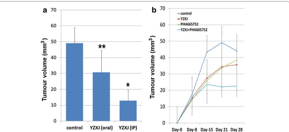
Using an in vivo A549 tumour model, it was shown that
YangZheng XiaoJi, delivered daily, had a significant inhib-itory effect on tumour growth (Fig. 6). The peritoneal
route appeared to be more effective than the oral route (Fig. 6a). The cMET kinase inhibitor, PHA665752 also had an effect on slowing down the growth, in a similar manner as YangZheng XiaoJi (Fig. 6b). A combination of
YangZheng XiaoJi and PHA665752 showed a more
pro-found inhibitory effect on the growth of A549 tumours (Fig. 6b, the oral route of delivery of all the agents after 3 weeks treatment).
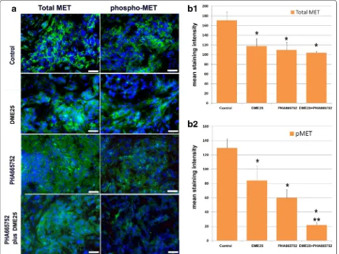
Immunofluorescence staining for HGF receptor and the receptor activation further demonstrated the effect of the treatment on the HGF receptor. As shown in Fig. 7a, left and b1, the three treatments resulted in some degree of reduction of the staining of total MET. However, when the activated receptor was examined, results revealed both YangZheng XiaoJi and PHA665752, and in particu-lar their combination, markedly reduced the intensity of
[image:6.595.54.540.89.335.2]the activation of MET (phospho-MET) (Fig. 7a, right and b2).
YangZheng XiaoJi and the interplay between HGF and EGFR‑TK complexes
In order to evaluate the possible interplay between Yang-Zheng XiaoJi and the HGF/EGF axis, we further evalu-ated the effect of the medicine on HGF and HGF/EGF induced cell migration and cell growth. In cell migra-tion assays, as shown in Fig. 8, the HGF/EGF combina-tion markedly induced an increase in the migracombina-tion of
A549 cells (Fig. 8a, b) and particularly in that of SKMES1 cells (Fig. 8c). As demonstrated earlier, YZXJ was able to significantly inhibit the migration of both cells, both HGF/EGF treated and untreated cells. Inhibitor to the HGF receptor (PHA665752) at the concentrations used, showed a significant inhibitory effect on HGF/EGF induced cell migration. The effect of EGFR inhibitor (AG490) was somewhat less effective. The combination of PHA665752 with YZXJ and to a certain degree AG490 had a stronger effect. However, the strongest effects were seen when PHA665752, AG490 and YZXJ were
Fig. 3 The effect of YangZheng XiaoJi and cMET kinase inhibitors, PHA665752 and SU11274 on HGF‑induced cell migration (A, B, C, D) and inva‑ sion (a, b, c, d), on both SKMES1 (top panel) and A549 (bottom panel) cells. Migration assayA both PHA665752 (10 nM) and SU11274 (20 nM) had a significant effect on HGF‑induced migration of SKMES1 cells. HGF concentration shown here is 50 ng/ml. *p < 0.01 cells with HGF/PHA665752 combination vs cells with control medium only; **p < 0.05 cells with HGF/SU11274 combination vs cells with HGF only. Ɵp = 0.059 cells with HGF alone vs cells with control medium. B The effect of the combination between DME25 and HGF inhibitors on SKMES1 cells. *p < 0.05 cells with HGF/ SU11274 (or HGF/PHA665752) combination vs cells with HGF only; **p < 0.05 cells with HGF/SU11274/DME25 (or HGF/PHA665752/DME25) combi‑ nation vs cells with HGF only; #p < 0.05 cells with HGF/PHA665752 combination vs cells with control medium only; Ɵp = 0.059 cells with HGF alone vs cells with control medium. C effect of HGF and HGF receptor inhibitors on the migration of A549 cells. *p < 0.01 cells with HGF/SU11274 (or HGF/ PHA665752) combination vs cells with HGF only; #p < 0.05 respective cells vs cells with control medium only. D effect of HGF, and the combina‑
tion of DME25 and HGF receptor inhibitors on the migration of A549 cells. *p < 0.01 cells with HGF/SU11274 (or HGF/PHA665752) combination vs cells with HGF only; **p < 0.05 cells with HGF/SU11274/DME25 (or HGF/PHA665752/DME25, or HGF/DME25) combination vs cells with HGF only;
#p < 0.05 respective cells vs cells with control medium only. Invasion assaya, b DME25 significantly potentiated the effect of HGF receptor inhibi‑
tors, PHA665752 (a) and SU121274 (b), on HGF‑induced invasion of SKMES1 cells. The effects were almost identical on both compounds. *p < 0.01 treatment plus DME25 vs respective treatment without DME25 control; **p < 0.05 treatment plus DME25 vs respective treatment without DME25 control; #p < 0.05 respective cells vs cells with control medium only. c, d Effects of DME25 on HGF and HGF receptor inhibitors, PHA665752 (c) and
[image:7.595.60.539.89.364.2]combined, and this effect was particularly strong in HGF/ EGF induced migration in SKMES1 cells.
Discussion
In the present study, we have described for the first time that a traditional Chinese medicine formula, YangZheng XiaoJi
has a profound direct effect on HGF induced aggressiveness of human lung cancer cells, both in vitro and in vivo. Fur-thermore, the medicine is also shown to inhibit the migra-tory effect induced by a combination of HGF and EGF, an axis of evil in clinical lung cancer in which they work syner-gistically in stimulating the progression of lung cancers.
HGF, and indeed its receptor cMET, are a pair of proteins that have been found to be frequently over-expressed in clinical lung cancers. In patients, raised lev-els of circulating HGF are frequently seen and, together with over-expression of HGF receptor, it is an evil pair of proteins causing the progression of lung cancer [8–21,
42]. HGF has some well established effects on cancer cells, including lung cancer cells. First, it is a power-ful cellular migration and cellular invasiveness inducing factor, which earned it’s other name ‘scatter factor’; sec-ond, HGF is a potent angiogenic and lymphangiogenic factor; third, it acts as a mitogen for cells, although this
[image:8.595.56.540.91.446.2]is mainly seen in normal hepatocytes. There has been compelling evidence to show that they are very credible targets in cancer treatment. Indeed, the past few years has witnessed the development of therapeutic methods, namely small molecules and neutralising antibodies, to target HGF and its receptor, with some now being used in patients. HGF receptor inhibitors have been shown
to reverse HGF induced EMT in lung cancer cells [19]. cMET over-expression has been shown to respond to cMET monoclonal (MAB) treatment as second line ther-apies by increasing overall survival and progression free survival [43]. In a recent study, PHA665752 has similarly been shown to block HGF induced effects on lung can-cer cells [28]. Blocking HGF/cMET pathways by way of
Fig. 5 YangZheng XiaoJi had an inhibitory effect on the activation of the HGF receptor, cMET in lung cancer cell, A549. Top images from immu‑ noblot assay. The phospho‑MET (pMET), total MET (cMET) and the loading control GAPDH were probed with the respective antibodies. Bottom
[image:9.595.63.537.86.534.2]small kinase inhibitors, neutralising antibodies [44] and antagonist [45] have been shown to be viable new treat-ments for lung cancer.
The current study has shown that the Chinese medi-cine, YangZheng XiaoJi, has a strong inhibitory effect on HGF induced cellular migration of the lung cancer cells tested. The study has further demonstrated that
YangZheng XiaoJi is able to work with the cMET inhib-itor in a synergistic manner in extending the inhibi-tory effects on the migration of cancer cells. The effect appears to be replicated in the in vivo models, in which the combination of YangZheng XiaoJi and PHA665752, the cMET inhibitor, can markedly increase the inhibi-tory effects seen by individual agents alone. Thus, the study has presented a coherent observation that Yang-Zheng XiaoJi, a traditional medicine currently being used in certain patients with cancer for example liver cancer, also has a therapeutic value in non-small cell lung cancer. In fact, in some small non-random stud-ies, some benefits to the patients with lung cancer have already been reported although with little mechanistic studies.
The effects of YangZheng XiaoJi on the in vivo growth of lung cancer are more likely to be via multiple actions. The present study has reported a direct effect of the med-icine on lung cancer cells. We have previously reported that the medicine also has a direct effect on angiogenesis in vitro and in vivo [10, 11]. Thus, it is plausible that the
anti-tumour effects, in vivo, are via the direct action on cancer and on the microenvironment, namely angiogen-esis. However, there have also been reports to show that patients who received the medicine have an improved immune function, for example favourable changes in NK cells and circulating cytokines. Thus, the anti-tumour and the therapeutic effect of the medicine are likely to be due to multiple factors.
The study has shown that application of YangZheng XiaoJi in tumour models has a significant effect on the levels of cMET, and most markedly activated cMET. In fact, both YangZheng XiaoJi and PHA665752 caused a reduction of cMET and phospho-cMET. The combi-nation of both agents almost completely blocked the appearance of phospho-cMET in lung tumours. These observations suggest that blocking the HGF recep-tor activation is a key mechanism by which YangZheng XiaoJi has its biological influence on lung cancer cells. It has been previously reported that YangZheng XiaoJi also impacts on the focal adhesion kinase (FAK), the AKT and sonic hedgehog (SHH) pathways in cancer cells and vas-cular endothelial cells. The broad effect on multiple sig-nalling pathways by YangZheng XiaoJi is not surprising, given that the medicine is a formula made of 16 herbal medicines. It is argued therefore that the medicinal for-mula of a mixture of ingredients would work on multiple targets. Future work to explore the nature of the ingredi-ents would be highly desirable.
[image:10.595.61.538.88.307.2]EGF receptor kinase inhibitors are now widely used in the treatment of lung cancer [7]. However, recent reports to show that HGF can trans-activate EGFR and make the anti-EGFR therapy less effective or indeed make lung cancer resistant to EGFR therapy in lung cancer [22–24] are very interesting indeed. Methods in targeting both have been suggested to be a viable option in enhancing the effect of anti-EGFR based therapies [46, 47]. Our study has shown that HGF and EGF had a profound effect on the migration of lung cancer cells. It is interest-ing to also note that AG490, an EGFR inhibitor has only marginal influence on this effect induced by HGF and EGF. We have shown that both YangZheng XiaoJi and PHA665752 are able to reduce the stimulation, and that a combination of the medicine, PHA665752 and AG490 almost completely abolished the effect of HGF/EGF.
This indicates that combined use of YangZheng XiaoJi
and HGF receptor inhibitor is an effective way to over-come resistance to anti-EGFR therapy in lung cancer and should be explored in clinical settings. Furthermore, it suggests that monitoring both HGF and EGF receptors are highly plausible in devising therapies for the patients.
Conclusion
The present study has shown that YangZheng XiaoJi has some significant effects on the migration and invasion of lung cancer cells and in vivo tumour growth. The effect is particularly profound on the functions induced by HGF and the HGF/EGF combination and is attributable to the inhibition of HGF receptor activation. The medi-cine is particularly effective when used together with the HGF receptor inhibitors. It is concluded that YangZheng Fig. 7 Immunofluorescence staining of total MET and phospho‑MET (pMET 1384) in lung tumours from the in vivo model. a Immunofluorescence images of total MET (left panel) and phospho‑MET (pMET 1384) (right panel) in lung tumours. Images were taken using an objective lens at ×40.
[image:11.595.57.542.90.453.2]XiaoJi has a strong effect on HGF-induced cell adhesion and migration, which may be important when consider-ing devisconsider-ing therapies in lung cancer and in the context of using the HGF/EGF receptor inhibitors in lung cancer.
Abbreviations
AG490: a small inhibitor to Epidermal Growth Factor Receptor; cMET: hepato‑ cyte Growth Factor Receptor; DME25: a soluble extract from YangZheng XiaoJi; ECIS: electric cell‑substrate impedance sensing; EGF: epidermal growth factor; EGFR: epidermal growth factor receptor; EVOS: a fl digital inverted fluores‑ cence microscope; HGF: hepatocyte growth factor; NSCLS: non‑small cell lung cancer; PHA665752: a small inhibitor to HGF receptor (cMET); SCLS: small cell lung cancer; SU11274: a small inhibitor to HGF receptor (cMET); TKI: tyrosine kinase inhibitor; YZXJ: YangZheng XiaoJi.
Authors contributions
WGJ, YG, CW, TM, SO, YW and MDM contributed to the conception and study design. WGJ, LY, FR, TAM, SO, PS, AJS, JL, LS and HPW participated in experimental design, experiments, data analysis and manuscript preparation. FR contributed to IFC experiments. All authors read and approved the final manuscript.
Author details
1 Cardiff University‑Peking University Cancer Institute, Cardiff University School
of Medicine, Henry Wellcome Building, Heath Park, Cardiff CF14 4XN, UK. 2 Yil‑
ing Medical Research Institute, No. 238 TianShan DaJie, Shijianzhuang, HeBei Province, China.
Acknowledgements
The authors wish to thank Cancer Research Wales, Albert Hung Foundation, and Ser Cymru Welsh Life Science Fund for supporting this work.
Compliance with ethical guidelines
Competing interests
The authors declare that they have no competing interest.
Received: 26 December 2014 Accepted: 14 August 2015
References
1. Jemal A, Center MM, De Santis C, Ward EM. Global patterns of cancer incidence and moralit rate and trends. Cancer Epidemiol Biomarker Prev. 2010;19:1893–907.
2. Goldstraw P, Ball D, Jett JR, Le Chevalier T, Lim E, Nicholson AG, Shepherd FA. Non‑small‑cell lung cancer. Lancet. 2011;378:1727–40.
3. Ellis L, Woods LM, Estève J, Eloranta S, Coleman MP, Rachet B. Cancer incidence, survival and mortality: explaining the concepts. Int J Cancer. 2014;135:1774–82.
Additional file
Additional file 1: Chemical finger printing of 12 batches of YangZheng XiaoJi, demonstrating the consistency of the formula.
4. Gadgeel SM, Kalemkerian GP. Racial differences in lung cancer. Cancer Metastasis Rev. 2003;22:39–46.
5. Lee CK, Brown C, Gralla RJ, Hirsh V, Thongprasert S, Tsai CM, Tan EH, Ho JC, da Chu T, Zaatar A, Osorio Sanchez JA, Vu VV, Au JS, Inoue A, Lee SM, Geb‑ ski V, Yang JC. Impact of EGFR inhibitor in non‑small cell lung cancer on progression‑free and overallsurvival: a meta‑analysis. J Natl Cancer Inst. 2013;105:595–605.
6. NSCLC Meta‑analysis Collaborative Group. Preoperative chemotherapy for non‑small‑cell lung cancer: a systematic review and meta‑analysis of individual participant data. Lancet. 2014;383:1561–71.
7. Wang QL, Xuo CM, Wu XP, Li YX, Bi XJ. Treatment of atypical gastric dys‑ plasia using Yangzheng Xiaoji. Chin J Diffic Compl Case. 2009;7:38–9. 8. Ye L, Ji K, Frewer N, Ji J, Jiang WG. Impact of Yangzheng Xiaoji on the
adhesion and migration of human cancer cells: the role of the AKT signal‑ ling pathway. Anticancer Res. 2012;32:2537–43.
9. Zhang SY, Gu CH, Gao XD, Wu YL. A random, double‑blindedand multicentre study of chemotherapy assisted Yangzhengxiaoji capsule on treating primary hepatic carcinoma. Chin J Diffic Compl Case. 2009;8:461–4.
10. Jiang WG, Ye L, Frewer N, Sanders AJ, Mason MD. Angiogenesis inhibitory effects of Yangzheng Xiaoji, the role of focal adhesion kinase, FAK path‑ way. Int J Oncol. 2012;41:1635–42.
11. Jiang WG, Ye L, Ji K, Ruge F, Wu Y, Gao Y, Ji JF, Mason MD. Anti‑tumour effects of Yangzheng Xiaoji in human osteosarcoma, the pivotal role of adhesion kinase (FAK) signalling. Oncol Rep. 2013;30:1405–13. 12. Nakamura T, Nishizawa T, Hagiya M, Seki T, Shimonishi M, Sugimura A,
Tashiro K, Shimizu S. Molecular cloning and expression of human hepato‑ cyte growth factor. Nature. 1989;342:440–3.
13. Rong S, Bodescot M, Blair D, Dunn J, Nakamura T, Mizuno K, Park M, Chan A, Aaronson S, Vande Woude GF. Tumorigenicity of the met proto‑ oncogene and the gene for hepatocyte growth factor. Mol Cell Biol. 1992;12:5152–8.
14. Suzuki Y, Sakai K, Ueki J, Xu Q, Nakamura T, Shimada H, Nakamura T, Matsumoto K. Inhibition of Met/HGF receptor and angiogenesis by NK4 leads to suppression of tumor growth and migration in malignant pleural mesothelioma. Int J Cancer. 2010;127:1948–57.
15. Jiang WG, Martin TA, Parr C, Davies G, Matsumoto K, Nakamura T. Hepato‑ cyte growth factor, its receptor, and their potential value in cancer thera‑ pies. Crit Rev Oncol Hematol. 2005;53:35–69.
16. Arriola E, Cañadas I, Arumí‑Uría M, Dómine M, Lopez‑Vilariño JA, Arpí O, Salido M, Menéndez S, Grande E, Hirsch FR, Serrano S, Bellosillo B, Rojo F, Rovira A, Albanell J. MET phosphorylation predicts poor outcome in small cell lung carcinoma and its inhibition blocks HGF‑induced effects in MET mutant cell lines. Br J Cancer. 2011;105:814–23.
17. Gelsomino F, Facchinetti F, Haspinger ER, Garassino MC, Trusolino L, De Braud F, Tiseo M. Targeting the MET gene for the treatment of non‑small‑ cell lung cancer. Crit Rev Oncol Hematol. 2014;89:284–99.
18. Dua R, Zhang J, Parry G, Penuel E. Detection of hepatocyte growth factor (HGF) ligand‑c‑MET receptor activation in formalin‑fixed paraffin embed‑ ded specimens by a novel proximity assay. PLoS One. 2011;6:e15932. doi:10.1371/journal.pone.0015932.
19. Cañadas I, Rojo F, Taus Á, Arpí O, Arumí‑Uría M, Pijuan L, Menéndez S, Zazo S, Dómine M, Salido M, Mojal S, García de Herreros A, Rovira A, Albanell J, Arriola E. Targeting epithelial‑to‑mesenchymal transition with Met inhibitors reverts chemoresistance in small cell lung cancer. Clin Cancer Res. 2014;20:938–50.
20. Feng Y, Minca EC, Lanigan C, Liu A, Zhang W, Yin L, Pennell NA, Farver C, Tubbs R, Ma PC. High MET receptor expression but not gene amplifica‑ tion in ALK 2p23 rearrangement positive non‑small‑cell lung cancer. J Thorac Oncol. 2014;9:646–53.
(See figure on previous page.)
21. Kabbout M, Garcia MM, Fujimoto J, Liu DD, Woods D, Chow CW, Mendoza G, Momin AA, James BP, Solis L, Behrens C, Lee JJ, Wistuba II, Kadara H. ETS2 mediated tumor suppressive function and MET onco‑ gene inhibition in human non‑small cell lung cancer. Clin Cancer Res. 2013;19:3383–95.
22. Gusenbauer S, Vlaicu P, Ullrich A. HGF induces novel EGFR functions involved in resistance formation to tyrosine kinase inhibitors. Oncogene. 2013;32:3846–56.
23. Nakagawa T, Takeuchi S, Yamada T, Nanjo S, Ishikawa D, Sano T, Kita K, Nakamura T, Matsumoto K, Suda K, Mitsudomi T, Sekido Y, Uenaka T, Yano S. Combined therapy with mutant‑selective EGFR inhibitor and Met kinase inhibitor for overcoming erlotinib resistance in EGFR‑mutant lung cancer. Mol Cancer Ther. 2012;11:2149–57.
24. Wang W, Li Q, Takeuchi S, Yamada T, Koizumi H, Nakamura T, Matsumoto K, Mukaida N, Nishioka Y, Sone S, Nakagawa T, Uenaka T, Yano S. Met kinase inhibitor E7050 reverses three different mechanisms of hepatocyte growth factor‑induced tyrosine kinase inhibitor resistance in EGFR mutant lung cancer. Clin Cancer Res. 2012;18:1663–71.
25. Masago K, Togashi Y, Fujita S, Sakamori Y, Okuda C, Kim YH, Mio T, Mishima M. Clinical significance of serum hepatocyte growth factor and epidermal growth factor gene somatic mutations in patients with non‑squamous non‑small cell lung cancer receiving gefitinib or erlotinib. Med Oncol. 2012;29:1614–21.
26. Han JY, Kim JY, Lee SH, Yoo NJ, Choi BG. Association between plasma hepatocyte growth factor and gefitinib resistance in patients with advanced non‑small cell lung cancer. Lung Cancer. 2011;74:293–9. 27. Kasahara K, Arao T, Sakai K, Matsumoto K, Sakai A, Kimura H, Sone T,
Horiike A, Nishio M, Ohira T, Ikeda N, Yamanaka T, Saijo N, Nishio K. Impact of serum hepatocyte growth factor on treatment response to epidermal growth factor receptor tyrosine kinase inhibitors in patients with non‑ small cell lung adenocarcinoma. Clin Cancer Res. 2010;16:4616–24. 28. Arriola E, Cañadas I, Arumí‑Uría M, Dómine M, Lopez‑Vilariño JA, Arpí O,
Salido M, Menéndez S, Grande E, Hirsch FR, Serrano S, Bellosillo B, Rojo F, Rovira A, Albanell J. MET phosphorylation predicts poor outcome in small cell lung carcinoma and its inhibition blocks HGF‑induced effects in MET mutant cell lines. Br J Cancer. 2011;105:814–23.
29. Siegfried JM, Weissfeld LA, Luketich JD, Weyant RJ, Gubish CT, Landreneau RJ. The clinical significance of hepatocyte growth factor for non‑small cell lung cancer. Ann Thorac Surg. 1998;66:1915–8.
30. Penuel E, Li C, Parab V, Burton L, Cowan KJ, Merchant M, Yauch RL, Patel P, Peterson A, Hampton GM, Lackner MR, Hegde PS. HGF as a circulating biomarker of onartuzumab treatment in patients with advanced solid tumors. Mol Cancer Ther. 2013;12:1122–30.
31. Wislez M, Rabbe N, Marchal J, Milleron B, Crestani B, Mayaud C, Antoine M, Soler P, Cadranel J. Hepatocyte growth factor production by neutro‑ phils infiltrating bronchioloalveolar subtype pulmonary adenocarcinoma: role in tumor progression and death. Cancer Res. 2003;63:1405–12. 32. Gherardi E, Stoker M. Hepatocyte growth factor–scatter factor: mito‑
gen, motogen, and met. Cancer Cell. 1991;3:227–32.
33. Martin TA, Jiang WG. Hepatocyte growth factor and its receptor signalling complex as targets in cancer therapy. Anticancer Agents Med Chem. 2010;10:2–6.
34. Ye L, Ji K, Ji JF, Jiang WG. Application of electric cell‑substrate impedance sensing in evaluation of traditional medicine on the cellular functions of gastric and colorecctal cancer cells. Cancer Metastasis Biol Treat. 2012;17:195–202.
35. Keese CR, Wegener J, Walker SR, Giaever I. Electrical wound‑healing assay for cells in vitro. Proc Natl Acad Sci USA. 2004;101:1554–9.
36. Stolwijk JA, Hartmann C, Balani P, Albermann S, Keese CR, Giaever I, Wegener J. Impedance analysis of adherent cells after in situ electropora‑ tion: non‑invasive monitoring during intracellular manipulations. Biosens Bioelectron. 2011;26:4720–7.
37. Jiang WG, Ye L, Sanders AJ, Ruge F, Kynaston HG, Ablin RJ, Mason MD. Prostate transglutaminase (TGase‑4, TGaseP) enhances the adhesion of prostate cancer cells to extracellular matrix, the potential role of TGase‑ core domain. J Transl Med. 2013;11:269.
38. Albini A, Benelli R. The chemoinvasion assay: a method to assess tumor and endothelial cell invasion and its modulation. Nat Protoc. 2007;2:504–11.
39. Martin TA, Parr C, Davies G, Watkins G, Lane J, Matsumoto K, Nakamura T, Mansel RE, Jiang WG. Growth and angiogenesis of human breast cancer in a nude mouse tumour model is reduced by NK4, a HGF/SF antagonist. Carcinogenesis. 2003;24:1317–23.
40. Jiang WG, Ye L, Ruge F, Sun PH, Sanders AJ, Ji K, Lane J, Zhang L, Satherley L, Weeks HP, Zhi X, Gao Y, Wei C, Wu Y, Mason MD. Expression of Sonic Hedgehog (SHH) in human lung cancer and the impact of YangZheng XiaoJi on SHH‑mediated biological function of lung cancer Cells and tumor growth. Anticancer Res. 2015;35:1321–31.
41. Davies G, Mason MD, Martin TA, Parr C, Watkins G, Lane J, Matsumoto K, Nakamura T, Jiang WG. The HGF/SF antagonist NK4 reverses fibroblast‑ and HGF‑induced prostate tumor growth and angiogenesis in vivo. Int J Cancer. 2003;106:348–54.
42. Takigawa N, Segawa Y, Maeda Y, Takata I, Fujimoto N. Serum hepatocyte growth factor/scatter factor levels in small cell lung cancer patients. Lung Cancer. 1997;17:211–8.
43. Giroux LÉ. A new drug in thoracic oncology: MetMab (onartuzumab). Rev Pneumol Clin. 2013;69:152–8.
44. Stabile LP, Rothstein ME, Keohavong P, Jin J, Yin J, Land SR, Dacic S, Luong TM, Kim KJ, Dulak AM, Siegfried JM. Therapeutic targeting of human hepatocyte growth factor with a single neutralizing monoclonal anti‑ body reduces lung tumorigenesis. Mol Cancer Ther. 2008;7:1913–22. 45. Kishi Y, Kuba K, Nakamura T, Wen J, Suzuki Y, Mizuno S, Nukiwa T, Matsu‑
moto K, Nakamura T. Systemic NK4 gene therapy inhibits tumor growth and metastasis of melanoma and lung carcinoma in syngeneic mouse tumor models. Cancer Sci. 2009;100:1351–8.
46. Nanjo S, Yamada T, Nishihara H, Takeuchi S, Sano T, Nakagawa T, Ishikawa D, Zhao L, Ebi H, Yasumoto K, Matsumoto K, Yano S. Ability of the Met kinase inhibitor crizotinib and new generation EGFR inhibitors to overcome resistance to EGFR inhibitors. PLoS One. 2013;8:e84700. doi:10.1371/journal.pone.0084700.
47. Luraghi P, Reato G, Cipriano E, Sassi F, Orzan F, Bigatto V, De Bacco F, Meni‑ etti E, Han M, Rideout WM 3rd, Perera T, Bertotti A, Trusolino L, Comoglio PM, Boccaccio C. MET signaling in colon cancer stem‑like cells blunts the therapeutic response to EGFR inhibitors. Cancer Res. 2014;74:1857–69.
Submit your next manuscript to BioMed Central and take full advantage of:
• Convenient online submission • Thorough peer review
• No space constraints or color figure charges • Immediate publication on acceptance
• Inclusion in PubMed, CAS, Scopus and Google Scholar • Research which is freely available for redistribution