R E S E A R C H
Open Access
Topoisomerase II alpha gene amplification is
a favorable prognostic factor in patients with
HER2-positive metastatic breast cancer treated
with trastuzumab
George Fountzilas
1*†, Christos Christodoulou
2†, Mattheos Bobos
3, Vassiliki Kotoula
3,4, Anastasia G Eleftheraki
5,
Ioannis Xanthakis
1, Anna Batistatou
6, George Pentheroudakis
7, Nikolaos Xiros
8, Irene Papaspirou
9,
Anna Koumarianou
8, Pavlos Papakostas
10, Dimitrios Bafaloukos
11, Dimosthenis V Skarlos
2and
Konstantine T Kalogeras
1,12Abstract
Background:The vast majority of patients with HER2-positive metastatic breast cancer (MBC) treated with
trastuzumab eventually develop resistance to this agent. There is an unmet need therefore, for identifying biological markers with possible prognostic/predictive value in such patients. The aim of this study was to investigate the prognostic role of topoisomerase II alpha gene (TOP2A) amplification and protein (TopoIIa) expression in patients treated with trastuzumab-containing regimens.
Methods:Formalin-fixed paraffin-embedded tumor tissue samples were retrospectively collected from 225 eligible patients treated with trastuzumab. Protein expression of ER, PgR, Ki67, PTEN, HER2 and TopoIIa were centrally assessed by immunohistochemistry.HER2andTOP2Agene amplification was evaluated by fluorescence in situ hybridization.PIK3CAmutations were identified by single nucleotide polymorphism genotyping. Survival was evaluated from the initiation of trastuzumab as 1st line treatment to the date of last follow-up or death.
Results:Among the 225 samples analyzed, only 137 (61%) were found to be HER2-positive.TOP2Awas amplified in 41% and deleted in 16% of such tumors.TOP2Agene amplification was more frequent in ER-negative tumors. TopoIIa protein expression was observed in the majority (65%) of the samples and was associated with ER-positive status, high Ki67 expression, presence of PTEN protein andPIK3CAmutations. Median follow-up for patients treated in the 1st line was 51 months. Survival was more prolonged with trastuzumab-containing treatment in
HER2-positive patients (50 months, log-rank, p=0.007).TOP2Anon-amplified or deleted tumors were associated with increased risk for death compared toTOP2Aamplified tumors (HR=2.16, Wald’s p=0.010 and HR=2.67, p=0.009, respectively). In multivariate analysis, a significant interaction ofTOP2Awith anthracycline treatment (either in the adjuvant or the 1st line setting) was observed for survival (Wald’s p=0.015). Among theTOP2Aamplified subgroup, anthracycline-treated patients were associated with decreased risk for death.
(Continued on next page)
* Correspondence:fountzil@auth.gr
†Equal contributors
1Department of Medical Oncology,“Papageorgiou”Hospital, Aristotle
University of Thessaloniki School of Medicine, 564 03, Thessaloniki, Macedonia, Greece
Full list of author information is available at the end of the article
(Continued from previous page)
Conclusions:TOP2Agene amplification was shown to be a favorable prognostic marker in HER2-positive MBC patients treated with trastuzumab, such an effect however, appears to rather be related to treatment with anthracyclines (predictive marker for benefit from anthracyclines). The results of the present retrospective study warrant validation in larger cohorts of patients treated in the context of randomized trials.
Keywords:Breast cancer, Topoisomerase II alpha, Fluorescence in situ hybridization, Gene amplification, Trastuzumab, Prognostic factors, Anthracyclines, Predictive factors
Background
Metastatic breast cancer (MBC) is an incurable disease. Chemotherapy or hormonal therapy mainly has a pallia-tive role, although newer agents may contribute to a significant prolongation of survival [1,2].HER2, a proto-oncogene located on chromosome 17q21.1, is amplified in approximately 20% of breast cancers and is associated with a number of adverse prognostic factors, such as axillary node involvement, advanced stage, hormone receptor (HR)-negativity and increased proliferation in-dices [3,4]. Trastuzumab (HerceptinW, Genentech, San Francisco, CA), a recombinant humanized monoclonal antibody against the HER2 protein, was found to pro-long progression-free survival (PFS) and overall survival (OS) of patients with MBC andHER2gene amplification or HER2 protein overexpression [5,6].
Nevertheless, it has become evident from numerous clinical trials that a considerable number of patients with MBC do not benefit from the administration of trastuzu-mab, either as a single agent or in combination with other systemic treatments. Moreover, in almost all patients who initially respond to trastuzumab-based treatments, tumor progression is eventually expected to occur. On the other hand, it is conceivable that any given targeted treatment is cost-effective only when it is administered exclusively to those patients who will derive the greatest benefit from it, sparing all other patients from unnecessary side effects. Therefore, there is an imperative need for identifying biological markers that will predict which patients are most likely to respond to trastuzumab-based treatments.
The topoisomerase II alpha gene (TOP2A) is located telomerically to HER2 at 17q21-q22 and encodes for topoisomerase II alpha (TopoIIa), a 170-kd cell cycle regulated protein [7]. The TOP2A gene is considered to be within the HER2 amplicon [8], although it is not included in the“smallest region of amplification”[9] and may follow a different fate than HER2 in terms of copy number alterations [10]. Topoisomerases II are consid-ered to be targets of anthracyclines [11], while TOP2A gene amplification has been linked to anthracycline sen-sitivity in patients with advanced breast cancer [12,13] or in patients with high-risk primary breast cancer receiving adjuvant chemotherapy [14-16] (reviewed in
[17]). On the other hand, the predictive value ofTOP2A has been refuted by several retrospective studies [18-20] however, flaws in the statistical design and the methodo-logical approaches used in these studies undermine the credibility of such opinions. In fact, a number of investi-gators believe that TopoIIa protein expression is more relevant thanTOP2A gene status in predicting response to anthracyclines [21].
It is noteworthy, that although extensive research efforts have been devoted to the evaluation of the poten-tial predictive role ofTOP2A gene status with respect to anthracycline responsiveness, little attention has been paid to TOP2A gene alterations with regard to the out-come of breast cancer patients following treatment with trastuzumab in advanced stages. The main objectives of the present study were to explore the impact of TOP2A gene status and TopoIIa protein expression on the out-come of MBC patients treated with trastuzumab-containing regimens and their possible interaction with anthracycline-containing treatment. The study was retro-spective in nature and was performed on archival tissue material (formalin-fixed paraffin-embedded, FFPE) from our Group’s Tumor Repository. In addition, based on the adverse prognostic effect of PIK3CA mutations and PTEN protein loss in the same patient cohort, as previ-ously shown [22], we investigated the association of these parameters with TOP2A gene and protein status.
Patients and methods
in patients with metastatic breast cancer”. All patients included in the study after 2005 provided written informed consent for the provision of biological material for future research studies before receiving any treatment. Waiver of consent was obtained from the Bioethics Committee for patients included in the study before 2005.
Tissue Material
FFPE tumor tissue samples were retrospectively col-lected from 246 breast cancer patients treated with trastuzumab-based regimens in the metastatic setting. Twenty-one cases were excluded for inadequate FFPE tumor tissue, thus decreasing the number of eligible/eva-luable patients to 225. A REMARK diagram for the translational research studies is provided in Figure 1. Representative hematoxylin-eosin stained sections from the tissue blocks were reviewed by a pathologist (M.B.). The most representative tumor areas were marked for the construction of tissue microarray (TMA) blocks, as previously described [22]. Each TMA block also con-tained cores from various neoplastic, non-neoplastic and reactive tissues serving as assay controls. Cases not represented, damaged or inadequate on the TMA sections were re-cut from the original blocks and these sections were used for protein and gene analysis.
Immunohistochemistry (IHC)
Immunohistochemical labeling was performed according to standard protocols on serial 2.5 μm thick sections from the original blocks or the TMA blocks. All cases were also stained for vimentin (clone V9, Dako,
Glostrup, Denmark) and cytokeratin 8/18 (clone 5D3, Novocastra™, Leica Biosystems, Newcastle, U.K), which were used as control stains for tissue immunoreactivity and fixation, as well as identification of tumor cells. Tis-sue samples negative for the above antibodies were excluded from the study. The staining procedures for es-trogen receptor (ER, clone 6F11, Novocastra™, Leica Biosystems), progesterone receptor (PgR, clone 1A6, Novocastra™, Leica Biosystems), HER2 (A0485 poly-clonal antibody, Dako) and Ki67 (clone MIB-1, Dako) were performed using a Bond Max™ autostainer (Leica Microsystems, Wetzlar, Germany), as previously described [23]. TopoIIa protein expression was evaluated using the KiS1 monoclonal antibody (Dako), as previously described [24] with slight modifications (antibody dilution: 1:200; detection system: Envision™, Dako). PTEN (phosphatase and tensin homologue deleted on chromosome 10) pro-tein expression was evaluated using the 6H2.1 monoclo-nal antibody (Dako), as previously described [25]. The evaluation of all IHC sections was done by experienced in breast cancer pathologists (M.B. and A.B.), blinded as to the patients’clinical characteristics and survival data. To assure optimal immunoreactivity, the sections of the TMA blocks were stained in one run for each antibody, shortly after mounting of the TMA sections on positively charged glass slides.
Interpretation of the IHC results
[image:3.595.60.540.471.706.2]ER and PgR, HER2, Ki67 and PTEN immunostaining was evaluated according to existing established criteria [26-29], as previously described [22]. Briefly, ER and PgR
were evaluated using the Histoscore method (max score: 400) and were considered positive if staining was present in ≥1% of tumor cell nuclei [26]; HER2 protein expres-sion was scored in a scale from 0 to 3+, the latter corre-sponding to uniform, intense membrane staining in >30% invasive tumor cells [27]; for Ki67, the expression was defined as low (<14%) and high (≥14%) based on the percentage of stained/unstained nuclei from the tumor areas [28]; and, PTEN protein expression (cytoplasmic, nuclear or both) was evaluated according to a stain-ing intensity scale from 0 (negative, no stainstain-ing) to 3 (intense staining), whereby tumors with PTEN scores of 0 or 1 were considered to have PTEN loss [29]. For TopoIIa immunostaining, a tumor was considered to be positive if moderate to intense nuclear staining was detected in >5% of tumor cells [30].
Fluorescencein situhybridization (FISH)
TMA sections or whole sections (5 μm thick) were cut for FISH analysis, using the ZytoLightW SPEC HER2/
TOP2A/centromere 17 (CEN17) triple color probe kit (ZytoVision, Bremerhaven, Germany). FISH was per-formed according to the manufacturer’s protocol with minor modifications. Four carcinoma cell lines (MDA-MB-231, MDA-MB-175, MDA-MB-453 and SK-BR-3) from the Oracle HER2 Control Slide (Leica Biosystems), with a known HER2 gene status, were also used as a control of the FISH assays and analyzed for HER2 and
TOP2Agenomic status.
FISH evaluation
For all probes, sequential (5 planes at 1.0 μm) digital images were captured using the Plan Apo VC 100x/1.40 oil objective (Nikon, Japan) using specific filters for each probe. The resulting images were reconstructed using specifically developed software for cytogenetics (XCyto-Gen, ALPHELYS, Plaisir, France). For the evaluation of
HER2/TOP2A/CEN17 status, non-overlapping nuclei from the invasive part of the tumor were randomly selected, according to morphological criteria using DAPI staining, and scored (M.B.). The virtual slides of HER2, ER or PgR stains, created as previously described [31], were used for selecting the invasive part of the tumor in each TMA. Twenty tumor nuclei were counted accord-ing to Press et al. [13]. The HER2 gene was considered to be amplified when the ratio of the respective gene probe/centromere probe was≥2.2 [27] and deleted when the ratio was <0.75. TheTOP2Agene was considered to be amplified when the ratio of the respective gene probe/CEN17 probe was≥2.0 and deleted when the ratio was <0.8 [32]. In cases with values at or near the cut-off (1.8-2.2 for amplifications and 0.7-0.9 for deletions), additional 20 or 40 nuclei were counted and the ratio was recalculated. In cases with a borderline ratio at 60
nuclei, additional FISH assays were performed in whole sections. All primary image data of the TMA and whole tumor sections have been digitally scanned and made publicly available at: http://www.hecog-images.gr/read-Dir.php?nextdir=TOP2AtrastuzumabMBC.
Single nucleotide polymorphism (SNP) genotyping for PIK3CA mutations
DNA was extracted from 182 FFPE whole tissue sections or macrodissected tissue fragments containing >70% tumor cells, using a fully automated isolation method based on silica-coated magnetic beads (Versant Tissue Preparation Reagents, Siemens Healthcare Diagnostics, Tarrytown, NY) in combination with a liquid handling robot, as previously described [33]. Mutation testing for
PIK3CA E542K and E545K (exon 9) and H1047R (exon 20) was accomplished with custom Taqman-MGB-SNP genotyping assays (duplex q-PCR for the detection of control DNA and mutant target in the same reaction), as previously described [22].
Statistical analysis
selection criteria with p<0.10, including in the initial step clinico-pathological parameters, such as age (>60 vs. 50– 60 vs. <50), menopausal status (post vs. pre), perform-ance status (1–2 vs. 0), number of metastatic sites (≥3 vs. <3), anthracycline treatment (yes vs. no), hormonal receptor status (ER/PgR) (positive vs. negative), Ki67 protein expression (high vs. low), HER2 status (positive vs. negative), TOP2A gene status (deleted vs. non-amplified vs. non-amplified) and TopoIIa protein expression (positive vs. negative). Multivariate analyses were per-formed in the total cohort and in the HER2-positive sub-group and were presented by forest plots. All tests were two-sided at the α=0.05 level of significance. No adjust-ment for multiple comparisons was performed. Results of this study were presented according to reporting recommendations for tumor marker prognostic studies [34]. The SPSS (version 15.0, IBM Corporation, Armonk, NY) and SAS (version 9.3, SAS Institute Inc., Cary, NC) software were used for statistical analysis.
Results
Among the 225 eligible patients with metastatic breast cancer treated with trastuzumab, only 137 (61%) were found to have centrally assessed HER2 gene amplifica-tion by FISH and/or 3+ HER2 protein overexpression by IHC (Figure 1). It is of note that all 225 patients were considered to be HER2-positive when assessed with IHC (and FISH in some cases) at the local laboratories and had therefore been treated with trastuzumab. Selected patient and tumor characteristics from the 225 patients, at trastuzumab initiation, are presented in Table 1.
Trastuzumab was given as 1st line treatment in 191 patients (85%), while in 15% of the patients trastuzumab was initiated later in the course of metastatic disease. The majority of the 1st line treated patients received it in combination with chemotherapy (186 patients, 97%), while the rest (5 patients, 3%) received trastuzumab as mono-therapy. Most of the patients received a taxane in the 1st line setting in addition to trastuzumab (137 patients, 72%), while 27 patients (14%) received anthracyclines.
[image:5.595.304.540.110.693.2]Median follow-up for all patients was 66 months, while for patients treated with trastuzumab in the 1st line median follow-up was 51 months. Totally, 137 patients died among all patients, while 151 of the 191 patients treated with tras-tuzumab in the 1st line demonstrated tumor progression. Median survival was significantly longer in HER2-positive patients treated with trastuzumab (median survival 50.4 months, 95% Confidence Interval [CI]: 39.4-61.4) compared to HER2-negative patients (median survival 35.3 months, 95% CI: 30.9-39.6, log-rank, p=0.007). TTP was 14 months (95% CI: 9.6-18.5) for HER2-positive patients treated with 1st line trastuzumab, as compared to 10.3 months (95% CI: 5.6-15.0) for HER2-negative patients. This difference was not statistically significant (log-rank, p=0.24), probably due
Table 1 Selected patient and tumor characteristics (at trastuzumab initiation) according to HER2 status
HER2 status
Positive Negative
N 137 88
Age (years)1
Median (range) 54.6 (28.4-95.0) 58.9 (31.8-78.8)
N (%) N (%)
Menopausal status
Premenopausal 43 (31.4) 26 (29.5)
Postmenopausal 94 (68.6) 62 (70.5)
Performance status
0 96 (70.1) 56 (63.6)
1 28 (20.4) 18 (20.5)
2 5 (3.6) 7 (8.0)
Unknown 8 (5.8) 7 (7.8)
History of adjuvant CT 75 (54.7) 55 (62.5)
Anthracycline containing 59 (43.1) 29 (33.0)
Taxane containing 36 (26.3) 17 (19.3)
CMF-like 42 (30.7) 30 (34.1)
History of adjuvant HT 61 (44.5) 42 (47.7)
History of adjuvant RT 52 (38.0) 33 (37.5)
Tumor grade (initial diagnosis)
1 4 (2.9) 2 (2.3)
2 48 (35.0) 36 (40.9)
3 75 (54.7) 42 (47.7)
Unknown 10 (7.3) 8 (9.1)
Site of metastases
Locoregional 45 (32.8) 28 (31.8)
Distant 117 (85.4) 78 (88.6)
Only locoregional 10 (7.3) 5 (5.7)
Only distant 82 (59.9) 55 (62.5)
Bones 54 (39.4) 37 (42.0)
Visceral 93 (67.9) 58 (65.9)
Number of metastatic sites
1 54 (39.4) 28 (31.8)
2 40 (29.2) 30 (34.1)
≥3 38 (27.7) 25 (28.4)
Unknown 5 (3.6) 5 (5.7)
History of 1st line CT 123 (89.8) 68 (77.3)
Anthracycline containing 18 (13.1) 9 (10.2)
Number of treatment lines with T
1 51 (37.2) 39 (44.3)
2 33 (24.1) 19 (21.6)
3 23 (16.8) 12 (13.6)
≥4 30 (21.9) 18 (20.5)
1
p=0.019;CTchemotherapy,HThormonal therapy,RTradiotherapy,
Ttrastuzumab.
to the small number of patients. ER, PgR, Ki67, PTEN and
PIK3CAdata were presented in detail in a previous publica-tion [22].
Associations between examined markers
TOP2A gene alterations were assessed in 221 tumors, 25% of which were amplified and 12% were deleted. Tumors that were notTOP2A amplified or deleted were analyzed as a separate group (TOP2A non-amplified tumors). As expected, TOP2A gene amplification and HER2-negative status were mutually exclusive. Represen-tative FISH images of the evaluated cell lines and inva-sive breast carcinoma cases are presented in Figure 2. The majority ofTOP2A gene deletions were seen in the HER2-positive group (21 of the 26 cases), while 43% of HER2-positive tumors were not amplified for TOP2A (Table 2). A significant association ofTOP2Agene status with HER2 status was found (Fisher’s exact test, p<0.001). Moreover,TOP2Agene amplification was more frequent in ER-negative tumors (28% in ER-negative vs. 24% in ER-positive, p=0.017), whereas no such associ-ation was found with Ki67 (p=0.47) or PTEN protein expression (p=0.12). TOP2A gene amplification was negatively associated with the presence ofPIK3CA muta-tions (28% in WT vs. 14% in mutated, p=0.035), the distribution however, ofPIK3CAmutations in the HER2-positive group did not differ between TOP2A amplified and non-amplified tumors (12% vs. 23%, p=0.25).
Concerning TopoIIa protein expression, the majority of cases were positive (125 of 193 cases assessed, 65%), but no association with HER2 status was found (p=0.65). TopoIIa protein positivity was associated with ER-positive status (72% in ER-ER-positive vs. 49% in ER-negative, p=0.005), high Ki67 expression (68% in high vs. 32% in low, p=0.004) and positive PTEN protein status (80% in positive vs. 58% in negative, p=0.003). The majority of the
PIK3CAmutations in this cohort (28 of 34) were encoun-tered in tumors with positive TopoIIa protein expression (p=0.015).
For 189 tumors, bothTOP2Agene status and TopoIIa protein expression were assessed (Figure 1), but no sig-nificant association was found between the two (p=0.11). More specifically, 37 among 49TOP2Aamplified tumors were TopoIIa-positive (76%), while 73 among 117
TOP2A non-amplified tumors were TopoIIa-positive (62%). TOP2A gene deletions were equally distributed according to TopoIIa protein expression (11 TopoIIa-negative cases vs. 12 TopoIIa-positive cases).
Effects of TOP2A gene alterations and TopoIIa protein expression on the outcome of MBC patients treated with trastuzumab
Survival analysis was performed in the 1st line treated subpopulation (191 of the 225 patients treated with
trastuzumab), as defined in the statistical analysis sec-tion. Since TOP2A gene amplification and HER2-negativity were found to be mutually exclusive, we examined in univariate analysis the association of
TOP2A gene status with outcome in the HER2-positive subgroup only. TOP2A gene status was not associated with TTP (Wald’s p=0.14), however, TOP2A non-amplified and deleted tumors were associated with increased risk for death (Hazard Ratio [HR]=2.16, 95% CI: 1.20-3.88, Wald’s p=0.010 and HR=2.67, 95% CI: 1.27-5.62, p=0.009, respectively) compared to amplified tumors (Table 3).
TopoIIa protein expression was not associated with ei-ther TTP (Wald’s p=0.80 for HER2-positive and p=0.98 for HER2-negative patients) or survival (Wald’s p=0.22 for HER2-positive and p=0.30 for HER2-negative patients). Tests for interaction of TopoIIa protein ex-pression with HER2 status were not significant (Wald’s p>0.05, for both TTP and survival). Interaction tests of TOP2A gene status with HER2 status were not applicable, since TOP2A gene amplification and HER2-negativity were found to be mutually exclusive.
Since, subgroups defined by ER/PgR status are of clin-ical interest, we also examined whether there was a sig-nificant interaction between ER/PgR status and TOP2A gene status or TopoIIa protein expression. No significant interactions were found (Wald’s p values >0.05).
Interaction of TOP2A gene alterations with anthracycline treatment
In the multivariate setting, we examined the predictive role of TOP2A gene expression to anthracycline treat-ment administered either in the adjuvant or the 1st line metastatic setting. Ninety-three patients received anthra-cyclines as adjuvant and/or 1st line treatment. The majority of them (64 cases, 69%) were HER2-positive. ConcerningTOP2Agene expression in the anthracycline-treated/HER2-positive subgroup, 25 patients were amp-lified, 29 were non-amplified and 10 cases had a
TOP2Adeletion.
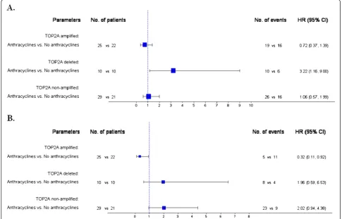
In parallel, for patients not treated with anthracyclines there were no significant differences in TTP between deleted and non-amplified versus amplified tumors (HR=0.76, 95% CI: 0.29-1.96, Wald’s p=0.57 and HR=1.23, 95% CI: 0.61-2.47, p=0.56, respectively)
[image:7.595.58.540.88.588.2](Figure 4A). For anthracycline-treated patients, deleted and non-amplified tumors were associated with increased risk for progression compared to amplified tumors (HR=3.42, 95% CI: 1.57-7.46, Wald’s p=0.002 and HR=1.83, 95% CI: 1.00-3.34, p=0.050, respectively)
(Figure 4B). Similarly, in terms of survival the same asso-ciations were observed. Among anthracycline-treated patients,TOP2A deleted and non-amplified tumors had increased risk for death compared to amplified tumors (HR=6.94, 95% CI: 2.26-21.34, Wald’s p=0.001 and HR=5.33, 95% CI: 2.02-14.12, p=0.001, respectively) (Figure 4D). In patients not treated with anthracyclines no such differences in survival were observed (Figure 4C). In the contrary, no significant differences in TTP and survival were observed between anthracycline- and non-anthracycline-treated patients in the HER2-positive sub-group when TOP2A gene status was not taken into account (log-rank, p=0.67 for TTP and p=0.57 for survival).
Repeating the analysis in HER2-positive patients that had received anthracyclines in the adjuvant setting (52 of the 64 anthracycline-treated/HER2-positive patients) the results were identical to the ones presented in Figure 3. Among theTOP2A deleted subgroup, adju-vant anthracycline-treated patients were associated with increased risk for progression (HR=3.25, 95% CI: 1.16-9.09, Wald’s p=0.025), while among the TOP2A ampli-fied subgroup, adjuvant anthracycline-treated patients
were associated with decreased risk for death (HR=0.27, 95% CI: 0.08-0.94, Wald’s p=0.041).
In multivariate analysis of the total cohort, the signifi-cance of TOP2A in anthracycline-treated patients remained, resulting in similar associations for both TTP and survival (Additional file 1 and Additional file 2) to the ones seen in the HER2-positive subgroup.
Finally, when examining in the models any TOP2A alteration (deletions or amplifications) versus non-amplification, the interaction with anthracycline treatment was significant only in the case of the HER2-positive subgroup in terms of survival (p=0.042), while among the anthracycline-treated patients, tumors withTOP2A altera-tions were associated with improved survival compared to the non-amplified tumors.
Discussion
To our knowledge, the present study is one of the first to evaluate the role of TOP2A gene amplification and TopoIIa protein expression in the outcome of patients treated with trastuzumab-based regimens for MBC. The most important evidence provided herein is that
[image:8.595.56.548.100.206.2]TOP2A gene amplification is a favorable prognostic
Table 3 Univariate Cox regression models for TOP2A expression according to HER2 status
HER2-positive HER2-negative
Events HR 95% CI Wald’s p Events HR 95% CI Wald’s p
TTP
TOP2A(FISH)
Deleted vs. Amplified 16 vs. 36 1.58 0.87-2.87 0.13
Non-amplified vs. Amplified 43 vs. 36 1.51 0.96-2.37 0.07
TopoIIa (IHC)
Positive vs. Negative 57 vs. 27 0.94 0.60-1.49 0.80 31 vs. 17 1.01 0.55-1.83 0.98
Survival
TOP2A(FISH)
Deleted vs. Amplified 12 vs. 17 2.67 1.27-5.62 0.009
Non-amplified vs. Amplified 33 vs. 17 2.16 1.20-3.88 0.010
TopoIIa (IHC)
Positive vs. Negative 38 vs. 19 0.71 0.41-1.24 0.22 23 vs. 14 0.70 0.36-1.37 0.30
[image:8.595.56.540.517.719.2]CIconfidence interval,HRhazard ratio,TTPtime to progression. Empty cells: Non-applicable.
Table 2 Association ofTOP2Aand TopoIIa with HER2 status
HER2 status Fisher’s exact p
Positive Negative
N % N %
TOP2A(FISH) n=221 Amplified 55 41.0 0 0 <0.001
Deleted 21 15.7 5 5.7
Non-amplified 58 43.3 82 94.3
TopoIIa (IHC) n=193 Negative 39 33.9 29 37.2 0.65
factor in HER2-positive patients treated with trastuzu-mab. Patients with HER2-positive/TOP2A non-amplified or deleted tumors did not seem to benefit from trastuzumab-based regimens and had an unfavorable out-come compared to TOP2Aamplified tumors, in line with recent reports on TOP2A gene dosage [35] and TOP2A gene amplification [36].
The role of the TOP2A gene has mainly been exam-ined in relation to anthracycline treatment. Co-amplification of HER2 and TOP2A was associated with favorable response to anthracycline-based therapy of lo-cally advanced breast cancer [12]. The results of our study did not appear, at first, to be associated with the administration of anthracyclines, since only 12% of our patients had received such treatment in the 1st line metastatic setting. However, 39% of all patients and 43% of the HER2-positive ones had received anthracyclines in the adjuvant setting. Upon further analysis of our data, taking into account adjuvant and/or 1st line anthracycline treatment, a significant interaction of
TOP2A with anthracycline treatment was observed both for TTP and survival. In terms of survival among the TOP2A amplified subgroup, anthracycline-treated
patients were associated with decreased risk for death. It appears therefore that the improvement in survival of theTOP2Aamplified subgroup treated with trastuzumab is probably due to the concurrent or previous exposure of the patients to anthracyclines, rather than the effect of the trastuzumab treatment itself. Furthermore, when
TOP2A amplified patients treated with anthracyclines in the adjuvant setting were analyzed separately, they were found to have decreased risk for death, suggesting that even history of adjuvant anthracycline treatment results in survival advantage ofTOP2Aamplified patients treated with trastuzumab.
[image:9.595.55.541.91.402.2]There is a very recent report from the Breast Cancer International Research Group (BCIRG) 006 trial [37] and an additional retrospective analysis of almost 5,000 patients [13] regarding the efficacy of trastuzumab in breast cancer patients with HER2 and TOP2A co-amplification. The first study is one of the largest rando-mized trials, which confirmed the role of trastuzumab in the adjuvant setting. The most interesting aspect of the BCIRG 006 trial is that it included a non-anthracycline regimen (docetaxel, carboplatin and tras-tuzumab), which was compared to AC-T (doxorubicin,
Figure 3Forest plots from multivariate Cox regression models in the HER2-positive patients. A: Time to progression (N=117). Among the
TOP2Adeleted subgroup, anthracycline-treated patients were associated with increased risk for progression.B: Survival (N=117). Among the
cyclophosphamide, followed by docetaxel) with or without trastuzumab. This study demonstrated that the DFS benefit conferred by AC-T without trastuzumab in HER2-positive breast cancer patients is actually restricted to TOP2A co-amplified malignancies, which constituted a subset (35%) of the HER2-positive cancers, and is virtually indistinguishable from the benefit achieved by the addition of trastuzumab. Importantly, this same benefit (found in theTOP2Aco-amplified sub-set) could also be attained by a non-anthracycline regi-men in combination with trastuzumab, thus avoiding the toxicities seen with anthracyclines. In our study, trastu-zumab was given to advanced-stage HER2-positive breast cancer patients in the metastatic setting, our find-ings should not therefore be compared to those of the BCIRG 006 trial [37].
Recent studies support the role of TopoIIa pro-tein expression, rather than TOP2A gene amplification, as a predictor of response to anthracycline-based
chemotherapy in the adjuvant setting [16]. It is of note, that TopoIIa protein overexpression has been reported in HER2-positive, as well as HER2-negative tumors, independently of TOP2A gene amplification [38]. The latter finding has also been shown in our study; TopoIIa protein overexpression however, was not associated with either TTP or survival.
[image:10.595.57.540.91.459.2]TopoIIa protein overexpression was however asso-ciated with ER-positive status and high Ki67 expression, partly in line with previous reports, since TopoIIa pro-tein expression had been shown to be associated with ER-positive status [39] and the Ki67 proliferation index [40]. To the best of our knowledge, the associations between TopoIIa and PTEN protein expression, as well asPIK3CAmutation presence are new findings in breast cancer tissue series, meriting further investigation for their biological importance. Of note, TopoIIa protein is upregulated in proliferating normal and cancer cells, in order to participate in the cell duplication process [41].
Hence, with the widely used cut-off of 5% positive neo-plastic cells to assess TopoIIa protein positivity, tumors are found to be positive for TopoIIa in the absence of underlying amplification of the corresponding gene.
Alterations of the TOP2A gene mostly happen in HER2-positive tumors, howeverTOP2A does not always follow the amplification fate or rate of theHER2 ampli-con, since it is not always included in the so called
“smallest region of amplification” next toHER2 [36,42], while it may also be deleted in the presence of HER2 amplification, as observed here and elsewhere [10]. Thus, at least in a subset of TOP2A amplified tumors, the mechanism drivingTOP2Aamplification may be dif-ferent than the one resulting in HER2 amplification [9,10,43], as shown by the far lower ratio of TOP2A signals in comparison to HER2 signals [10]. In addition,
TOP2A may also be amplified or deleted in the absence of HER2 amplification, further supporting the view of distinct and possibly multiple mechanisms, resulting in alterations of this gene. The absence of TOP2A amplifi-cation and the presence of deletions may practically have the same unfavorable impact on the outcome of HER2-positive patients, as shown in this study. With respect to gene deletions, it should be noted that the way markers are scored with FISH on FFPE sections it is unavoidable to obtain false positive results (deletions), due to nuclear truncations that interfere with the number of fluorescent signals to be counted per nucleus in a mostly unpredict-able manner. Hence, although TOP2A gene deletions may indeed occur, the results concerning this FFPE-FISH marker, in the present and in the previously pub-lished studies, should be interpreted with caution.
In most of the published series,TOP2A gene amplifi-cation or deletion was a rare event in HER2-negative patients [44]. Only in four studies [10,16,18,45], the rate of TOP2A alterations was considerably greater than the 1% to 2% range reported in all other studies. In line with the majority of the published data we did not find HER2-negative patients withTOP2Agene amplification.
Conclusions
In conclusion our study is one of the first to examine the role of theTOP2A gene in the field of trastuzumab-based treatment in MBC. We have evaluated the role of
TOP2A gene amplification and TopoIIa protein expres-sion and we have shown that TOP2A gene amplifica-tion is a favorable prognostic factor in HER2-positive MBC patients treated with trastuzumab, such an effect however, appears to rather be related to treatment with anthracyclines. In advanced-stage HER2-positive breast cancer patients treated with trastuzumab,TOP2A ampli-fication appears to be a strong predictive factor for improved survival in patients with concurrent or pre-vious exposure to anthracyclines. Nevertheless, given the
small size and the retrospective nature of our study, these data have to be viewed as hypothesis generating and need to be further explored and validated in larger cohorts of patients treated in the context of randomized trials. We are currently investigating these associations in patients included in a large adjuvant phase III trial conducted by our Group.
Additional files
Additional file 1:Forest plots from multivariate Cox regression models in the total study population: time to progression (A, n=181) and survival (B, n=172).
Additional file 2:Kaplan-Meier curves for time to progression (TTP, A-B) and survival (C-D) according toTOP2Agene status in the total study population stratified by anthracycline treatment.
Abbreviations
CEN17: Centromere 17; CI: Confidence interval; ER: Estrogen receptor; FFPE: Formalin-fixed paraffin-embedded; HER2: Human epidermal growth factor receptor 2; HR: Hazard ratio; IHC: Immunohistochemistry; Ki67: Antigen Ki67; MBC: Metastatic breast cancer; OS: Overall survival;
PIK3CA: Phosphoinositide-3-kinase, catalytic, alpha polypeptide;
PFS: Progression-free survival; PgR: Progesterone receptor; PTEN: Phosphatase and tensin homolog deleted on chromosome 10; SNP: Single nucleotide polymorphism; TOP2A: Topoisomerase II alpha (gene amplification); TopoIIa: Topoisomerase II alpha (protein expression); TTP: Time to progression; TMA: Tissue microarray.
Competing interests
The authors declare that they have no competing interests.
Authors’contributions
GF conceived of the study, participated in its design and coordination, participated in the clinical management of the patients, contributed to the collection of the tumor tissue samples analyzed in the study and drafted the manuscript. CC conceived of the study, participated in its design and coordination, participated in the clinical management of the patients, contributed to the collection of the tumor tissue samples analyzed in the study and drafted the manuscript. MB carried out the TMA construction and the IHC and FISH assays and helped to draft the manuscript. VK carried out the molecular studies and helped to draft the manuscript. AGE performed the statistical analysis and helped to draft the manuscript. IX participated in the clinical management of the patients and contributed to the collection of the tumor tissue samples analyzed in the study. AB carried out the immunoassays. GP participated in the clinical management of the patients and contributed to the collection of the tumor tissue samples analyzed in the study. NX participated in the clinical management of the patients and contributed to the collection of the tumor tissue samples analyzed in the study. IP carried out the immunoassays. AK participated in the clinical management of the patients and contributed to the collection of the tumor tissue samples analyzed in the study. PP participated in the clinical management of the patients and contributed to the collection of the tumor tissue samples analyzed in the study. DB participated in the clinical management of the patients and contributed to the collection of the tumor tissue samples analyzed in the study. DVS participated in the clinical management of the patients and contributed to the collection of the tumor tissue samples analyzed in the study. KTK conceived of the study, participated in its design and coordination and drafted the manuscript. All authors read and approved the final manuscript.
Acknowledgements
This work has been presented in part at the 33rdAnnual San Antonio Breast
data management and Stella Dallidou for secretarial assistance. Supported by a Hellenic Cooperative Oncology Group research grant (HE TR_10).
Author details
1Department of Medical Oncology,“Papageorgiou”Hospital, Aristotle
University of Thessaloniki School of Medicine, 564 03, Thessaloniki, Macedonia, Greece.2Second Department of Medical Oncology,
“Metropolitan”Hospital, Athens, Greece.3Laboratory of Molecular Oncology, Hellenic Foundation for Cancer Research, Aristotle University of Thessaloniki School of Medicine, Thessaloniki, Greece.4Department of Pathology, Aristotle University of Thessaloniki School of Medicine, Thessaloniki, Greece.5Section of Biostatistics, Hellenic Cooperative Oncology Group, Data Office, Athens, Greece.6Department of Pathology, Ioannina University Hospital, Ioannina, Greece.7Department of Medical Oncology, Ioannina University Hospital, Ioannina, Greece.8Oncology Section, Second Propaedeutic Department of Internal Medicine, University General Hospital“Attikon”, Athens, Greece. 9Histopathology Department,“Alexandra”Hospital, Athens, Greece. 10
Department of Medical Oncology,“Hippokration”Hospital, Athens, Greece. 11First Department of Medical Oncology,“Metropolitan”Hospital, Athens,
Greece.12Translational Research Section, Hellenic Cooperative Oncology Group, Data Office, Athens, Greece.
Received: 31 May 2012 Accepted: 16 October 2012 Published: 23 October 2012
References
1. Dafni U, Grimani I, Xyrafas A, Eleftheraki AG, Fountzilas G:Fifteen-year trends in metastatic breast cancer survival in Greece.Breast Cancer Res Treat2010,119:621–631.
2. Gennari A, Conte P, Rosso R, Orlandini C, Bruzzi P:Survival of metastatic breast carcinoma patients over a 20-year period: a retrospective analysis based on individual patient data from six consecutive studies.
Cancer2005,104:1742–1750.
3. Andrulis IL, Bull SB, Blackstein ME, Sutherland D, Mak C, Sidlofsky S, Pritzker KP, Hartwick RW, Hanna W, Lickley L,et al:neu/erbB-2 amplification identifies a poor-prognosis group of women with node-negative breast cancer. Toronto Breast Cancer Study Group.Journal of clinical oncology: official journal of the American Society of Clinical Oncology1998,
16:1340–1349.
4. Slamon DJ, Godolphin W, Jones LA, Holt JA, Wong SG, Keith DE, Levin WJ, Stuart SG, Udove J, Ullrich A,et al:Studies of the HER-2/neu proto-oncogene in human breast and ovarian cancer.Science1989,
244:707–712.
5. Cobleigh MA, Vogel CL, Tripathy D, Robert NJ, Scholl S, Fehrenbacher L, Wolter JM, Paton V, Shak S, Lieberman G, Slamon DJ:Multinational study of the efficacy and safety of humanized anti-HER2 monoclonal antibody in women who have HER2-overexpressing metastatic breast cancer that has progressed after chemotherapy for metastatic disease.Journal of clinical oncology: official journal of the American Society of Clinical Oncology
1999,17:2639–2648.
6. Slamon DJ, Leyland-Jones B, Shak S, Fuchs H, Paton V, Bajamonde A, Fleming T, Eiermann W, Wolter J, Pegram M,et al:Use of chemotherapy plus a monoclonal antibody against HER2 for metastatic breast cancer that overexpresses HER2.N Engl J Med2001,344:783–792.
7. Tsai-Pflugfelder M, Liu LF, Liu AA, Tewey KM, Whang-Peng J, Knutsen T, Huebner K, Croce CM, Wang JC:Cloning and sequencing of cDNA encoding human DNA topoisomerase II and localization of the gene to chromosome region 17q21-22.Proc Natl Acad Sci U S A1988,
85:7177–7181.
8. Jacobson KK, Morrison LE, Henderson BT, Blondin BA, Wilber KA, Legator MS, O’Hare A, Van Stedum SC, Proffitt JH, Seelig SA, Coon JS:
Gene copy mapping of the ERBB2/TOP2A region in breast cancer.Genes Chromosomes Cancer2004,40:19–31.
9. Arriola E, Marchio C, Tan DS, Drury SC, Lambros MB, Natrajan R, Rodriguez-Pinilla SM, Mackay A, Tamber N, Fenwick K,et al:Genomic analysis of the HER2/TOP2A amplicon in breast cancer and breast cancer cell lines.Laboratory investigation; a journal of technical methods and pathology2008,88:491–503.
10. Nielsen KV, Muller S, Moller S, Schonau A, Balslev E, Knoop AS, Ejlertsen B:
Aberrations of ERBB2 and TOP2A genes in breast cancer.Mol Oncol2010,
4:161–168.
11. Capranico G, Butelli E, Zunino F:Change of the sequence specificity of daunorubicin-stimulated topoisomerase II DNA cleavage by
epimerization of the amino group of the sugar moiety.Cancer Res1995,
55:312–317.
12. Coon JS, Marcus E, Gupta-Burt S, Seelig S, Jacobson K, Chen S, Renta V, Fronda G, Preisler HD:Amplification and overexpression of topoisomerase IIalpha predict response to anthracycline-based therapy in locally advanced breast cancer.Clinical cancer research: an official journal of the American Association for Cancer Research2002,8:1061–1067.
13. Press MF, Sauter G, Buyse M, Bernstein L, Guzman R, Santiago A, Villalobos IE, Eiermann W, Pienkowski T, Martin M,et al:Alteration of topoisomerase II-alpha gene in human breast cancer: association with responsiveness to anthracycline-based chemotherapy.Journal of clinical oncology: official journal of the American Society of Clinical Oncology2011,
29:859–867.
14. Di Leo A, Gancberg D, Larsimont D, Tanner M, Jarvinen T, Rouas G, Dolci S, Leroy JY, Paesmans M, Isola J, Piccart MJ:HER-2 amplification and topoisomerase IIalpha gene aberrations as predictive markers in node-positive breast cancer patients randomly treated either with an anthracycline-based therapy or with cyclophosphamide, methotrexate, and 5-fluorouracil.Clin Cancer Res2002,8:1107–1116.
15. Tanner M, Isola J, Wiklund T, Erikstein B, Kellokumpu-Lehtinen P, Malmstrom P, Wilking N, Nilsson J, Bergh J:Topoisomerase IIalpha gene amplification predicts favorable treatment response to tailored and dose-escalated anthracycline-based adjuvant chemotherapy in HER-2/ neu-amplified breast cancer: Scandinavian Breast Group Trial 9401.
J Clin Oncol2006,24:2428–2436.
16. O’Malley FP, Chia S, Tu D, Shepherd LE, Levine MN, Bramwell VH, Andrulis IL, Pritchard KI:Topoisomerase II alpha and responsiveness of breast cancer to adjuvant chemotherapy.J Natl Cancer Inst2009,101:644–650. 17. Pritchard KI, Messersmith H, Elavathil L, Trudeau M, O’Malley F, Dhesy-Thind
B:HER-2 and topoisomerase II as predictors of response to
chemotherapy.Journal of clinical oncology: official journal of the American Society of Clinical Oncology2008,26:736–744.
18. Bartlett JM, Munro A, Cameron DA, Thomas J, Prescott R, Twelves CJ:Type 1 receptor tyrosine kinase profiles identify patients with enhanced benefit from anthracyclines in the BR9601 adjuvant breast cancer chemotherapy trial.Journal of clinical oncology: official journal of the American Society of Clinical Oncology2008,26:5027–5035. 19. Bartlett JM, Munro AF, Dunn JA, McConkey C, Jordan S, Twelves CJ,
Cameron DA, Thomas J, Campbell FM, Rea DW,et al:Predictive markers of anthracycline benefit: a prospectively planned analysis of the UK National Epirubicin Adjuvant Trial (NEAT/BR9601).Lancet Oncol2010,
11:266–274.
20. Di Leo A, Desmedt C, Bartlett JM, Piette F, Ejlertsen B, Pritchard KI, Larsimont D, Poole C, Isola J, Earl H,et al:HER2 and TOP2A as predictive markers for anthracycline-containing chemotherapy regimens as adjuvant treatment of breast cancer: a meta-analysis of individual patient data.Lancet Oncol2011,12:1134–1142.
21. Oakman C, Moretti E, Sotiriou C, Viale G, Di Leo A:Re: Topoisomerase II alpha and responsiveness of breast cancer to adjuvant chemotherapy.
J Natl Cancer Inst2009,101:1735–1736. author reply 1736–1737. 22. Razis E, Bobos M, Kotoula V, Eleftheraki AG, Kalofonos HP, Pavlakis K,
Papakostas P, Aravantinos G, Rigakos G, Efstratiou I,et al:Evaluation of the association of PIK3CA mutations and PTEN loss with efficacy of trastuzumab therapy in metastatic breast cancer.Breast Cancer Res Treat
2011,128:447–456.
23. Fountzilas G, Kourea HP, Bobos M, Televantou D, Kotoula V, Papadimitriou C, Papazisis KT, Timotheadou E, Efstratiou I, Koutras A,et al:Paclitaxel and bevacizumab as first line combined treatment in patients with metastatic breast cancer: the hellenic cooperative oncology group experience with biological marker evaluation.Anticancer Res2011,
31:3007–3018.
24. Christodoulou C, Kostopoulos I, Kalofonos HP, Lianos E, Bobos M, Briasoulis E, Gogas H, Razis E, Skarlos DV, Fountzilas G:Trastuzumab combined with pegylated liposomal doxorubicin in patients with metastatic breast cancer. phase II Study of the Hellenic Cooperative Oncology Group (HeCOG) with biomarker evaluation.Oncology2009,
76:275–285.
Gemcitabine combined with gefitinib in patients with inoperable or metastatic pancreatic cancer: a phase II Study of the Hellenic Cooperative Oncology Group with biomarker evaluation.Cancer Invest
2008,26:784–793.
26. Hammond ME, Hayes DF, Dowsett M, Allred DC, Hagerty KL, Badve S, Fitzgibbons PL, Francis G, Goldstein NS, Hayes M,et al:American Society of Clinical Oncology/College Of American Pathologists guideline recommendations for immunohistochemical testing of estrogen and progesterone receptors in breast cancer.Journal of clinical oncology: official journal of the American Society of Clinical Oncology2010,
28:2784–2795.
27. Wolff AC, Hammond ME, Schwartz JN, Hagerty KL, Allred DC, Cote RJ, Dowsett M, Fitzgibbons PL, Hanna WM, Langer A,et al:American Society of Clinical Oncology/College of American Pathologists guideline recommendations for human epidermal growth factor receptor 2 testing in breast cancer.Journal of clinical oncology: official journal of the American Society of Clinical Oncology2007,25:118–145.
28. Cheang MC, Chia SK, Voduc D, Gao D, Leung S, Snider J, Watson M, Davies S, Bernard PS, Parker JS,et al:Ki67 index, HER2 status, and prognosis of patients with luminal B breast cancer.J Natl Cancer Inst
2009,101:736–750.
29. Gori S, Sidoni A, Colozza M, Ferri I, Mameli MG, Fenocchio D, Stocchi L, Foglietta J, Ludovini V, Minenza E,et al:EGFR, pMAPK, pAkt and PTEN status by immunohistochemistry: correlation with clinical outcome in HER2-positive metastatic breast cancer patients treated with trastuzumab.Annals of oncology: official journal of the European Society for Medical Oncology/ESMO2009,20:648–654.
30. Bhargava R, Lal P, Chen B:HER-2/neu and topoisomerase IIa gene amplification and protein expression in invasive breast carcinomas: chromogenic in situ hybridization and immunohistochemical analyses.
Am J Clin Pathol2005,123:889–895.
31. Fountzilas G, Ciuleanu E, Bobos M, Kalogera-Fountzila A, Eleftheraki AG, Karayannopoulou G, Zaramboukas T, Nikolaou A, Markou K, Resiga L,et al:
Induction chemotherapy followed by concomitant radiotherapy and weekly cisplatin versus the same concomitant chemoradiotherapy in patients with nasopharyngeal carcinoma: a randomized phase II study conducted by the Hellenic Cooperative Oncology Group (HeCOG) with biomarker evaluation.Annals of oncology: official journal of the European Society for Medical Oncology/ESMO2012,23:427-435.
32. Knoop AS, Knudsen H, Balslev E, Rasmussen BB, Overgaard J, Nielsen KV, Schonau A, Gunnarsdottir K, Olsen KE, Mouridsen H, Ejlertsen B:
retrospective analysis of topoisomerase IIa amplifications and deletions as predictive markers in primary breast cancer patients randomly assigned to cyclophosphamide, methotrexate, and fluorouracil or cyclophosphamide, epirubicin, and fluorouracil: Danish Breast Cancer Cooperative Group.Journal of clinical oncology: official journal of the American Society of Clinical Oncology2005,23:7483–7490. 33. Bohmann K, Hennig G, Rogel U, Poremba C, Mueller BM, Fritz P,
Stoerkel S, Schaefer KL:RNA extraction from archival formalin-fixed paraffin-embedded tissue: a comparison of manual, semiautomated, and fully automated purification methods.Clin Chem2009,
55:1719–1727.
34. McShane LM, Altman DG, Sauerbrei W, Taube SE, Gion M, Clark GM:
Reporting recommendations for tumor marker prognostic studies (REMARK).J Natl Cancer Inst2005,97:1180–1184.
35. Zaczek A, Markiewicz A, Jaskiewicz J, Pienkowski T, Rhone P, Jassem J, Welnicka-Jaskiewicz M:Clinical evaluation of developed PCR-based method with hydrolysis probes for TOP2A copy number evaluation in breast cancer samples.Clin Biochem2010,43:891–898.
36. Lamy PJ, Fina F, Bascoul-Mollevi C, Laberenne AC, Martin PM, Ouafik L, Jacot W:Quantification and clinical relevance of gene amplification at chromosome 17q12-q21 in human epidermal growth factor receptor 2-amplified breast cancers.Breast cancer research: BCR2011,
13:R15.
37. Slamon D, Eiermann W, Robert N, Pienkowski T, Martin M, Press M, Mackey J, Glaspy J, Chan A, Pawlicki M,et al:Adjuvant trastuzumab in HER2-positive breast cancer.N Engl J Med2011,365:1273–1283. 38. Callagy G, Pharoah P, Chin SF, Sangan T, Daigo Y, Jackson L, Caldas C:
Identification and validation of prognostic markers in breast cancer with the complementary use of array-CGH and tissue microarrays.
J Pathol2005,205:388–396.
39. Koren R, Rath-Wolfson L, Ram E, Itzhac OB, Schachter B, Klein B, Gal R, Dreznik Z:Prognostic value of Topoisomerase II in female breast cancer.
Oncol Rep2004,12:915–919.
40. Lynch BJ, Guinee DG Jr, Holden JA:Human DNA topoisomerase II-alpha: a new marker of cell proliferation in invasive breast cancer.Hum Pathol
1997,28:1180–1188.
41. Turley H, Comley M, Houlbrook S, Nozaki N, Kikuchi A, Hickson ID, Gatter K, Harris AL:The distribution and expression of the two isoforms of DNA topoisomerase II in normal and neoplastic human tissues.Br J Cancer
1997,75:1340–1346.
42. Kauraniemi P, Kallioniemi A:Activation of multiple cancer-associated genes at the ERBB2 amplicon in breast cancer.Endocr Relat Cancer2006,
13:39–49.
43. Sircoulomb F, Bekhouche I, Finetti P, Adelaide J, Ben Hamida A, Bonansea J, Raynaud S, Innocenti C, Charafe-Jauffret E, Tarpin C,et al:Genome profiling of ERBB2-amplified breast cancers.BMC Cancer2010,10:539.
44. Slamon DJ, Press MF:Alterations in the TOP2A and HER2 genes: association with adjuvant anthracycline sensitivity in human breast cancers.J Natl Cancer Inst2009,101:615–618.
45. Nielsen KV, Ejlertsen B, Moller S, Jorgensen JT, Knoop A, Knudsen H, Mouridsen HT:The value of TOP2A gene copy number variation as a biomarker in breast cancer: Update of DBCG trial 89D.Acta Oncol2008,
47:725–734.
doi:10.1186/1479-5876-10-212
Cite this article as:Fountzilaset al.:Topoisomerase II alpha gene
amplification is a favorable prognostic factor in patients with HER2-positive metastatic breast cancer treated with trastuzumab. Journal of Translational Medicine201210:212.
Submit your next manuscript to BioMed Central and take full advantage of:
• Convenient online submission
• Thorough peer review
• No space constraints or color figure charges
• Immediate publication on acceptance
• Inclusion in PubMed, CAS, Scopus and Google Scholar
• Research which is freely available for redistribution