RESEARCH
Characterization of
Plasmodium relictum
,
a cosmopolitan agent of avian malaria
Gediminas Valkiūnas
1*, Mikas Ilgūnas
1, Dovilė Bukauskaitė
1, Karin Fragner
2, Herbert Weissenböck
2,
Carter T. Atkinson
3and Tatjana A. Iezhova
1Abstract
Background: Microscopic research has shown that Plasmodium relictum is the most common agent of avian malaria. Recent molecular studies confirmed this conclusion and identified several mtDNA lineages, suggesting the existence of significant intra-species genetic variation or cryptic speciation. Most identified lineages have a broad range of hosts
and geographical distribution. Here, a rare new lineage of P. relictum was reported and information about biological
characters of different lineages of this pathogen was reviewed, suggesting issues for future research.
Methods: The new lineage pPHCOL01 was detected in Common chiffchaff Phylloscopus collybita, and the parasite
was passaged in domestic canaries Serinus canaria. Organs of infected birds were examined using histology and
chro-mogenic in situ hybridization methods. Culex quinquefasciatus mosquitoes, Zebra finch Taeniopygia guttata,
Budgeri-gar Melopsittacus undulatus and European goldfinch Carduelis carduelis were exposed experimentally. Both Bayesian and Maximum Likelihood analyses identified the same phylogenetic relationships among different, closely-related
lineages pSGS1, pGRW4, pGRW11, pLZFUS01, pPHCOL01 of P. relictum. Morphology of their blood stages was
com-pared using fixed and stained blood smears, and biological properties of these parasites were reviewed.
Results: Common canary and European goldfinch were susceptible to the parasite pPHCOL01, and had markedly variable individual prepatent periods and light transient parasitaemia. Exo-erythrocytic and sporogonic stages were
not seen. The Zebra finch and Budgerigar were resistant. Neither blood stages nor vector stages of all examined P.
relictum lineages can be distinguished morphologically.
Conclusion: Within the huge spectrum of vertebrate hosts, mosquito vectors, and ecological conditions, different
lineages of P. relictum exhibit indistinguishable, markedly variable morphological forms. Parasites of same lineages
often develop differently in different bird species. Even more, the variation of biological properties (parasitaemia dynamics, blood pathology, prepatent period) in different isolates of the same lineage might be greater than the variation in different lineages during development in the same species of birds, indicating negligible taxonomic value of such features. Available lineage information is excellent for parasite diagnostics, but is limited in predictions about relationships in certain host-parasite associations. A combination of experiments, field observations, microscopic and
molecular diagnostics is essential for understanding the role of different P. relictum lineages in bird health.
Keywords: Plasmodium relictum, Birds, Morphological and molecular characterization, Review
© The Author(s) 2018. This article is distributed under the terms of the Creative Commons Attribution 4.0 International License
(http://creativecommons.org/licenses/by/4.0/), which permits unrestricted use, distribution, and reproduction in any medium,
provided you give appropriate credit to the original author(s) and the source, provide a link to the Creative Commons license,
and indicate if changes were made. The Creative Commons Public Domain Dedication waiver (http://creativecommons.org/
publicdomain/zero/1.0/) applies to the data made available in this article, unless otherwise stated.
Background
Plasmodium relictum is an invasive blood parasite, which causes malaria in many species of birds from all over the word [1–4]. Naive birds often experience severe disease
and even mortality during malaria infection [5, 6], but some bird species and their populations appear to be relatively resistant and can tolerate this infection [7–11]. This parasite was the first recognized and described agent of avian malaria [12], likely due to its high preva-lence in a wide range of different avian hosts and because morphological characteristics of its mature blood stages are so distinctive in blood films. Mature stages typically
Open Access
*Correspondence: gedvalk@ekoi.lt
possess prominent nuclei and cytoplasm, numerous pig-ment granules and markedly influence the position of the nuclei of their host erythrocytes, causing lateral shifts in their position. Numerous synonymous names of this organism exist [7, 13]. These names were suggested for distinguishing the morphologically similar or even iden-tical blood stages, which were reported in different avian hosts and/or different geographical areas [13–15]. Micro-scopic examination of blood films, the main avian malaria diagnostic tool used in the 20th Century, has identified P. relictum as the most common agent of avian malaria with reports from over 300 species of birds belonging to 11 orders from all over the world [1, 7, 16, 17]. Recent molec-ular studies have supported this conclusion and uncov-ered significant genetic diversity among different isolates of P. relictum, suggesting existence of intra-species genetic variation or even cryptic speciation [2, 18–22].
Partial sequences of mitochondrial cytochrome b gene (cytb) have been successfully used in distinguishing differ-ent lineages of avian malaria parasites, and they are excelldiffer-ent molecular markers for disease diagnostics [2, 4, 19, 23–25]. Over 100 closely related cytb lineages of avian Plasmodium were deposited in GenBank and MalAvi database (http://
mbio-serv2.mbioekol.lu.se/Malavi) and many of them
may belong to P. relictum. However, few of these molecu-lar lineages are supported by microscopic examination of well-fixed and stained blood smears and the small genetic difference in cytb sequences alone cannot be considered as final proof that closely related lineages belong to the same morpho-species. For example, some morphologically dis-tinct haemosporidian species differ in their partial cytb sequences just by a few nucleotide bases [26, 27]. Currently, only four lineages (pSGS1, pGRW4, pGRW11, pLZFUS01) have been linked to P. relictum based on morphological characters of their blood stages, and these data are helpful for distinguishing this infection in blood films [28–30].
During the past 15 years, much data have been col-lected about host, geographical distribution, vectors, virulence, and other biological characters of P. relictum based on cytb lineages [2, 3, 8, 19, 30–34]. This provides opportunities to examine patterns in the biology and pathology of avian Plasmodium infection at the level of these specific lineages. This study characterizes a new cytb lineage of P. relictum (pPHCOL01), makes com-prehensive comparisons of morphological characters of blood stages of all known lineages of this parasite, and reviews their biological features to help identify some new directions for future avian malaria research.
Methods
Collection of blood and tissue samples
Fieldwork was carried out at the Ventės Ragas Ornitho-logical Station, Lithuania between 4 and 18 May, 2017.
Twenty-three Common chiffchaffs Phylloscopus collybita were caught with mist nets and large stationary traps. The blood was taken by puncturing the brachial vein. Three blood films were prepared immediately after with-drawal of the blood, air-dried using a battery-operated fan, fixed in absolute methanol and stained with Giemsa. About 30 μl of whole blood was taken in heparinized microcapillaries and stored in SET buffer (0.05 M Tris, 0.15 M NaCl, 0.5 M EDTA, pH 8.0) at ambient tempera-ture while in the field and then maintained at − 20 °C in the laboratory.
To detect and isolate the Plasmodium parasite strain, the blood films from each captured bird were quickly examined microscopically in the field, as previously described [35]. One naturally infected Common chiff-chaff was detected, with parasitaemia of 0.1%. Ten blood films were prepared for microscopic examination from this bird. Additionally, blood was also collected in hep-arinized microcapillaries and used to expose two unin-fected domestic Common canaries Serinus canaria forma domestica by sub-inoculation into their pectoral muscle of about 250 μl of a freshly prepared mixture of infected blood, 3.7% sodium citrate (anticoagulant) and 0.9% saline (4:1:5) [36].
Parasitaemia developed in both exposed canaries, and blood of these birds was passaged as described above in three additional canaries. Two Zebra finches
Taeniopy-gia guttate, one Budgerigar Melopsittacus undulatus
and two European goldfinches Carduelis carduelis were also exposed, as described above. Six uninfected canar-ies were used as controls. Blood of all control and experi-mental birds was tested by microscopic examination and PCR-based methods (see description below) twice before the experiment to ensure that they were uninfected with malaria parasites.
Two canaries were observed for 57 and 94 days post exposure (dpe) and then euthanized for histology and chromogenic in situ hybridization research. Two Euro-pean goldfinches were observed for 127 dpe. All remain-ing birds were observed for 131 dpe. Post-exposure blood samples were taken for microscopic examination and PCR-based testing as described above once every 4 days during the first post-exposure month, once every week during the second month and once every 1–2 weeks dur-ing the remaindur-ing experiment time. All experimental and control birds were kept indoors in a vector-free room under natural light–dark photoperiod. They were fed a standard diet for seed-eating bird species.
formalin, embedded in paraffin and processed using tra-ditional histologic methods [7]. Histological sections of 4 μm were obtained, stained with haematoxylin–eosin, mounted in BioMount (BioGnost, Croatia) and exam-ined microscopically. One smear of bone marrow was prepared from the tibia bone of each bird, air dried, fixed with absolute methanol and stained with Giemsa.
Experimental infection and investigation of Culex
quinquefasciatus mosquitoes
Laboratory-reared Culex quinquefasciatus mosquitoes were maintained and exposed to canaries infected with the isolated Plasmodium sp., as previously described [32]. Briefly, insects were kept in cages (45 × 45 × 45 cm) under 65–70% relative humidity, 16/8 h light/dark photo-period and 26 ± 1 °C. One experimentally infected canary was used as the donor of parasites for infecting mosqui-toes. Eleven female mosquitoes took blood meals on this canary. Parasitaemia was 0.02% with few visible mature gametocytes. Preparations of midgut contents (6 insects were dissected 24–48 h post exposure), one preparation of midgut wall (one insect on 12 dpe) and 4 preparations of salivary glands (on 15, 16 and 18 dpe) were prepared and examined, as previously described [32].
Microscopic examination
Detailed microscopic analysis was carried out with vari-ous Olympus light microscopes equipped with Olympus digital cameras and imaging software. Preparations of blood stages of the lineages pSGS1, pGRW11, pGRW4, and pLZFUS01 were from collections of voucher speci-mens which have been deposited at P. B. Šivickis Labo-ratory of Parasitology, Nature Research Centre Vilnius. These were blood films from canaries whose were exposed experimentally to the parasite lineages pSGS1 (parasitaemia varied between 0.6 and 1.8%, preparation accession number 48979–48981 NS), pGRW11 (1.1–6%, 48982–48984 NS), pGRW4 (0.2–2.1%, 48985–48987 NS), and the lineage pLZFUS01 (0.5%, 48694–48696 NS) from the blood of a naturally infected Red-backed shrike Lanius collurio (for exposure description see [30]). Addi-tionally, preparations with the Hawaiian isolate of P. rel-ictum (pGRW4) were used. These were (1) 12 blood films from two individual canaries that were exposed experi-mentally by inoculation of infected blood (parasitaemia varies between 0.6 and 10%, accession nos. 48988–48999 NS) (for infection details see [5]), (2) 11 blood films from one naturally infected Apapane Himatione sanguinea (parasitaemia 22%, accession nos. 49000–49010 NS).
Blood films from each infected bird were examined and the observed blood stages were morphologically com-pared by skilled parasitologists of avian malaria parasites at the P. B. Šivickis Laboratory of Parasitology. At least
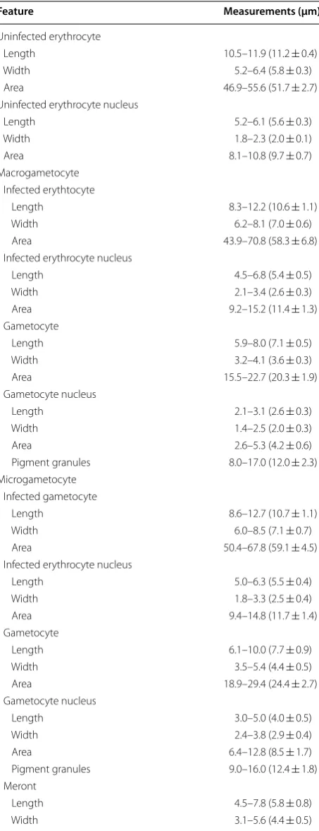
100 fields were studied at high magnification (1000×) in each preparation. Intensity of parasitaemia was estimated as a percentage by actual counting of the number of para-sites per 10,000 erythrocytes. The morphometric features studied (Table 1) were those defined in [7]. The analyses were carried out using the ‘Statistica 7’ package as previ-ously described [7].
In situ hybridization
Chromogenic in situ hybridization (ISH) was applied to increase detectability of tissue stages of the para-sites. Organs (the same as for histological examination) from one naturally infected Common chiffchaff and two experimentally infected canaries (57 and 94 dpe, respec-tively) were tested using a previously described ISH pro-tocol [37]. 3 μm paraffin-embedded tissue sections of all these organs were prepared. The sections were deparaffi-nized, subjected to proteolytic treatment with proteinase K (Roche, Basel, Switzerland) 6 μg/ml in Tris-buffered saline at 37 °C for 50 min. For hybridization, the slides with tissue sections were incubated overnight at 40 °C with hybridization mixture and a final probe concen-tration of 100 ng/ml. The used oligonucleotide probe
(sequence: 5′
-TTTAATAACTCGTTATATATATCAGT-GTAGCAC-3′) was labelled with digoxigenin at the 3′
end (Eurofins MWG Operon, Ebersberg, Germany). This probe is specific to detect avian Plasmodium parasites [6, 37]. The digoxigenin-labelled hybrids were detected by incubating the slides with anti-digoxigenin-AP Fab fragments (Roche) (1:200) for 1 h at room tempera-ture (RT). Visualization of the reaction was carried out using the colour substrates 5-bromo-4-chloro-3-indolyl phosphate (BCIP) and 4-nitro blue tetrazolium chloride (NBT) (Roche). A positive control (sections of a lung of a Eurasian blackbird Turdus merula naturally infected with
Plasmodium vaughani, which was proven to be positive
by previous ISH) was used to assure that the protocol worked. Preparations were examined microscopically by skilled parasitologists and pathologists; at least 50 fields of each preparation were studied at low magnification (400×), and then each preparation was examined for 10–15 min at high magnification (1000×).
Molecular and phylogenetic analysis
DNA template (2 µl). Negative controls (nuclease-free water) were used after each seven samples to detect possible false amplifications, and one positive control (extracted parasite DNA from a blood sample, which was confirmed positive during previous PCR testing) was used to evaluate the success of PCR if none of the sam-ples would have been amplified.
Temperatures for the PCR were as described in the original protocols. The success of the performed PCR was evaluated by running electrophoresis on a 2% aga-rose gel. Successfully amplified DNA was precipitated using 11 µl of 8 M NH 4Ac, 37 µl of 96% and 150 µl of 70% ethanol. After centrifugation, the supernatant was discarded, the samples were air-dried overnight, and then 16 µl of nuclease-free water was added on the precipi-tated DNA. Big Dye Terminator V3.1 Cycle Sequencing Kit and ABI PRISM™ 3100 capillary sequencing robot
(Applied Biosystems, Foster City, California) were used for sequencing. Sequences were edited and aligned using BioEdit software [40]. Absence of double-base calling in sequence electropherograms was used as an indication of single infections [41]. Nucleotide BLAST (megablast algorithm) (http://blast.ncbi.nlm.nih.gov/Blast.cgi) was used to compare our amplified sequences with sequences deposited in the GenBank.
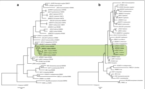
Molecular phylogenetic analysis was carried out using Bayesian and Maximum Likelihood algorithms. Sequences for the phylogenetic analysis were collected from GenBank and double-checked in MalAvi database [19]. Plasmodium falciparum was used as an outgroup. GenBank accession numbers and codes of the lineages are provided in the phylogenetic trees (Fig. 1). Bayes-ian phylogenetic tree (Fig. 1a) was constructed using MrBayes version 3.1 [42] software. The General Time Reversible Model (GTR) was used as suggested by the software MrModeltest 2.2 (https://github.com/nylander/ MrModeltest2). Analysis was run for a total of 10 million generations with a sampling frequency of every 100 gen-erations. Before the construction of the consensus tree, 25% of the initial trees were discarded as ‘burn in’ period. The tree was visualized using the software FigTree v1.4.3
(http://tree.bio.ed.ac.uk/software/figtree/). Maximum
[image:4.595.59.286.134.724.2]Likelihood tree (Fig. 1b) was constructed using the MEGA 7.0 [43] software; it was performed with 1,000 Table 1 Morphometry of blood stages and host cells
of Plasmodium (Haemamoeba) relictum (pPHCOL01) from the blood of Common chiffchaff Phylloscopus colly-bita (n = 21)
Feature Measurements (μm)a
Uninfected erythrocyte
Length 10.5–11.9 (11.2 ± 0.4)
Width 5.2–6.4 (5.8 ± 0.3)
Area 46.9–55.6 (51.7 ± 2.7) Uninfected erythrocyte nucleus
Length 5.2–6.1 (5.6 ± 0.3)
Width 1.8–2.3 (2.0 ± 0.1)
Area 8.1–10.8 (9.7 ± 0.7)
Macrogametocyte Infected erythtocyte
Length 8.3–12.2 (10.6 ± 1.1) Width 6.2–8.1 (7.0 ± 0.6) Area 43.9–70.8 (58.3 ± 6.8) Infected erythrocyte nucleus
Length 4.5–6.8 (5.4 ± 0.5) Width 2.1–3.4 (2.6 ± 0.3) Area 9.2–15.2 (11.4 ± 1.3) Gametocyte
Length 5.9–8.0 (7.1 ± 0.5) Width 3.2–4.1 (3.6 ± 0.3) Area 15.5–22.7 (20.3 ± 1.9) Gametocyte nucleus
Length 2.1–3.1 (2.6 ± 0.3) Width 1.4–2.5 (2.0 ± 0.3)
Area 2.6–5.3 (4.2 ± 0.6)
Pigment granules 8.0–17.0 (12.0 ± 2.3) Microgametocyte
Infected gametocyte
Length 8.6–12.7 (10.7 ± 1.1) Width 6.0–8.5 (7.1 ± 0.7) Area 50.4–67.8 (59.1 ± 4.5) Infected erythrocyte nucleus
Length 5.0–6.3 (5.5 ± 0.4) Width 1.8–3.3 (2.5 ± 0.4) Area 9.4–14.8 (11.7 ± 1.4) Gametocyte
Length 6.1–10.0 (7.7 ± 0.9) Width 3.5–5.4 (4.4 ± 0.5) Area 18.9–29.4 (24.4 ± 2.7) Gametocyte nucleus
Length 3.0–5.0 (4.0 ± 0.5) Width 2.4–3.8 (2.9 ± 0.4) Area 6.4–12.8 (8.5 ± 1.7) Pigment granules 9.0–16.0 (12.4 ± 1.8) Meront
Length 4.5–7.8 (5.8 ± 0.8) Width 3.1–5.6 (4.4 ± 0.5)
a Minimum and maximum values are provided, followed in parentheses by the
arithmetic mean and standard deviation
Table 1 continued
Feature Measurements (μm)a
bootstrap replications using the GTR model and the same dataset as during the Bayesian analysis.
The new sequence of lineage pPHCOL01 was deposited in GenBank (accession MG724747). Genetic differences between different lineages of P. relictum were calculated using the Jukes–Cantor model of substitution, as imple-mented in the programme MEGA 7.0 [43].
Results
Parasite lineage identification and susceptibility of experimental birds
Single infections of P. relictum (cytb lineage pPHCOL01) was identified in the donor Common chiffchaff both by microscopic examination of blood films and PCR-based amplification and sequencing. All exposed canaries were susceptible and developed a single infection with the same malaria parasite, as determined both by micro-scopic examination of blood films and PCR-based test-ing. Parasitaemia developed in one exposed European goldfinch. Two Zebra finches, one Budgerigar and one
European goldfinch were resistant. All control canaries remained non-infected during this study.
Phylogenetic analysis
The reported lineage of P. relictum (pPHCOL01) was new. It clustered with other morphologically character-ized lineages of P. relictum (pSGS1, pGRW4, pGRW11, pLZFUS01) in both phylogenetic analyses, supporting the close phylogenetic relationships among them (Fig. 1a, b). Genetic differences among five lineages of P. relictum varied between 0.2% (minimum, the lineages pSGS1 and pGRW11) and 3% (maximum, the lineages pSGS1 and pLZFUS01).
Characterization of Plasmodium (Haemamoeba) relictum
(pPHCOL01)
See (Fig. 2, Table 1).
DNA sequence
Mitochondrial cytb lineage pPHCOL01 (new lineage, 479 bp, GenBank accession MG724747).
hHIICT1 H. belopolskyi DQ630006
pANLA2 P. multivacuolaris FJ389157
hRB1 H. lanii KR049256
lGALLUS08 Leucocytozoon schoutedeni DQ676823 pANLAT07 P. multivacuolaris FJ404720
hSYAT1 H. parabelopolskyi AY831750 pCOLL4 DQ368374
P. falciparum AJ298787
pPHCOL01P. relictum
pALEDIA02 P. parahexamerium EU810634
hFREMIN01 H. iwa JF833050
hHAECOL1 H. columbae AF495554 pTFUS05 P. lutzi KC138226
pSPMAG01 P. tejerai JX272844
pDENPET03 P. nucleophilum HF543647 pANLAT01 P. globularis FJ404710
pGRW11 P. relictum AY831748
lSISKIN2 L. sp. AY393796 pGRW4 P. relictum AF254975
pPADOM16 P. rouxi HM146901
hCOLBUC01 H. multipigmentatus GU296227 pGALLUS2 P. juxtanucleare AB250415 pTFUS06 P. unalis KC771248
pANLA1 P. globularis EU770151
pGRW6 P. elongatum DQ368381
pGALLUS01 P. gallinaceum AY099029
pSEIAUR01 P. cathemerium DQ838988 pGRW2 P. ashfordi AF254962
hRW1 Haemoproteus payevskyi DQ630009 pSW2 P. homonucleophilum KC342643
pTURDUS1 AF495576
pLZFUS01 P. relictum AB308046 pALDI1 P. parahexamerium FJ389155
pBAEBIC02 P. homopolare KJ482708
pCYOL1 P. megaglobularis EU770152
pSYAT05 Plasmodium vaughani DQ847271
pCOLL6 P. delichoni KU529943
pSGS1 P. relictum AF495571
pLINN1 P. matutinum KY287235
pSW2 P. homonucleophilum
100 71 77 99 98 100 100 100 98 99 93 95 78 73 94 72 0.05
pTFUS06 P. unalis
pSYAT05 Plasmodium vaughani
pALEDIA02 P. parahexamerium
pALDI1 P. parahexamerium
pBAEBIC02 P. homopolare
pANLAT01 P. globularis
pANLA1 P. globularis
pPADOM16 P. rouxi
pANLA2 P. multivacuolaris
pANLAT07 P. multivacuolaris
pGALLUS2 P. juxtanucleare
pDENPET03 P. nucleophilum
pGRW2 P. ashfordi
pCOLL6 P. delichoni
pCOLL4
pGALLUS01 P. gallinaceum
pTURDUS1 pSEIAUR01 P. cathemerium
pCYOL1 P. megaglobularis
pLZFUS01 P. relictum
pPHCOL01 P. relictum pSGS1 P. relictum
pGRW11 P. relictum
pGRW4 P. relictum
pSPMAG01 P. tejerai
pLINN1 P. matutinum
pTFUS05 P. lutzi
pGRW6 P. elongatum
hCOLBUC01 H. multipigmentatus
hHAECOL1 H. columbae
hFREMIN01 H. iwa
hHIICT1 H. belopolskyi
hRB1 H. lanii
hRW1 Haemoproteus payevskyi
hSYAT1 H. parabelopolskyi
lGALLUS08 Leucocytozoon schoutedeni
lSISKIN2 L. sp. P. falciparum 0.93 0.77 0.92 0.91 0.98 0.91 0.78 0.92 0.77 0.98 0.98 1 1 1 1 1 1 1 1 1 1 1 1 1 1 1 1 MG724747 0.03
a
b
Fig. 1 Bayesian (a) and Maximum Likelihood (b) phylogeny of 29 mitochondrial cytochrome b lineages of Plasmodium species, seven lineages of
Haemoproteus spp. and two lineages of Leucocytozoon spp. One lineage of Plasmodium falciparum is used as an outgroup. Codes of the lineages
[image:5.595.58.540.88.383.2]Avian hosts
Common chiffchaff Phylloscopus collybita is a natural host. Other natural avian hosts are unknown. Two Zebra finches and one Budgerigar that were exposed by sub-inoculation of infected blood were resistant. The most similar Plasmodium parasite lineages were reported only in sub-Saharan birds by Loiseau et al. [44] (the lin-eage PV40, accession HQ022817, 2 bp difference, avian host was not reported), Beadell et al. [2] (the lineage P27, accession DQ659568, 7 bp difference, the host is the Cameroon sunbird Cyanomitra oritis) and Lutz et al. [45] (the lineage P_AFR110, accession KM056570, 7 bp differ-ence, the host is the Miombo tit Parus griseiventris).
Vectors
Remain unidentified. Sporogonic development was not observed in Culex quinquefasciatus mosquitoes.
Site of infection
Red blood cells; no other data.
Representative blood films
Voucher specimens (accession numbers 48965–48974
NS, Phylloscopus collybita, 7 May 2017, parasitaemia
0.1%, collected by D. Bukauskaitė, and 48975–48978 NS, Serinus canaria, 2–6 June, 2017, collected by M. Ilgūnas) were deposited in Nature Research Centre, Vilnius, Lithuania.
Prevalence
The overall prevalence was 1 of 23 (4.3%) in Common chiffchaff at the study site.
Parasitaemia and virulence
Canaries are susceptible, with long-lasting (up to 65 dpe), but light parasitaemia (< 0.01%) reported in the majority of exposed birds. One of two exposed European gold-finches developed very light (0.001%) and long-lasting (up to 127 dpe) parasitaemia. In all positive birds, para-sitaemia was transient, i.e., it was not seen during all days of testing. In experimentally exposed birds, the maxi-mum reported parasitaemia was 0.02%, and it was seen in one canary. The parasitaemia remained light or even declined into latency approximately 1–2 weeks after the first parasites seen in blood films in all positive birds, with a few parasites appearing in the circulation during entire observation time.
All blood stages (trophozoites, growing and mature meronts, growing and mature gametocytes) were reported in the peripheral circulation of naturally infected Common chiffchaff, experimentally exposed canaries and one goldfinch. This indicates asynchronous development in the blood. Mortality was not reported among exposed birds, and they appeared healthy. Clinical signs of disease were not observed during this study, and it is probable that susceptible inoculated birds can toler-ate this infection.
The prepatent period varied markedly, with first para-sites observed in the peripheral circulation 9, 14, 31, and 49 dpe in different canaries. Prepatent period was 11 dpe in one European goldfinch.
Morphology of blood stages of the new lineage para-sites was the same in the Common chiffchaff and the experimentally exposed canaries and one goldfinch. Description of blood stages of this infection is given from preparations with parasitaemia of 0.1% in Common chiff-chaff (Fig. 2).
Trophozoites
Figure 2a–d. Develop mainly in mature erythrocytes (Fig. 2b, d), but sometimes were also seen in polychro-matic erythrocytes (Fig. 2a, c). Earliest trophozoites usually are of irregular form, often amoeboid in outline (Fig. 2a, b); they only slightly displace nuclei in infected erythrocytes laterally. Advanced trophozoites pos-sess prominent nuclei and cytoplasm, but lack vacuoles (Fig. 2c, d); they were often attached to the host cell nuclei (Fig. 2d), which are slightly displaced. Pigment granules are roundish, small (< 0.5 µm), few, dark-brown, and usually grouped.
Erythrocytic meronts
Figure 2e–k. Develop in mature erythrocytes. Young growing meronts possess plentiful cytoplasm and large nuclei (Fig. 2e); size of the nuclei and amount of the cyto-plasm markedly decrease as the parasites mature (com-pare Fig. 2e with Fig. 2f–h). Vacuoles are absent from both developing and mature meronts. Pigment gran-ules are small (< 0.5 µm), dark-brown or black, usually grouped in young meronts (Fig. 2e), clumped and diffi-cult to calculate in mature meronts (Fig. 2g–k). Mature meronts produce up to 22 merozoites (Table 1), which are usually arranged haphazardly (Fig. 2j, k). Grow-ing and mature meronts markedly displace nuclei of
(See figure on next page.)
Fig. 2 Plasmodium relictum (lineage pPHCOL01) from the blood of Common chiffchaff Phylloscopus collybita. a–d—trophozoites; e–k
infected erythrocytes (Fig. 2e–k) and sometimes enucle-ate the host cells. Meronts were uncommon in peripheral circulation.
Macrogametocytes
Figure 2l–t. Predominate in peripheral circulation; they develop in mature erythrocytes. Growing and mature gametocytes are markedly variable in form, with round-ish (Fig. 2o, p), oval (Fig. 2m, r–t) and various irregu-lar shapes (Fig. 2n) present. Numerous growing and mature gametocytes adhere to the nuclei of erythrocytes (Fig. 2m, o, p, r, s). Gametocytes adhering to the erythro-cyte nuclei predominate, but the gametoerythro-cytes, which do not touch nuclei of erythrocytes were also seen (Fig. 2n, q). Small vacuoles were reported in the cytoplasm occa-sionally (Fig. 2l, p, s). Parasite nucleus is prominent, of irregular shape; nucleolus is readily visible (Fig. 2o). Pig-ment granules are small (< 0.5 µm) or of medium size (0.5–1 µm), black or dark-brown, mainly roundish or oval (Fig. 2n, p, q), but elongate pigment granules were seen occasionally (Fig. 2r); pigment granules are scattered in the cytoplasm (Fig. 2n, r, t) or sometimes grouped (Fig. 2o, q). Gametocytes markedly deform the infected red blood cells and displace their nuclei toward one of poles of the host cells; they often enucleate the infected cells (Fig. 2t).
Microgametocytes
Figure 2u–y. General configuration and other features are as for macrogametocytes, with usual haemosporidian sexual dimorphic characters, which are the pale staining of the cytoplasm and the diffuse large nuclei. Irregular-shape mature gametocytes are common (Fig. 2w–y).
Remarks
Examination of all blood films with the P. relictum lin-eages pSGS1, pGRW4, pGRW11, pLZFUS01 (Fig. 3) revealed the morphological identity of trophozoites, mer-onts and macro- and microgametocytes of these parasites in all infections that were examined. Number of merozo-ites in mature erythrocytic meronts of all parasite line-ages and different isolates of the same lineage is markedly variable during development in the same and different species of avian hosts; it varied between 10 and 32 mero-zoites, but most often reported to be between 12 and 24 merozoites in all examined infections. These lineages of
P. relictum cannot be distinguished based on this char-acter. Additionally, the main morphological forms of blood stages reported in parasites of the new lineage pPHCOL01 (Fig. 2) were seen in blood films with single infection of all other lineage of P. relictum in the same and different species of avian hosts (Fig. 3). Variation in shape of each blood stage of P. relictum occurs, but all observed morphological forms of blood stages (Figs. 2a–y, 3a–x) were seen in parasites belonging to each examined para-site lineage. In other words, the morphological forms of all blood stages (trophozoites, growing and mature mer-onts, growing and mature gametocytes) in all examined P. relictum lineages were indistinguishable.
Interestingly, two different isolates of the lineage GRW4 (the Hawaiian strain and the strain isolated in Europe) produced indistinguishable trophozoites, mer-onts and gametocytes during development in canar-ies. Additionally, extensive microscopic examination showed that morphological and morphometric charac-ters of blood stages of the widespread lineages pGRW4 and pSGS1 were variable during development in same and different avian hosts, and they markedly overlapped among these lineages. In other words, blood stages of the lineages pSGS1 and pGRW4 were indistinguishable from each other during their development in canary and other avian hosts (see Additional file 1: Figure S1, Additional file 2: Figure S2, Additional file 3: Figure S3, Additional file 4: Figure S4).
Exo‑erythrocytic development
Exo-erythrocytic stages were readily visible in a posi-tive control, assuring that the ISH protocol worked. Microscopic examination of the histological sections stained with H&E and the same organ sections treated for ISH did not reveal tissue stages of the parasite lineage pPHCOL01.
Sporogonic development
Development of ookinetes, oocysts and sporozoites was not observed in exposed Culex quinquefasciatus mosquitoes. Only eleven mosquitoes were exposed to pPHCOL01 lineage. Because parasitaemia was barely detectable at sub-microscopic levels in all exposed exper-imental birds, we were unable to repeat mosquito-infec-tion experiments.
(See figure on next page.)
Fig. 3 Plasmodium relictum lineages pSGS1 (a, e, i, m, q, u), pGRW4 (b, f, j, n, r, v), pGRW11 (c, g, k, o, s, w) and pLZFUS01 (d, h, l, p, t, x). a–d—early
Discussion
Relationship of pPHCOL01 to other lineages of Plasmodium
relictum
This study demonstrate that the new rare lineage pPHCOL01 can be linked to P. relictum on both mor-phological and molecular grounds and provide new data about specificity and development of this infection in experimentally infected avian hosts. This is the first study to compare morphology of blood stages of different lin-eages of P. relictum using the same methodology. Para-sites of all examined lineages are typical representatives of sub-genus Haemamoeba, whose inclusive species pro-duce large erythrocytic meronts and gametocytes, both of which markedly influence host cell nuclear position (Figs. 2, 3). Morphological forms of blood stages of the parasite lineage pPHCOL01 found in Common chiffchaff, canary and European goldfinch were indistinguishable morphologically. Extensive comparison of blood stages of other P. relictum lineages gave the same results (Figs. 2,
3; Additional file 1: Figure S1, Additional file 2: Figure S2, Additional file 3: Figure S3, Additional file 4: Figure S4). Morphological characters, which might be used for distinguishing different lineages of P. relictum, were not found because of marked variability of these features dur-ing each sdur-ingle infection in all parasite lineages. These data are in accordance with former morphological obser-vations on blood stages of the lineages pSGS1, pGRW4 and pLZFUS01 accessed during experimental exposure of different avian hosts [28–30]. The lineages pSGS1, pGRW4, pGRW11, pLZFUS01, and pPHCOLL01 of P.
relictum belong to the same P. relictum morphotype.
Interestingly, the same is true for sporogonic stages of the lineages pSGS1, pGRW11 and pGRW4, which complete development in Culex pipiens forma molestus mosqui-toes synchronously and produce morphologically indis-tinguishable ookinetes, oocysts and sporozoites at same conditions [32, 33, 46].
None of the bird species that were experimentally infected with lineage pPHCOL01 was good host for investigating dynamics of parasitaemia, and they can-not be recommended for experimental research aimed at studying blood stage infections. Canaries were sus-ceptible, but parasitaemia was transient and light. Zebra finches and one Budgerigar were resistant. Interestingly, the available field observations indicate that the lat-ter two avian species are likely resistant to other P.
rel-ictum lineages as well. Zebra finches and Budgerigars
have never been reported as host of Plasmodium para-sites by microscopic examination of blood films (this method provides opportunities to visualize blood stages), and probably might resist or tolerate many species of malaria parasites [7, 16, 17]. It is worth noting that Baron et al. [47] reported the lineage pGRW4 in New Zealand
budgerigars, indicating that these birds were exposed naturally, but provided no information about whether this parasite completed its life cycle and produced eryth-rocytic infection in this host species. Development of this parasite might be abortive in Budgerigars, as is the case in many haemosporidian infections [48]. Thus, both Zebra finches and Budgerigars might be excellent model hosts for better understanding mechanisms of innate resistance during avian malaria.
Biological variation within Plasmodium relictum
Molecular techniques that amplify parasite cytb genes provide new opportunities to readily distinguish geneti-cally different isolates of P. relictum and to identify infections caused by these parasites in avian hosts. This was impossible during the pre-molecular era of malaria research. Numerous molecular studies reported P. relic-tum in naturally infected birds [19, 23, 24, 34, 49], result-ing in a solid body of information about the occurrence of these parasite lineages in various avian hosts and eco-systems all over the world (Table 2). However, compara-tive research on development and virulence of P. relictum lineages in different avian hosts and vectors has lagged behind and remains uncommon. This missing informa-tion is an obstacle to developing a better understand-ing of the biological properties of infections caused by different P. relictum lineages, limits the ability to pre-dict disease outbreaks, and makes it more difficult to develop adequate steps for improving bird health and conservation.
Experimental research is essential for better under-standing the biology of malaria parasites [5, 8, 11, 33,
50–55]. Controlled experimental studies with P. relictum are relatively easy to design due to availability of labo-ratory-friendly experimental vertebrate hosts (canaries and some species of other common birds), laboratory-colonized susceptible mosquitoes (species of the Culex pipiens complex) and worldwide high prevalence in many wild bird species (donors of natural infections). This makes P. relictum a convenient and even unique model organism to approach numerous questions about mecha-nisms of host-parasite interactions, including the immu-nological aspects during malaria infections [56–58], the ecology and evolution of host-parasite associations [25,
59–63], the host adaptations to tolerate malaria infec-tions [10, 31, 47, 64, 65], patterns of mosquito transmis-sion [32, 46, 53, 66–68] and many other questions.
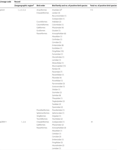
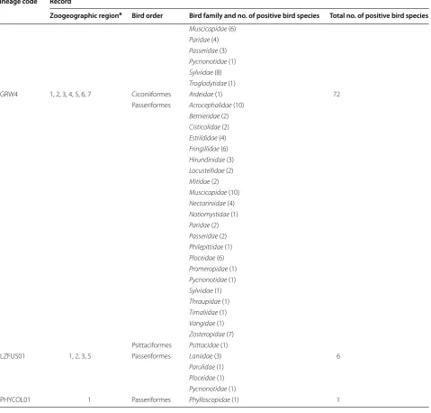
Table 2 Polymerase chain reaction-based reports of Plasmodium relictum lineages in avian hosts
Lineage code Record
Zoogeographic regiona Bird order Bird family and no. of positive bird species Total no. of positive bird species
pSGS1 1, 2, 4, 5, 6 Anseriformes Anatidae (2)b 115
Charadriiformes Laridae (3)
Recurvirostridae (1)
Scolopacidae (1)
Ciconiiformes Ardeidae (2) Columbiformes Columbidae (1) Galliformes Phasianidae (4) Gruiformes Gruidae (1) Passeriformes Acrocephalidae (6)
Alaudidae (1)
Certhiidae (1)
Corvidae (5)
Emberizidae (8)
Estrildidae (1)
Fringillidae (10)
Furnariidae (1)
Hirundinidae (1)
Laniidae (1)
Motacillidae (1)
Muscicapidae (15)
Paridae (9)
Passeridae (7)
Passerellidae (1)
Ploceidae (4)
Prunellidae (1)
Pycnonotidae (3)
Scotocercidae (1)
Sittidae (1)
Sturnidae (2)
Sylviidae (8)
Thaupidae (1)
Troglodytidae (2)
Turdidae (2)
Tyrannidae (2)
Procellariiformes Procellariidae (1) Sphenisciformes Spheniscidae (1) Strigiformes Strigidae (1) Trochiliformes Trochilidae (2)
pGRW11 1, 2, 6 Charadriiformes Scolopacidae (1) 41 Galliformes Phasianidae (2)
Passeriformes Acrocephalidae (3)
Alaudidae (1)
Cettiidae (1)
Corvidae (3)
Emberizidae (1)
Fringillidae (3)
Hirundinidae (2)
[image:11.595.57.543.102.718.2]known about this biological variation is given in the fol-lowing sections.
Pathology
The pathology of known lineages of P. relictum is highly variable in host species or incompletely known. For example, the same P. relictum lineage might cause severe disease in one species of avian host, but other bird species
might be tolerant or even resistant [5, 8, 50, 69]. Experi-mental observations show that the same isolate of pSGS1 behave markedly differently in different species of birds, with the susceptibility ranging from complete resistance to light subclinical (< 0.1%) and high (> 10%) parasitaemia [8, 69]. The variation in parasitaemia dynamics and maxi-mum intensity are often great in different individuals of the same bird species infected with pSGS1 parasite [55].
Modified from MalAvi database (http://www.iucnredlist.org/details/103843725/0)
a Zoogeographic regions: 1—Palaearctic, 2—Afrotropic, 3—Nearctic, 4—Neotropic, 5—Indo-Malay, 6—Australasian, 7—Oceanic (borders of the regions were
considered according to http://users.tamuk.edu/kfjab02/Biology/Mammalogy/mammalogy_zoogeography.htm)
b Number of species is given in parenthesis Table 2 continued
Lineage code Record
Zoogeographic regiona Bird order Bird family and no. of positive bird species Total no. of positive bird species
Muscicapidae (6)
Paridae (4)
Passeridae (3)
Pycnonotidae (1)
Sylviidae (8)
Troglodytidae (1)
pGRW4 1, 2, 3, 4, 5, 6, 7 Ciconiiformes Ardeidae (1) 72 Passeriformes Acrocephalidae (10)
Bernieridae (2)
Cisticolidae (2)
Estrildidae (4)
Fringillidae (6)
Hirundinidae (3)
Locustellidae (2)
Mitidae (2)
Muscicapidae (10)
Nectariniidae (4)
Notiomystidae (1)
Paridae (2)
Passeridae (2)
Philepittidae (1)
Ploceidae (6)
Promeropidae (1)
Pycnonotidae (1)
Sylviidae (1)
Thraupidae (1)
Timaliidae (1)
Vangidae (1)
Zosteropidae (7)
Psittaciformes Psittacidae (1)
pLZFUS01 1, 2, 3, 5 Passeriformes Laniidae (3) 6
Parulidae (1)
Ploceidae (1)
Pycnonotidae (1)
[image:12.595.61.541.107.565.2]Similarly, the susceptibility of same bird species to dif-ferent isolates of the same P. relictum lineage also might be markedly different. For example, Hawaiian isolates of pGRW4 readily infect canaries, with maximum para-sitaemia ranging from light (about 0.1%) to high (up to 30% and greater) reported in birds exposed by inocula-tion of infected blood ([70], CTA, pers comm.). How-ever, this bird species was either resistant or had mainly light (< 0.1%) and transient parasitaemia, which rapidly turned to chronic or even latent stages of infection after exposure to European isolates of the same parasite line-age by the same mode of infection ([11], GV, unpublished observation).
It remains unclear why different geographical isolates of the same lineage of P. relictum (pGRW4) behave so differently in the same species of birds. The differences between different geographic isolates of P. relictum line-ages might be due to different clonal intra-lineage genetic diversity, which is great in Hawaiian strains of the line-age pGRW4, but remains insufficiently documented in European isolates of the same lineage [21, 31]. Marked variation in the susceptibility of same experimental bird species to different parasite lineages provide opportu-nities to use this host-parasite model system for com-parative research aimed at a better understanding of the genetic mechanisms of tolerance and virulence during parasitic infections.
Without question, the lineages pSGS1 and pGRW4 are virulent in birds and can cause marked blood pathology and even mortality in susceptible hosts [5, 8, 11, 29, 50,
69]. The negative effects of P. relictum (pSGS1) on bird physiological parameters and behaviour are documented due to delicate experimental studies [54, 55]. Obser-vations of infected, naive birds in zoos and rehabilita-tion centres provided evidence of the severity of disease caused by these and related parasite lineages in wild birds [71–74]. These studies are the basis of understanding the predictions and conclusions of field observations about negative influence of P. relictum on population decline or even extinction, particularly on oceanic islands [63, 75–
78]. However, to evaluate the true virulence of a malaria parasite lineage in certain avian host species, experimen-tal and field observations are needed, ideally in each tar-geting host-parasite system separately.
Even though there are numerous reports of exo-eryth-rocytic stages of P. relictum from the pre-molecular research era [1, 7, 13, 84], information about these stages and associated tissue pathology in avian hosts is still absent for parasites of all lineages of P. relictum. This is an obstacle to understanding of the mechanisms of persis-tence in birds, as well as, the association between tissue merogony and pathogenicity caused by different parasite lineages in different avian hosts. This study shows that
exo-erythrocytic stages of P. relictum can be difficult to find during chronic infections even in experimentally infected birds with visible parasitaemia. This indicates that large multinuclear tissue stages, which are easy to see under light microscopy [6, 13], might persist for a short time and their development might be markedly dependent on the stage of infection. Application of in situ hybridization methods is promising in the investigation of tissue merogony of haemosporidians [6, 37, 78], but may not be sensitive enough to detect uninuclear hypno-zoite-like intracellular stages should they occur in P. relic-tum, as is the case in human Plasmodium vivax infection. This suggests application of more sensitive immunofluo-rescent diagnostic techniques in parallel with traditional histology and in situ hybridization methods in research of exo-erythrocytic development of different lineage par-asites [1, 6, 35, 37, 78].
Observation of parasites in blood films and deter-mination of morphological characters of their blood stages remain important not only in identification of haemosporidian species [11, 27, 79], but also for distin-guishing competent and abortive haemosporidian infec-tions, which might have different consequences for the bird health. During abortive infections, the parasites might circulate within avian hosts as sporozoites or even undergo partial development within non-erythroid tis-sues, providing templates for PCR amplification, but the parasite would not be able to complete its life cycle due to an inability to enter red blood cells. This would result in absence of gametocytes and other blood stages in the circulation, but severe disease might occur due to damage of internal organs [48]. In the latter case, a positive PCR signal might be obtained, but parasitaemia would be absent or barely detectable due to difficulties in microscopic detection of remnants of tissue stages in the circulation [80–82]. This highlights the relevance of microscopic detection of blood stages and knowledge about morphological features of haemosporidians in pathology and epidemiological studies when used in par-allel with molecular diagnostic tools.
Pre‑patent period and parasitaemia
This study demonstrated that prepatent period of infec-tion is markedly variable in different bird species and individuals of the same species during blood-induced infection of the lineage pPHCOL01. The prepatent period is often about 1 week after the blood-induced infections of pSGS1, but varies markedly in different species of avian hosts and even individuals of the same species even after the same mode and dose of infection, and it might be as long as several weeks after infected blood-induced exposure, indicating the possibility of parasite persistence in internal organs [7, 8, 13, 69, this study].
In all investigated lineages of P. relictum, parasitae-mia was asynchronous, with trophozoites, growing and mature meronts as well as gametocytes present in the same blood films at the same time in all species of exposed birds at any stage of parasitaemia [8, 29, 30, 33,
70, this study]. This provides opportunities to design vec-tor research with all lineages at any stage of parasitaemia using susceptible avian hosts as donors of infections to expose mosquitoes, but all work carried out to date with different lineages has failed to demonstrate significant differences.
Host range
An interesting finding of this study is that canaries may not be suitable experimental hosts for all lineages of P. relictum and possibly not even isolates of the same age. Information about susceptibility of canaries to line-age pLZFUS01 is absent; further experimental studies are needed. This study indicates that canaries can tolerate the pPHCOL01 infection, during which light transient parasitaemia occurs and signs of illness have not been reported. Canaries are good experimental hosts for the lineages pSGS1, pGRW11 and pGRW4 due to long-last-ing parasitaemia (usually, several months before latency, with infected birds maintaining infections for several years, with occurring seasonal relapses).
However, infectivity and patterns of development of different lineages and even different isolates of the same lineage might be different, sometimes significantly in canary [11, 70]. A moderate to high (> 0.1% and greater) long-lasting (several months) parasitaemia usually devel-ops during infections with lineages pSGS1 and pGRW11 in canaries exposed by inoculation of infected blood [22,
32, 46]. The same is true for the parasite lineage pGRW4 during development in canaries, but not for all its iso-lates. For example, the Hawaiian and European isolates of the lineage pGRW4 develop differently in canaries. Hawaiian pGRW4 isolates develop naturally in canaries when caged birds are exposed in habitats with active nat-ural transmission and can develop high (up to 30% and higher) long-lasting parasitaemia after sub-inoculation of infected blood, although significant individual variation
is present ([70], CTA, unpublished data). Attempts to induce a long-lasting parasitaemia (several weeks or longer) and gametocytaemia exceeding 0.01% with Euro-pean isolates of lineage pGRW4 were either completely unsuccessful (compete resistance was recognized in nine exposed birds) or only partially successful with extremely light transient parasitaemia (few gametocytes reported after examination of 100 microscopic fields at high mag-nification in four birds) ([11], GV, unpublished data). In other words, the canary is not a good host for experimen-tal studies of erythrocytic infections with the European isolates of the lineage pGRW4, but can be used in experi-ments with the Hawaiian isolate. Experimental studies with other geographical isolates of P. relictum (pGRW4) infection have not been performed. Due to relative resist-ance of canaries to European isolates of lineage pGRW4, Eurasian siskin Carduelis spinus has been used in experi-ments with this parasite lineage, and this species is an excellent experimental host [33].
Hybridization and gene flow
The lineages pSGS1, pGRW4, pGRW11, pLZFUS01, and pPHCOL01 of P. relictum are closely related based on similarities in cytb sequence (Fig. 1) and cannot be dis-tinguished by morphology (Figs. 2, 3, Additional file 1: Figure S1, Additional file 2: Figure S2, Additional file 3: Figure S3, Additional file 4: Figure S4). Do these lineages represent distinct species of the P. relictum group or are they different genetic variants of the same morpho-spe-cies? Do parasites of these lineages maintain the ability to mate? Does the available information provide oppor-tunities to approach answering these questions? This study and available experimental observations [28–30,
32, 33, 46] show that morphological data both of blood and vector stages cannot help in distinguishing parasites of the lineages pSGS1, pGRW4, pGRW11, pLZFUS01, pPHCOL01, indicating that they might belong to the same P. relictum morphotype, but some of them also might represent cryptic species of the P. relictum group.
in 4 investigated nuclear genes and may represent cryp-tic species. These lineages of Haemoproteus majoris are closely related and differ by only 1–6 substitutions over the 479 bp of sequenced cytb gene (0.2–1.3% difference). By contrast, an experimental observation in vivo [22] has demonstrated that parasites of the closely related lineages pSGS1 and pGRW11 can mate in mosquitoes Culex pipi-ens forma molestus and produce hybrid oocysts. Genetic differences between these lineages in the cytb gene are small (0.2%). According to hybridization experiments [22], the parasites of the lineages pSGS1 and pGRW11 are different variants of the same species, but informa-tion about hybridizainforma-tion of other lineages of P. relictum and other avian haemosporidian parasites is absent.
It is worth noting that partial sequences of merozoite surface protein 1 (msp1) gene were determined in 3 P.
relictum lineages (pSGS1, pGRW11, pGRW4) in samples
collected from different geographic sites using nuclear markers [21]. All three lineages were from markedly ran-domly sampled birds, with unclear geographical origin of infection. Four different alleles were reported in the lineage pSGS1, and three of them were shared with the lineage pGRW11, indicating possible hybridization. This is in accordance with the available experimental observa-tions [22]. However, five different alleles were revealed in the lineage pGRW4 [21], suggesting the lack of gene flow between parasites of this lineage and the lineages pSGS1 and pGRW11. However, due to the markedly random sampling (many lineage isolates came from different spe-cies of African migrants with unclear geographical origin of infection), it is difficult to rule out that the reported genetic difference might reflect strain varieties, but not species differences. Additionally, due to common co-infections of malaria parasites in naturally infected hosts and possible selective amplification of different lineages using general primers [87], it is possible that some samples contained co-infections of different line-ages. Because of this, the possibility to create between-lineage nuclear gene artefacts cannot be ruled out as well. In other words, the quality of the haemosporidian sequences should be carefully considered if samples from wildlife are used [88].
Plasmodium relictum is a unique among malaria
para-sites in regard to the enormous range of its avian hosts and mosquito species involved in its transmission. There-fore, direct in vivo experimental hybridization of differ-ent P. relictum lineages [22] would be most useful if they involved lineage isolates which are transmitted at the same site by the same mosquito species as this would make experimental studies closer to real epidemiological situations that are observed in wildlife.
Geographic distribution and prevalence
Data about vertebrate host and geographical distribu-tion of different P. relictum lineages are summarized in Table 2. The lineages pLZFUS01, pPHCOL01 of P. rel-ictum have been reported occasionally, mainly in birds wintering or resident in tropical countries where trans-mission occurs [30, this study). The parasite lineage pGRW4 has both broad host and worldwide geographi-cal distribution, but is rare in Europe [2, 11, 21, 33]. The lineage pSGS1 and pGRW11 are also broadly distributed, but neither has been reported in several extensive studies in the mainland Americas [2, 21, 89–91]. However, Mar-zal et al. [3] found P. relictum (pSGS1) in 8 native bird species belonging to two orders in Peru, and Quillfeldt et al. [92] reported this parasite in seabirds on Falkland Islands, indicating presence of transmission, at least in South America.
The reported differences in geographical distribution of the lineages pSGS1 and pGRW11 on the one hand, and GRW4 on the other hand are difficult to explain bearing in mind the enormously broad range of their suscepti-ble avian hosts (Tasuscepti-ble 2) and mosquito vectors, such as the globally distributed Culex pipiens, Culex quinque-fasciatus and other mosquito species of the Culex pipi-ens complex, which are of global distribution [93–95]. It is worth noting that recent experimental studies have demonstrated complete sporogony of the pGRW4 para-sites from European birds, in cosmopolitan Culex pipiens forma molestus mosquitoes at relatively low tempera-tures. This indicates that there are no obstacles prevent-ing transmission of this infection in Europe durprevent-ing the warm period of the year [33]. The following explana-tions of the observed phylogeographic data are worth discussion.
others, particularly in cases of co-infections of different lineages. Relatively simple experimental studies using the protocol by Bernotienė et al. [87] might be helpful in answering this question. Co-infections of malaria para-sites are common and even predominate in some bird populations [87, 96, 97]. This information is essential for better understanding of true distribution of P. relictum lineages both by hosts and geographically. Application of specific primers might contribute to better understand-ing patterns of geographical distribution of these invasive bird infections.
Second, parasite prevalence data depend on both force of infection and the longevity of infection. If local trans-mission is occurring, the low prevalence of GRW4 infec-tion in European bird populainfec-tions might be a result of (1) mortality of some European birds due to this infec-tion, as is the case with some endemic Hawaiian birds [1]; (2) resistance and ability of some bird species to tolerate the pGRW4 malaria infection [11]; or, (3) a combination of these two factors. Naive Hawaiian and New Zealand endemic birds suffer mortality from infection with P. rel-ictum pGRW4 [5, 50, 75, 77, 99, 100], but introduced bird species are less susceptible and might tolerate this dis-ease [5, 50, 70]. Little is known about the virulence of the pGRW4 infection in resident European birds and other birds worldwide [33]. Preliminary observations indicate that several European bird species (Fringilla coelebs, Sylvia atricapilla, Passer domesticus) can resist pGRW4 strains, which were isolated from African migrating Great read warblers Acrocephalus arundinaceus [11]. Further experimental studies and application of lineage specific primers might provide more certain informa-tion about distribuinforma-tion of these parasite lineages, their co-existence in the same avian hosts and study sites, and better understanding infections in bird health.
Vector research
The list of mosquito species, which are susceptible to P. relictum includes over 20 species [7, 13], however, information about vectors at parasite lineage levels is insufficient [101]. Widespread Culex pipiens, Culex quinquefasciatus and Culex tarsalis mosquitoes are excel-lent vectors for pSGS1, pGRW4, pGRW11 [22, 32, 33, 46,
52, 102–105], but data about vectors of the pLZFUS01 and pPHCOLL01 parasites are absent. It is interesting to note that mosquitoes belonging to three genera, Aedes albopictus, Wyeomyia mitchellii and Culex quinquefas-ciatus, are susceptible to the pGRW4 parasite, and the sporogony was completed in all these mosquito species, but prevalence varied significantly between species. The latter mosquito is the main vector, but other mosquito species might be involved in transmission as well [106]. However, it worth mentioning that, while sporogony was
completed in a small fraction of Wyeomyia mitchellii, the authors [106] did express doubt in the viability of aber-rant sporozoites in this mosquito species.
Culex quinquefasciatus is absent in Lithuania. This
insect was used in experiments because the new P. relic-tum lineage (pPHCOL01) was isolated from a bird spe-cies wintering in Africa where Culex quinquefasciatus is widespread [93, 94]. Sporogony of the parasite lineage pPHCOL01 was not initiated in Culex quinquefasciatus probably because the donor bird has light gametocytae-mia (single gametocytes were seen in donor canaries dur-ing mosquito exposure), and that might have been the main obstacle.
Numerous mosquitoes were incriminated as possible P. relictum vectors using microscopic methods, but mainly only oocysts were reported in the majority of the stud-ied insects, and the development of sporozoites were accessed in a few species [101]. This questions the con-clusions about true possibility and involvement of mos-quitoes belonging to different genera to act as effective vectors of P. relictum in wildlife. More delicate studies, including the observation of sporozoites in the salivary gland are needed to reach conclusions about ability of certain mosquito species to act as vectors. It is important to note that even presence of sporozoites of Plasmodium parasites in salivary glands does not always guarantee that the insects can transmit infection by bite. For exam-ple, sporozoites of Plasmodium hermani were reported in mosquito Wyeomyia vanduzeei, and these sporozo-ites were used successfully to induce infection in turkeys by syringe inoculation, but this mosquito was unable to transmit infection by bite [107]. This example calls for more delicate vector studies for better understanding transmission of avian haemosporidians. Determination of vectors is time consuming in wildlife studies where diversity of blood-sucking dipteran insects is high. The PCR-based reports of P. relictum lineages in wild-caught dipteran insects markedly speed search for possible vec-tors by indicating significant links between insects, avian hosts and parasites [103, 104, 108–118], but cannot prove that sporozoites develop and can be transmitted by the PCR-positive insects. The observation of Plasmodium spp. sporozoites in salivary glands and the studies of transmission by mosquito bites remain the gold stand-ards for determining vector competence. Combination of molecular diagnostic, experimental procedures and microscopic tools remain essential in haemosporidian vector research [33, 46, 101, 106, 119–121].
Conclusion
Plasmodium relictum is a unique species among the
of avian hosts and mosquito vectors. These character-istics make the various P. relictum lineages exceptional model organisms for better understanding ecological and genetic mechanisms that make generalist pathogens so successful.
Five lineages of P. relictum (pSGS1, pGRW4, pGRW11, pLZFUS01, pPHCOL01) have been identified and par-tially characterized. Parasites of these lineages are phylogenetically closely related, and they cannot be dis-tinguished using morphological characters of their blood or vector stages. Available data show that the same line-ages develop markedly differently in different avian hosts. Remarkably, variation among biological properties (pre-patent period, parasitaemia dynamics, blood pathology) between different isolates of the same lineage might be greater than the variation between different lineages dur-ing their development in the same species of avian host. This indicates the negligible value of these features for diagnosing specific parasite lineages. Currently, the lin-eages of P. relictum can be readily distinguished mainly through mtDNA sequences.
Malaria caused by P. relictum is of particular impor-tance for bird health. Controlled laboratory experimen-tal studies show that the lineages pSGS1 and pGRW4 are virulent in birds and can cause marked blood pathology and even mortality in susceptible hosts. However, the exo-erythrocytic stages and tissue pathology caused by them in avian hosts is unknown for parasites of all line-ages of P. relictum. This is a prominent obstacle for devel-opment of the effective prevention and treatment options for birds.
Certainly, more research is needed on biology of P. rel-ictum lineages. The existence of still unclear geographi-cally related limitations in transmission of the most prevalent lineages pSGS1 and pGRW4 has been often suspected in explanation of the restricted distribution of these parasites globally. However, methodological issues in the diagnosis of these parasite lineages remain and limit our ability to study co-infections in broadly dis-tributed lineages of P. relictum. The information about frequency of co-infection occurrence in lineages of P.
rel-ictum is inadequate. Mainly general primers have been
applied in PCR-based detection and phylogeographic studies of P. relictum, and this method is insufficiently sensitive in determining haemosporidian co-infections. It is predicted that available information about both host and geographical distribution of these lineages might be significantly updated if more sensitive diagnostic tools are applied for distinguishing co-infections of these and other P. relictum lineages.
Although closely related lineages of P. relictum can hybridize, within-species diversity may also indicate the presence of possible cryptic speciation in the P. relictum
group. Speciation processes have been insufficiently addressed in experimental parasitology studies mainly because of difficulties in accessing and measuring mate-recognition signals in parasites. By focusing on the extra-cellular sexual process of oogamy, which can be readily visualized both in vivo and in vitro, and the development of oocysts possessing numerous copies of nuclear genes, experimental hybridization can be readily accessed using haemosporidian parasite lineages [22, 33]. Methodolo-gies of between-lineage hybridization of avian Plasmo-dium parasites as well as sister Haemoproteus species have been developed [22, 85]. It is important to gain more information about true range of cryptic speciation in pathogens, particularly due to increasingly frequent outbreaks of zoonotic infections, which appear after host switching, leading to the emergence of new diseases [1,
6, 34, 75]. Such studies would also provide directions on how to approach future taxonomic reconstructions on species levels in the genus Plasmodium and other hae-mosporidians. Phylogenetic analysis based on partial cytb sequences placed different lineages of P. relictum in a tight cluster. Importantly, parasites of these lineages often occur in sympatry in many cases thus, are conveni-ent model organisms to answer questions about the range of cryptic speciation in wildlife malaria and other related haemosporidian parasites.
Additional files
Additional file 1: Figure S1. Mature erythrocytic meronts of the lineage pGRW4 of Plasmodium relictum in Hawaiian (a–t) and European (u–x) isolates during development in naturally infected Apapane Himatione sanguinea (a–h) and experimentally infected domestic canary Serinus canaria (i–x). Note that the size and shape of mature meronts, number of nuclei in them, influence of the meronts on host cells are markedly variable and overlap in both isolates. Meronts of both isolates cannot be distinguished by morphological characters and patterns of their influence on host cells during their development in the same and different avian hosts. Furthermore, meronts of the lineages pGRW4 cannot be distin-guished from meronts of the lineage pSGS1 (see Additional file 2: Figure S2). Arrowheads—pigment granules. Giemsa-stained thin blood films. Scale bar = 10 μm.
Additional file 2: Figure S2. Mature erythrocytic meronts of the lineage pSGS1 of Plasmodium relictum in European isolate during development in experimentally infected Eurasian siskin Carduelis spinus (a–h) and domestic canary Serinus canaria (i–t). Note that size and shape of mature meronts, number of nuclei in them, influence of meronts on host cells are markedly variable. Meronts of this parasite lineage cannot be distinguished by mor-phological characters and patterns of their influence on host cells during their development in different avian hosts. Furthermore, meronts of the lineages pSGS1 cannot be distinguished from meronts of the lineage pGRW4 (see Additional file 1: Figure S1). Arrowheads—pigment granules. Giemsa-stained thin blood films. Scale bar = 10 μm.