R E S E A R C H
Open Access
Lentiviral Nef suppresses iron uptake in a strain
specific manner through inhibition of Transferrin
endocytosis
Herwig Koppensteiner
1,2†, Kristin Höhne
1,2†, Marcos Vinicius Gondim
1, Francois-Xavier Gobert
3, Miriam Widder
2,6,
Swantje Gundlach
2, Anke Heigele
4, Frank Kirchhoff
4, Michael Winkler
5, Philippe Benaroch
3and Michael Schindler
1,2*Abstract
Background:Increased cellular iron levels are associated with high mortality in HIV-1 infection. Moreover iron is an important cofactor for viral replication, raising the question whether highly divergent lentiviruses actively modulate iron homeostasis. Here, we evaluated the effect on cellular iron uptake upon expression of the accessory protein Nef from different lentiviral strains.
Results:Surface Transferrin receptor (TfR) levels are unaffected by Nef proteins of HIV-1 and its simian precursors but elevated in cells expressing Nefs from most other primate lentiviruses due to reduced TfR internalization. The SIV Nef-mediated reduction of TfR endocytosis is dependent on an N-terminal AP2 binding motif that is not required for downmodulation of CD4, CD28, CD3 or MHCI. Importantly, SIV Nef-induced inhibition of TfR endocytosis leads to the reduction of Transferrin uptake and intracellular iron concentration and is accompanied by attenuated lentiviral replication in macrophages.
Conclusion:Inhibition of Transferrin and thereby iron uptake by SIV Nef might limit viral replication in myeloid cells. Furthermore, this new SIV Nef function could represent a virus-host adaptation that evolved in natural SIV-infected monkeys.
Keywords:HIV, SIV, Nef, Iron homeostasis, AIDS, Lentiviral replication, Macrophages
Background
Iron is an essential element in the human body and in-volved in cellular proliferation and immune response [1]. Since free iron generates harmful reactive oxygen species iron homeostasis is tightly regulated. Dietary iron is re-sorbed by the Divalent Metal Transporter into entero-cytes. Loaded on Transferrin (Tf ) iron enters the blood stream and is taken up by target cells via the Transferrin Receptor I (TfR). Internalization of TfR can be antago-nized by the hemochromatis protein (HfE). Thus, cells can regulate iron uptake by TfR expression or by the rate
of TfR internalization through HfE [1,2]. Iron export from cells is also controlled by two antagonizing mecha-nisms. Ferroportin loads cellular iron on plasma Tf and this process is suppressed by the hormone hepcidine [3]. All these mechanisms have the consequence that only trace amounts of iron, the so called labile iron pool (LIP) [4], is redox-active. Long term storage of iron is within Ferritin, mainly in hepatocytes [1,5].
Dysregulation of iron homeostasis is a hallmark of many diseases including AIDS [1-3]. Elevated cellular iron loads are associated with high HIV-1 titers and fas-ter progression to AIDS and iron is important in various steps of HIV-1 propagation [2,6-8]. Hence, viruses have evolved mechanisms to increase cellular iron [2]. The HIV-1 Nef protein was proposed to downregulate HfE, leading to enhanced uptake of iron loaded Tf [9]. * Correspondence:michael.schindler@helmholtz-muenchen.de
†Equal contributors
1
Institute of Virology, Helmholtz Zentrum Munich, German Research Center for Environmental Health, Neuherberg, Germany
2
Heinrich Pette Institute, Leibniz Institute for Experimental Virology, Hamburg, Germany
Full list of author information is available at the end of the article
HIV-1 Nef is considered as a pathogenicity factor con-tributing to AIDS progression [10]. It enhances viral rep-lication and infectivity and mediates immune evasion by multiple functions, including downmodulation of CD4 and MHCI. Nef is highly variable and not only present in HIV-1, but also in simian immunodeficiency viruses (SIV) and HIV-2 [10]. HIV-1 causes AIDS in humans and it was demonstrated that SIVcpz leads to immuno-deficiency in chimpanzees [11]. HIV-2 is usually less pathogenic than HIV-1 but causes AIDS post an extended chronical phase [12]. Within the natural simian hosts, SIVs do not cause disease due to a well balanced virus-host coevolution. Hallmarks of non-pathogenic SIV infec-tions are high viral loads in the absence of chronic and generalized immune activation [13]. Notably, Nef proteins derived from non-pathogenic infections prevent activation of infected T cells by removal of the CD3 molecule from the cell surface [14]. Thus, SIV Nef could be considered as a“persistence”factor in contrast to its HIV-1 counterpart.
We hypothesized that lentiviral Nefs may also differ in their ability to manipulate cellular iron uptake. Interest-ingly, we found that Nef-mediated degradation of HfE is not a conserved feature of HIV-1 and other lentiviral Nefs. In contrast, we demonstrate inhibition of TfR intern-alization by most SIV Nef proteins including the lentivi-ruses which are non-pathogenic in their natural simian hosts. This phenotype results in reduced cellular iron levels and attenuated lentiviral replication in macrophages.
Negative regulation of cellular iron stores by Nef in in-fected cells might be an additional strategy to achieve non-harmful and persistent virus-host coexistence.
Results
SIV Nef increases the cell surface expression of TfR
We assessed if modulation of receptors involved in cel-lular iron uptake is a conserved feature of different lenti-viral Nef variants. Macrophages regulate iron turnover in vivo[1,15]. Therefore, we infected the myeloid cell line THP-1 with HIV-1 coexpressing Nef and eGFP (HIV-NIG) [14] via an internal ribosomal entry site (IRES) and measured cell surface expression of TfR and HfE by flow cytometry (Figure 1). Both remained largely unchanged upon expression of HIV-1 Nef (Figure 1A). In contrast, Nef from SIVmac239 or the distantly related SIVblu caused up to threefold increase in TfR levels without modifying HfE surface levels (Figure 1A).
[image:2.595.57.537.439.676.2]Drakesmith and colleagues reported degradation of HfE by HIV-1 SF2 Nef in HeLa cells transfected to ex-press HfE [9]. Although HIV-1 NA7, NL4-3 and SF2 Nef are highly similar, we considered the possibility that sub-tle alterations in Nef might be sufficient to allow HfE degradation. Consistent with the results from HIV-1 in-fected THP-1 cells (Figure 1A), NL4-3, NA7 or 239 Nef had no or only marginal effects on cell surface HfE upon cotransfection in 293 T cells with HfE cDNA (Figure 1B). 293 T cells transfected with the HfE plasmid showed a
strong increase in cell surface staining, demonstrating the specificity of the HfE antibody and SF2 Nef down-modulated approximately 40% of cell surface HfE under these experimental conditions (Figure 1B). In agreement with its reported capacity to induce HfE degradation [16], expression of the HCMV US2 protein led to a dras-tic reduction of HfE surface levels (>90%) (Figure 1B). Thus, modulation of HfE is not a conserved feature of lentiviral Nefs. Conversely, SIV Nef enhances cell surface levels of TfR.
Most SIV Nef proteins increase cell surface TfR
To gain insights into the Nef-induced modulation of the
TfR, we compared 31 nef alleles from HIV-1 M, O and
N, SIVcpz, and the HIV-1 precursors SIVgsn/mus/mon (Group 1) as well as HIV-2, SIVsmm and divergent SIV species (Group 2, see also Additional file 1: Table S1). This collection faithfully represents a cross section of phylogenetically clustered and evolutionary related
lenti-viral nef alleles [10,14]. Isogenic HIV-1 NIG proviral
constructs only differing in their respective nef ORF
were used to generate virus stocks and infect PBMC, MDM and THP-1 cells (Figure 2). HIV-1 and related Nefs (Group 1) modulated TfR only marginally (1.39 ± 0.08; n = 13) in PBMCs (Figure 2A). In contrast, most other Nefs (Group 2) caused a marked increase in TfR expression
at the cell surface (2.45 ± 0.18; n = 18). Of note, the only HIV-2 Nef that upregulated TfR is clone 60415 K (2.56 ±0.5, compare Additional file 1: Table S1) isolated from an apathogenic HIV-2 infection [17].
Given the critical role of macrophages in iron turnover in vivo, we next examined the effects of Nef in THP-1
cells and MDMs. We found that all nef alleles that
modulated TfR surface levels in PBMCs were also ac-tive in THP-1 cells and MDMs (Figure 2B). Next, we tested in PBMCs whether Nef-induced upregulation of TfR surface levels correlates with other Nef functions (Figure 2C). Downmodulation of CD28 and MHCI by Nef did not correlate with TfR surface levels. In con-trast, TfR upregulation exhibited a positive correlation with CD3 downmodulation and a negative one with CD4
downmodulation, although the R2values were fairly low
[image:3.595.58.539.422.628.2](R2= 0.370 for CD3 and R2= 0.222 for CD4; Figure 2C). These observations are in line with two previously re-ported observations: (i) CD3 is also modulated by Nef in a lineage-dependent manner [18] and (ii) TfR and CD4 are both internalized through a mechanism involving the cla-thrin adaptor protein 2 (AP2) [19,20]. However, in both cases we could also identify Nef proteins which were se-lectively defective in one of these functions or active in both. Thus, overlapping but distinct Nef regions seem to be involved in modulation of TfR, CD3 and CD4.
Our data reveal that Nef modulates TfR in a lineage dependent manner. HIV-1 and its simian precursors are inactive in this function whereas most other lentiviruses upregulate TfR.
Tyrosine 28 in SIV Nef is required for TfR upregulation
To identify regions in Nef responsible for the increase in cell surface TfR, we tested a previously characterized
[image:4.595.58.543.197.637.2][21] panel of SIVmac239 Nef mutants. PBMCs were in-fected with the HIV-1 NIG variants and analysed for modulation of several receptors (Figure 3A). The pheno-type of the different 239 Nef variants for CD4, CD28, CD3 and MHCI modulation was as expected [21], al-though in this set of experiments Nef dependent modu-lation of CD4 is difficult to assess, since Vpu and Env also reduce CD4 expression in HIV-1 infected PBMC
[22]. The YE and Y223changes did not impair TfR
upreg-ulation whereas the EDR and Δ64-67 mutants were
at-tenuated (Figure 3A). Of note, the truncated 239 Nef E93* selectively increased cell surface TfR. Thus, a region
within the first 93 amino acids of SIVmac239 Nef is re-sponsible for TfR upregulation.
To identify the Nef residues responsible for TfR
upreg-ulation we generated a panel of eighteen 239 Nef E93*
mutants with serial deletions. Most deletions did not affect the capability of 239 Nef to increase TfR expres-sion. However, a set of five mutants that lacked AA 26–29 were inactive (Figure 3A). This region comprises a tyro-sine which is present in all Nef variants capable to
in-crease TfR expression and is part of a canonical AP2μ
binding site (Yxxϕ; ϕ is a bulky hydrophobic AA side
chain; see Additional file 2: Figure S2 and [20,23]). Indeed,
introduction of the “YGRL” motif in full-length NL4-3
Nef conferred the ability to increase cell surface expres-sion of TfR in PBMC (Figure 3B). Conversely, mutation of 239 Nef Y28to F (Figure 3B) or A (not shown) selectively
disrupted the upregulation of TfR. In conclusion, a tyro-sine based N-terminal AP2μbinding motif in Nef is crit-ical for TfR upregulation.
Strong Nef binding to AP2μis associated with TfR upregulation
The canonical pathway of TfR internalization is by AP2 [20,23]. Hence, Nef-mediated AP2 sequestration might im-pede TfR uptake. To assess if lentiviral Nefs differentially
interact with AP2μ we generated an AP2μ-eCFP fusion
and Nef-eYFP fusion expression vectors and measured binding with a FACS-based FRET assay [24] (Figure 4A).
Coexpression of HIV-1 and SIVcpz Nefs with AP2μ-eCFP
did generally not result in FRET signals, implicating the ab-sence of direct interaction with AP2μ(Figure 4A). In
con-trast, all HIV-2 and SIV Nefs showed FRET with AP2μ,
although with considerable differences. Values higher than 40% of FRET + cells were exclusively observed for Nefs able to upregulate TfR whereas HIV-2 and SIV Nef vari-ants unable to upregulate TfR or containing inactivating mutations exerted significant lower FRET (Figure 4A). Of note, the inactive HIV-2 Ben and SIVsm FFm1 as well as 239 Nef with the mutated YRGL motif still interacted with
AP2μ, albeit mean FRET was lower than 35% in all
mea-surements. This suggests (i) that other Nef residues con-tribute to AP2μbinding and (ii) that a certain threshold of AP2μbinding by Nef is required for increased TfR expres-sion. In addition, differential Nef binding to other AP2 subunits (thanμ) could also have an impact on TfR upreg-ulation [25].
To further examine the role of AP2μ in Nef-mediated
TfR upregulation, we performed AP2μ knock-down by
shRNA. 293 T cells were transduced with self-inactivating lentiviral vectors expressing GFP as infection marker and
a shRNA against AP2μor a scrambled shRNA. Three days
[image:5.595.56.540.456.643.2]later, cells were transfected with pCG Nef expression vec-tors containing the fluorescence protein mtagBFP [26]. This strategy allows to specifically identify by FACS
Figure 4Nef binding to AP2μis associated with TfR upregulation. (A)293 T cells were cotransfected with different Nef-YFP fusion protein vectors and AP2μ-CFP. 24 hours later the amount of cells scoring FRET + was measured by flow cytometry as described before [24]. CFP + YFP is a negative control of 293 T cells which are cotransfected with CFP and YFP expressing plasmids. CFP-YFP is a positive control of 293 T cells that express a CFP-YFP fusion protein. Mean values and SD are calculated from four to nine independent transfections.(B)293 T cells were transfected with lentiviral vectors coexpressing GFP and a shRNA against AP2μor the GFP only expressing vector control. 72 hours later the cells were
cells that simultaneously express the shRNA and Nef (Figure 4B). Only 239 Nef increased TfR in the
pres-ence of the control shRNA (black bars). Upon AP2μ
knock-down, TfR accumulated at the cell surface irre-spective of functional Nef expression. Altogether the data suggest that Nef sequesters AP2 resulting in re-duced TfR internalization and surface accumulation.
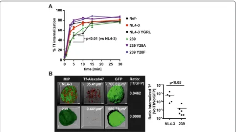
SIV Nef inhibits Transferrin internalization
Delayed TfR turnover should have direct impact on the internalization of Tf. Therefore, 293 T cells were trans-fected with pCG-Nef-IRES-GFP vectors and the amount of internalized Tf versus totally bound Tf was assessed in GFP/Nef expressing cells by FACS (Figure 5A). SIV-mac239 Nef expression slowed down Tf internalization whereas disruption of the AP2μbinding motif (Y28F/A)
abrogated this effect. In contrast, NL4-3 Nef had only minor effects on Tf internalization whereas introduction of the YGRL motif phenocopied SIVmac239 Nef.
To assess the consequence of delayed Tf internalization for the total amount of cellular Tf, we infected THP-1 cells with HIV-1 NIG and added Alexa647-Tf for 5 minutes.
After removal of cell surface Alexa647-Tf by acid wash, z-stacks of GFP expressing/HIV-1 infected cells were acquired to reconstruct 3-dimensional images (Figure 5B). The total cellular volume was calculated using GFP fluor-escence as a surrogate marker. Similarly, we calculated the volume of cell internal Tf from the Alexa647 fluores-cence. Mean volume ratios [Tf/GFP] were 0.0572 (±0.0271 SEM; n = 5) for NL4-3 compared to 0.0015 (±0.0009 SEM; n = 6) for 239 Nef. This reflects a 38-fold reduction in the total volume of cell-associated Tf in SIVmac239 Nef ex-pressing cells (Figure 5B) (p = 0.0043 Mann–Whitney test and p = 0.0497 Students T test). Thus, SIV Nef expression delays internalization of TfR and Tf which might reduce intracellular iron concentrations.
SIV Nef lowers intracellular iron levels and attenuates viral replication in primary macrophages
[image:6.595.57.540.373.644.2]The very small proportion of cellular free redox-active iron, called the labile iron pool (LIP), is subjected to rapid changes upon alterations in iron uptake or release [4]. Dir-ect effDir-ects of Nef on the LIP were estimated using the green fluorescent dye calcein-acetoxymethylester (CA-AM) [4].
Calcein is quenched by free redox active iron. Thus, a de-crease in Calcein fluorescence indicates an inde-crease in the LIP. Unfortunately, due to the high Calcein background fluorescence and the small amount of labile iron, this assay exhibits a very low signal to noise ratio. Hence we expected small changes in fluorescence and the need for multiple biological replicates in order to achieve signifi-cant differences.
THP-1 cells were infected with HIV-NIG expressing eCFP instead of GFP. 48 hours later the LIP was ana-lyzed post CA-AM staining in infected/eCFP expressing cells. We repeated the experiment eight times with two to three independent virus stocks and calculated the LIP relative to the Calcein fluorescence of SIVmac239 Nef
positive cells (Figure 6A). THP-1 expressing no Nef or NL4-3 Nef showed a trend towards higher labile iron. However, Calcein fluorescence scattered strongly and dif-ferences were not significant. In contrast, SIVmac239 Nef expressing cells contained less labile iron than the Y28F/A
mutants (Figure 6A) which do not block TfR internaliza-tion and Tf uptake. Despite the strong Calcein background fluorescence we measured an increase in mean values of
13.0% (239 Nef vs Y28F; n = 22) and 14.1% (239 Nef vs
Y28F; n = 22) which is highly significant (p < 0.0001).
Hence, Nef proteins that inhibit Tf uptake lower the amount of redox active iron within the cell.
Macrophages are important HIV-1 target cells in vivo
[image:7.595.56.541.272.638.2]and play a key role in cellular iron metabolism [1,15].
Since iron is an essential cofactor for multiple steps in the viral replication cycle [2], we speculated that differ-ences in cellular iron uptake due to Nef expression might influence the capacity of HIV-1 to replicate in pri-mary macrophages. MDMs were infected with isogenic R5-tropic HIV-1 just differing in thenefcoding sequence and virus production was monitored over twenty days (Figure 6B and C). As expected from previous results [27], NL4-3 Nef expression had no influence on HIV-1 spread and propagation in macrophages. In contrast, 239 Nef suppressed HIV-1 replication in macrophages and this effect was dependent on Y28(Figure 6B and C).
Therefore, 239 Nef might attenuate lentiviral replication in macrophages by lowering the intracellular iron pool.
Discussion
Our study suggests that manipulation of cellular iron uptake is a strategy of primate lentiviruses to regulate replication in myeloid cells. Moreover, the Transferrin receptor pathway is not inhibited by HIV-1 and its pre-cursors but by most other SIV Nefs.
Previous work suggested HIV-1 Nef mediated down-regulation of HfE [9] and TfR [28]. We show that HIV-1 Nefs have little if any effect on cell surface TfR while most SIV Nef proteins caused an up to three fold upreg-ulation (Figure 2 and Additional file 1: Table S1). At first, these discrepancies seem surprising. However, most previous results were performed in HeLa cells overex-pressing Nef and quantification was mainly performed by confocal microscopy [28]. In contrast, the majority of our experiments were performed using flow cytometry and conducted in HIV-1 infected PBMCs and macro-phages. Furthermore, our finding that HIV-1 Nef does only marginally manipulate TfR is in line with others [18,29,30]. For detection of HfE, Drakesmith and col-leagues used HfE overexpressing HeLa cells and HIV-1 SF2 Nef [9]. We mimicked this experiment and included HCMV US2 as positive control (Figure 1B) [16]. HfE was modulated by SF2 Nef (~40%), but not by any other Nef protein. In contrast, HCMV US2 degraded more than 90% of HfE. Thus, downregulation of HfE is a less pronounced and non-conserved Nef function.
Conversely, plasma membrane levels of TfR increased up to 3.5 fold post SIV Nef expression in infected PBMCs and were on average elevated 2.5 fold which is in the range of the well characterized Nef functions CD4 and MHC-I downregulation (Figure 2, Additional file 1: Table S1; also reviewed here [10]). Mechanistically, sev-eral lines of evidence suggest that SIV Nef upregulates TfR by competition for AP2: (i) We could identify a
con-served Yxxϕ sequence which is a canonical AP2μ
bind-ing motif [20,23] and present in the N-terminus of all Nefs upregulating TfR (Additional file 2: Figure S2). (ii)
Mutation of Y28 selectively disrupts Nef-mediated TfR
increase (Figure 3B). (iii) Insertion of the Yxxϕin HIV-1 Nef confers upregulation of TfR (Figure 3B). (iv) The capacity to increase cell surface TfR is associated with
strong Nef binding to AP2μ (Figure 4A) and (v)
knock-down of AP2 by shRNA increases TfR irrespective of Nef expression (Figure 4B). Thus, the data indicates that
SIV Nef sequesters AP2 from TfR and“upregulation” of
TfR is an effect of the decelerated internalization of the receptor and decreased cellular Tf uptake (Figure 5A). Importantly, steady state expression of TfR at the PM might strongly underestimate the magnitude of repressed cellular Tf uptake reflected by our 3D reconstructions (Figure 5B). Thus, lentiviral Nef proteins inhibiting TfR internalization will strongly suppress cellular Tf and iron uptake.
What are the consequences of dysregulated iron homeostasis in lentiviral infections? Iron is important for an effective immune response [1-3]. However there is some controversy because increased iron levels correl-ate with severe HIV-1 progression [31-36], whereas pro-gressive HIV-1 infection is also associated with anemia and therefore iron depletion [37,38]. Furthermore, HIV-1 patients with elevated cellular iron stores showed strongly reduced survival probabilities [6-8,39,40].
In this context it is noteworthy that generalized unspe-cific immune activation is a hallmark of progressive HIV-1 infection [13]. Thus, infected immune cells are in a state of hyperactivation and cellular iron is increased upon inflammation which is beneficial for retroviral rep-lication [5]. Multiple steps in the lentiviral life cycle are iron dependent. Among them provision of nucleotides, NF-κB activation [41], Tat dependent transcription [42], Rev mediated mRNA export [43] and final steps of as-sembly and release [44] (reviewed in [2,8]). Conceivably, iron chelationin vitroinhibits HIV-1 infection and repli-cation [41-43,45]. Considering the high iron need of HIV-1 in conjunction with the appearance of opportun-istic infections that also consume iron for growth, anemia and iron depletion in progressive HIV-1 infec-tion is not surprising [46]. Whether dietary iron supple-mentation at that stage might be helpful for the host or rather deleterious is a question of high relevance that is currently not yet answered [47].
Setting up iron limiting experimental conditionsex vivo is difficult. Nevertheless we could demonstrate the direct reduction of labile iron by HIV-1 expressing SIVmac 239 Nef. This phenotype correlated with an attenuated course of viral replication in macrophages (Figure 6). In contrast,
HIV-1 replication and CD4+ T cell depletion in ex vivo
was only inhibited by Nefs from lentiviruses that are highly divergent from HIV-1 including those which are most likely apathogenic in their natural simian hosts. These SIVs preferentially use CCR5 for cell entry and since their genomes express Vpx, the cellular tropism of the virus is greatly expanded allowing efficient infection of myeloid cells [48]. Under these conditions it might have been necessary to maintain a function that limits excessive replication in myeloid/macrophage cells. Of note, robust SIV replication in macrophages was observed in the SIV macaque model and has also been associated with pro-gression of simian AIDS [49]. In this context, the SIV Nef mutants which are selectively defective for block of TfR internalization identified herein are valuable tools to clar-ify the impact of altered cellular iron on pathogenicity and viral loads in a model of natural SIV-infected monkeys.
Reduction of cellular iron was only observed for SIV Nef. In general, HIV-1 and related Nefs had only mar-ginal effects on TfR and HfE cell surface levels. There-fore HIV-1 does either not manipulate cellular iron or has evolved differential mechanisms to dysregulate iron homeostasis. Given the importance of iron for lentiviral growth, it will be of high interest to investigate in future studies possible alternative mechanisms by which HIV-1 could alter cell associated iron.
Conclusions
Herein we establish inhibition of TfR internalization as a novel function that is exerted by most SIV Nef proteins in primary T cells and macrophages. We identify an
YXXϕAP-2 binding motif in the N-terminus of Nef that
is sufficient and necessary to confer this function. Nef competes with AP-2 for TfR binding resulting in reduced internalization of Transferrin and therefore iron delivery into the cell. In myeloid cells we could demonstrate the direct reduction of iron uptake leading to attenuated lentiviral replication in this cell type. We postulate that reduction of cellular iron uptake by SIV Nef is a func-tion that evolved to regulate viral replicafunc-tion in macro-phages and this could have an impact on lentiviral pathogenicity.
Methods
Proviral constructs and plasmids
HIV-1 pBR-NL4-3 IRES-eGFP proviral constructs ex-pressing different lentiviral Nefs have been described previously (NL4-3 NIG) [14]. R5-tropic derivatives of these constructs were generated by subcloning of thenef
ORF viaHpaIandMluIinto pBR-NL4-3 V3
92th014.12-IRES-eGFP [50]. Similarly, HIV-1 NIG variants express-ing different SIVmac 239 Nef mutants were generated by PCR amplifications from pCG-vector templates that were
published previously [21]. The SIVmac239 Nef Y28A/F
mutants were generated by splice overlap extension PCR
and the YGRL sequence was introduced at position 28 in NL4-3 Nef by primer mutagenesis. CMV-driven pCG plasmids coexpressing Nef and GFP via an IRES have been
described before [51]. pCG-SF2-nef-IRES GFP and the
pCG-HCMV-US2-IRES GFP were generated by
amplifica-tion of the specific reading frame with primers introdu-cing 5′XbaIand 3′MluIrestriction sites and subsequent standard restriction and ligation procedures. Truncated pCG-SIVmac 239 Nef variants with different in frame de-letions were generated by PCR amplification and ligation of published deletion mutants [19] with primers introdu-cing a 5′XbaIsite and a premature stop codon at aa pos-ition 93 followed by a 3′MluIsite. Fusion protein vectors peCFP and peYFP, peCFP-eYFP, NL4-3 Nef-YFP and SIV-mac 239 Nef-YFP have been described before [24].
Plas-mids expressing AP2μand HfE with a C-terminal CFP-tag
were constructed by PCR-amplification of Ap2μand HfE
from a HeLa cDNA library. Lentiviral Nef-YFP fusion pro-teins were amplified using the HIV-1 NIG proviral vectors as templates. The according ligation procedure has been described [24]. All PCR derived inserts were sequenced to confirm sequence identity.
Cell culture, transfection and HIV-1 infection
293 T cells were maintained in DMEM (Gibco) and THP-1 cells in RPMI (Gibco) with standard supple-ments. Primary human monocyte derived macrophages (MDM) and primary blood derived mononuclear cells (PBMC) were isolated and cultured as described [14,50]. HIV-1 virus stocks were generated by calcium phosphate transfection of 293 T cells [14]. For infection experi-ments HIV-1 stocks were quantified by p24-ELISA [27]. To assess HIV-1 replication in macrophages 25.000 cells were seeded in 48 well plates in 1% serum conditions and infected with 50 ng p24. Six hours later cells were washed and new media was added. Aliquots were taken in two to three days intervals and virus production was quantified by p24 ELISA. For the flow cytometric mea-surements of cell surface receptor modulation 2*10^5 macrophages or 1*10^6 PBMC were seeded in 35 mm Greiner dishes or six well plates and infected with 200 ng p24. Macrophages were harvested five days later by 10 mM EDTA treatment. PBMCs were usually ana-lysed 48 hours post infection. HIV-1 infection experi-ments of human lymphoid tissue (HLT) was done as already described [27].
Flow cytometry
anti-HFE (Abnova; polyclonal), anti-CD3 (Caltag; UCHT1; APC), CD4 antidody (Caltag; RPA-T4; APC), anti-HLA-ABC (Dako; W6/32; PE). Post staining cells were washed twice and fixed with 2% PFA/PBS. At least 2.000 infected cells were measured with a FACS Canto II (BDBioscience). Fold modulation of cell surface receptor modulation was calculated as before [14,51]. Flow cyto-metric measurement of FRET and the according gating strategy were performed as already described [24]. Cellular iron content was measured by the use of the fluorescent dye CA-AM which is quenched by free cellular iron [4].
Internalization assay
Tf internalization assays were performed essentially as previously described [18]. In brief, cells were harvested and incubated with Tf-Alexa647 for 30 min on ice, washed, and shifted to 37°C for various periods of time in culture medium supplemented with 20 mM HEPES. The medium was removed by washing, and half the
samples were washed in 25 mM glycine-HCl–125 mM
NaCl (pH 2.8) and rapidly neutralized with 25 mM Tris (pH 10). Samples were then washed and analyzed by flow cytometry. Mean fluorescence intensities (MFI) of GFP-positive cells in FL4 were determined. The ratio of the intracellular MFI (acid wash resistant) to the total MFI at each time point was plotted as a function of time.
Image acquisition, analysis and software
Infected THP-1 cells were incubated with Alexa647 la-beled Tf for 5 minutes. Then we exposed the cells to the acidic washing procedure as described above and finally fixed them with 2% PFA. THP-1 cells were mounted on objective slides with Mowiol and z-stacks of infected GFP expressing cells were acquired with a Zeiss LSM510 Meta. 3D reconstruction of z-stacks was done with Bit-plane Imaris V6.4.2. We also used this software to calcu-late the volume of the cells by GFP expression and the volume of internalized Tf by Alexa647 fluorescence. In general, images were never modified apart from enhan-cing contrast and/or brightness. Statistical analyses were performed using the GraphPad Prism V5 software pack-age. Statistical tests used were the unpaired two-tailed T test and the Mann–Whitney test and regression analyses with p-value calculations.
Additional files
Additional file 1: Table S1.Modulation of TfR and previously described PBMC surface receptors by lentiviral Nef proteins.
Additional file 2: Figure S2.Alignment of the N-terminus of Nef variants analyzed. The first 70 aminoacids of all lentiviral Nefs analyzed in this study were aligned. The tyrosine of the putative AP2-binding motif Yxxϕis marked in red. All Nef variants inactive in TfR upregulation are grouped by
the red square whereas Nef proteins inhibiting TfR uptake are surrounded by the green square.
Additional file 3: Figure S3.SIV 239 Nef with or without mutated Y28 motif does not affect HIV-1 replication and CD4+ T cell depletion in
ex vivoinfected human lymphoid tissue (HLT). Cumulative p24 production over 13 days (left) and CD4+ T cell depletion at the end of the culture period (right) in tissues of five donors infected with the indicated R5-tropic HIV-1 NL4-3 variants. Shown are mean values +/- SEM.
Competing interests
The authors declare that they have no competing interests.
Authors’contributions
Designed research: HK, KH, AH, PB, MS; Performed research: HK, KH, MVG, FXG, MW, SG, AH, MS; Contributed reagents: HK, MWK, FK, PB, MS; Analyzed data: HK, PB, MS; Wrote the manuscript: MS. All authors read and approved the final manuscript.
Acknowledgements
The authors thank Ulrike Protzer for constant support and encouragement and Ute Finkel for technical assistance. This work was funded by a grant of the German Research Foundation (DFG) to MS (SCHI 1073/2-1).
Author details 1
Institute of Virology, Helmholtz Zentrum Munich, German Research Center for Environmental Health, Neuherberg, Germany.2Heinrich Pette Institute, Leibniz Institute for Experimental Virology, Hamburg, Germany.3Institut Curie, INSERM U932, Paris, France.4Ulm University Medical Center, Institute of Molecular Virology, Ulm, Germany.5German Primate Center, Göttingen, Germany.6Current address: Martin Luther University Halle-Wittenberg, Internal Medicine IV, Halle, Germany.
Received: 12 August 2013 Accepted: 20 December 2013 Published: 2 January 2014
References
1. Johnson EE, Wessling-Resnick M:Iron metabolism and the innate immune response to infection.Microbes Infect2012,14:207–216.
2. Drakesmith H, Prentice A:Viral infection and iron metabolism.Nat Rev Microbiol2008,6:541–552.
3. Drakesmith H, Prentice AM:Hepcidin and the iron-infection axis.Science2012, 338:768–772.
4. Prus E, Fibach E:Flow cytometry measurement of the labile iron pool in human hematopoietic cells.Cytometry A2008,73:22–27.
5. Xu M, Kashanchi F, Foster A, Rotimi J, Turner W, Gordeuk VR, Nekhai S: Hepcidin induces HIV-1 transcription inhibited by ferroportin.
Retrovirology2010,7:104.
6. McDermid JM, Jaye A, van der Loeff MF S, Todd J, Bates C, Austin S, Jeffries D, Awasana AA, Whittlex AA, Prentice A:Elevated iron status strongly predicts mortality in West African adults with HIV infection.J Acquir Immune Defic Syndr2007,46:498–507.
7. McDermid JM, van der Loeff MF, Jaye A, Hennig BJ, Bates C, Todd J, Sirugo G, Hill AV, Whittle HC, Prentice AM:Mortality in HIV infection is independently predicted by host iron status and SLC11A1 and HP genotypes, with new evidence of a gene-nutrient interaction.Am J Clin Nutr2009,90:225–233.
8. Nekhai S, Kumari N, Dhawan S:Role of cellular iron and oxygen in the regulation of HIV-1 infection.Future Virol2013,8:301–311.
9. Drakesmith H, Chen N, Ledermann H, Screaton G, Townsend A, Xu XN: HIV-1 Nef down-regulates the hemochromatosis protein HFE, manipulating cellular iron homeostasis.Proc Natl Acad Sci U S A
2005,102:11017–11022.
10. Kirchhoff F, Schindler M, Specht A, Arhel N, Munch J:Role of Nef in primate lentiviral immunopathogenesis.Cell Mol Life Sci2008,65:2621–2636. 11. Keele BF, Jones JH, Terio KA, Estes JD, Rudicell RS, Wilson ML, Li Y, Learn GH,
12. Nyamweya S, Hegedus A, Jaye A, Rowland-Jones S, Flanagan KL, Macallan DC: Comparing HIV-1 and HIV-2 infection: lessons for viral immunopathogenesis.
Rev Med Virol2013,23(4):221–240.
13. Chahroudi A, Bosinger SE, Vanderford TH, Paiardini M, Silvestri G:Natural SIV hosts: showing AIDS the door.Science2012,335:1188–1193. 14. Schindler M, Munch J, Kutsch O, Li H, Santiago ML, Bibollet-Ruche F,
Muller-Trutwin MC, Novembre FJ, Peeters M, Courgnaud V,et al:Nef-mediated suppression of T cell activation was lost in a lentiviral lineage that gave rise to HIV-1.Cell2006,125:1055–1067.
15. Koppensteiner H, Brack-Werner R, Schindler M:Macrophages and their relevance in human immunodeficiency virus type I infection.Retrovirology
2012,9:82.
16. Vahdati-Ben Arieh S, Laham N, Schechter C, Yewdell JW, Coligan JE, Ehrlich R:A single viral protein HCMV US2 affects antigen presentation and intracellular iron homeostasis by degradation of classical HLA class I and HFE molecules.Blood2003,101:2858–2864.
17. Gao F, Yue L, Robertson DL, Hill SC, Hui H, Biggar RJ, Neequaye AE, Whelan TM, Ho DD, Shaw GM,et al:Genetic diversity of human immunodeficiency virus type 2: evidence for distinct sequence subtypes with differences in virus biology.J Virol1994,68:7433–7447.
18. Toussaint H, Gobert FX, Schindler M, Banning C, Kozik P, Jouve M, Kirchhoff F, Benaroch P:Human immunodeficiency virus type 1 nef expression prevents AP-2-mediated internalization of the major histocompatibility complex class II-associated invariant chain.J Virol
2008,82:8373–8382.
19. Lock M, Greenberg ME, Iafrate AJ, Swigut T, Muench J, Kirchhoff F, Shohdy N, Skowronski J:Two elements target SIV Nef to the AP-2 clathrin adaptor complex, but only one is required for the induction of CD4 endocytosis.EMBO J1999,18:2722–2733.
20. Traub LM:Sorting it out: AP-2 and alternate clathrin adaptors in endocytic cargo selection.J Cell Biol2003,163:203–208.
21. Schindler M, Munch J, Brenner M, Stahl-Hennig C, Skowronski J, Kirchhoff F: Comprehensive analysis of nef functions selected in simian immunodeficiency virus-infected macaques.J Virol2004,78:10588–10597.
22. Wildum S, Schindler M, Munch J, Kirchhoff F:Contribution of Vpu, Env, and Nef to CD4 down-modulation and resistance of human immunodeficiency virus type 1-infected T cells to superinfection.
J Virol2006,80:8047–8059.
23. Owen DJ, Evans PR:A structural explanation for the recognition of tyrosine-based endocytotic signals.Science1998,282:1327–1332. 24. Banning C, Votteler J, Hoffmann D, Koppensteiner H, Warmer M, Reimer R,
Kirchhoff F, Schubert U, Hauber J, Schindler M:A flow cytometry-based FRET assay to identify and analyse protein-protein interactions in living cells.PLoS One2010,5:e9344.
25. Chaudhuri R, Lindwasser OW, Smith WJ, Hurley JH, Bonifacino JS: Downregulation of CD4 by human immunodeficiency virus type 1 Nef is dependent on clathrin and involves direct interaction of Nef with the AP2 clathrin adaptor.J Virol2007,81:3877–3890.
26. Subach OM, Gundorov IS, Yoshimura M, Subach FV, Zhang J, Gruenwald D, Souslova EA, Chudakov DM, Verkhusha VV:Conversion of red fluorescent protein into a bright blue probe.Chem Biol2008,15:1116–1124. 27. Schindler M, Rajan D, Banning C, Wimmer P, Koppensteiner H, Iwanski A,
Specht A, Sauter D, Dobner T, Kirchhoff F:Vpu serine 52 dependent counteraction of tetherin is required for HIV-1 replication in macrophages, but not in ex vivo human lymphoid tissue.
Retrovirology2010,7:1.
28. Madrid R, Janvier K, Hitchin D, Day J, Coleman S, Noviello C, Bouchet J, Benmerah A, Guatelli J, Benichou S:Nef-induced alteration of the early/ recycling endosomal compartment correlates with enhancement of HIV-1 infectivity.J Biol Chem2005,280:5032–5044.
29. Brugger B, Krautkramer E, Tibroni N, Munte CE, Rauch S, Leibrecht I, Glass B, Breuer S, Geyer M, Krausslich HG,et al:Human immunodeficiency virus type 1 Nef protein modulates the lipid composition of virions and host cell membrane microdomains.Retrovirology2007,4:70.
30. Turk G, Gundlach S, Carobene M, Schindler M, Salomon H, Benaroch P: Single Nef proteins from HIV type 1 subtypes C and F fail to upregulate invariant chain cell surface expression but are active for other functions.
AIDS Res Hum Retroviruses2009,25:285–296.
31. Gordeuk VR, Delanghe JR, Langlois MR, Boelaert JR:Iron status and the outcome of HIV infection: an overview.J Clin Virol2001, 20:111–115.
32. Costagliola DG, de Montalembert M, Lefrere JJ, Briand C, Rebulla P, Baruchel S, Dessi C, Fondu P, Karagiorga M, Perrimond H,et al:Dose of desferrioxamine and evolution of HIV-1 infection in thalassaemic patients.Br J Haematol1994,87:849–852.
33. Salhi Y, Costagliola D, Rebulla P, Dessi C, Karagiorga M, Lena-Russo D, de Montalembert M, Girot R:Serum ferritin, desferrioxamine, and evolution of HIV-1 infection in thalassemic patients.J Acquir Immune Defic Syndr Hum Retrovirol1998,18:473–478.
34. Salmon-Ceron D, Fontbonne A, Saba J, May T, Raffi F, Chidiac C, Patey O, Aboulker JP, Schwartz D, Vilde JL:Lower survival in AIDS patients receiving dapsone compared with aerosolized pentamidine for secondary prophylaxis of Pneumocystis carinii pneumonia. Study Group.
J Infect Dis1995,172:656–664.
35. Delanghe JR, Langlois MR, Boelaert JR, Van Acker J, Van Wanzeele F, van der Groen G, Hemmer R, Verhofstede C, De Buyzere M, De Bacquer D,et al: Haptoglobin polymorphism, iron metabolism and mortality in HIV infection.AIDS1998,12:1027–1032.
36. de Monye C, Karcher DS, Boelaert JR, Gordeuk VR:Bone marrow macrophage iron grade and survival of HIV-seropositive patients.
AIDS1999,13:375–380.
37. Sullivan PS, Hanson DL, Chu SY, Jones JL, Ward JW:Epidemiology of anemia in human immunodeficiency virus (HIV)-infected persons: results from the multistate adult and adolescent spectrum of HIV disease surveillance project.Blood1998,91:301–308.
38. O'Brien ME, Kupka R, Msamanga GI, Saathoff E, Hunter DJ, Fawzi WW: Anemia is an independent predictor of mortality and immunologic progression of disease among women with HIV in Tanzania.J Acquir Immune Defic Syndr2005,40:219–225.
39. Friis H, Gomo E, Nyazema N, Ndhlovu P, Krarup H, Madsen PH, Michaelsen KF:Iron, haptoglobin phenotype, and HIV-1 viral load: a cross-sectional study among pregnant Zimbabwean women.
J Acquir Immune Defic Syndr2003,33:74–81.
40. Gordeuk VR, Onojobi G, Schneider MF, Dawkins FW, Delapenha R, Voloshin Y, von Wyl V, Bacon M, Minkoff H, Levine A,et al:The association of serum ferritin and transferrin receptor concentrations with mortality in women with human immunodeficiency virus infection.Haematologica2006,91:739–743.
41. Sappey C, Boelaert JR, Legrand-Poels S, Forceille C, Favier A, Piette J:Iron chelation decreases NF-kappa B and HIV type 1 activation due to oxidative stress.AIDS Res Hum Retroviruses1995,11:1049–1061. 42. Debebe Z, Ammosova T, Jerebtsova M, Kurantsin-Mills J, Niu X, Charles S,
Richardson DR, Ray PE, Gordeuk VR, Nekhai S:Iron chelators ICL670 and 311 inhibit HIV-1 transcription.Virology2007,367:324–333.
43. Hoque M, Hanauske-Abel HM, Palumbo P, Saxena D, D'Alliessi Gandolfi D, Park MH, Pe'ery T, Mathews MB:Inhibition of HIV-1 gene expression by ciclopirox and deferiprone, drugs that prevent hypusination of eukaryotic initiation factor 5A.Retrovirology2009,6:90.
44. Zimmerman C, Klein KC, Kiser PK, Singh AR, Firestein BL, Riba SC, Lingappa JR:Identification of a host protein essential for assembly of immature HIV-1 capsids.Nature2002,415:88–92.
45. Debebe Z, Ammosova T, Breuer D, Lovejoy DB, Kalinowski DS, Kumar K, Jerebtsova M, Ray P, Kashanchi F, Gordeuk VR,et al:Iron chelators of the di-2-pyridylketone thiosemicarbazone and 2-benzoylpyridine thiosemicarbazone series inhibit HIV-1 transcription: identification of novel cellular targets–iron, cyclin-dependent kinase (CDK) 2, and CDK9.Mol Pharmacol2011,79:185–196.
46. McDermid JM, Hennig BJ, van der Sande M, Hill AV, Whittle HC, Jaye A, Prentice AM:Host iron redistribution as a risk factor for incident tuberculosis in HIV infection: an 11-year retrospective cohort study.BMC Infect Dis2013,13:48.
47. Hummelen R, Hemsworth J, Reid G:Micronutrients, N-acetyl cysteine, pro-biotics and prepro-biotics, a review of effectiveness in reducing HIV progres-sion.Nutrients2010,2:626–651.
48. Laguette N, Sobhian B, Casartelli N, Ringeard M, Chable-Bessia C, Segeral E, Yatim A, Emiliani S, Schwartz O, Benkirane M:SAMHD1 is the dendritic-and myeloid-cell-specific HIV-1 restriction factor counteracted by Vpx.
Nature2011,474:654–657.
50. Koppensteiner H, Banning C, Schneider C, Hohenberg H, Schindler M: Macrophage internal HIV-1 is protected from neutralizing antibodies.
J Virol2012,86:2826–2836.
51. Schindler M, Wurfl S, Benaroch P, Greenough TC, Daniels R, Easterbrook P, Brenner M, Munch J, Kirchhoff F:Down-modulation of mature major histocompatibility complex class II and up-regulation of invariant chain cell surface expression are well-conserved functions of human and simian immunodeficiency virus nef alleles.J Virol2003,77:10548–10556.
doi:10.1186/1742-4690-11-1
Cite this article as:Koppensteineret al.:Lentiviral Nef suppresses iron uptake in a strain specific manner through inhibition of Transferrin endocytosis.Retrovirology201411:1.
Submit your next manuscript to BioMed Central and take full advantage of:
• Convenient online submission
• Thorough peer review
• No space constraints or color figure charges
• Immediate publication on acceptance
• Inclusion in PubMed, CAS, Scopus and Google Scholar
• Research which is freely available for redistribution