R E V I E W
Open Access
Graphene based scaffolds effects on stem cells
commitment
Eriberto Bressan
1†, Letizia Ferroni
2†, Chiara Gardin
2, Luca Sbricoli
1, Luca Gobbato
1, Francesco Saverio Ludovichetti
1,
Ilaria Tocco
3, Amedeo Carraro
4*, Adriano Piattelli
5and Barbara Zavan
2*Abstract
Graphene is a flat monolayer of carbon atoms, arranged in a two-dimensional hexagonal structure, with extraordinary electrical, thermal, and physical properties. Moreover, the molecular structure of graphene can be chemically modified with molecules of interest to promote the development of high-performance devices. Although carbon derivatives have been extensively employed in industry and electronics, their use in regenerative medicine is still in an early phase. Study prove that graphene is highly biocompatible, has low toxicity and a large dosage loading capacity. This review describes the ability of graphene and its related materials to induce stem cells differentiation into osteogenic, neuronal, and adipogenic lineages.
Keywords:Graphene, Tissue engineering, Stem cells, Oosteogenic differentiation, Neuronal differentiation, Adipogenic differentiation
Introduction
An outburst of research on regenerative medicine has re-cently emerged to develop nanostructured materials as smart interfaces to be used for cellular studies and regen-erative medicine. Tissue regeneration is a demanding field in terms of development of biomaterials since it requires a variety of fabrication scales, ranging from signal transduc-tion levels to macroscopic tissue recapitulatransduc-tion. Current nanoscale research is focusing primarily on new materials that might be manufactured at high production volume and thus be associated with significant human application. The final goal is a better understanding of the complexity that entails the native extracellular matrix (ECM) through the creation of in vitro models, leading to engineered scaffolds specifically designed to modulate cells differenti-ation. Many authors have demonstrated that cell shape, morphology, attachment, proliferation, and migration can be controlled by cell-material interactions [1-4]. The rele-vant properties of biomaterials in the modulation of cell
behavior are not limited to substrate rigidity, topography or roughness; the density and distribution of adhesive li-gands, and the chemistry and the substrate elasticity may also induce the up-regulation of neurogenic, myogenic, and osteogenic markers in human mesenchymal stem cells (MSCs) [5].
In the search for brand new factors which may have an influence on cell behavior, much attention has been re-cently given to environmental components [6,7], and par-ticularly on graphene [8]. Graphene is a flat monolayer of carbon atoms, arranged in a two-dimensional hexagonal structure [9]. The carbon atom has a valence of four, which determines the number of possible covalent bonds between carbon atoms within a molecule. Carbon allotropes differ according to the types of linkages that form between the carbon atoms to create macromolecular structures, for ex-ample rolled graphene sheets represent carbon nanotubes (CNT), with extraordinary electrical, thermal, and physical properties. Also, the molecular structure of graphene can be chemically modified, enabling the attachment of differ-ent molecules of interest; this feature promotes the devel-opment of high-performance devices.
Although carbon derivatives have been extensively employed in industry and electronics, their use in regenera-tive medicine is still in an early phase [10]. The strategies developed so far to apply carbon-based nanomaterials to * Correspondence:[email protected];[email protected]
†Equal contributors
4General Surgery and Liver Transplant Unit Department of General Surgery and Odontoiatrics, University Hospital of Verona, P.le A. Stefani 1, 37126 Verona, Italy
2
Department of Biomedical Sciences, University of Padova, via Giuseppe Colombo 3, 35131 Padova, Italy
Full list of author information is available at the end of the article
© 2014 Bressan et al.; licensee BioMed Central Ltd. This is an Open Access article distributed under the terms of the Creative Commons Attribution License (http://creativecommons.org/licenses/by/4.0), which permits unrestricted use, distribution, and reproduction in any medium, provided the original work is properly credited. The Creative Commons Public Domain Dedication waiver (http://creativecommons.org/publicdomain/zero/1.0/) applies to the data made available in this article, unless otherwise stated.
tissue engineering and cell differentiation are suspension of nanomaterials into cell culture media or coating nanoma-terials for in vitro stem cell culture. The second strategy is widely accepted to modulate stem cell behaviors since nanomaterial-coated substrates are able to provide a unique physical framework, comparable to natural ECM, for stem cell [11,12]. Graphene can improve the performance of a broad range of devices and can be used as an “any-shape” biocompatible single-atomic thick scaffold to enhance stem cells differentiation thanks to its unique physical, chemical and mechanical characteristics [13]. Specifically, graphene is highly biocompatible, has low toxicity and a large dosage loading capacity, making it a potential efficient carrier for therapeutic proteins [14].
The exceptional properties of graphene and its poten-tial different applications have led to the development of composite materials that include few-layer-graphene (FLG), ultrathin graphite, graphene oxide (GO), reduced graphene oxide (rGO), and graphene nanosheets (GNS),
comprising a broad set of “graphene-family
nanomater-ials”(GFNs) (Figure 1) [15-17].
Some of them, like GO substrates, have been already demonstrated to stimulate human MSCs to differentiate into adypocites [13,18], to induce the differentiation of neural stem cells (NSCs) into neurons in three dimen-sional (3D) porous structure [19] and of induced pluri-potent stem cells into the endodermal lineage [9]. It was
also reported that graphene scaffolds may be considered as great substrates to induce bone formation from hu-man MSCs [3]. We further highlight how the properties of graphene are being exploited for stem cell differenti-ation in tissue engineering, comprehensively surveying recent experimental works featuring graphene and gra-phene derivatives (Table 1).
Graphene and tissue engineering Osteogenic differentiation
Bone tissue engineering promises to restore bone defects that are caused by severe trauma, congenital malforma-tions, etc. Many researchers are studying the ways to confer a pro-osteodifferentiation or osteoinductive cap-ability on implants or scaffold materials, where osteo-genesis of seed cells is promoted. Graphene provides a new kind of coating material that may confer the pro-osteodifferentiation capability on implants and scaffold materials by surface modification. Here, we review re-cent studies on the effects of graphene on surface modi-fications of implants or scaffold materials. The ability of graphene to improve the biological properties of scaffold materials, and its ability to promote the adhesion, prolif-eration, and osteogenic differentiation of MSCs or osteo-blasts have been demonstrated in several studies [20].
[image:2.595.59.539.426.704.2]Graphene as a coating material for biocompatible sur-faces has proved to be a positive and safe model to
Figure 1Representation of some members of the graphene-family nanomaterials.Few-layered graphene(A), graphene nanosheet(B), graphene oxide (GO)(C), and reduced graphene oxide (rGO)(D)[15].
Bressanet al. Journal of Translational Medicine2014,12:296 Page 2 of 15
Table 1 Commitment of stem cells on different graphene substrates
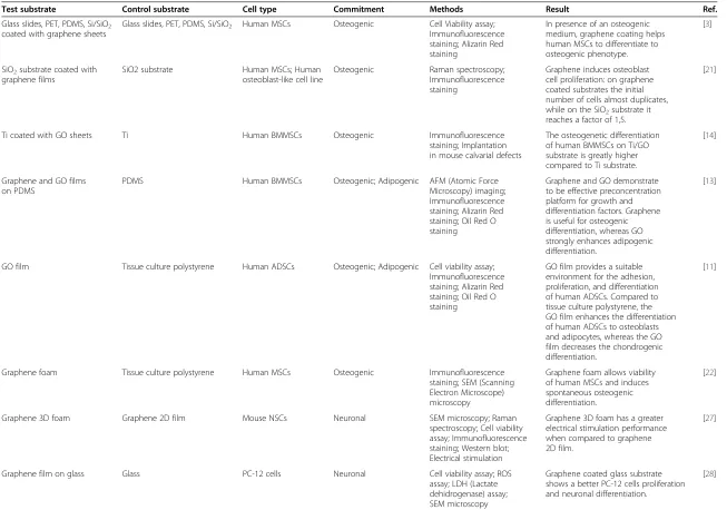
Test substrate Control substrate Cell type Commitment Methods Result Ref.
Glass slides, PET, PDMS, Si/SiO2
coated with graphene sheets
Glass slides, PET, PDMS, Si/SiO2 Human MSCs Osteogenic Cell Viability assay;
Immunofluorescence staining; Alizarin Red staining
In presence of an osteogenic medium, graphene coating helps human MSCs to differentiate to osteogenic phenotype.
[3]
SiO2substrate coated with
graphene films
SiO2 substrate Human MSCs; Human osteoblast-like cell line
Osteogenic Raman spectroscopy; Immunofluorescence staining
Graphene induces osteoblast cell proliferation: on graphene coated substrates the initial number of cells almost duplicates, while on the SiO2substrate it
reaches a factor of 1,5.
[21]
Ti coated with GO sheets Ti Human BMMSCs Osteogenic Immunofluorescence staining; Implantation in mouse calvarial defects
The osteogenetic differentiation of human BMMSCs on Ti/GO substrate is greatly higher compared to Ti substrate.
[14]
Graphene and GO films on PDMS
PDMS Human BMMSCs Osteogenic; Adipogenic AFM (Atomic Force Microscopy) imaging; Immunofluorescence staining; Alizarin Red staining; Oil Red O staining
Graphene and GO demonstrate to be effective preconcentration platform for growth and differentiation factors. Graphene is useful for osteogenic differentiation, whereas GO strongly enhances adipogenic differentiation.
[13]
GO film Tissue culture polystyrene Human ADSCs Osteogenic; Adipogenic Cell viability assay; Immunofluorescence staining; Alizarin Red staining; Oil Red O staining
GO film provides a suitable environment for the adhesion, proliferation, and differentiation of human ADSCs. Compared to tissue culture polystyrene, the GO film enhances the differentiation of human ADSCs to osteoblasts and adipocytes, whereas the GO film decreases the chondrogenic differentiation.
[11]
Graphene foam Tissue culture polystyrene Human MSCs Osteogenic Immunofluorescence staining; SEM (Scanning Electron Microscope) microscopy
Graphene foam allows viability of human MSCs and induces spontaneous osteogenic differentiation.
[22]
Graphene 3D foam Graphene 2D film Mouse NSCs Neuronal SEM microscopy; Raman spectroscopy; Cell viability assay; Immunofluorescence staining; Western blot; Electrical stimulation
Graphene 3D foam has a greater electrical stimulation performance when compared to graphene 2D film.
[27]
Graphene film on glass Glass PC-12 cells Neuronal Cell viability assay; ROS assay; LDH (Lactate dehidrogenase) assay; SEM microscopy
Graphene coated glass substrate shows a better PC-12 cells proliferation and neuronal differentiation.
[28]
Bressan
et
al.
Journal
of
Translationa
lMedici
ne
2014,
12
:296
Page
3
o
f
1
5
http://ww
w.translationa
l-medicin
e.com/conten
Table 1 Commitment of stem cells on different graphene substrates(Continued)
Graphene coated glass Glass Human NSCs Neuronal Immunofluorescence staining; Electrical stimulation
Graphene substrate is an excellent cell-adhesion layer during the differentiation process and induces the differentiation of human NSCs more toward neurons than glial cells.
[19]
Graphene film Tissue culture polystyrene Mouse NSCs Neuronal AFM imaging; Immunofluorescence staining; Electrical stimulation
NSCs seeded on graphene film differentiate and form functional neuronal networks.
[33]
Fluorinated graphene Graphene Human BMMSCs Neuronal Immunofluorescence staining; AFM imaging;
Fluorinated graphene enhances cell adhesion and proliferation of human BMMSCs. It exhibits a neuro-inductive effect via spontaneous cell polarization.
[29]
rGO/TiO2 GO/TiO2and TiO2 Human NSCs Neuronal flash photo stimulation;
Immunofluorescence staining
After flash photo stimulation, human NSCs proliferate more on rGO/TiO2
that on GO/TiO2and TiO2.The
neuronal differentiation of human NSCs on rGO/TiO2substrate is greatly
higher compared to GO/TiO2and TiO2
substrate.
[34]
GO Graphene, CNTs Mouse ESCs Neuronal Immunofluorescence staining; Real time PCR
GO substrate demonstrates an important enhancement of dopamine neurons differentiation whereas the GR and the CNTs do not show any important promotion on dopamine neurons differentiation.
[32]
Bressan
et
al.
Journal
of
Translationa
lMedici
ne
2014,
12
:296
Page
4
o
f
1
5
http://ww
w.translationa
l-medicin
e.com/conten
obtain osteoblasts starting from MSCs and pre-osteoblasts [17]. Kalbacova et al. made the first observations plating osteoblasts on two different substrates, silica (SiO2) and
graphene-coated SiO2[21]. After 48 hours incubation the
cells were homogenously covering the graphene substrate, while appearing as separate spots on the SiO2surface. The
results were supported by fluorescent imaging, revealing that in the graphene substrate the initial number of osteo-blasts almost duplicated, compared to a 1,5 increase factor in the SiO2 film. Nonetheless, these initial observations
tended to be controversial, considering the study of Nayak et al. [3]. When graphene was used as a coating agent on polydimethylsiloxane (PDMS), polyethilenterefta-late (PET), glass and Si/SiO2, it did not demonstrate to
in-fluence the shape or structure of seeded cells compared to uncoated surfaces. Furthermore, these authors investi-gated the osteogenic potential of graphene on MSCs.
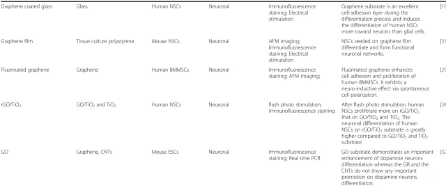
MSCs cultured on uncoated Si/SiO2, PDMS and PET
sur-faces showed a CD-44 positive staining and a complete negative osteocalcin (OCN) staining. On the other hand, OCN was significantly represented when cells were cul-tured on the same graphene-coated surfaces (Figure 2). The results were confirmed by the detection of a greater extent of calcium deposition in the second cells popula-tion, confirming the role played by graphene on the induc-tion of osteogenic differentiainduc-tion.
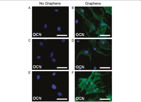
Further studies as the one by Lee et al. showed a positive correlation between culture on graphene substrate and osteogenic differentiation [13]. In particular, the study re-vealed the ability of graphene substrates to act as a precon-centration platform for osteogenic differentiation factors, such as dexamethasone and beta-glicerophosphate. The osteogenic differentiation was also visualized by Alizarin Red staining (Figure 3).
More recently, the alternative coating with GO has started to be explored. The study by La et al. investi-gated the potential of GO-coated titanium (Ti) in load-ing and releasload-ing bone morphogenic protein type 2 (BMP-2) [14]. The osteogenic differentiation of human bone marrow MSCs (hBMMSCs) cultured on Ti and Ti/ GO substrates was then tested and immunocytochemical staining for OCN showed a higher osteogenetic potential at 2 and 3 weeks of hBMMSCs on Ti/GO substrate compared to Ti substrate without coating. The same study performed an in vivo validation through implant-ation of Ti and Ti/GO substrates, with or without BMP-2, into mice calvaria defects. After a 8 weeks follow up, micro computed tomography imaging and the histo-logical analysis confirmed that no new bone formation was observed without the use of BMP-2, but the Ti/GO/ BMP-2 implant substrates showed a greater bone forma-tion compared to Ti/BMP-2 substrates.
The osteogenic differentiation potential of MSCs on graphene substrates was confirmed also by the study of
Crowder et al., where a 3D graphene structure was used to induce osteogenic differentiation [22]. The authors remarked that the foam shape structure used was par-ticularly suitable as a substrate to induce MSCs to differ-entiate into bone lineage.
The effect of graphene on MSC osteogenic commitment has been also studied by Duan et al. [23] who compared carbon nanomaterials (CNMs), such as CNT, with gra-phene. Their combinations with nanofibrous polymeric scaffolds, which mimic the morphology of natural ECM of bone, arouse indeed keen interest in bone tissue engineer-ing. The hypothesis of the authors was that the sheet-like graphene might have stronger enhancement in regulating osteocompatibility than tubular multiwall CNT composite scaffolds, because the former provided more contacting surface to cells than the latter when they were at the same content. Therefore, composite nanofibrous scaffolds were prepared by using poly-L-lactide (PLLA) and graphene as starting materials. Briefly, graphenes were added into PLLA-tetrahydrofuran (THF) solutions, and thermal-induced phase separation (TIPS) technique was applied to induce the nanofibrosis of PLLA. To this end, CNMs were incorporated into nanofibrous PLLA scaffolds by TIPS technique. The CNMs-containing composite nanofibrous
scaffolds were biologically evaluated by both in vitro
co-culture of hBMMSCs and in vivo implantation. The
nanofibrous structure itself demonstrated significant enhancement in cell adhesion, proliferation and osteo-genic differentiation of hBMMSCs, and with the incorpor-ation of CNMs, the composite nanofibrous scaffolds further promoted osteogenic differentiation of hBMMSCs significantly. Between the two CNMs, graphene showed stronger effect in promoting osteogenic differentiation of
hBMMSCs than CNT. The results ofin vivoexperiments
revealed that the composite nanofibrous scaffolds had both good biocompatibility and strong ability in inducing osteo-genesis. CNMs could remarkably enhance the expression of osteogenesis-related proteins as well as the formation of type I collagen. Similarly, the graphene-containing com-posite nanofibrous scaffolds demonstrated the strongest effect on inducing osteogenesis in vivo. These findings demonstrated that CNMs-containing composite nanofi-brous scaffolds were obviously more efficient in promoting osteogenesis than pure polymeric scaffolds [23].
The same positive results have been observed by Tavarty et al. [24] that hypothesized that incorporating GO with an osteoinductive material could synergistically direct the differentiation of human MSCs toward osteo-genic lineage. Calcium phosphates (CaP) such as hy-droxyapatite (HAp) are biomimetic biomaterials that are well-recognized for their osteoconductivity (facilitating bone formation) and osteoinductivity (facilitating the osteogenic differentiation of human MSCs). To validate the above hypothesis, the authors synthesized a novel
Bressanet al. Journal of Translational Medicine2014,12:296 Page 5 of 15
biocompatible GO-CaP nanocomposite and evaluated its capability of inducing the osteogenic differentiation in hu-man MSCs. The GO-CaP nanocomposite was fabricated using GO microflakes, uniquely structured highly osteoin-ductive CaP nanoparticles, and pluronics polymeric coat-ing. The osteoinductive properties of GO microflakes, CaP, and GO-CaP on human MSCs were evaluated by quantita-tive measurements on bone nodule formation and the immunofluorescence imaging of osteoblast biomarkers. GO-CaP exhibited osteogenic capability that was superior to individual or combined effects of GO and CaP. To evaluate the materials’osteoinductive capability, GO, CaP and GO-CaP were introduced to human MSCs and their osteogenic commitment has been evaluated. Results re-vealed that treatments with GO, CaP and GO-CaP in osteogenic medium induced significantly larger quantity of calcium than control at all the time points, while no calcifi-cation was observed in negative controls. GO-CaP nano-composites exhibited superior osteoinductivity to CaP or
GO, inducing much larger amount of mineralization than control. Phosphate assay was also performed from the de-position in parallel plates after 2 and 3 weeks of treatment. The amount of phosphate among all the groups followed the sequence of GO-CaP > CaP > > GO > control, consist-ent with the outcome from calcium quantification. Surpris-ingly, GO microflakes at low concentration (0,5 mg/mL) increased calcification up to 50% more than the control at 3 and 4 weeks. The osteogenic differentiation of human MSCs was verified moreover through immunofluorescence staining of osteoblast markers: alkaline phosphatase (ALP) and OCN after 2 weeks of treatments. In good agree-ments with Alizarin Red staining, ALP activities and OCN expression level followed the sequence of GO-CaP > CaP > > GO > control, affirming the potential of GO-CaP in directing human MSCs differentiation toward osteo-genic lineage [24].
[image:6.595.60.538.90.435.2]Owing to the superior mechanical properties and low coefficient of thermal expansion, graphene has been Figure 2Immunostaining of MSCs seeded for 15 days in osteogenic differentiation medium on different substrates.Si/SiO2(A,B), PDMS
(C,D), and PET(E,F)substrates were coated or not with graphene. Cells are stained with DAPI (blue) and OCN (green). MSCs growing on Si/SiO2
(A), PDMS(C), and PET(E)without graphene show OCN negative staining. Once these substrates are coated with graphene (B,D,Frespectively), cells are positive for OCN, indicating osteogenic differentiation. Scale bars are 100μm [3].
Bressanet al. Journal of Translational Medicine2014,12:296 Page 6 of 15
widely used in the reinforcement of ceramics. Xie et al. [25] studied that various ratios of graphene (0,5 wt%, 1,5 wt% and 4 wt%) reinforced with graphene calcium silicate (CS) for load-bearing implant surface modifica-tion. Surface characteristics of the graphene-calcium sili-cate (GC) composite coatings were characterized by scanning electron microscopy. Results showed that the graphene plates (less than 4 wt% in the coatings) were embedded in the CS matrix homogeneously. The sur-faces of the coatings showed a hierarchical hybrid nano-microstructure, which is believed to be beneficial to the behaviors of the cell and early bone fixation of the implants. Wear resistance measured by a pin-on-disc model exhibited an obvious enhancement with the adop-tion of graphene plates. The weight losses of the GC coatings decreased with the increase of graphene con-tent. However, too high graphene content (4 wt% or more) made the composite coatings porous and the wear resistance decreased dramatically. The weight loss was only 1,3 ± 0,2 mg for the GC coating containing 1,5 wt% graphene (denoted as GC1,5) with a load of 10 N and sliding distance of 500 m, while that of the pure CS
coating reached up to 28,6 ± 0,5 mg. In vitro
cytocom-patibility of the GC1,5 coating was evaluated using a hBMMSCs culture system. The proliferation and ALP, osteopontin and OCN osteogenesis-related gene
expression of the cells on the GC1,5 coating did not de-teriorate with the adoption of graphene. Conversely, even better adhesion of the hBMMSCs was observed on the GC1,5 coating than on the pure CS coating. All of the results indicate that the GC1,5 coating is a good can-didate for load-bearing implants [25].
Neuronal differentiation
Inducing human NSCs to differentiate into neurons is a critical challenge to reach an important biomedical goal, since a promising opportunity in therapies for neural re-generation could arise [26,27].
Park et al. tested the graphene substrate as a promoter of human NSCs differentiation into neurons [19]. The most remarkable data were observed when comparing the human NSCs seeded on a graphene and a glass substrate after 30 days. Immunofluorescence showed a greater de-gree of cell attachment along with cell differentiation rate into neurons on the graphene substrate, whereas more glial cells than neurons were found on the glass surface. The analysis was performed counting the immunopositive cells for GFAP (a glial cell marker) and TUJ1 (a neuronal cell marker).
[image:7.595.57.540.87.348.2]Other results indicating that graphene-based sub-strates can promote neural differentiation came from the study by Hong et al. [28]. After culturing PC-12 cells on Figure 3Alizarin Red staining of MSCs seeded on PDMS, graphene, and GO substrates.A higher amount of Alizarin Red, which is an indicator of osteogenic differentiation, is found in MSCs cultured on graphene(C)for 12 days in osteogenic differentiation medium than that cultured on GO(E)and PDMS(A)in the same conditions. In presence of basal medium, MSCs do not show staining for Alizarin Red in none of the 3 substrates(B,D,F)[13].
Bressanet al. Journal of Translational Medicine2014,12:296 Page 7 of 15
uncovered or graphene-covered glass substrates, the au-thors observed a better cellular adherence along with higher cell proliferation and neural differentiation on the graphene-coated substrate.
In the attempt of influencing in a significant way stem cell differentiation, several declination of graphene scaf-folds were studied. Wang et al. tested MSCs on fluorinated sheets of graphene, observing a strong enhancement of neuronal differentiation when compared to cells seeded on graphene [29]. This indicated how fluorinated gra-phene may be a good engineered platform to enhance neuronal differentiation. Li et al. used a new 3D scaffold based on graphene, a graphene foam, which regulated mice NSCs behavior supporting their growth and keeping cells at an active proliferation state [27]. Its porous struc-ture showed to be a good substrate for NSCs adhesion, probably due to its irregular surface which improved mechanical adhesion [30]. The scaffold was also proved to be highly biocompatible since no cytotoxicity was ob-served and cell viability was not affected. Moreover, the 3D structure had a greater electrical stimulation perform-ance when compared to a 2D graphene structure, and electrical stimulation has been proven to induce neural differentiation [31].
The study by Yang et al. investigated the capability of GO, graphene and CNTs to induces dopamine neural dif-ferentiation of mouse embryonic stem cells (ESCs) [32]. ESCs were seeded in all the 3 substrates and a stromal cell-derived inducing activity method was used. After 14 days of differentiation, the GO substrate demonstrated an important enhance of dopamine neural differentiation while the graphene and CNTs did not show any important promotion of dopamine neural differentiation.
Another important characteristic of graphene could be represented by its capability to form a functional neural network. In the study by Tang et al., neurospheres were seeded on graphene substrates and after a 14 days cul-ture the process of network formation was made clear by beta-tubulin immunostaining [33]. Newly formed neuritis started to form synapses. This result confirmed how graphene could be considered as a valid substrate to promote neural activity (Figure 4).
Stimulating human NSCs to differentiate into neurons rather than glia is a another key point in order to obtain a neural regeneration result [29]. The study by Akhavan and Ghaderi specifically concentrated on testing human NSCs differentiating behavior on different substrates (TiO2, GO/TiO2, and rGO/TiO2), with and without flash
photo stimulation [34]. When the 3 substrate went under flash photo stimulation, the number of cells in-creased by a factor of 1,5 on the rGO/TiO2substrate, by
a rate of 48% in the GO/TiO2substrate and by a rate of
24% on the TiO2substrate. No differences were detected
without flash stimulation. When the differentiation rate
was analyzed it was found that the rGO/TiO2 substrate
was the more beneficiary of the flash photo stimulation: an 88% decrease in the ratio of glial cells and an 81% in-crease in the ratio of neuronal cells. In the other 2 sub-strates the difference was less evident: 25% of neuronal cells increase rate on the GO/TiO2substrate and 15% on
the TiO2 substrate. Overall, flash photo stimulation of
graphene-based substrate provide both a greater cell pro-liferation and a greater human NSCs differentiation into neurons rather that glia cells. However, these results could be obtained when the flash photo stimulation of human NSCs was at an optimum concentration of a biocompat-ible hole scavenger, and at an optimum flash intensity.
For central nervous system (CNS) regeneration, the selective differentiation of NSCs into either neurons or oli-godendrocytes (as opposed to astrocytes) is highly desirable. Several approaches have been employed to guide differenti-ation into neurons, however, oligodendrocyte differentidifferenti-ation has proven to be much more elusive, resulting in only a small percentage of the differentiated cell population. The primary approach to guide oligodendrocyte differentiation has focused on either developing culture media containing a combination of growth factors or the forced expression of key oligodendrocyte-promoting transcription factors. In this view the developing a biomaterials-approach to achieve effi-cient differentiation of NSCs into mature oligodendrocytes, while eliminating the potential adverse or variable side-effects from growth factors and viral gene vectors, would be highly beneficial.
Shah et al. [35] developed a graphene-based nanomater-ial for the design of hybrid nanofibrous scaffolds to guide NSCs differentiation into oligodendrocytes. The authors demonstrated as the use of GO was an effective coating material in combination with electrospun nanofibers for the selective differentiation of NSCs into oligodendrocytes. By varying the amount of GO coating on the nanofibers, they observed a GO concentration-dependent change in the expression of key neural markers, wherein coating with a higher concentration of GO was seen to promote differentiation into mature oligodendrocytes. Further in-vestigation into the role of GO-coating on the nanofibrous scaffolds showed the overexpression of a number of key integrin-related intracellular signaling molecules that are known to promote oligodendrocyte differentiation in nor-mal development. In their studies, polycaprolactone (PCL) was electrospun onto a metallic collector and then trans-ferred to glass substrates for cell culture using a medical grade adhesive. Nanofibers with an average diameter of 200–300 nm were generated, which is a fiber size range that has been reported to be favorable for oligodendrocyte culture, potentially due to the close morphological resem-blance to axons. Thin-layered GO was then synthesized and then deposited on the PCL nanofiber surface. Among the various cell-signaling proteins, the authors examined
Bressanet al. Journal of Translational Medicine2014,12:296 Page 8 of 15
the expression of focal adhesion kinase (FAK), Akt, integrin-linked kinase (ILK) and Fyn kinase (Fyn), which have been found to mediate cytoskeletal remodeling and process extension during oligodendrocyte development. The researchers found that NSCs cultured on the GO coated surfaces enhanced the gene expression of all of these factors. These signaling molecules exhibited the same trend in expression, wherein the GO-coated glass showed higher expression than PCL, and PCL-GO showed the strongest level of expression with a 2,6-fold increase in FAK and about a 1,7-fold increase in Akt, ILK and Fyn. Additionally, treating the cells grown on PCL-GO scaf-folds with cell signaling inhibitors showed a significant de-crease in gene expression of mature oligodendrocyte markers, which provides further evidence for the potential role of such cellular signaling in the observed oligodendro-cyte differentiation. Collectively, this data supports the role of GO-coating in the upregulation of these down-stream molecules in the integrin signaling pathway and may explain, at least in part, the enhanced oligodendrocyte differentiation of NSCs on hybrid scaffolds. Data obtained suggest that the GO-coating on the nanofiber scaffolds may promote oligodendrocyte differentiation through specific microenvironmental interactions which activate integrin-related intracellular signaling. Overall, Shah et al. demonstrated the capability of a unique graphene-nanofiber hybrid scaffold to provide instructive physical cues that lead to the selective differentiation of NSCs into mature oligo-dendrocytes, without introducing differentiation inducers in
the culture media. The ability to selectively guide stem cell differentiation by merely changing the properties of an underlying biomaterial scaffold is a valuable ap-proach for tissue engineering, which can help comple-ment or potentially eliminate the use of exogenous differentiation. Moreover, their hybrid scaffold is excep-tional in that it combines the well-established properties of nanofibers and graphene-based nanomaterials. For instance, nanofibers have been shown to provide ideal top-ography for fabricating nerve guidance conduits, directing neurite outgrowth and promoting axonal regeneration. On the other hand, graphene-based nanomaterials provide permissive surfaces for protein and cell adhesion, as well as high conductivity to mediate electrical stimulation for supporting neuronal electrophysiology [35].
[image:9.595.63.538.90.323.2]GO based nanoparticles has been used also to drive the commitment of ESCs into dopamine neurons. Yang et al. [36] studied the effects of CNTs, GO and graphene nano-particles on the dopamine neural differentiation of mouse ESCs. The dopamine neural differentiation of the ESCs was examined by immunocytochemistry and real-time PCR showing that only GO could effectively promote dopamine neuron differentiation after induction of a stro-mal cell-derived inducing activity and further enhance dopamine neuron-related gene expression compared with cells treated with no nanoparticle control, and the other two nanoparticles (CNTs and graphene). In conclusion, authors suggest that GO is a promising nanomaterial-based technical platform to effectively enhance dopamine Figure 4Immunostaining of neurospheres deriving from NSCs seeded on graphene films up to14 days.Cells are stained with DAPI (blue) and beta-tubulin (green) at day 1(A), day 3(B), day 7(C), and day 14(D). NSCs differentiated on graphene substrates display neurites growing to various directions and distances, indicating the development of neural networks [29].
Bressanet al. Journal of Translational Medicine2014,12:296 Page 9 of 15
neural differentiation of ESCs, which can be potentially applied for cell transplantation therapy.
Novel important applications of graphene in neurosci-ence has been found in the end by Song at al. [37], who studied the anti-inflammatory effects of three-dimensional graphene foams cultured with microglial cells. Nanoma-terials are increasingly used in medical diagnosis and treat-ment due to their unique mechanical, optical, electrical, and magnetic properties. However, studies have revealed that most of the nanomaterials could initiate some form of inflammation both in vitro and in vivo, then lead to other biological effects, as expected of any foreign particu-late. Current studies mostly focus on the pulmonary in-flammation caused by nanomaterials insults, while little is known about the neuroinflammatory effects. Given that some nanomaterials (quantum dots, CNTs, graphene, etc.) have been attempted to be used in neuroscience, the neu-roinflammation should be considered. External insults range from hypoxia and ischemia to a number of bacterial and viralinfections, all of which elicit a characteristic neu-roinflammatory reaction in the brain. Microglia, astro-cytes, and peripheral macrophages are key players mediating this response. In the brain, most of the damage caused by nanomaterials is mediated by the microglia, a macrophage-like, phagocytic cell that is normally inactive unless confronted by potentially damaging xenobiotics. In response to certain cues such as brain injury or immuno-logical stimuli, however, microglia are readily activated. Graphene has been at the forefront of nanotechnology and advanced materials sciences due to its intriguing phys-ical and chemphys-ical features. Especially, it has been utilized in a variety of biomedical applications. Recently, Li et al. [38] discovered the great potentials of using graphene for neural interfacing, as it could promote neurite sprouting and outgrowth in primary culture of hippocampal neu-rons, enhance the neural performances in the network dif-ferentiated by NSCs, direct stem cell differentiation, and be used as electric field stimulator for effective cerebral blood volume enhancement. Meanwhile, graphene foams were found to greatly induce NSCs differentiation to neur-onal lineage and were proposed as a neural scaffold for NSC-based therapy. These pioneering works demonstrate the capability of graphene for applications in CNS. How-ever, to the best of our knowledge, there is no report regarding the possible neuroinflammatory effects of gra-phene, which should be well addressed before any further clinical applications. In this work, the researchers report the neuroinflammatory responses of microglia under the
presence of graphene by in vitro culturing and justify
whether this graphene-induced neuroinflammatory effects are detrimental or beneficial to the neural cells. The im-portance of this work is the elucidation of the pro- and/or anti-inflammatory effects of graphene and pave the way for the applications of graphene in biomedicine. The
graphene, especially 3D graphene, supported microglia growth and showed comparable biocompatibility to the commercial tissue culture polystyrene substrates. Despite of the similar proinflammatory responses in the microglia without lipopolysaccharide (LPS) activation, 3D graphene evoked much milder neuroinflammation in the microglia after LPS activation in comparison to 2D graphene, sug-gesting that the topographical structures of the materials might affect the inflammatory behaviors. Furthermore, the unique topographical structures of 3D-graphene may re-strict the morphological transformation of microglia under over-activation, leading to the anti-inflammatory effects.
Adipogenic differentiation
Graphene and GO substrates have been also used for in-vestigating their effects on the adipogenic differentiation of MSCs. In particular, in the study of Lee et al., MSCs were plated on graphene or GO sheets in presence of adipogenic differentiation medium for 14 days [13]. The cells were then stained with Oil Red O and counted. The results showed a strong suppression of adipogenesis on graphene substrates; on the contrary, GO was a strong enhancer. This difference was probably due to the ability of graphene to denaturate insulin: GO did not denaturate insulin which therefore could maintain its role of mediator of fatty acid synthesis.
Kim et al. evaluated GO potential in differentiating hu-man adipose-derived stem cells (ADSCs) into adipocytes, demonstrating a higher adipogenesis on the GO sub-strate when compared to the control (tissue culture polystyrene) [11]. This study also documented how GO could be considered a unique substrate, allowing the at-tachment and proliferation of ADSCs and modulating cell differentiation not only toward the adipogenic line but also toward the osteogenic and epithelial pheno-types. On the contrary, the GO films resulted in de-creased chondrogenic differentiation of the ADSCs.
Periodontal ligament stem cells
In regenerative dentistry, stem cell-based therapy often re-quires a scaffold to deliver cells or growth factors to the injured site. GO and silk fibroin (SF) are promising bioma-terials for tissue engineering as they are both non toxic and promote cell proliferation. A field that could be im-proved by the availability of an effective film scaffold would be the reparation of periodontal tissues. The peri-odontium, which is composed of four dental tissues (i.e., gingiva, alveolar bone, cementum, and periodontal ment [PDL]), is constantly maintained by periodontal liga-ment stem cells (PDLSCs) owing to their great capacity of differentiation into cementoblasts, odontoblasts, and fi-broblasts. PDL plays a key role in the attachment of teeth to the jaw; in the most drastic cases of periodontitis, which is associated with a chronic inflammation process, PDL
Bressanet al. Journal of Translational Medicine2014,12:296 Page 10 of 15
destruction could lead to the loss of the tooth [39]. With the aim of expanding tissue engineering therapy to as many patients as possible, acellular biomaterials may be employed as a novel approach to heal periodontal site by the active recruitment of autologous cells into the PDL scaffold, thus providing an in situ regeneration in cases of periodontitis. So, in order to make the evaluation of the mentioned SF-GO composite film as scaffold, human den-tal stem cells were chosen by Rodriguez-Lonzano et al. as cellular model to test the the performance of GO and SF. In their study the authors evaluated the effects of the novel biomaterials GO and SF on PDLSCs phenotype, ad-hesion, proliferation rate and viability. Biocompatibility of scaffolds is a prerequisite for generating cell-biomaterial constructs and for their successful clinical application. GO and fibroin-based biomaterials have been previously stud-ied for several tissue engineering based-therapies [39], but they have never been tested in conjunction with mesen-chymal stem cells isolated from PDL. The morphology of PDLSCs cultured for different times on GO and fibroin coated surfaces by staining of actin cytoskeleton showed that PDLSCs cultured on fibroin displayed lower amounts of F-actin, lower spreading and delayed growth. By con-trast, GO or GO plus fibroin-coated surfaces significantly improved F-actin content, cell spreading and growth rate from 96 h of culture when compared to fibroin alone. Fur-thermore, it has been also studied the proliferation rate of PDLSCs on GO and fibroin-based biomaterials by MTT assays. Results confirmed that after 7 days of culture, PDLSCs showed a high cell proliferation rate in presence of GO, although it was slightly lower than on plastic, whereas fibroin or GO-fibroin biomaterials supported a discrete proliferation. In addition, to evaluate the possible cellular cytotoxic effect of the different biomaterials employed as well as changes on the expression of mesen-chymal surface markers, the researchers characterized their surface molecule expression pattern by flow cytome-try. Culture of PDLSCs on fibroin, GO or GO plus fibroin did not significantly alter the level of expression of CD73, CD90 or CD105 after 24, 48, 72, 96 or 168 h compared to expression levels displayed by PDLSCs cultured on plastic. Thus, the biomaterials employed in this study were able to maintain the mesenchymal phenotype of PDLSCs.
Cardiomyogenic differentiation
As well reported above, graphene has drawn attention as a substrate for stem cell culture and has been reported to stimulate the differentiation of multipotent adult stem cells. Recently, Lee et al. [40] reported that graphene enhances the cardiomyogenic differentiation of human ESCs at least in part, due to nanoroughness of graphene coated with vitronectin (VN). Human ESCs were cul-tured on either VN-coated glass or VN-coated graphene for 21 days. The cells were also cultured on glass coated
with Matrigel, which is a substrate used in conventional, directed cardiomyogenic differentiation systems. Results confirmed that the culture of human ESCs on graphene promoted the expression of genes involved in the step-wise differentiation into mesodermal and endodermal lineage cells and subsequently cardiomyogenic differenti-ation compared with the culture on glass or Matrigel. In addition, the culture on graphene enhanced the gene ex-pression of cardiac-specific extracellular matrices. The authors concluded in the end that graphene may provide a new platform for the development of stem cell therap-ies for ischemic heart diseases by enhancing the cardio-myogenic differentiation of human ESCs.
The same results have been reched by Park et al. [41] that demonstrated the use of MSCs culture to promote cardiomyogenic differentiation. Also in this case gra-phene exhibited no sign of cytotoxicity for stem cell culture. MSCs were committed toward cardiomyogenic lineage by simply culturing them on graphene. The au-thors speculated that this may be attributed, at least par-tially, to the regulation of expression levels of ECM and signaling molecules.
iPSCs
The successful reprogramming of somatic cells into in-duced pluripotent stem cells (iPSCs) by Takahashi and Yamanaka in 2006 [42] was seen as a landmark event in disease research. This new technology promised to pro-vide a powerful tool for modeling human pathology that could be used to understand the underlying causes of various human diseases. The following years saw a stream of new and improved approaches for converting somatic cells into more differentiated cell types.
In an interesting study by Yoo et al. [43], the authors reported that graphene promotes the reprogramming of mouse somatic fibroblasts into iPSCs.
The generation of ESCs-like cells from somatic cells by ectopic expression of defined factors is an approach com-monly known as cell reprogramming. Moreover, because iPSCs generation is known to be a multiple-step process mediated by overexpression of the transcription factors Oct4, Sox2, Klf4, and cMyc, the generation of iPSCs is very inefficient, and cell reprogramming is considered a stochastic process in which successive barriers must be overcome to reach a state of pluripotency. In particular, one of the first noticeable changes during the reprogram-ming of somatic fibroblasts is their transformation into tightly packed clusters of rounded cells in a process that resembles a mesenchymal-to-epithelial-transition (MET). More interestingly, recent studies found that microtopo-graphy substrate affects the MET, improving reprogram-ming efficiency. The authors examinated whether somatic fibroblasts should be efficiently reprogrammed into the pluripotent state on graphene-based substrate. Fitstly they
Bressanet al. Journal of Translational Medicine2014,12:296 Page 11 of 15
carachterised the monolayer graphene film by Raman spectroscopy, then they studied whether the graphene substrate enhances cell reprogramming by seeding Oct4-GFP knock-in (KI) mouse embryonic fibroblasts (MEFs) onto both the control and graphene-coated substrate in MEF medium. One day after plating, MEFs were trans-duced using specific vectors expressing Oct4, Sox2, Klf4, and cMyc transcription factor. The MEFs plated on the graphene substrate began to form colonies 10 days after viral infection, exibiting a significant increase in colonies undergoing reprogramming on graphene substrate. To quantify the reprogramming efficiency, they performed FACS analysis for Oct4-GFP-positive iPSCs derived from Oct4-GFP KI MEFs in both the control substrate and graphene-coated substrate. Fifteen days after doxycycline (dox) induction, the graphene substrate cultures had a sig-nificant increase in the number of GFP-cells and quantita-tive real-time PCR analysis showed that pluripotency marker genes, including Oct4, Nanog, Sox2 and Esrrb were markedly elevated in graphene-coated substrate cul-tures compared to uncoated substrate culcul-tures. The pluri-potent state of graphene-induced iPSCs was assessed by immunostaining of pluripotency marker. Consistent with these results, they observed that the graphene-coated sub-strate significantly increased the number of Oct4, SSEA1 and Nanog colonies. Moreover, in order to analyze the dif-ferentiation potential of graphene mediated iPSCs, their capacity for direct differentiation into the cell types of the three germ layers has been tested. Immunocytochemical staining and real-time PCR showed that the differentiated cells were positive. In the end the further developmental potency of the graphene induced iPSC lines was investi-gated by SCID mice. Four weeks after injection, teratomas were readily visible and histological analysis confirmed the presence of cell types derived from all three embryonic germ layers validating the pluripotency of these graphene induced iPS cells. In order to examine whether graphene substrate affects chromatin state during reprogramming, they measured the enrichment level of histone modifica-tions that mark active (histone 3 lysine 4 tri-methylation, H3K4me3) in the graphene induced cell reprogramming. Surprisingly, they found a dramatic increase of H3K4me3 expression on graphene substrate and that the transcrip-tion start sites of Oct4 and Nanog were enriched for H3K4me3 in graphene surface-induced reprogramming cultures. Taken together, these results suggest that the graphene-coated substrate specifically promotes the MET process in cell reprogramming without affecting the EMT process. In this study, the autors showed that the epigen-etic reprogramming of somatic fibroblasts is enhanced on graphene substrate providing a potential method for the efficient generation of iPSCs. Interestingly, they found a specific induction of MET by the graphene substrate dur-ing reprogrammdur-ing and an accumulation of H3K4me3,
which facilitates increased Oct4 and Nanog occupancy. These data indicate a unique role for graphene substrates in facilitating cell reprogramming [43].
Carbon nanoparticles such as zero dimensional (0D) ful-lerenes, one dimensional (1D) CNTs, and recently two di-mensional (2D) graphene have been investigated for applications in therapeutics, bioimaging, and regenerative medicine. MSCs are currently being widely investigated to repair, regenerate, and restore damaged tissues. Nanoparti-cles have been employed to deliver growth factors or genes into MSCs to manipulate their differentiation [44]. The development of graphene nanoparticles for MSC applica-tions necessitates thorough examination of their effects and interactions with these cells to identify potential therapeutic doses. To date, very few studies have investi-gated the cytotoxicity of graphene nanoparticle formula-tions with specific focus on progenitor cells or MSCs. Zhang et al. examined the toxicity of graphene quantum dots, single reduced graphene sheets with diameters in the range of 5–10 nm, on three progenitor cell types: neuro-spheres cells, pancreas progenitor cells, and cardiac progenitor cells [45]. Akhavan et al. employed umbilical cord-derived MSCs and investigated the size-dependent cytotoxicity of graphene oxide nanoplatelets and reduced graphene oxide nanoplatelets (prepared using the modi-fied Hummer’s method) [46]. Graphene nanoparticles, de-pending on the synthesis method, can exhibit different morphologies, chemical properties, and physical proper-ties. Thus, it is necessary to systematically investigate the effects that graphene nanoparticles with different morph-ologies, synthesized by various methods, have on MSC viability and differentiation. In an interesting study of Taluktat et al. [44], the dose- and time-dependent effects were investigated of three graphene nanoparticles on the viability and differentiation of human MSCs. The initial cytotoxicity screening over a broad range of concentra-tions (0–300μg/mL) and time points (one and three days) was performed on ADSCs and BMMSCs to identify a range of potentially safe doses. ADSCs were then employed to investigate whether these graphene nanoparticles at a potentially safe low and high dose affect the differential capabilities of MSCs. The cytotoxicity and differentiation studies together allowed identification of the range of doses for the three-graphene nanoparticle formulations that do not elicit any significantly adverse outcomes on the viability and differentiation capabilities of MSCs. Results confirmed that graphene nano-onions (GNOs), graphene oxide nanoribbons (GONRs), and graphene oxide nanopla-telets (GONPs) elicited a dose-dependent (0–300 μg/mL), but not a time-dependent (24 and 72 h) cytotoxic response on ADSCs and BMMSCs. For all three nanoparticles, con-centrations of less than 50 mg/mL showed no significant differences compared to untreated controls. The adipogenic and osteogenic differentiation potential of ADSCs was not
Bressanet al. Journal of Translational Medicine2014,12:296 Page 12 of 15
adversely affected after treatment with a low (10 mg/mL) or high (50 mg/mL) concentration. GNOs and GONPs were internalized by ADSCs, while GONRs were not. The results suggest that GNOs, GONRs, and GONPs at con-centrations of less than 50 mg/mL for 24 or 72 h could be considered potentially safe incubation conditions for ex vivo labeling for MSCs. The results open avenues for use of these graphene nanoparticle formulations for ex vivo la-beling of MSCs for applications in regenerative medicine.
Conclusions
The literature on the biological interactions of graphene family is growing rapidly, and includes studies primarily motivated by biomedical applications, and environmental health and safety. As with other biomaterials, the issue of potential toxicity arises not only in biomedical applications, but also in non-biomedical products where unintended oc-cupational, consumer, and environmental exposures can occur. The exact mechanism of GO and the derived com-posites in SCs differentiation is still unresolved. It has been generally hypothesized that the surface characteristics of graphene family nanomaterials such as nanotopography, surface stiffness, and large absorption capacity influence the molecular pathways that control the fate of stem cells. Both G and GO were reported acting as preconcentrators for chemicals, proteins and growth factors on their surface to promote cell differentiation.
In this view, we have reported a review of the inter-national literature to produce a state-of-the-art update of graphene applied to tissue engineering and stem cells.
Many studies confirmed that graphene and its related materials are able to induce human stem cells differenti-ation into specific lineages. Materials coated with gra-phene or GO or even 3D gragra-phene foam were capable to guarantee viability and to induce osteogenic differentiation of stem cells when compared with traditional substrates or scaffolds [3,11,13,14,21,22]. In terms of neuronal regener-ation, graphene and GO showed the same promising dif-ferentiative potential. Furthermore, a real cellular and electrical network could be structurally and functionally created on these materials [19,27-29,32-34]. As well as in osteogenic and neuronal differentiation, GO can control adipogenesis [11,13].
Even if we are still uncertain about potential in vivo applications, mostly due to open questions around tox-icity, graphene clearly appears to be a step forward in the field of tissue engineering.
In this light it is interesting the results found by Chen et al. that express an expert opinion based on sciento-metric patterns revealed by CiteSpace without prior work-ing experience in the regenerative medicine field. Emerging trends and new developments have been identi-fied based on structural and temporal properties derived from the relevant publications. The detected surge of the
keyword graphene in the literature of regenerative medi-cine led the authors to investigate the nature and context of its use in regenerative medicine. The investigation re-vealed a rapidly increasing number of studies that specific-ally used graphene and GO in regenerative medicine research because of their desirable surface properties for, among other applications, culturing and maintaining SCs. Similarly a detected burst of citations provides insightful guidance for navigating through the fast-changing [47].
There is also an emerging literature on potential health risks. Despite the popular image of graphene as a large-area substrate coating, many graphene-family ma-terials are dry powders at some point in their processing, and in this form pose the most significant exposure risk through inhalation. Of particular concern are FLG sam-ples directly following the thermal exfoliation step or after washing and drying. There is need for measure-ment of airborne dust levels in research laboratories, and in pilot and full-scale manufacturing facilities. GFNs are high-surface-area materials with corresponding high potential to cause adsorptive and quenching artifacts in biological assays [48]. Adsorption on carbon surfaces is generally favored for molecules with low solubility, par-tial hydrophobicity, or positive charge (for the common case of negatively charged GFNs). The biological conse-quences may include (i) micronutrient depletion, (ii) ar-tifacts in assays that rely on dye-based molecular probes (iii) the capacity to carry small molecule drug cargoes, and (iv) synergistic or antagonistic toxic effects when GFNs coexist with small molecule toxicants, whose bioa-vailabilty can be increased or decreased as they partition to graphene surfaces. More work is needed in this area, and authors must be skeptical of standard assays without extensive controls for possible interference. As a final note,in the area of toxicity, there have been a number of studies reported, but the field is too young and the lit-erature too limited to reach conclusions about potential hazards sufficient for risk assessment or regulation. Nano-GO has been reported to be biocompatible in a number of the studies focused on biomedical applica-tions, at least under the limited conditions covered by such studies. Other studies have reported adverse bio-logical responses, including cytotoxicity using human lung epithelial cells and fibroblasts [49,50]. Cellular up-take of GFNs has been shown for macrophages and hu-man lung epithelial cells in some studies although there have been no studies exploring the mechanism of uptake and intracellular fate. These sheet-like GFNs may physic-ally perturb cytoskeletal organization, mitosis, organelle integrity, and impair cell motility and secretion. A po-tential toxicity pathway for GFNs is oxidative stress [51,52] it is not clear whether oxidant generation is re-lated to reactive edge sites or an indirect response of tar-get cells to nanomaterials. It is clear that lateral size is a
Bressanet al. Journal of Translational Medicine2014,12:296 Page 13 of 15
key variable in cell uptake, while layer number affects deposition and surface area, and surface chemistry has a large affect on adsorption and dispersibility. Molecular dyanamics modeling of interactions between GFNs and cell membranes should provide valuable information about uptake mechanisms. Systematic investigation of toxico-logical endpoints using a defined set of carbon nanoma-terials including carbon black, CNT, and GFNs will be important to develop structure-activity relations. Because graphenes form a material family with wide variation in properties, the graphene-bio field will benefit greatly in the long run, if its authors show diligence in characterizing their materials, and describing them according to layer number, lateral size, surface chemistry rather thanad hoc sample names.
Abbreviations
ADSCs:Adipose-derived stem cells; BMP-2: Bone morphogenic protein type 2; CaP: Calcium phosphates; CNMs: Carbon nanomaterials; CNS: Central nervous system; CNT: Carbon nanotube; CS: Calcium silicate; dox: Doxycycline; ECM: Extracellular matrix; ESCs: Embryonic stem cells; FAK: Focal adhesion kinase; FLG: Few-layer-graphene; Fyn: Fyn kinase; GC: graphene-calcium silicate; GFNs: Graphene-family nanomaterials”; GNOs: Graphene nano-onions; GNS: Graphene nanosheets; GO: Graphene oxide; GONPs: Graphene oxide nanoplatelets; GONRs: Graphene oxide nanoribbons; H3K4me3: Histone 3 lysine 4 tri-methylation; HAp: Hydroxyapatite; hBMMSCs: Human bone marrow MSCs; ILK: Integrin-linked kinase; iPSCs: Induced pluripotent stem cells; KI: Knock-in; LPS: Lipopolysaccharide; MET: Mesenchymal-to-epithelial-transition; MSCs: Mesenchymal stem cells; NSCs: Neural stem cells; OCN: Osteocalcin; PCL: Polycaprolactone; PDL: Periodontal ligament; PDLSCs: Periodontal ligament stem cells; PDMS: Polydimethylsiloxane; PET: Polyethilentereftalate; PLLA: Poly-L-lactide; rGO: Reduced graphene oxide; SF: Silk fibroin; SiO2: Silica; THF: Tetrahydrofuran; Ti: Titanium; TIPS: Thermal-induced phase separation; VN: Vitronectin.
Competing interests
The authors declare that they have no competing interests.
Authors’contributions
EB and LF contributed equally to draft the manuscript coordinating the work , AC performed introduction; LS, FL, IT, and CG contribute in all commitment section; LG contributed on discussion section. AP revised and critically appraised the manuscript. BZ revised, critically appraised and provided overall supervision for the project. All authors read and approved the final manuscript.
Acknowledgements
This research was supported by funds from University of Padua, Progetto di Ateneo awarded to Barbara Zavan.
Author details
1Department of Neurosciences, University of Padova, via Giustiniani 2, 35131 Padova, Italy.2Department of Biomedical Sciences, University of Padova, via Giuseppe Colombo 3, 35131 Padova, Italy.3Institute of Plastic Surgery, University Hospital of Padova, via Giustiniani 2, 35131 Padova, Italy.4General Surgery and Liver Transplant Unit Department of General Surgery and Odontoiatrics, University Hospital of Verona, P.le A. Stefani 1, 37126 Verona, Italy.5Department of Medical, Oral and Biotechnological Sciences, University of Chieti-Pescara, Via dei Vestini 1, 66100 Chieti, Italy.
Received: 3 September 2014 Accepted: 10 October 2014
References
1. Bressan E, Carraro A, Ferroni L, Gardin C, Sbricoli L, Guazzo R, Stellini E, Roman M, Pinton P, Sivolella S, Zavan B:Nanotechnology to drive stem cell commitment.Nanomedicine (Lond)2013,8:469–486.
2. McMurray RJ, Gadegaard N, Tsimbouri PM, Burgess KV, McNamara LE, Tare R, Murawski K, Kingham E, Oreffo RO, Dalby MJ:Nanoscale surfaces for the long-term maintenance of mesenchymal stem cell phenotype and multipotency.Nat Mater2011,10:637–644.
3. Nayak TR, Andersen H, Makam VS, Khaw C, Bae S, Xu X, Ee PL, Ahn JH, Hong BH, Pastorin G, Özyilmaz B:Graphene for controlled and accelerated osteogenic differentiation of human mesenchymal stem cells.ACS Nano
2011,5:4670–4678.
4. Reilly GC, Engler AJ:Intrinsic extracellular matrix properties regulate stem cell differentiation.J Biomech2010,43:55–62.
5. Engler AJ, Sen S, Sweeney HL, Discher DE:Matrix elasticity directs stem cell lineage specification.Cell2006,126:677–689.
6. Discher DE, Mooney DJ, Zandstra PW:Growth factors, matrices, and forces combine and control stem cells.Science2009,324:1673–1677.
7. Spradling A, Drummond-Barbosa D, Kai T:Stem cells find their niche.
Nature2001,414:98–104.
8. Novoselov KS, Geim AK, Morozov SV, Jiang D, Zhang Y, Dubonos SV, Grigorieva IV, Firsov AA:Electric field effect in atomically thin carbon films.Science2004,306:666–669.
9. Chen GY, Pang DW, Hwang SM, Tuan HY, Hu YC:A graphene-based platform for induced pluripotent stem cells culture and differentiation.Biomaterials
2012,33:418–427.
10. Mimeault M, Batra SK:Great promise of tissue-resident adult stem/progenitor cells in transplantation and cancer therapies.Adv Exp Med Biol2012, 741:171–186.
11. Kim J, Choi KS, Kim Y, Lim KT, Seonwoo H, Park Y, Kim DH, Choung PH, Cho CS, Kim SY, Choung YH, Chung JH:Bioactive effects of graphene oxide cell culture substratum on structure and function of human adipose-derived stem cells.J Biomed Mater Res A2013,101:3520–3530.
12. Mooney E, Dockery P, Greiser U, Murphy M, Barron V:Carbon nanotubes and mesenchymal stem cells: biocompatibility, proliferation and differentiation.Nano Lett2008,8:2137–2143.
13. Lee WC, Lim CH, Shi H, Tang LA, Wang Y, Lim CT, Loh KP:Origin of enhanced stem cell growth and differentiation on graphene and graphene oxide.ACS Nano2011,5:7334–7341.
14. La WG, Park S, Yoon HH, Jeong GJ, Lee TJ, Bhang SH, Han JY, Char K, Kim BS:Delivery of a therapeutic protein for bone regeneration from a substrate coated with graphene oxide.Small2013,9:4051–4060. 15. Jastrzębska AM, Kurtycz P, Olszyna AR:Recent advances in graphene
family materials toxicity investigations.J Nanopart Res2012,14:1320. 16. Kim KS, Zhao Y, Jang H, Lee SY, Kim JM, Kim KS, Ahn JH, Kim P, Choi JY,
Hong BH:Large-scale pattern growth of graphene films for stretchable transparent electrodes.Nature2009,457:706–710.
17. Sanchez VC, Jachak A, Hurt RH, Kane AB:Biological interactions of graphene-family nanomaterials: an interdisciplinary review.Chem Res Toxicol2012,25:15–34.
18. Ku SH, Park CB:Myoblast differentiation on graphene oxide.Biomaterials
2013,34:2017–2023.
19. Park SY, Park J, Sim SH, Sung MG, Kim KS, Hong BH, Hong S:Enhanced differentiation of human neural stem cells into neurons on graphene.
Adv Mater2011,23:H263–267.
20. Gu M, Liu Y, Chen T, Du F, Zhao X, Xiong C, Zhou Y:Is graphene a promising nano-material for promoting surface modification of implants or scaffold materials in bone tissue engineering?Tissue Eng Part B Rev
2014,20:477–491.
21. Kalbacova M, Broz A, Kong J, Kalbac M:Graphene substrates promote adherence of human osteoblasts and mesenchymal stromal cells.Carbon
2010,48:4323–4329.
22. Crowder SW, Prasai D, Rath R, Balikov DA, Bae H, Bolotin KI, Sung HJ: Three-dimensional graphene foams promote osteogenic differentiation of human mesenchymal stem cells.Nanoscale2013,5:4171–4176.
23. Duan S, Yang X, Mei F, Tang Y, Li X, Shi Y, Mao J, Zhang H, Cai Q:Enhanced osteogenic differentiation of mesenchymal stem cells on poly(l-lactide) nanofibrous scaffolds containing carbon nanomaterials.J Biomed Mater Res A2014, in press.
24. Tatavarty R, Ding H, Lu G, Taylor RJ, Bi X:Synergistic acceleration in the osteogenesis of human mesenchymal stem cells by graphene oxide-calcium phosphate nanocomposites.Chem Commun (Camb)2014,50:8484–8487. 25. Xie Y, Li H, Zhang C, Gu X, Zheng X, Huang L:Graphene-reinforced
calcium silicate coatings for load-bearing implants.Biomed Mater2014, 9:025009.
Bressanet al. Journal of Translational Medicine2014,12:296 Page 14 of 15
26. Orive G, Anitua E, Pedraz JL, Emerich DF:Biomaterials for promoting brain protection, repair and regeneration.Nat Rev Neurosci2009,10:682–692. 27. Li N, Zhang Q, Gao S, Song Q, Huang R, Wang L, Liu L, Dai J, Tang M,
Cheng G:Three-dimensional graphene foam as a biocompatible and conductive scaffold for neural stem cells.Sci Rep2013,3:1604. 28. Hong SW, Lee JH, Kang SH, Hwang EY, Hwang YS, Lee MH, Han DW,
Park JC:Enhanced neural cell adhesion and neurite outgrowth on graphene-based biomimetic substrates.Biomed Res Int2014,2014:212149. 29. Wang Y, Lee WC, Manga KK, Ang PK, Lu J, Liu YP, Lim CT, Loh KP:
Fluorinated graphene for promoting neuro-induction of stem cells.
Adv Mater2012,24:4285–4290.
30. Li X, Macewan MR, Xie J Dr, Siewe D, Yuan X, Xia Y:Fabrication of Density Gradients of Biodegradable Polymer Microparticles and Their Use in Guiding Neurite Outgrowth.Adv Funct Mater2010,20:1632–1637. 31. Chang KA, Kim JW, Kim JA, Lee SE, Kim S, Suh WH, Kim HS, Kwon S, Kim SJ,
Suh YH:Biphasic electrical currents stimulation promotes both proliferation and differentiation of fetal neural stem cells.PLoS One2011, 6:e18738.
32. Yang D, Li T, Xu M, Gao F, Yang J, Yang Z, Le W:Graphene oxide promotes the differentiation of mouse embryonic stem cells to dopamine neurons.
Nanomedicine (Lond)2014, doi:10.2217/nnm.13.197
33. Tang M, Song Q, Li N, Jiang Z, Huang R, Cheng G:Enhancement of electrical signaling in neural networks on graphene films.Biomaterials
2013,34:6402–6411.
34. Akhavan O, Ghaderi E:Flash photo stimulation of human neural stem cells on graphene/TiO2 heterojunction for differentiation into neurons.
Nanoscale2013,5:10316–10326.
35. Shah S, Yin PT, Uehara TM, Chueng ST, Yang L, Lee KB:Guiding stem cell differentiation into oligodendrocytes using graphene-nanofiber hybrid scaffolds.Adv Mater2014,11:3673–3680.
36. Yang D, Li T, Xu M, Gao F, Yang J, Yang Z, Le W:Graphene oxide promotes the differentiation of mouse embryonic stem cells to dopamine neurons.
Nanomedicine (Lond)2014, in press.
37. Song Q, Jiang Z, Li N, Liu P, Liu L, Tang M, Cheng G:Anti-inflammatory effects of three-dimensional graphene foams cultured with microglial cells.Biomaterials2014,35:6930–6940.
38. Li N, Zhang X, Song Q, Su R, Zhang Q, Kong T:The promotion of neurite sprouting and outgrowth of mouse hippocampal cells in culture by graphene substrates.Biomaterials2011,32:9374–9382.
39. Rodríguez-Lozano FJ, García-Bernal D, Aznar-Cervantes S, Ros-Roca MA, Algueró MC, Atucha NM, Lozano-García AA, Moraleda JM, Cenis JL: Effects of composite films of silk fibroin and graphene oxide on the proliferation, cell viability and mesenchymal phenotype of periodontal ligament stem cells.J Mater Sci Mater Med.2014, in press.
40. Lee TJ, Park S, Bhang SH, Yoon JK, Jo I, Jeong GJ, Hong BH, Kim BS: Graphene enhances the cardiomyogenic differentiation of human embryonic stem cells.Biochem Biophys Res Commun2014,12:174–180. 41. Park J, Park S, Ryu S, Bhang SH, Kim J, Yoon JK, Park YH, Cho SP, Lee S,
Hong BH, Kim BS:Graphene-regulated cardiomyogenic differentiation process of mesenchymal stem cells by enhancing the expression of extracellular matrix proteins and cell signaling molecules.Biomaterials
2014,35:4863–4877.
42. Takahashi K, Yamanaka S:Induction of pluripotent stem cells from mouse embryonic and adult fibroblast cultures by defined factors.Cell2006, 126:663–676.
43. Yoo J, Kim J, Baek S, Park Y, Im H, Kim J:Cell reprogramming into the pluripotent state using graphene based substrates.Biomaterials2014, 35:8321–8329.
44. Talukdar Y, Rashkow JT, Lalwani G, Kanakia S, Sitharaman B:The effects of graphene nanostructures on mesenchymal stem cells.Biomaterials2014, 35:4863–4877.
45. Zhang M, Bai L, Shang W, Xie W, Ma H, Fu Y:Facile synthesis of watersoluble,highly fluorescent graphene quantum dots as a robust biological label for stem cells.J Mater Chem2012,22:7461e7. 46. Akhavan O, Ghaderi E, Akhavan A:Size-dependent genotoxicity of
graphene nanoplatelets in human stem cells.Biomaterials2012, 33:8017–8025.
47. Chen C, Dubin R, Kim MC:Emerging trends and new developments in regenerative medicine: a scientometric update (2000–2014).Expert Opin Biol Ther2014,14:1295–1317.
48. Nel AE, Madler L, Velegol D, Xia T, Hoek EM, Somasundaran P, Klaessig F, Castranova V, Thompson M:Understanding biophysicochemical interactions at the nano-bio interface.Nat Mater2009,8:543–557. 49. Chang Y, Yang ST, Liu JH, Dong E, Wang Y, Cao A, Liu Y, Wang H:In vitro
toxicity evaluation of graphene oxide on A549 cells.Toxicol Lett2011, 200:201–210.
50. Sanchez VC, Jachak A, Hurt RH, Kane AB:Biological interactions of graphene-family nanomaterials: an interdisciplinary review.
Chem Res Toxicol2012,13:15–34.
51. Yang X, Wang Y, Huang X, Ma Y, Huang Y, Yang R, Duana H, Chen Y: Multi-functionalized graphene oxide based anticancer drug-carrier with dual-targeting function and pH-sensitivity.J Mater Chem2010, 21:3448–3454.
52. Zhang Y, Ali SF, Dervishi E, Xu Y, Li Z, Casciano D, Biris AS:Cytotoxicity effects of graphene and single-wall carbon nanotubes in neural phaeochromocytoma-derived PC12 cells.ACS Nano2010,4:3181–3186.
doi:10.1186/s12967-014-0296-9
Cite this article as:Bressanet al.:Graphene based scaffolds effects on stem cells commitment.Journal of Translational Medicine201412:296.
Submit your next manuscript to BioMed Central and take full advantage of:
• Convenient online submission
• Thorough peer review
• No space constraints or color figure charges
• Immediate publication on acceptance
• Inclusion in PubMed, CAS, Scopus and Google Scholar
• Research which is freely available for redistribution
Submit your manuscript at www.biomedcentral.com/submit
Bressanet al. Journal of Translational Medicine2014,12:296 Page 15 of 15
![Figure 1 Representation of some members of the graphene-family nanomaterials. Few-layered graphene (A), graphene nanosheet (B), grapheneoxide (GO) (C), and reduced graphene oxide (rGO) (D) [15].](https://thumb-us.123doks.com/thumbv2/123dok_us/8322803.298044/2.595.59.539.426.704/representation-graphene-nanomaterials-graphene-graphene-nanosheet-grapheneoxide-graphene.webp)