R E S E A R C H A R T I C L E
Open Access
Diagnostic role of fine needle aspiration cytology
(FNAC) in the evaluation of salivary gland swelling:
an institutional experience
Samreen Naz
1, Atif Ali Hashmi
1, Amna khurshid
1, Naveen Faridi
1, Muhammad Muzzammil Edhi
2,
Anwar Kamal
1and Mehmood Khan
3*Abstract
Background:Fine needle aspiration cytology (FNAC) is a cytodiagnostic method based on morphologic findings of
individual and small group of cells aspirated using a fine needle. The aim of the present study is to evaluate the spectrum of salivary gland lesions in our setting and to assess the diagnostic accuracy of FNAC for salivary gland lesions.
Methods:The study involved 187 cases of parotid and submandibular swellings of patients who underwent FNAC at our institution. Thirty one (31) patients with a FNAC diagnosis of neoplastic lesion subsequently underwent excision biopsies. The results of FNAC and final histology were compared and accuracy of FNAC was determined.
Results:Mean age of patients was 42 (±21) years and male to female ratio was 1:1. Chronic sialadenitis was the most common non-neoplastic lesion (33.8%) followed by acute and chronic sialadenitis (29.7%) and chronic granulomatous inflammation (27.0%). Pleomorphic adenoma was the most common benign neoplasm and non-Hodgkin’s lymphoma was the most common malignant lesion (38.9%) followed by acinic cell (27.8%) and adenoid cystic carcinoma (16.7%). Total 31 patients subsequently underwent surgical excision, out of which 21 were benign and 9 were malignant, 20 cases (64.5%) were of pleomorphic adenoma, 3 cases (9.6%) of acinic cell carcinoma, 2 cases (6.4%) each of warthin tumor, adenoid cystic carcinoma and non-hodgkin lymphoma and 1 case (3.2%) each of mucoepidermoid carcinoma and mucinous adenocarcinoma. The overall accuracy of FNAC in our study was found to be 83.8% with 77.7% sensitivity and 86.3%, specificity. The revised sensitivity and specificity after adjusting verification bias were 68.5% and 91% respectively. False negative diagnosis was rendered in mucoepidermoid carcinoma and acinic cell carcinoma whereas false positive diagnosis was given in cases of pleomorphic adenoma.
Conclusion:We found a good concordance between FNAC and histology, however pleomorphic adenoma may
impart a diagnostic challenge when inadequately aspirated and therefore we advice either immunohistochemical studies (if cell block material is available) or repeat aspiration in difficult cases.
Keywords:Salivary gland, Fine needle aspiration (FNAC), Parotid gland, Submandibular gland
Background
Fine needle aspiration cytology (FNAC) is a cytodiag-nostic method based on morphologic findings of indi-vidual and small group of cells aspirated using a fine needle. FNAC was introduced in 1920’s and soon it gained wide acceptance among clinicians due to ease of its performance and rapidity of diagnosis [1]. Nowadays
FNAC has become a cornerstone diagnostic tool in head and neck swellings [2,3].
The role of FNAC in suspected salivary gland swellings is two folds. Firstly to confirm the origin as preauricular and submandibular lymph node swellings can mimic salivary gland neoplasm clinically and secondly to get a preliminary diagnosis about the nature of the disease process before embarking on definite management plan. FNAC is a reliable method to differentiate between inflammatory and neoplastic lesions. FNAC diagnosis of neoplastic process even when benign usually lead to * Correspondence:mehmoodkhan955@yahoo.com
3Dhaka Medical College, Dhaka, Bangladesh
Full list of author information is available at the end of the article
surgical excision. Although diagnostic accuracy of FNAC in the assessment of salivary gland swellings has been studied in various studies, it has not been widely assessed in our set up [4-7]. The aim of the present study is to evaluate the spectrum of salivary gland le-sions in our setting and to assess the diagnostic accuracy of FNAC for salivary gland lesions.
Methods
This is a retrospective observational study performed in the department of histopathology, Liaquat National Hospital for duration of four (4) years. The study involved 187 cases who presented with parotid and submandibular swellings. The study was approved by Liaquat National hospital and medical college research and ethical review committee. FNAC was performed using a 22 – 23 gauge needle at-tached to a disposable syringe with plunger under aseptic conditions. Smears were performed and slides were stained with haemotoxylin and eosin, Papanicolou and giemsa methods. Remaining material was processed as cell block preparation and processed according to standard guide-lines. Thirty one (31) patients with a FNAC diagnosis of neoplastic lesion subsequently underwent excision biopsies. After gross examination of specimens, the representative sections were processed and examined by H & E methods. Special and immunohistochemical stains were performed when necessary. The results of FNAC and final histology were compared and accuracy of FNAC was determined.
Statistical analysis
The cytological and histological analysis was reported in terms of frequencies and percentages. Furthermore, diagnostic accuracy of FNAC for salivary gland swellings was measured using Histopathology as Gold Standard. Sensitivity, Specificity, NPV and PPV were calculated using formulas listed below.
PPV¼ number of true positives
number of true positives þ number of false positives
¼number of true positives
number positives calls
NPV¼ number of true negatives
number of true negatives þnumber of false negatives
¼number of true negatives
number negative calls
accuracy ¼ number of true positivesþnumber of true negatives number of true positives þ false positives
þfalse negativesþ true negatives
sensitivity ¼ number of true positives
number of true positives þnumber of false negatives
specificity ¼ number of true negativesnumber of true negativesþnumber of false positives
In addition, Sensitivity and Specificity were also ad-justed for verification bias.
Sens ¼ ðT ¼ 1=D ¼ 1Þ ¼
n1s1 s1þr1 n1s1 s1þr1þ
n2s2 s2þr2
Spe ¼ p Tð ¼ 0=D ¼ 0Þ ¼
n2s2 s2þr2 n1s1 s1þr1þ
n2s2 s2þr2
Results
[image:2.595.56.291.101.183.2]The mean age of patients was 42 (±21) years and male to female ratio was 1:1. Out of 187 patients who under-went FNAC, 8 cases were non-diagnostic due to lack of adequate material, 74 were non-neoplastic/inflammatory. In 49 cases a diagnosis of neoplastic process was ren-dered, out of which 23 were benign and an unequivocal diagnosis of malignancy was given in 18 (9.6%) cases as shown in Table 1. Chronic sialadenitis was the most common non-neoplastic lesion (33.8%) followed by acute Table 1 Cytologic diagnostic categories
Cytologic diagnostic category Frequency (n) Percentage (%)
Non-diagnostic 8 4.3%
Inflammatory/non-neoplastic 74 39.6%
Benign neoplasm 64 34.2%
Suspicious for malignancy 23 12.2%
[image:2.595.305.537.101.156.2]Malignant 18 9.6%
Table 2 Cytologic diagnosis of non-neoplastic lesions
Cytological diagnosis Frequency (n) Percentage (%)
Chronic granulomatous inflammation 20 27.0%
Chronic sialadenitis 25 33.7%
Acute on chronic sialdenitis 22 29.7%
Benign cystic lesion 7 9.4%
[image:2.595.305.538.638.733.2]Total 74 39.5%
Table 4 Cytologic diagnosis of malignant neoplasms
Cytological diagnosis Frequency (n) Percentage (%)
Mucoepidermoid carcinoma 2 11.1%
Acinic cell carcinoma 5 27.7%
Adenoid cystic carcinoma 3 16.6%
Lymphoproliferative disorder 7 38.8%
Papillary adenocarcinoma 1 5.5%
Total 18 9.6%
Table 3 Cytologic diagnosis of benign neoplasms
Cytological diagnosis Frequency (n) Percentage (%)
Pleomorphic adenoma 57 89.0%
Warthin tumor 7 10.9%
[image:2.595.58.291.649.733.2]and chronic sialadenitis (29.7%) and chronic granuloma-tous inflammation (27.0%) (Table 2). Pleomorphic aden-oma was the most common benign neoplasm (Table 3) and non-Hodgkin’s lymphoma was the most common malignant lesion (38.9%) followed by acinic cell (27.8%) and adenoid cystic carcinoma (16.7%) (Table 4).
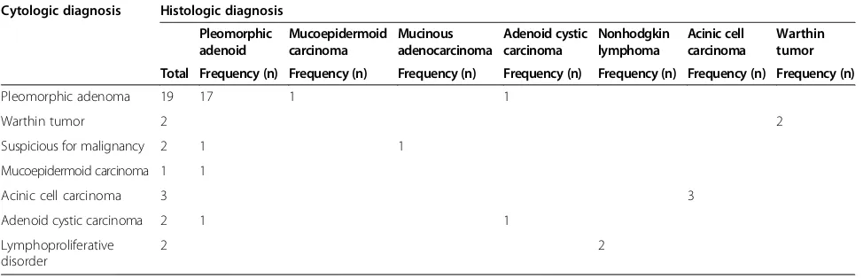
Out of these 187 cases, 31 patients subsequently underwent surgical excision/histologic evaluation, out of which 21 were benign and 9 were malignant, Total 31 patients subsequently underwent surgical excision, out of which 21 were benign and 9 were malignant, 20 cases (64.5%) were of pleomorphic adenoma, 3 cases (9.6%) of acinic cell carcinoma, 2 cases (6.4%) each of warthin tumor, adenoid cystic carcinoma and non-hodgkin lymph-oma and 1 case (3.2%) each of mucoepidermoid carcin-oma and mucinous adenocarcincarcin-oma. Table 5 shows the comparison of cytologic and histologic diagnosis. Overall accuracy of FNAC in our study was found to be 83.8% with 77.7% sensitivity and 86.3%, specificity as shown in Table 6. The revised sensitivity and specificity after adjust-ing verification bias were 68.5% (37.9-99.2) and 91% (82.7-99.3) respectively (Table 7).
Discussion
Swelling of salivary glands, specifically parotid and sub-mandibular gland presents as a common problem and being readily visible creates havoc among patients. In addition parotid/submandibular swellings also remain a
diagnostic challenge among clinicians. FNAC provides a convenient way to obtain a tissue based diagnosis and therefore has now become a diagnostic test of choice to solve this dilemma. Our study explains the role of this procedure in our setup to diagnose salivary gland lesions and the spectrum of disease pathology in our population.
[image:3.595.56.543.101.258.2]Literature review revealed a wide variation in the sen-sitivity and specificity of FNAC for salivary gland swelling in different populations and setups [8-10]. Zerpa et al. studied 93 cases of parotid gland tumors, revealing a sensitivity and specificity of 57% and 95% respectively [11]. On the other hand, Pastore et al. found a sensitivity and specificity of 83% and 93% respectively. They evalu-ated 357 cases of salivary gland lesions [12]. Similarly Jaein et al. revealed 92.8% sensitivity and 93.9% specificity in a study involving 80 cases of salivary gland swellings, out of which 14 cases were of malignant salivary gland neo-plasms [13]. Kim et al. found a diagnostic accuracy of FNAC to be 92% in differentiating malignant from benign salivary gland tumors [14]. Fakhry et al. evaluated 249 par-otid tumors, out of which 75% were benign and 25% were malignant. The sensitivity and specificity to detect malig-nancy was assessed to be 80% and 89.5% respectively. They found 16 false positive results, among which warthin’s tumor and pleomorphic adenoma were most common, while false negative diagnosis were given in cases of lymphomas and mucoepidermoid carcinomas. The diagnostic accuracy for benign and malignant tu-mors was 16% and 44% respectively [15].
Table 5 Comparison of cytologic and histologic diagnosis
Cytologic diagnosis Histologic diagnosis
Pleomorphic adenoid
Mucoepidermoid carcinoma
Mucinous adenocarcinoma
Adenoid cystic carcinoma
Nonhodgkin lymphoma
Acinic cell carcinoma
Warthin tumor
Total Frequency (n) Frequency (n) Frequency (n) Frequency (n) Frequency (n) Frequency (n) Frequency (n)
Pleomorphic adenoma 19 17 1 1
Warthin tumor 2 2
Suspicious for malignancy 2 1 1
Mucoepidermoid carcinoma 1 1
Acinic cell carcinoma 3 3
Adenoid cystic carcinoma 2 1 1
Lymphoproliferative disorder
[image:3.595.56.292.658.733.2]2 2
Table 6 Diagnostic accuracy of FNAC for Salivary gland swelling
Sensitivity 77.7%
Specificity 86.3%
Positive predictive value 70%
Negative predictive value 90.4%
[image:3.595.305.538.680.734.2]Accuracy 83.3%
Table 7 Revised sensitivity and specificity after adjusting verification bias
95% confidence interval
Upper limit Lower limit
Sensitivity 68.5% 37.9% 99.2%
We found an overall diagnostic accuracy of FNAC to be 83.8%. There were two cases of false negative diagno-sis. These two cases were one each of mucoepidermoid carcinoma and acinic cell carcinoma which were initially diagnosed as pleomorphic adenoma on FNAC. Pleo-morphic adenoma is a biphasic neoplasm. The epithelial component may present a variety of histologic patterns including squamous and ductal structures and they may sometimes exhibit significant cytologic atypia. Lack of stromal component in the aspirated material may lead to a false positive diagnosis especially that of low grade mucoepidermoid carcinoma which may show diversity in the morphologic patterns of epithelial components in-cluding squamous, intermediate and mucinous cells. There were 3 cases of false positive diagnosis. In one of that case there were extensive squamous elements without any other component and therefore a diagnosis of neo-plastic lesion, suspicious for malignancy was given with a possibility of metastatic squamous cell carcinoma and mucoepidermoid carcinoma. The final histology revealed the diagnosis of pleomorphic adenoma. The other two cases with false negative diagnosis were that of mucoepi-dermoid and adenoid cystic carcinoma which were inaccurately diagnosed on FNAC as pleomorphic aden-oma. Adenoid cystic carcinoma is a close differential of pleomorphic adenoma and basal cell adenoma. This dif-ferentiation is very important as the surgical management is different. Adenoid cystic carcinoma shows basement membrane like material which may be misinterpreted as stromal component. The epithelial component of adenoid cystic carcinoma is usually very bland leading to inaccur-ate impression of benignancy. Immunohistochemical studies on cell block material may be very helpful in this distinction including stains for basal lamina and CD117 stain which is positive in adenoid cystic carcinoma.
A few studies were also conducted in Pakistan, evalu-ating the role of FNAC in salivary gland pathology. A study conducted in Pakistan including 129 cases of par-otid gland lesions, revealed 98 benign and 31 malignant neoplasms. Pleomorphic adenoma was the most com-mon benign tumor while mucoepidermoid carcinoma being the most common malignant diagnosis [16]. In our study, although pleomorphic adenoma was the most common benign tumor, however the most common ma-lignant tumor was acinic cell carcinoma.
Conclusion
In conclusion, we found a good concordance between FNAC and final histology, however pleomorphic adenoma may impart a diagnostic challenge when inadequately as-pirated and therefore either immunohistochemical studies should be done if cell block material is available or repeat aspiration should be advised before embarking on thera-peutic excision.
Abbreviations
FNAC:Fine needle aspiration cytology; H& E: Hematoxylin and eosin.
Competing interests
The authors declare that they have no competing interests.
Authors’contributions
SN and AAA: main authors of manuscript, have made substantial contributions to design of data. AK: have made substantial contributions to analysis and interpretation of data. NF and MME: Involved in drafting and revision of the manuscript. AK and MK: have been involved in drafting the manuscript. All authors read and approved the final manuscript.
Acknowledgments
We gratefully acknowledge all staff members of Pathology department, Liaquat national hospital and medical college, Karachi, Pakistan for their help and cooperation.
Author details
1Department of Histopathology, Liaquat National Hospital and Medical
College, Karachi, Pakistan.2Liaquat National Hospital and Medical College, Karachi, Pakistan.3Dhaka Medical College, Dhaka, Bangladesh.
Received: 7 September 2014 Accepted: 11 March 2015
References
1. Dudheon LS, Patrick CV. A new method for the rapid microscopical diagnosis of tumors. Br J Surg. 1927;15:250–61.
2. Rajbhandari M, Dhakal P, Shrestha S, Sharma S, Shrestha S, Pokharel M, et al. The correlation between fine needle aspiration cytology histopahology of head and neck lesion in kathmandu university hospital. Kathmandu Univ Med J (KUMJ). 2013;11(44):296–9.
3. Poorey VK, Tyagi A. Accuracy of fine needle aspiation cytology in head and neck masses. Indian J Otolaryngol Head Neck Surg. 2014;66(2):182–6. 4. Diaz KP, Gerhard R, Domingues RB, Martins LL, Prado Ribeiro AC, Lopes MA,
et al. High diagnistic accuracy and reproducibility of fine needle aspiration cytology for diagnosing salivary gland tumors : cytohistologic correlation in 182 cases. Oral Surg Oral Med Oral Pathol Oral Radiol. 2014;118(2):226–35. 5. Huq AH, Aktaruzzaman M, Habib MA, Islam MS, Amin S. A comperative
study between fine needle aspiration cytology findings and histopathological report of major salivary gland neoplasm in a tertiary hospital of bangladesh. Bangladesh Med Res Counc Bull. 2013;39(2):69–73. 6. Kechagias N, Ntomouchtsis A, Valeri R, Patrikidou A, Kitikidou K, Xirou P,
et al. Fine needle aspiration cytology of salivary gland tumors: a 10 years retrospective analysis.”. Oral Maxilofac Surg. 2012;16(1):35–40.
7. Singh Nanda KD, Meht A, Nanda J. Fine needle aspiration cytology : a reliable tool in the diagnosis of salivary gland lesions. J Oral Pathol Med. 2012;41(1):106–12.
8. Murai N, Taniguchi Z, Takahashi Y, Kuboshima F, Tateya I. A study of salivary gland aspiration cytology reporting: guideline validity. Nihon Jibiinkoka Gakkai Kaiho. 2011;114(7):615–9.
9. Piccioni LO, Fabiano B, Gemma M, Sarandria D, Bussi M. Fine needle aspiration cytology in the diagnosis of parotid lesions. Acta Otorhinolaryngol Ital. 2011;31(1):1–4.
10. Singh A, Haritwal A, Murali B. Correlation between cytology and histopathology of th salivary gland. Australas Med J. 2011;4(2):66–71. 11. Zerpa Zerpa V, Cuesta Gonzales MT, Agostini Porras G, Marcano Acuna M,
EstellesFerriol E, Dalmau GJ. Diagnostic accuracy of fine needle aspiration cytology in parotid tumors. Acta Otorrinolaringol Esp. 2014;65(3):157–61. 12. Pastore A, Borin M, Malagutti N, Di Laora A, Becati D, Delazer AL, et al.
Preopertive assisment of salivary gland neoplasm with fine needle aspiration cytology and echography: a retrospective analysis of 357 cases. Int J Immunopathol Pharmacol. 2013;26(4):965–71.
13. Jain R, Gupta R, Kudesia M, Sing S. Fine needle aspiration cytology in the diagnosis of salivary gland lesions: a study with histologic comparison. Cytojournal. 2013;10:5.
15. Fakhry N, Antonini F, Michel J, Penicaud M, Mancini J, Lagier A, et al. Fine needle aspiration cytology in the management of parotid masses: evaluation of 249 patient. Eur Ann Otorhinolaryngol Head Neck Dis. 2012;129(3):131–5.
16. Ali NS, Akhtar S, Junaid M, Awan S, Aftab K. Diagnostic accuracy of fine needle aspiration cytology in parotid lesions. ISRN Surg. 2011;2011:721525.
Submit your next manuscript to BioMed Central and take full advantage of:
• Convenient online submission
• Thorough peer review
• No space constraints or color figure charges
• Immediate publication on acceptance
• Inclusion in PubMed, CAS, Scopus and Google Scholar
• Research which is freely available for redistribution