RESEARCH
Association of the peripheral blood levels
of circulating microRNAs with both recurrent
miscarriage and the outcomes of embryo
transfer in an in vitro fertilization process
Qian Yang
1†, Wen‑Wen Gu
1†, Yan Gu
2, Na‑Na Yan
3, Yan‑Yan Mao
1, Xing‑Xing Zhen
1, Jian‑Mei Wang
2, Jing Yang
3,
Hui‑Juan Shi
1*, Xuan Zhang
1*and Jian Wang
1Abstract
Background: Implantation failure is not only a major cause of early pregnancy loss, but it is also an obstacle to assisted reproductive technologies. The identification of potential circulating biomarkers for recurrent miscarriage (RM) and/or recurrent implantation failure would contribute to the development of novel diagnosis and prediction techniques.
Methods: MiR (miR‑23a‑3p, 27a‑3p, 29a‑3p, 100‑5p, 127‑3p and 486‑5p) expression in the villi, decidual tissues and peripheral blood plasma and serum were validated by qPCR, and the localization of miRs in the villi and decidual tissues of RM and normal pregnancy (NP) women were detected by in situ hybridization. The invasiveness of HTR8/ SVneo cells was determined using a Transwell assay. The predictive values of miRs for RM and the outcome of IVF‑ET were respectively calculated by the receiver operating characteristic analysis.
Results: The signals of six miRs were observed in the villi and decidual tissues of RM and NP women. The villus miR‑ 27a‑3p, miR‑29a‑3p and miR‑100‑5p were significantly up‑regulated, whereas miR‑127‑3p and miR‑486‑5p appeared to be down‑regulated in RM women compared to NP women. The invasiveness of HTR8/SVneo cells transfected with miR‑23a‑3p mimics was evidently weakened, whereas that of cells transfected with miR‑127‑3p mimics was obvi‑ ously enhanced. The peripheral blood plasma levels of miR‑27a‑3p, miR‑29a‑3p, miR‑100‑5p and miR‑127‑3p were significantly increased, whereas that of miR‑486‑5p was remarkably decreased in RM compared to NP women. By contrast, serum miR‑23a‑3p and miR‑127‑3p were significantly decreased, whereas that of miR‑486‑5p was remark‑ ably increased. The combination of six plasma miRs levels discriminated RM with a sensitivity of 100% and a specificity of 83.3%, whereas that of six serum miRs levels showed a sensitivity of 78.3% and a specificity of 93.1%. In the IVF‑ET cohort, the significantly decreased peripheral blood plasma levels of miR‑23a‑3p, miR‑27a‑3p, miR‑100‑5p and miR‑ 127‑3p, and the serum levels of miR‑100‑5p and miR‑486‑5p, in addition to the significantly increased serum level of miR‑27a‑3p, were found to be associated with the failure of ET. Moreover, the combination of plasma miR‑23a‑3p, miR‑27a‑3p, miR‑29a‑3p, miR‑100‑5p, miR‑127‑3p and miR‑486‑5p levels discriminated the outcome of IVF‑ET with a sensitivity of 68.1% and a specificity of 54.1%, whereas the combination of plasma miR‑127‑3p and miR‑486‑5p levels showed a sensitivity of 50.0% and a specificity of 75.3%.
© The Author(s) 2018. This article is distributed under the terms of the Creative Commons Attribution 4.0 International License (http://creativecommons.org/licenses/by/4.0/), which permits unrestricted use, distribution, and reproduction in any medium, provided you give appropriate credit to the original author(s) and the source, provide a link to the Creative Commons license, and indicate if changes were made. The Creative Commons Public Domain Dedication waiver (http://creativecommons.org/ publicdomain/zero/1.0/) applies to the data made available in this article, unless otherwise stated.
Open Access
*Correspondence: shihuijuan2011@163.com; xuanzhang@sippr.org.cn †Qian Yang and Wen‑Wen Gu contributed equally to this work 1 Key Laboratory of Reproduction Regulation of NHFPC, Shanghai Institute of Planned Parenthood Research, Pharmacy School, Fudan University, Shanghai, China
Background
Successful embryo implantation, which is a crucial step for the establishment and maintenance of pregnancy in mammals, requires a harmonized interaction between invaded blastocysts and receptive endometrium [1]. Implantation failure is not only a major cause of early pregnancy loss, but it is also an obstacle to the significant improvement of assisted reproductive technologies [2, 3].
Recurrent miscarriage (RM) can be defined as 2 or more consecutive pregnancy losses prior to the 20th week of gestation in humans. RM has an incidence of 1–2% in pregnant women, and the etiology of 68% of RM cases is unexplained [4]. Because the exact pathogenic mechanisms underlying RM remain unclear, its clinical management still lacks powerful means of diagnosis and prediction and effective therapeutic techniques [5]. The widespread application of assisted reproductive tech-nologies, particularly in vitro fertilization (IVF), has ben-efited numerous infertility patients over the past decades; however, implantation failure after embryo transfer (ET), in particular recurrent implantation failure, has fettered the further advance of the IVF success rate [6, 7]. Thus, the identification and evaluation of potential circulating biomarkers for RM and/or recurrent implantation fail-ure would undoubtedly contribute to the development of novel diagnosis and prediction techniques.
MicroRNAs (miRs) are single-stranded small non-cod-ing RNA sequences of usually 19–25 nucleotides (nts) in length that participate in the post-transcriptional regu-lation of gene expression in a variety of biological and physiological processes [8]. It has been widely reported that miRs are involved in embryo implantation, and the dysfunctions of miRs are associated with implantation failure, resulting in RM and recurrent implantation fail-ure [9, 10]. In particular, because miRs are extracellularly stable and can be detected in human plasma, and because the alteration of such circulating miRs might occur in response to physiological or pathological status [11], they are therefore identified as a new class of non-invasive biomarkers.
Recently, we established RM-related villus miR pro-files by deep-sequencing identification using three pairs of tissue samples collected from RM patients and well-matched normal pregnancy (NP) women. A number of miRs were screened to potentially be differentially expressed in the villus tissues of RM patients; however,
only a small part of them that met our designated crite-ria of P < 0.05 and fold change > 1.5 (or 2.0) were reported, including miR-100-5p and miR-486-5p [12, 13]. By searching the human serum database (http://www.exiqo n.com/ plate-layout-files), we found that in addition to 100-5p and 486-5p, another four miRs, miR-23a-3p, miR-27a-3p, miR-29a-3p and miR-127-3p, which were also screened to be differentially expressed in RM patients with fold changes ranging from 1.1 to 1.5, and P < 0.05 (Additional file 1: Table S1), are presented in human peripheral blood. More interestingly, miR-23a-3p [14], miR-27a-3p [15], miR-29a-3p [16] and miR-127-3p [17] have been reported by other labs to be involved in human pathological pregnancy.
Thus, in the present study, the differential expressions of these six miRs (miR-23a-3p, miR-27a-3p, miR-29a-3p, miR-100-5p, miR-127-3p and miR-486-5p) in the villi of RM patients were validated by real-time quantitative PCR, and their expression localization in the villus and decidual tissues of RM patients and NP women were also determined by in situ hybridization. The effects of their over-expressions on the invasive activities of HTR8/ SVneo were individually assessed by the transfection of their specific mimics in vitro. Finally, their plasma or serum levels in peripheral blood were detected and compared between RM patients and NP women and between pregnant and non-pregnant women undergo-ing IVF treatment, with the aim of identifyundergo-ing circulatundergo-ing miRs biomarkers related to both RM and the outcomes of IVF-ET.
Methods Sample collection
Human villus and decidual tissues and peripheral blood samples of RM patients and NP women were collected from April to August 2017 at the Department of Gyne-cology and Obstetrics, the Second Hospital of Tianjin Medical University, Tianjin, China. The current preg-nancy losses of RM patients were objectively confirmed by transvaginal ultrasound examination. Classical risk factors including abnormal parental karyotypes, uter-ine anatomical abnormalities, infectious diseases, luteal phase defects, diabetes mellitus, thyroid dysfunction, and hyperprolactinemia were excluded. In parallel, NP women, who had no history of miscarriage and were undergoing legal elective abortions, were enrolled and
Conclusions: Circulating miR‑23a‑3p, miR‑27a‑3p, miR‑29a‑3p, miR‑100‑5p, miR‑127‑3p and miR‑486‑5 might be involved in RM pathogenesis and present potential diagnostic biomarkers for RM. Meanwhile, these miRs, in particular miR‑127‑3p and miR‑486‑5p, provide promising prediction indexes for the outcomes of IVF‑ET.
checked for classical risk factors for early pregnancy losses. All samples were collected after informed consent was obtained. Then, the decidual and villus tissues were frozen and stored at − 80 °C. The venous blood plasma and serum samples were freshly prepared and subse-quently stored at − 80 °C.
Peripheral blood samples of IVF-ET women were col-lected from June to July 2017 at the Reproductive Medi-cine Center, Renmin Hospital of Wuhan University, Hubei Province, China. Participants who were under-going IVF with ICSI due to male factor infertility were enrolled, and peripheral blood samples were collected prior to ET on the day of ET after informed consent was obtained. The participants were followed up till the end of the first trimester to observe the outcome of the cur-rent ET treatment.
RNA extraction from villus tissues
Total RNA was extracted respectively from five pairs of villus samples from RM patients and NP women using TRIzol reagent according to the manufacturer’s protocol (Invitrogen, Carlsbad, CA). The concentration of total RNA products was measured by NanoDrop (Thermo Sci-entific, Wilmington, DE), and RNA integrity was checked with a Bioanalyzer2100 (Agilent, Santa Clara, CA).
Real‑time quantitative PCR for MiRs in placental villus tissues
Total RNA products extracted from villus tissues were used to validate the differential expression of miRs between the RM patients and NP women by real-time quantitative PCR analysis. Total RNA was reversely tran-scribed using miRNA specific reverse primers (Ribobio, Guangzhou, China) to obtain cDNA. Real-time PCR was performed using the FastStart Universal SYBR Green Master Kit (Roche Diagnostics, USA) according to the manufacturer’s description and was analyzed using an ABI 7900 HT (Applied Biosystems, Foster, CA). All miRs assay primers were purchased commercially (Ribobio). Primer efficiencies were determined by the standard curve. The relative expression of miRs was calculated by the efficiency-corrected ΔΔCt method and normalized to the endogenous control snRNA U6. Each sample in each group was measured in triplicate, and the experiment was repeated at least three times.
In situ hybridization
Two pairs of villus and decidual samples (RM1 and RM2 in the RM group and NP1 and NP2 in the NP group) were chosen randomly for the in situ hybridization analy-sis. Frozen 10-μm serial sections were rehydrated and fixed in 4% paraformaldehyde for 20 min, treated with protease K for 5 min at room temperature, and followed
by hybridization with LNA microRNA probes (Exiqon, Copenhagen, Denmark) specifically against miRNAs at 55 °C overnight. The sections were sequentially washed in 5×, 2×, and 0.2× saline-sodium citrate buffer. After blocking, the sections were incubated with alkaline phosphatase–conjugated anti-digoxin antibodies (1:200, Roche, Indianapolis, IN) at 4 °C overnight. BCIP/NBT (Promega, Madison, WI) was used as the substrate to vis-ualize the stained signals according to the manufacturer’s instructions. The scramble miRNA (non-specific control) probe was used as the negative control.
After that, the human decidua slides were incubated with HLA-G antibodies (Abcam, 1:800) or IgG antibod-ies at 4 °C overnight to identify which cell type expresses these miRNAs in decidua. Further incubation with horse-radish peroxidase-conjugated secondary antibodies (Vec-tor, 1:500) was visualized with DAB (Dako Cytomation, Glostrup, Denmark) solution containing 0.03% H2O2 as
the substrate. The human villi and decidua slides were counterstained with Nuclear Fast Red (Beijing Dingguo Changsheng Biotechnology, Beijing, China) and mounted with neutral balsam. The probe sequences are presented in Additional file 1: Table S2.
HTR‑8/SVneo cell culture and treatment
The HTR8/SVneo trophoblast cell line was derived from human first-trimester placenta [18]. The cells were kindly provided by Dr. Hongmei Wang, Institute of Zoology, Chinese Academy of Sciences, China, and cultured in RPMI-1640 medium supplemented with fetal bovine serum (FBS; Gibco, Carlsbad, CA), 100 U/ml penicil-lin (Gibco) and 100 µg/ml streptomycin (Gibco) under standard culture conditions (37 °C in a 5% humidified CO2 incubator). The cells used for transfection were
plated 30,000 per well in the 24-well plate (Corning, NY, USA) using 500 μl of medium. The mimics and inhibi-tors of miRs and their negative control (mimic-NC/ inhibitor-NC) were purchased from Ribobio. When the cells reached 70% confluence, they were transiently trans-fected by the mimics (10 µM) and mimic-NC (10 µM), respectively, using Lipofectamine™ 2000 (Thermo scien-tific) according to the manufacturer’s instruction. After 6 h, the cells were washed with serum-free and antibi-otics culture medium and continued being cultured in complete medium (1 ml/well) overnight.
Transwell assay of HTR‑8/SVneo cell invasiveness
inhibitor transfection, the HTR8/SVneo cells were har-vested with serum-free medium, 2 × 10⁵ cells were
seeded into the upper compartment of the prepared inserts, and medium supplemented with 25% FBS (Gibco) was added to the lower compartment to induce migration. After 24 h of incubation at 37 °C with 5% CO2,
the cells remaining inside the inserts were removed using a cotton swab. The membranes were then fixed with 4% paraformaldehyde, stained with 0.1% crystal violet, washed with ddH2O and observed under the microscope.
Then, the cells were lysed with methanol, and the absorb-ance at a wavelength of 560 nm was measured using a UV spectrophotometer for quantification. The experiments in each group were repeated three times under the same conditions.
Blood plasma and serum separation and RNA extraction
Whole venous blood samples used for plasma separa-tion were collected in EDTA-containing tubes and then immediately centrifuged at 1600×g for 10 min to separate the plasma samples. The plasma samples were carefully transferred into new tubes and stored at − 80 °C until use. To prepare the serum samples, the whole blood was col-lected in blood collection tubes without anticoagulants and placed at room temperature (15–25 °C) for 30 min to complete clotting. Then, the blood sample tubes were centrifuged for 10 min at 1900×g (3000 rpm) under 4 °C. The yellow upper serum phase was carefully transferred to a new tube without disturbing the intermediate buffy coat layer (containing white blood cells and platelets) and kept frozen in aliquots at − 80 °C. Total RNA was extracted from the plasma or serum using a miRNeasy Serum/Plasma Kit (Qiagen, Suzhou, China) following the manufacturer’s instructions. The quantity and qual-ity of obtained miRNA was measured with a NanoDrop ND-1000 Spectrophotometer (Thermo scientific).
Real‑time quantitative PCR for MiRs in plasma and serum
Total RNAs extracted from plasma and serum were reversely transcribed using miRNA specific reverse primer (Ribobio) to obtain cDNA. Real-time PCR was performed using the FastStart Universal SYBR Green Master (Roche Diagnostics, Basel, Switzerland) accord-ing to the manufacturer’s description and analyzed usaccord-ing an ABI 7900 HT (Applied Biosystems). All miRs assay primers used in this study were purchased commer-cially (Ribobio). Primer efficiencies were determined by a standard curve. The relative miRNAs’ expressions were calculated by the efficiency-corrected ΔΔCt method and normalized to the exogenous control cel-miR-39-3p [19]. Each sample was analyzed in duplicate, and the mean was used to determine the miRNA levels.
Statistical analysis
All continuous parametric values were presented as the mean ± SEM, as determined from at least three independent experiments. Statistical significance was assessed using a one-way ANOVA. P < 0.05 was consid-ered statistically significant. The statistical analysis was conducted using SPSS 19.0 software (SPSS Software, Chi-cago, IL, USA).
Because some of the six miRNA expressions were related to the clinical pregnancy outcomes of IVF-ET and RM pathogenesis, we analyzed the ability of the com-bination of the six miRNA expressions in plasma and serum to predict the clinical pregnancy outcome for IVF-ET and potential diagnostic biomarkers for RM using receiver operating characteristic (ROC) curves and cal-culating the AUC with the 95% confidence intervals after a logistic regression. The sensitivity and specificity of the optimal cut-off were calculated. The statistical tests were performed using Stata 15.0 (StataCorp, TX, USA). The results were considered significant when P < 0.05.
Results
Clinical characteristics of enrolled participants
In total, 16 RM patients who had experienced at least two consecutive spontaneous early miscarriages before the 12th gestational week were recruited, and informa-tion about their personal history of thromboembolic dis-eases and previous early pregnancy losses were inquired. Meanwhile, 29 matched NP women were concurrently enrolled. No significant differences in the average age and gestational week at sampling were observed between RM patients and NP women (Table 1).
[image:4.595.305.540.647.724.2]For the IVF-ET cohort, a total of 146 women under-going IVF-ET were enrolled. By the end of the first trimester, 72 women were diagnosed by imaging with ultrasonography as clinically pregnant (Pregnancy), whereas another 74 women were diagnosed as non-pregnant after the ET (Failure). Fifty women in the Pregnancy group and 53 participants in the Failure group have never received the ET treatment before,
Table 1 Clinical characteristics of the recruited recurrent miscarriage (RM) patients (n = 16) and normal pregnant (NP) women (n = 29)
Clinical characteristics RM (mean ± SD) NP (mean ± SD) P value
Age (years) 29.9 ± 0.84 28.0 ± 0.92 0.208
and 22 women in the Pregnancy group and 21 partici-pants in the Failure group had received ET treatment at least once. No significant differences in the average age, number of transferred embryos, infertility duration, BMI, or peripheral blood concentrations of FSH and LH were observed between the Pregnancy and Failure groups (Table 2).
Validation of differential villus miR expressions
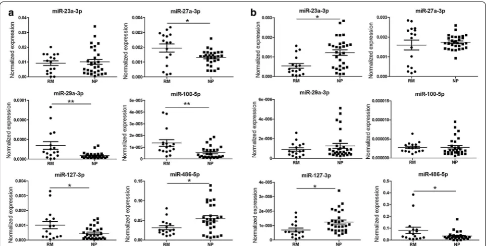
In our previous study, the differential villus expression of miR-100-5p in RM patients was validated by qPCR [13]. MiR-23a-3p, 27a-3p, 29a-3p and miR-127-3p were also screened out by deep-sequencing, but they were not listed as RM-related differentially villus expressed miRs in our published data because their fold changes were less than 2.0 (Additional file 1: Table S1). In this study, we validated the differential villus expression of 23a-3p, 27a-3p, 29a-3p, 127-3p and 486-5p and that of miR-100-5p (as a positive control) by qPCR. As expected, the villus expressions of miR-27a-3p, miR-29a-3p and miR-100-5p were significantly up-regulated in RM patients, and the villus expression of miR-23a-3p in RM patients also showed an increasing tendency, whereas the expressions of miR-127-3p and miR-486-5p were
decreased in RM patients, but no significant differences were observed (Fig. 1a).
Villus and decidual expression localization of miRs
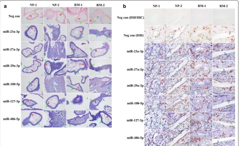
In situ hybridization analyses were performed to local-ize the expressions of miRs using 2 paired samples of RM patients (RM1 and RM2) and NP women (NP1 and NP2) (Additional file 1: Table S3). HLA-G protein signals were detected as the biomarker of extravillous tropho-blast cells by an immunohistochemistry analysis to con-firm whether the decidual tissues were from implantation sites. The results showed that positive signals of these miRs were detected in the villi of early pregnancy and were widely localized in cytotrophoblast cells, syncy-tiotrophoblasts and column cytotrophoblasts (Fig. 2a). In decidual tissues, the positive miRNA signals were observed not only in maternal decidual stromal cells but also in embryonic interstitial trophoblast cells and endo-vascular trophoblast cells (Fig. 2b), indicating that the expressions of these miRs were not villus tissue-specific at the maternal–fetal interface during early pregnancy.
Effects of miRs on HTR8/SVneo cell invasion
The results of the Transwell assay showed that the migrated cell number of miR-23a-3p mimic-trans-fected HTR8/SVneo cells was significantly reduced, whereas that of miR-127-3p mimic-transfected cells was
Table 2 Clinical characteristics of the recruited women undergoing IVF-ET treatment History of embryo
transfer treatment Clinical characteristics Outcome of embryo transfer P value
Failure Pregnancy
n Mean ± SD n Mean ± SD
1st time Age (years) 53 33.8 ± 0.73 50 31.0 ± 0.57 0.0040
Number of transferred embryo 1.98 ± 0.6312 1.98 ± 0.3488 0.9788
Infertility duration (years) 5.74 ± 0.69 4.89 ± 0.50 0.3234
BMI (kg/m2) 22.1 ± 0.48 21.9 ± 0.32 0.7683
FSH (mIU/ml) 7.49 ± 0.43 8.14 ± 0.43 0.2831
LH (mIU/ml) 3.85 ± 0.32 4.37 ± 0.44 0.3418
2nd–4th time Age (years) 21 31.2 ± 0.77 22 31.6 ± 1.15 0.8275
Number of transferred embryo 1.95 ± 0.47 1.86 ± 0.99 0.4334
Infertility duration (years) 3.95 ± 0.63 5.91 ± 0.82 0.0670
BMI (kg/m2) 21.4 ± 0.51 22.4 ± 0.50 0.1875
FSH (mIU/ml) 6.74 ± 0.41 6.45 ± 0.38 0.6063
LH (mIU/ml) 3.92 ± 0.68 5.32 ± 0.78 0.1864
Total Age (years) 74 31.0 ± 0.59 72 32.5 ± 0.58 0.6326
Number of transferred embryo 1.99 ± 0.45 1.93 ± 0.46 0.3360
Infertility duration (years) 4.93 ± 0.43 5.41 ± 0.54 0.4898
BMI (kg/m2) 22.0 ± 0.29 22.0 ± 0.36 0.9880
FSH (mIU/ml) 7.50 ± 0.37 7.50 ± 0.31 0.9936
[image:5.595.56.546.453.726.2]significantly increased compared to the corresponding control treated cells (Fig. 1b, c). This result indicates an inhibitory effect of miR-23a-3p and a stimulating effect of miR-127-3p on the cell invasiveness in vitro. However, there was no significant difference in the migrated cell number of miRNA inhibitor-transfected HTR8/SVneo cells (Additional file 1: Fig. S1).
Differential levels of miRs in the peripheral blood of RM patients
Peripheral blood samples were obtained from 16 RM patients and 29 NP women (Table 1), and the results of the qPCR analysis showed that compared to NP women, the plasma levels of miR-27a-3p, miR-29a-3p, miR-100-5p and miR-127-3p were significantly ele-vated, whereas the level of miR-486-5p was remarkably decreased in RM patients, but no significant differences in plasma miR-23a-3p levels were reported (Fig. 3a). In serum, the levels of miR-23a-3p and miR-127-3p were decreased, whereas the level of miR-486-5p was elevated in RM patients compared to NP women; however, no sig-nificant differences in the serum levels of miR-27a-3p, miR-29a-3p and miR-100-5p were detected (Fig. 3b).
Predictive value of miRs for the potential diagnostic biomarkers of RM
To appraise the predictive value of these circulating miRs as potential diagnostic biomarkers of RM, the lev-els of miRs (23a-3p, 27a-3p, 29a-3p, miR-100-5p, miR-127-3p and miR-486-5p) in peripheral blood plasma or serum were calculated using logistic models. The ROC analysis for miRs in plasma indicated that the performance of Model 1 (combination of all six miRs) and Model 2 (combination of only 127a-3p and miR-486-5p) for predicting RM reached 0.96 (0.92–1.00) and 0.90 (0.81–0.99), respectively. Model 1 has a sensitivity of 100% and a specificity of 83.3% with 89.6% correctly clas-sified, whereas Model 2 has a sensitivity of 88.9% and a specificity of 80.0% with 83.3% correctly classified. These preliminary results suggest that the plasma levels of miR-127a-3p and miR-486-5p could serve as predictive factors for RM (Table 3 and Fig. 4a).
The ROC analysis for miRs in serum indicated that the performance of Model 1 (combination of all six miRs) and Model 2 (combination of miR-23a-3p, miR-29a-3p and miR-486-5p) for predicting RM reached 0.87 (0.76– 0.98) and 0.85 (0.72–0.98), respectively. Model l has a sensitivity of 78.3% and a specificity of 93.1% with 86.5%
[image:6.595.59.540.86.337.2]correctly classified, whereas Model 2 has a sensitivity of 82.1% and a specificity of 89.7% with 86.5% correctly clas-sified. This indicated that the serum levels of miR-23a-3p, miR-29a-3p and miR-486-5p could also serve as predic-tive factors for RM (Table 4 and Fig. 4b).
Association of the peripheral blood levels of miRs with the outcomes of IVF‑ET
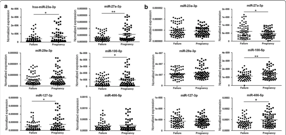
To preliminarily evaluate the potential of these circu-lating miRs to be used as clinical indexes to predict the outcome of IVF-ET, the plasma and serum levels of miR-23a-3p, miR-27a-3p, miR-29a-3p, miR-100-5p, miR-127-3p and miR-486-5p were compared between 72 pregnant and 74 non-pregnant women after ET treatment during an IVF cycle. The results showed that compared to pregnant women, the plasma levels of miR-23a-3p, miR-27a-3p, miR-100-5p and miR-127-3p were significantly decreased in non-pregnant women (Fig. 5a), and the serum level of miR-27a-3p was significantly ele-vated, whereas the levels of miR-100-5p and miR-486-5p were remarkably decreased in non-pregnant women
(Fig. 5b). We also attempted to take the history of ET treatment into account; however, there was no signifi-cant difference in the plasma (Additional file 1: Figs. S2A, S3A) or serum (Additional file 1: Figs. S2B, S3B) levels of any miRs.
Predictive value of miRs for the outcome of IVF‑ET
To appraise the predictive value of these circulating miRs for the clinical outcome of IVF-ET, the levels of miRs (23a-3p, 27a-3p, 29a-3p, miR-100-5p, miR-127-3p and miR-486-5p) in peripheral blood plasma or serum were calculated using logistic models. The plasma levels of 127a-3p and miR-486-5p were shown to significantly predict the outcome of IVF-ET; however, in serum, none of the selected miRs could significantly predict the outcome (Addi-tional file 1: Table S4). The ROC analysis for miRs in plasma indicated that the performance of both Model 1 (combination of all six miRs) and Model 2 (combina-tion of miR-127a-3p and miR-486-5p) for predicting clinical pregnancy reached 0.62 (0.53–0.71). Model 1
[image:7.595.57.540.88.382.2]has a sensitivity of 68.1% and a specificity of 54.1% with 61% correctly classified, whereas Model 2 has a sensi-tivity of 50% and a specificity of 75.3% with 62.3% cor-rectly classified (Table 5 and Additional file 1: Fig. S4A). The ROC curve analysis for miRNAs in serum indi-cated that the performance of the combination of the six miRs for predicting clinical pregnancy reached 0.53 (0.44–0.61) with a sensitivity of 27.8% and a specificity of 85.1% (Additional file 1: Table S5, Fig. S4B).
Furthermore, we took the history of IVF-ET treat-ment into account to evaluate the prediction for clini-cal pregnancy outcomes of IVF-ET by the six miRNAs in plasma and serum after fitting logistic models. When adjusting for the number of transplantation treatments, the ROC curve analysis for miRNAs in plasma indicated that the performance of the full model (combination of all six miRNAs) and the reduced model (combination of miR-127a-3p and miR-486-5p) for predicting clinical
Fig. 3 Comparison of the relative levels of miR‑23a‑3p, miR‑27a‑3p, miR‑29a‑3p, miR‑100‑5p, miR‑127‑3p and miR‑486‑5p in plasma (a) and serum (b) of peripheral blood between RM patients and NP controls. *Significantly different from control (P < 0.05); **significantly different from control (P < 0.01). NP normal pregnant women, RM recurrent miscarriage patients
Table 3 Receiver operating characteristic analysis in potential diagnostic biomarkers of RM in plasma
* No significant difference in ROC area between Model 1 and Model 2
a Model 1, Cut-off value = 0.213, (miR-23a-3p, miR-27a-3p, miR-29a-3p, miR-100-5p, miR-127a-3p, miR-486-5p) (6.312687, 6.228964, 13.70655, 13.10668, 9.521886, 4.800646)
b Model 2, Cut-off value = 0.336, (miR-100-5p, miR-486-5p) (13.27899, 4.726676)
Roc index Prediction for clinical pregnancy outcome
Model 1a (combination of the six miRNAs) Model 2b (combination of miR‑127a‑3p and miR‑ 486‑5p)
AuROC (95% CI)* 0.96 (0.92, 1.00) 0.90 (0.81, 0.99)
Sensitivity (%) 100 88.9
Specificity (%) 83.3 80.0
Positive predictive value (%) 78.3 72.7
Negative predictive value (%) 100 92.3
Correctly classified (%) 89.6 83.3
[image:8.595.57.540.87.331.2] [image:8.595.67.540.402.541.2]pregnancy reached 0.61 (0.52–0.71). The full model has a sensitivity of 68.1% and a specificity of 51.4% with 59.9% correctly classified, and the reduced model has a sensitiv-ity of 50% and a specificsensitiv-ity of 70% with 59.9% correctly classified (Additional file 1: Table S6).
The ROC curve analysis for miRNAs in serum indi-cated that the performance of the combination of all six miRNAs for predicting clinical pregnancy reached 0.52 (0.42–0.61) with a sensitivity of 12.5% and a specificity of 97.1% (Additional file 1: Table S7). No expression level of miRNAs was shown to be able to significantly pre-dict the clinical pregnancy outcome in either plasma or
serum when stratified according to a history of IVF-ET treatment.
Discussion
The present exploratory study validated the differen-tial villus expressions of 23a-3p, 27a-3p, miR-29a-3p, miR-100-5p, miR-127-3p and miR-486-5p in RM patients compared to NP women and localized their expression in the villus and decidual tissues of early pregnancy. The expression profiles of these miRs were also investigated in the peripheral blood of RM patients and women undergoing the IVF-ET procedure, and the
Fig. 4 Predictive value assessment by ROC analysis in the plasma and serum of RM patients. a ROC curve of miRs for peripheral blood plasma. Curves of Model 1 (combination of all 6 miRs: miR‑23a‑3p, miR‑27a‑3p, miR‑29a‑3p, miR‑100‑5p, miR‑127‑3p and miR‑486‑5p) and Model 2 (combination of 2 miRs: miR‑127a‑3p and miR‑486‑5p) for peripheral blood plasma; b ROC curves of miRs (combined 6 miRs) for peripheral blood serum. Curves of Model 1 (combination of all 6 miRs: miR‑23a‑3p, miR‑27a‑3p, miR‑29a‑3p, miR‑100‑5p, miR‑127‑3p and miR‑486‑5p) and Model 2 (combination of 3 miRs: miR‑23a‑3p, miR‑29a‑3p and miR‑486‑5p) for peripheral blood serum
Table 4 Receiver operating characteristic analysis in potential diagnostic biomarkers of RM in serum
* No significant difference in ROC area between Model 1 and Model 2
a Model 1, Cut-off value = 0.396, (miR-23a-3p, miR-27a-3p, miR-29a-3p, miR-100-5p, miR-127a-3p, miR-486-5p) (8.986689, 6.043169, 14.62186, 13.73859, 12.26344, 6.515767)
b Model 2, Cut-off value = 0.382, (miR-23a-3p, miR-29a-3p, miR-48s5p) (5.41586, 14.80947, 2.419319)
Roc index Prediction for clinical pregnancy outcome
Model 1a (combination of the six miRNAs) Model 2b (combination of miR‑ 23a‑3p, miR‑29a‑3p and miR‑ 486‑5p)
AuROC (95% CI)* 0.87 (0.76, 0.98) 0.85 (0.72, 0.98)
Sensitivity (%) 78.3 82.1
Specificity (%) 93.1 89.7
Positive predictive value (%) 90.0 86.4
Negative predictive value (%) 84.4 86.7
Correctly classified (%) 86.5 86.5
[image:9.595.59.540.89.249.2] [image:9.595.57.547.350.493.2]predictive value of these miRs for the outcome of IVF-ET was calculated preliminarily.
We demonstrated that, consistent with the previous results of deep-sequencing analysis, the villus expres-sion levels of miR-27a-3p, miR-29a-3p and miR-100-5p were significantly up-regulated in RM patients com-pared to NP women, and an up-regulation trend of miR-23a-3p expression, in addition to down-regulated
trends of miR-127-3p and miR-486-5p expressions, were observed, although no significant difference was counted. The expressions of these six miRs were localized both in maternal DSCs and fetal trophoblast cells.
miR-23a has been shown to promote trophoblast cell apoptosis and its expression level is up-regulated in the placenta of PE patients [20], but the role of miR-23a-3p in RM patients has not yet been revealed. Given that
Fig. 5 Comparison of the relative levels of miR‑23a‑3p, miR‑27a‑3p, miR‑29a‑3p, miR‑100‑5p, miR‑127‑3p and miR‑486‑5p in the plasma (a) and serum (b) of peripheral blood between pregnant women (n = 72) and non‑pregnant women (n = 74) after embryo transfer in IVF‑ET cycle. *Significantly different from control (P < 0.05); **significantly different from control (P < 0.01). Failure, non‑pregnant women after ET treatment; Pregnancy, pregnant women after ET treatment
Table 5 Receiver operating characteristic analysis in prediction for outcome of IVF-ET by the selected miRs in peripheral blood plasma
* No significant difference in ROC area between Model 1 and Model 2
a Model 1: Cut-off value = 0.469; 23a-3p, 22.112194); 27a-3p, 28.546087); 29a-3p, 23.40597); 100-5p, 28.019193); 127-3p, 27.498337); (miR-486-5p, 12.804071)
b Model 2: Cut-off value = 0.533; (miR-127-3p, 28.653163); (miR-486-5p, 14.214297)
Roc index Prediction for clinical pregnancy outcome
Model 1a (combination of the six miRs) Model 2b (combination of miR‑127a‑3p and miR‑ 486‑5p)
AuROC (95% CI)* 0.62 (0.53, 0.71) 0.62 (0.53, 0.71)
Sensitivity (%) 68.1 50.0
Specificity (%) 54.1 75.3
Positive predictive value (%) 59.0 65.5
Negative predictive value (%) 63.5 60.4
Correctly classified (%) 61.0 62.3
[image:10.595.57.539.88.316.2] [image:10.595.58.540.415.555.2]miR-23-3p inhibits type II collagen expression in chon-drocytes [21] and that the increased expression of type II collagen can promote cell migration [22], miR-23a-3p might suppress trophoblast cell invasion. Coinciden-tally, the inhibitory effect of miR-23a-3p over-expres-sion on HTR8/SVneo cell invasiveness was observed in this study.
The association between miR-27a polymorphisms and RM was recently identified [23]. Here, a significantly increased villus expression of miR-27a-3p in RM patients was also validated. However, although miR-27a-3p over-expression promotes the migration and invasion of hepatocellular carcinoma [24] and nasopharyngeal car-cinoma cells [25], no obvious effect of miR-27a-3p over-expression on the invasiveness of HTR8/SVneo cells was observed here. Very recently, it was reported that miR-27a-3p over-expression in human granulosa-like tumor cells inhibits cell proliferation and promotes cell apopto-sis [26]. Thus, ectopic miR-27a-3p in placental villi might dysregulate the proliferation and apoptosis of trophoblast cells, resulting in pregnancy loss.
The expression of miR-29a at the placental creta sites is evidently lower compared to non-creta sites, and its over-expression induces HTR8/SVneo cell apoptosis [27]. Thus, it is reasonable for us to speculate that the signifi-cantly increased expression of miR-29a-3p in the pla-centa villi of RM patients observed in this study might be involved in the RM pathogenesis by leading to excessive trophoblast cell apoptosis during early pregnancy.
The role of miR-100-5p in reproduction remains mostly unknown, but the stimulating effect of miR-100-5p over-expression on neuronal cell apoptosis was reported [28]. Herewith, the villus miR-100-5p expression was detected to be significantly up-regulated in RM patients; thus, we supposed that similar to miR-29-3p, the dysregulation of miR-100-5p expression might also disturb trophoblast cell apoptosis, resulting in early pregnancy loss.
MiR-127-3p was reported to consistently induce the migration and invasion of glioblastoma cells [29]; this study showed that HTR8/SVneo cell invasive activity was effectively promoted by miR-127-3p over-expression. The decreasing trend of its villus expression in RM patients suggested that it might be involved in RM pathogenesis by reducing trophoblast cell invasion.
MiR-486-5p was another miRNA that showed a decreasing trend of villus expression in RM patients, and its lower expression level in the cumulus cells of poly-cystic ovary syndrome patients was also reported [30]. In lung cancer cells, miR-486-5p over-expression could inhibit cell proliferation and migration, but it induced cell apoptosis [31], indicating that insufficient functions of miR-486-5p might lead to implantation failure by the dysregulation of trophoblast cell activities.
We also observed the effect of these miRNA inhibitors on cell invasive activities, but no significant differences were identified. Because there are several important requirements for an miRNA inhibitor to achieve effective downregulation of a targeted miRNA, such as stability, specificity and affinity [32, 33], further studies must be performed using synthetic inhibitors with high efficacy or transfection with lentiviral plasmid vector.
Given that circulating miRs could reflect pathologi-cal status and be detectable in peripheral blood [34] and that several differentially expressed miRs in the placenta of complicated pregnancies could be detected in mater-nal peripheral blood [35], we speculated that these six miRs might also represent various peripheral blood levels between RM patients and NP women. In addi-tion, although both blood plasma and serum are com-monly used to detect the circulating miRs, it has been suggested that serum is better than plasma for use in research because hemolysis can influence the circulating miR abundance [36]; we thus determined the plasma and serum levels of six detected miRs in this study.
Encouragingly, the results showed that compared to NP women, the plasma levels of miR-27a-3p, miR-29a-3p, miR-100-5p and miR-127-3p in RM patients were sig-nificantly increased, whereas the plasma miR-486-5p level was remarkably decreased. By contrast, the serum levels of miR-23a-3p and miR-127-3p were significantly lower, whereas the serum miR-486-5p level was remark-ably higher. The higher plasma levels of miR-27a-3p, miR-29a-3p and miR-100-5p, and the lower level of miR-486-5p, were in consistent with their differential vil-lus expression levels. An opposite relationship between plasma levels and serum levels of 23a-3p, miR-127-3p and miR-486-5p was observed, and we thought this might be caused by different resources of these miRs.
difference in the types of miRNAs between the plasma and serum may be indicative of the difference in the con-fined location of miRNAs in peripheral blood. The poten-tial of circulating miRNAs as blood-based biomarkers for RM is promising. Importantly, it was expected that combining multiple miRNAs into the miRNA profile may provide greater accuracy than can be expected from the assessment of a single miRNA [37].
Furthermore, given that implantation failure after IVF-ET treatment has been a limiting factor for the improve-ment of the IVF success rate and that miR-29a has been explored as a potential predictive biomarker for out-comes of the IVF process [38], we also observed an asso-ciation between the peripheral blood levels of these six miRs and the outcomes of embryo transfer in an IVF cycle. The distinctly lower plasma levels of miR-23a-3p, miR-27a-3p, miR-100-5p and miR-127-3p, and the serum levels of miR-100-5p and miR-486-5p, in addition to the remarkably higher serum level of miR-27a-3p, were observed to be correlated with the failure of ET, showing an almost inverse change compared to the RM cohort. This could be explained by the different sampling time. Samples from the RM cohort were collected after the implantation failure, of which fetal factors were included, whereas samples of the IVF-ET cohort were collect prior to embryo transfer and lacked fetal factors. Alterations in peripheral blood levels of these six circulating miRs are summarized in Table 6.
In particular, the combination of plasma levels of these six miRs discriminated the outcome of IVF-ET with a sensitivity of 68.1% and a specificity of 54.1%, whereas the combination of plasma miR-127-3p and miR-486-5p
levels showed a slightly lower sensitivity (50.0%) but a notable higher specificity (75.3%), providing potential biomarkers to efficiently predict the outcomes of IVF-ET treatment. When the IVF-ET participants were further sub-grouped according to the history of embryo trans-fer treatment and the prediction for clinical pregnancy outcomes of IVF-ET by the six miRNAs in plasma and serum was determined after fitting logistic models, no miRNA expression levels were able to predict the clini-cal pregnancy outcome significantly in both plasma and serum. This might be due to the limited number of IVF-ET patients when sub-grouped according to the his-tory of embryo transfer treatment. We should increase the case number to strength our conclusion in future investigations.
Conclusions
This exploratory study suggested that peripheral blood levels of circulating 23a-3p, 27a-3p, miR-29a-3p, miR-100-5p, miR-127-3p and miR-486-5 were associated with both RM and the outcomes of embryo transfer to varying degrees, thereby presenting potential diagnostic or predictive biomarkers for RM and the out-comes of IVF-ET treatment. Further validation of these preliminary findings might lead to the development of novel non-invasive diagnostic and predictive means for the improvement of clinical infertility management. There were several limitations in this study. The low abundance of these circulating miRs, inadequate sample sizes, and potentially insufficient utilization of available data should influence the confidence level of this study, calling for a large-scale multicenter trial in the future.
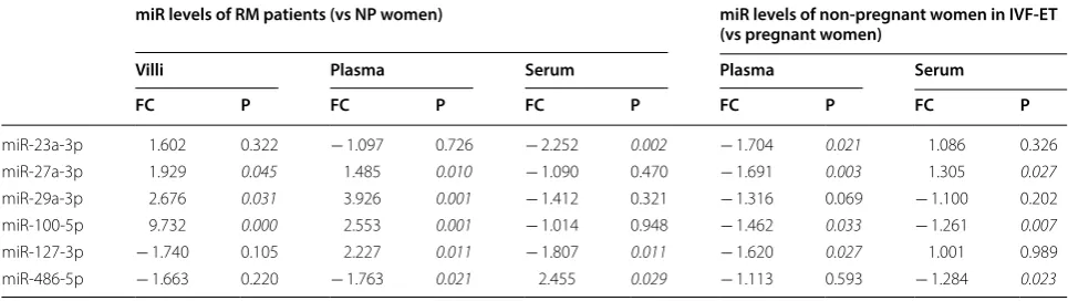
Table 6 Alterations in peripheral blood levels of miRs
In RM and NP group: Positive fold change (FC) means up-regulated in RM patients and negative fold change means down regulated in RM patients compared to NP women. In IVF-ET group: Positive fold change (FC) means up-regulated in pregnant women in IVF-ET and negative fold change means down regulated in non-pregnant women in IVF-ET compared to non-pregnant women. Results were considered significant when P < 0.05
miR levels of RM patients (vs NP women) miR levels of non‑pregnant women in IVF‑ET (vs pregnant women)
Villi Plasma Serum Plasma Serum
FC P FC P FC P FC P FC P
miR‑23a‑3p 1.602 0.322 − 1.097 0.726 − 2.252 0.002 − 1.704 0.021 1.086 0.326
miR‑27a‑3p 1.929 0.045 1.485 0.010 − 1.090 0.470 − 1.691 0.003 1.305 0.027
miR‑29a‑3p 2.676 0.031 3.926 0.001 − 1.412 0.321 − 1.316 0.069 − 1.100 0.202
miR‑100‑5p 9.732 0.000 2.553 0.001 − 1.014 0.948 − 1.462 0.033 − 1.261 0.007
miR‑127‑3p − 1.740 0.105 2.227 0.011 − 1.807 0.011 − 1.620 0.027 1.001 0.989
[image:12.595.56.539.514.650.2]Abbreviations
RM: recurrent miscarriage; MicroRNAs: miRs; IVF‑ET: in vitro fertilization‑ embryo transfer.
Authors’ contributions
QY and WWG contributed to the experiment and data analysis. YG, NNY, JMW and JY contributed to the sample collection and clinical diagnosis. XXZ per‑ formed the PCR analysis. YYM was involved in the data analysis. JW, HJS and XZ were involved in the study design and drafting the paper. All authors read and approved the final manuscript.
Author details
1 Key Laboratory of Reproduction Regulation of NHFPC, Shanghai Institute of Planned Parenthood Research, Pharmacy School, Fudan University, Shang‑ hai, China. 2 The Second Hospital of Tianjin Medical University, Tianjin, China. 3 Renmin Hospital of Wuhan University, Wuhan, China.
Acknowledgements
The authors thank American Journal Experts in the language editing of this manuscript.
Competing interests
The authors declare that they have no competing interests.
Availability of data and materials
The datasets used and/or analyzed during the current study are available from the corresponding author on reasonable request.
Consent for publication
Not applicable.
Ethics approval and consent to participate
All experiments involving humans were approved by the Medical Ethics Com‑ mittees of The Second Hospital of Tianjin Medical University (KY2017K002), Renmin Hospital of Wuhan University (WDRY2016‑K017), and the Shanghai Institute of Planned Parenthood Research (Ref # 2013‑7, 2013‑12), and we have also obtained consents to publish the research data derived from these collected samples from the recruited subjects.
Funding
This study was support by Grants from the Natural Science Foundation of China (No. 81701445, 81571513) and the Tianjin Municipal Science & Technol‑ ogy Committee (No. 17JCYBJC26400).
Publisher’s Note
Springer Nature remains neutral with regard to jurisdictional claims in pub‑ lished maps and institutional affiliations.
Received: 8 February 2018 Accepted: 23 June 2018
References
1. Cha J, Sun X, Dey SK. Mechanisms of implantation: strategies for success‑ ful pregnancy. Nat Med. 2012;18:1754–67.
2. Evans J, Salamonsen LA, Winship A, Menkhorst E, Nie G, Gargett CE, Dimi‑ triadis E. Fertile ground: human endometrial programming and lessons in health and disease. Nat Rev Endocrinol. 2016;12:654–67.
3. Norwitz ER, Schust DJ, Fisher SJ. Implantation and the survival of early pregnancy. N Engl J Med. 2001;345:1400–8.
Additional file
Additional file 1. Additional figures and tables.
4. Vaiman D. Genetic regulation of recurrent spontaneous abortion in humans. Biomed J. 2015;38:11–24.
5. Brezina PR, Kutteh WH. Classic and cutting‑edge strategies for the management of early pregnancy loss. Obstet Gynecol Clin North Am. 2014;41:1–18.
6. Ledee N, Petitbarat M, Chevrier L, Vitoux D, Vezmar K, Rahmati M, Dubanchet S, Gahery H, Bensussan A, Chaouat G. The uterine immune profile may help women with repeated unexplained embryo implantation failure after in vitro fertilization. Am J Reprod Immunol. 2016;75:388–401.
7. Guo F, Zhou M, Zhang A. Advances in the treatment of recurrent implan‑ tation failure. Reprod Dev Med. 2017;1:123–6.
8. Bartel DP. MicroRNAs: genomics, biogenesis, mechanism, and function. Cell. 2004;116:281–97.
9. Galliano D, Pellicer A. MicroRNA and implantation. Fertil Steril. 2014;101:1531–44.
10. Liang J, Wang S, Wang Z. Role of microRNAs in embryo implantation. Reprod Biol Endocrinol. 2017;15:90.
11. Mitchell PS, Parkin RK, Kroh EM, Fritz BR, Wyman SK, Pogosova‑Agadjan‑ yan EL, Peterson A, Noteboom J, O’Briant KC, Allen A, et al. Circulating microRNAs as stable blood‑based markers for cancer detection. Proc Natl Acad Sci USA. 2008;105:10513–8.
12. Gu Y, Zhang X, Yang Q, Wang J, He Y, Sun Z, Zhang H, Wang J. Aberrant placental villus expression of miR‑486‑3p and miR‑3074‑5p in recurrent miscarriage patients and uterine expression of these microRNAs during early pregnancy in mice. Gynecol Obstet Invest. 2015;81:112–7. 13. Wang JM, Gu Y, Zhang Y, Yang Q, Zhang X, Yin L, Wang J. Deep‑
sequencing identification of differentially expressed miRNAs in decidua and villus of recurrent miscarriage patients. Arch Gynecol Obstet. 2016;293:1125–35.
14. Hromadnikova I, Kotlabova K, Hympanova L, Krofta L. Gestational hypertension, preeclampsia and intrauterine growth restriction induce dysregulation of cardiovascular and cerebrovascular disease associ‑ ated microRNAs in maternal whole peripheral blood. Thromb Res. 2016;137:126–40.
15. Yang S, Li H, Ge Q, Guo L, Chen F. Deregulated microRNA species in the plasma and placenta of patients with preeclampsia. Mol Med Rep. 2015;12:527–34.
16. Jairajpuri DS, Malalla ZH, Mahmood N, Almawi WY. Circulating micro‑ RNA expression as predictor of preeclampsia and its severity. Gene. 2017;627:543–8.
17. Rodosthenous RS, Burris HH, Sanders AP, Just AC, Dereix AE, Svensson K, Solano M, Tellez‑Rojo MM, Wright RO, Baccarelli AA. Second trimester extracellular microRNAs in maternal blood and fetal growth: an explora‑ tory study. Epigenetics. 2017;12:804–10.
18. Graham CH, Hawley TS, Hawley RG, MacDougall JR, Kerbel RS, Khoo N, Lala PK. Establishment and characterization of first trimester human trophoblast cells with extended lifespan. Exp Cell Res. 1993;206:204–11. 19. Mooren FC, Viereck J, Kruger K, Thum T. Circulating microRNAs as
potential biomarkers of aerobic exercise capacity. Am J Physiol Heart Circ Physiol. 2014;306:H557–63.
20. Li L, Hou A, Gao X, Zhang J, Zhang L, Wang J, Li H, Song Y. Lentivirus‑ mediated miR‑23a overexpression induces trophoblast cell apoptosis through inhibiting X‑linked inhibitor of apoptosis. Biomed Pharmacother. 2017;94:412–7.
21. Kang L, Yang C, Song Y, Liu W, Wang K, Li S, Zhang Y. MicroRNA‑23a‑3p promotes the development of osteoarthritis by directly targeting SMAD3 in chondrocytes. Biochem Biophys Res Commun. 2016;478:467–73. 22. Clarke CJ, Berg TJ, Birch J, Ennis D, Mitchell L, Cloix C, Campbell A, Sump‑
ton D, Nixon C, Campbell K, et al. The initiator methionine tRNA drives secretion of type II collagen from stromal fibroblasts to promote tumor growth and angiogenesis. Curr Biol. 2016;26:755–65.
23. Rah H, Chung KW, Ko KH, Kim ES, Kim JO, Sakong JH, Kim JH, Lee WS, Kim NK. miR‑27a and miR‑449b polymorphisms associated with a risk of idiopathic recurrent pregnancy loss. PLoS ONE. 2017;12:e0177160. 24. Li JM, Zhou J, Xu Z, Huang HJ, Chen MJ, Ji JS. MicroRNA‑27a‑3p inhibits
cell viability and migration through down‑regulating DUSP16 in hepato‑ cellular carcinoma. J Cell Biochem. 2017;119:5143–52.
•fast, convenient online submission •
thorough peer review by experienced researchers in your field • rapid publication on acceptance
• support for research data, including large and complex data types •
gold Open Access which fosters wider collaboration and increased citations maximum visibility for your research: over 100M website views per year •
At BMC, research is always in progress.
Learn more biomedcentral.com/submissions
Ready to submit your research? Choose BMC and benefit from: 26. Wang M, Sun J, Xu B, Chrusciel M, Gao J, Bazert M, Stelmaszewska J, Xu
Y, Zhang H, Pawelczyk L, et al. Functional characterization of MicroRNA‑ 27a‑3p expression in human polycystic ovary syndrome. Endocrinology. 2018;159:297–309.
27. Gu Y, Bian Y, Xu X, Wang X, Zuo C, Meng J, Li H, Zhao S, Ning Y, Cao Y, et al. Downregulation of miR‑29a/b/c in placenta accreta inhibits apoptosis of implantation site intermediate trophoblast cells by targeting MCL1. Placenta. 2016;48:13–9.
28. Ye X, Luo H, Chen Y, Wu Q, Xiong Y, Zhu J, Diao Y, Wu Z, Miao J, Wan J. MicroRNAs 99b‑5p/100‑5p regulated by endoplasmic reticulum stress are involved in abeta‑induced pathologies. Front Aging Neurosci. 2015;7:210. 29. Jiang H, Hua D, Zhang J, Lan Q, Huang Q, Yoon JG, Han X, Li L, Foltz G,
Zheng S, Lin B. MicroRNA‑127‑3p promotes glioblastoma cell migration and invasion by targeting the tumor‑suppressor gene SEPT7. Oncol Rep. 2014;31:2261–9.
30. Shi L, Liu S, Zhao W, Shi J. miR‑483‑5p and miR‑486‑5p are down‑ regulated in cumulus cells of metaphase II oocytes from women with polycystic ovary syndrome. Reprod Biomed Online. 2015;31:565–72. 31. Yi Y, Lu X, Chen J, Jiao C, Zhong J, Song Z, Yu X, Lin B. Downregulated
miR‑486‑5p acts as a tumor suppressor in esophageal squamous cell carcinoma. Exp Ther Med. 2016;12:3411–6.
32. Stenvang J, Kauppinen S. MicroRNAs as targets for antisense‑based therapeutics. Expert Opin Biol Ther. 2008;8:59–81.
33. Robertson B, Dalby AB, Karpilow J, Khvorova A, Leake D, Vermeulen A. Specificity and functionality of microRNA inhibitors. Silence. 2010;1:10. 34. Chen X, Ba Y, Ma L, Cai X, Yin Y, Wang K, Guo J, Zhang Y, Chen J, Guo X,
et al. Characterization of microRNAs in serum: a novel class of biomarkers for diagnosis of cancer and other diseases. Cell Res. 2008;18:997–1006. 35. Jiang H, Wen Y, Hu L, Miao T, Zhang M, Dong J. Serum microRNAs as
diagnostic biomarkers for macrosomia. Reprod Sci. 2015;22:664–71. 36. Hamam R, Hamam D, Alsaleh KA, Kassem M, Zaher W, Alfayez M,
Aldahmash A, Alajez NM. Circulating microRNAs in breast cancer: novel diagnostic and prognostic biomarkers. Cell Death Dis. 2017;8:e3045. 37. Creemers EE, Tijsen AJ, Pinto YM. Circulating microRNAs: novel biomark‑
ers and extracellular communicators in cardiovascular disease? Circ Res. 2012;110:483–95.