Int. J. Electrochem. Sci., 7 (2012) 5565 - 5573
International Journal of
ELECTROCHEMICAL
SCIENCE
www.electrochemsci.org
Size controlled synthesis of uniform Li
2MnSiO
4nanospheres
and their electrochemical behaviors in lithium-ion batteries
Xiangzhi Kong1, Tao Mei1, Zheng Xing1, Na Li1, Zhengqiu Yuan1, Yongchun Zhu1,* and Yitai Qian1,2,* 1
Hefei National Laboratory for Physical Science at Microscale and Department of Chemistry, University of Science and Technology of China, Hefei, Anhui 230026, P.R. China
2
School of Chemistry and Chemical Engineering, Shandong University, Jinan 250100, P.R. China *
E-mail: ytqian@ustc.edu.cn
Received: 16 April 2012 / Accepted: 14 May 2012 / Published: 1 June 2012
The uniform Li2MnSiO4 nanospheres with diameters of ~500 and 300 nm have been selectively prepared via monodisperse spherical SiO2 precursors with corresponding sizes by solid state method. Furthermore, Li2MnSiO4/C composites were obtained by annealing Li2MnSiO4 nanospheres with glucose at 400 °C. Electrochemical measurements show that spherical Li2MnSiO4/C with a diameter of ~300 nm exhibits first discharge capacity of 145 mAh g-1, together with a stable discharge capacity of 121 mAh g-1 after 50 cycles, which are higher than that with a diameter of ~500 nm.
Keywords: Li2MnSiO4; Nannospheres; SiO2; Size control; Lithium-ion batteries
1. INTRODUCTION
The orthosilicates, Li2MSiO4 (where M = Mn2+, Fe2+, Co2+, Ni2+) [1-8], have been recognized as one of the most promising candidates in lithium-ion batteries (LIBs) due to its low cost, high safety, structure stability, and environmental friendliness. The most attractive feature is that Li2MSiO4 have a high theoretical capacity of 333 mAh g-1 by the insertion/extraction of two lithium ions per formula unit.
nanosized particles (100-200 nm). The as-obtained products presented a discharge capacity of 98 mAh g-1 after 15 cycles under 10 mA g-1 [13]. Aravindan et al. prepared Li2MnSiO4 by solid state method using SiO2 as silicon source, and irregular particulate morphology with some sort of aggregation was observed. When the carbon contents were 42 and 12%, discharge capacities of 140 and 25 mAh g-1 after 40 cycles at 0.05 C were obtained, respectively [14].
As we known that the electrochemical performance of materials depends significantly on the particle size, so it is important to control the particle size and study its effects on cell performance [15]. While some groups have reported the synthesis of nanocrystalline Li2MnSiO4, size controlled synthesis of Li2MnSiO4 and investigation of the relationship between electrochemical performance and the particle size have been less carried out due to the difficulties in controlling the particle size. In particular, tailoring the particle morphology and size in solid state synthesis is more complicated than other methods because of the experiment being conducted at high temperature, which may usually results in irregular particulate morphology and strongly aggregation.
Herein, we have devised a size controlled synthesis of uniform Li2MnSiO4 nanospheres by solid state method, where monodisperse SiO2 spheres with different sizes were used as precursors. Li2MnSiO4 nanospheres with diameters of ~500 and 300 nm have been prepared successfully. Li2MnSiO4 nanospheres were coated by carbon layers using glucose as carbon source to improve the low inherent electronic conductivity. The influence of particles size on electrochemical performance of Li2MnSiO4/C has been studied, which shows that spherical Li2MnSiO4/C with a diameter of ~300 nm displays higher reversible capacity and better cycle stability than that with a diameter of ~500 nm.
2. EXPERIMENTAL
All the chemical reagents used here were analytical grade, and were used without further purification.
2.1 Synthesis of SiO2 spheres
Monodisperse SiO2 solid spheres with diameters of ~500 and 300 nm were performed by a slightly modified Stöber process [16,17].
Synthesis of ~500 nm SiO2 spheres: 4.5 mL of TEOS was rapidly added into the mixture of 9 mL of ammonium hydroxide (28%), 24.75 mL of distilled water and 61.75 mL of absolute ethanol under stirring and kept for 3 h. The white precipitates were centrifuged and washed with absolute ethanol several times. The products were dried in a vacuum at 60 °C for 3 h.
2.2 Synthesis of Li2MnSiO4 and Li2MnSiO4/C nanospheres
Stoichiometric amount of LiOH•H2O, SiO2 (as-prepared), MnCO3 were fined ground using mortar and pestle and fired at 400 °C for 4 h in air. Then the intermediate products were ground and then calcined at 700 °C for 12 h in flowing argon (5wt%, H2). The as-prepared samples were labeled as Sample 1 (~500 nm in diameter) and Sample 2 (~300 nm in diameter) respectively.
The as-prepared Sample 1 and Sample 2 were mixed with 40wt% glucose and carbonized at 400 °C for 4 h under Ar (5wt%, H2) flow to obtain Li2MnSiO4/C composites and were labeled as Sample 3 and Sample 4, respectively.
2.3 Materials characterization
ray powder diffraction (XRD) patterns of the products were recorded on a Philips X’pert X-ray diffractometer with Cu Kα radiation (λ=1.54182 Å). The microstructure was observed with a field-emitting scanning electron microscope (SEM, JEOL-JSM-6700F), a transmission electron microscope (TEM, H7650), and a high-resolution transmission electron microscope (HRTEM, JEOL-2010) with an accelerating voltage of 200 kV. Raman spectrums were carried out on a JY LABRAM-HR confocal laser micro-Raman spectrometer using Ar+ laser excitation with a wavelength of 514.5 nm. X-ray Photoelectronic Spectrum measurements (XPS) were performed by using a VGESCA-LABMKIIX-ray photoelectronic spectrometer. Elemental analyses were carried out on a vario EL-Ⅲ elemental analyzer (Germany).
2.4 Electrochemical measurements
Charge/discharge tests were carried out using CR 2016 coin-type cells. The active material, acetylene black and polyvinylidene fluoride (PVDF) with a weight ratio of 70:20:10 were mixed homogeneously with N-methyl-pyrrolidone, the obtained slurry was pasted on Al foil and dried at 100 °C for 12 h in vacuum. The electrode sheet typically had an active material of ~1 mg cm−2. The coin cell was then assembled in an argon-filled glove box (Mikrouna, Super 1220/750/900, China) and was consist of Li2MnSiO4 (cathode), Celgard 2400 (separator), and lithium foil (anode). 1 mol L-1 solution of LiPF6 dissolved in ethylene carbonate/dimethyl carbonate (EC/DMC) (1:1 volume ratio) was used as the electrolyte. Galvanostatic charge/discharge measurements were performed in a potential range of 1.5-4.8 V at room temperature on a LAND-CT2001A instrument.
3. RESULTS AND DISCUSSION
[image:4.596.106.490.134.398.2]
5.3794 Å and c = 5.0009 Å, which are in good agreement with values published by previous literatures [9]. Additional peaks are observed in the diffractograms, which have been identified as MnO.
Figure 1. XRD patterns of as-prepared Li2MnSiO4 (Sample 1, 2) and Li2MnSiO4/C (Sample3, 4).
[image:4.596.119.477.462.737.2]
Obtaining a phase pure Li2MnSiO4 material by solid state method is still a challenge, the impurities phases like Li2SiO3, MnO or Mn2SiO4 are also present in previous reports [6,11]. XRD patterns of Sample 3 and Sample 4 indicate that there are no peaks for carbon which could attribute to the amorphous nature or low quantity of carbon. The carbon contents in Sample 3 and Sample 4 were determined to be about 15.18 and 14.96wt% respectively by elementary analysis.
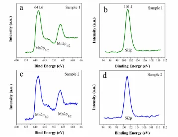
The Mn 2p and Si 2p XPS spectra of Sample 1 and Sample 2 are shown in Fig. 2. The binding energy of Mn 2p3/2 (641.6 eV) is consistent with that of Mn2+, indicating that the divalent state of manganese in our samples. The binding energy of Si 2p (101.1 eV) is in line with that of Si4+ in polysiloxane, indicating the formation of the orthosilicate structure [SiO4] [18].
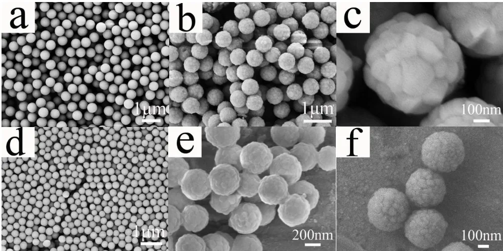
Figure 3. Typical SEM images of (b, c) Sample 1, (e, f) Sample 2 and corresponding monodisperse spherical SiO2 precursors with a size of about (a) 500 nm, (d) 300 nm.
[image:5.596.49.544.249.496.2]
Figure 4. TEM images of (a) Sample 3, (b) Sample 4, and (c) HRTEM images of Sample 3.
Li2MnSiO4/C composites were observed by TEM and HRTEM. Fig. 4a, b show the TEM images of Sample 3 and Sample 4, in which the core-shell structure could be observed. Li2MnSiO4 nanospheres are uniformly coated by carbon layers. The HRTEM image of Sample 3 (inset of Fig. 4c) further confirm that the structure are constructed by Li2MnSiO4 core and carbon shell. The strong contrast between the light edge and dark center of the composite provide evidence of its core-shell structure. Crystal lattice stripes of Sample 3 (Fig. 4c) are observed with d-spacing of 5.4 Å, which corresponds to the (010) plane of orthorhombic Li2MnSiO4 crystals.
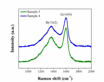
Figure 5. Raman spectra of Sample 3 and Sample 4.
[image:6.596.119.471.389.666.2]
respectively. The D band (1362 cm-1) comes from disordered carbon, while the G band (1600 cm-1) is related to the graphite. The ID/IG ratios of Sample 3 and Sample 4 are found to be 1.87 and 1.79 respectively, demonstrating that a mass of the coated carbon is fairly amorphous. The Raman datas agree with the HRTEM images and XRD patterns.
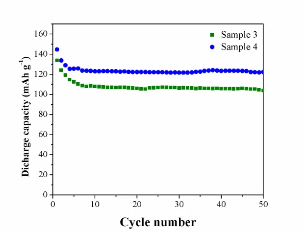
Figure 6. The cycling performances of Sample 3 and Sample 4 measured at 0.05 C in room temperature.
The electrochemical performances of as-prepared Li2MnSiO4/C nanospheres were investigated. Fig. 6c shows the discharge capacities and cyclic performances of Sample 3 and Sample 4 at 0.05 C in room temperature. In the voltage range of 1.5-4.8 V, the initial discharge capacities of the samples are 134 and 145 mAh g−1 respectively. The capacities fade fast at first, and keep stable after 5 cycles. After 50 cycles, the capacities retentions of the samples are 99 and 121 mAh g−1, corresponding to 74 and 83% of those in the initial cycle respectively, which demonstrate stable cycle behavior. This result suggests that Li2MnSiO4/C nanospheres with a diameter of ~300 nm display higher discharge capacity and better cycle stability than that with a diameter of ~500 nm. The reasons could attribute to the materials with smaller size providing short pathways for quick lithium ion, electronic conduction within the nanoparticles and larger surface area for contracting with electrolyte, which can improve the rate capability and cycling stability of materials [12].
[image:7.596.143.454.159.396.2]
conclude that the Li2MnSiO4 electrode material which consist of large spherical agglomerates of nanosized primary particles may exhibit both enhanced electrochemical performance and volume energy density [21]. Further studies on optimizing the size and spherical morphology of Li2MnSiO4 are in progress.
4. CONCLUSIONS
Monodisperse spherical SiO2 precursors with different sizes were used to control the size of Li2MnSiO4. Li2MnSiO4 nanospheres with diameters of ~500 and 300 nm have been successfully synthesized by a solid state method. SEM images exhibit that Li2MnSiO4 approximately retains the uniform spheral morphology and size of corresponding SiO2 precursor. Li2MnSiO4 nanospheres were annealed with glucose at 400 °C to prepare the Li2MnSiO4/C composites. Electrochemical measurements show that Li2MnSiO4/C nanospheres with a diameter of ~300 nm display higher discharge capacity and better cycle stability than that with a diameter of ~500 nm.
In this paper, we provide an effective method to prepare Li2MnSiO4 products with regular spherical morphology and different particle sizes. It is notable that various morphologies and sizes of SiO2 have been studied in a mass of reports [16,22,23]. Li2MnSiO4 with more morphologies and sizes can be expected, such as hollow spheres and nanowires et al.
ACKNOWLEDGEMENTS
This work was financially supported by the National Natural Science Fund of China (No. 91022033), the 973 Project of China (No. 2011CB935901)
References
1. A. Nyten, A. Abouimrane, M. Armand, T. Gustafsson and J. O. Thomas, Electrochem. Commun., 7 (2005) 156-160
2. R. Dominko, M. Bele, A. Kokalj, M. Gaberscek and J. Jamnik, J. Power Sources, 174 (2007) 457-461
3. C. Lyness, B. Delobel, A. R. Armstrong and P. G. Bruce, Chem. Commun., (2007) 4890-4892 4. Z. L. Gong, Y. X. Li, G. N. He, J. Li and Y. Yang, Electrochem. Solid St., 11 (2008) A60-A63 5. N. Kuganathan and M. S. Islam, Chem. Mater., 21 (2009) 5196-5202
6. W. G. Liu, Y. H. Xu and R. Yang, J. Alloy Compd., 480 (2009) L1-L4
7. C. Sirisopanaporn, C. Masquelier, P. G. Bruce, A. R. Armstrong and R. Dominko, J. Am. Chem. Soc., 133 (2011) 1263-1265
8. K. D. M. Dinesh Rangappa, Nano. Lett., 12 (2012) 1146-1151
9. R. Dominko, M. Bele, M. Gaberscek, A. Meden, M. Remskar and J. Jamnik, Electrochem. Commun., 8 (2006) 217-222
10. C. Deng, S. Zhang, B. L. Fu, S. Y. Yang and L. Ma, Mater. Chem. Phys., 120 (2010) 14-17 11. K. Karthikeyan, V. Aravindan, S. B. Lee, I. C. Jang, H. H. Lim, G. J. Park, M. Yoshio and Y. S.
Lee, J. Power Sources, 195 (2010) 3761-3764
14. V. Aravindan, K. Karthikeyan, K. S. Kang, W. S. Yoon, W. S. Kim and Y. S. Lee, J. Mater. Chem., 21 (2011) 2470-2475
15. S. H. Choi, J. W. Son, Y. S. Yoon and J. Kim, J. Power Sources, 158 (2006) 1419-1424 16. T. Zhang, Q. Zhang, J. Ge, J. Goebl, M. Sun, Y. Yan, Y. Liu, C. Chang, J. Guo and Y. Yin, J.
Phys. Chem. C, 113 (2009) 3168-3175
17. Q. Yu, P. Wang, S. Hu, J. Hui, J. Zhuang and X. Wang, Langmuir, 27 (2011) 7185-7191 18. Y. X. Li, Z. L. Gong and Y. Yang, J. Power Sources, 174 (2007) 528-532
19. J. Liu, T. E. Conry, X. Y. Song, L. Yang, M. M. Doeff and T. J. Richardson, J. Mater. Chem., 21 (2011) 9984-9987
20. J. Ying, C. Jiang and C. Wan, J. Power Sources, 129 (2004) 264-269 21. X. Xiao, J. Lu and Y. Li, Nano Research, 3 (2010) 733-737
22. W. Stöber, A. Fink and E. Bohn, J. Colloid Interf. Sci., 26 (1968) 62-69
23. N. I. Kovtyukhova, T. E. Mallouk and T. S. Mayer, Adv. Mater., 15 (2003) 780-785
![The Synthesis and Electrochemical Property of Spinel Cathode Material Li1.05[(Mn1.99Gd0.01)0.91(Mn1.8Co0.2)0.09]O4 for Lithium- Ion Batteries](data:image/gif;base64,R0lGODlhAQABAIAAAP///wAAACH5BAEAAAAALAAAAAABAAEAAAICRAEAOw==)