This is a repository copy of TMPyP functionalised chitosan membrane for efficient sunlight driven water disinfection.
White Rose Research Online URL for this paper: http://eprints.whiterose.ac.uk/120518/
Version: Accepted Version Article:
Majiya, H, Chowdhury, KF, Stonehouse, NJ orcid.org/0000-0003-1146-5519 et al. (1 more author) (2019) TMPyP functionalised chitosan membrane for efficient sunlight driven water disinfection. Journal of Water Process Engineering, 30. 100475. ISSN 2214-7144
https://doi.org/10.1016/j.jwpe.2017.08.013
Crown Copyright © 2017 Published by Elsevier Ltd. This manuscript version is made available under the CC-BY-NC-ND 4.0 license
http://creativecommons.org/licenses/by-nc-nd/4.0/
[email protected] https://eprints.whiterose.ac.uk/
Reuse
This article is distributed under the terms of the Creative Commons Attribution-NonCommercial-NoDerivs (CC BY-NC-ND) licence. This licence only allows you to download this work and share it with others as long as you credit the authors, but you can’t change the article in any way or use it commercially. More
information and the full terms of the licence here: https://creativecommons.org/licenses/
Takedown
If you consider content in White Rose Research Online to be in breach of UK law, please notify us by
1
TMPyP functionalised chitosan membrane for
efficient sunlight driven water disinfection
Hussaini Majiyaa, Kaniz F Chowdhurya, Nicola Stonehouseb and Paul Millnera#
aSchool of Biomedical Sciences, University of Leeds, UK
bSchool of Molecular and Cellular Biology, University of Leeds, UK
# address correspondence to Paul Millner, [email protected]
Key Words: photodynamic inactivation, singlet oxygen, TMPyP, water disinfection, MS2
Abstract word count: 237 words
Importance word count:
2 Abstract
Sunlight-driven water disinfection system could help provide clean water to some of the
I chitosan membranes were modified by pyromellitic
dianhydride in order to introduce carboxyl groups and facilitate adsorption of the highly basic
photosensitiser - 5, 10, 15, 20-tetrakis (1-methyl-4-pyridinio) porphyrin tetra p-toluene
sulfonate (TMPyP). The physico-chemical properties of these modified membranes were
investigated by microscopy, absorption spectroscopy and Midland surface blotting
approaches. The chitosan membrane-TMPyP composite showed photodynamic inactivation
of bacteriophage MS2 and E. coli BL21. For photodynamic inactivation under stationary
conditions, complete inactivation of MS2 was observed after 90 min illumination at a light
intensity of 32 mW cm-2 (which equates to around 3% of bright mid-day time sunlight under
clear sky conditions in sub-Saharan Africa). For a flowing system, complete inactivation of
MS2 was observed for sample flowing at 0.33 ml/min and passed over the surface of the
modified membranes while being illuminated. Reduction of approximately 3 log PFU/ml were
observed for samples passed just once over the membranes under the same conditions. With
E. coli, under the same stationary conditions, a reduction of 3 logCFU/ml was observed. Each
TMPyP functionalised chitosan membrane was used at least three times for the
photodynamic inactivation of both MS2 and E. coli BL21 without any detectable loss of
inactivation capacity. The re-usability of the membranes will reduce cost and increase the
3
1. Introduction
Conventional methods of waste water treatment and disinfection are costly and complex
because of intensive use of chemicals, manpower and energy, as well as the centralised
nature of their infrastructures and operations. As such, it is not affordable in most instances
in rural areas of developing countries (Loeb et al., 2016). Water-borne pathogens such as
Cryptosporidium parvum, cysts of Entamoeba hystolytica, Giardia lamblia and enteric viruses
are resistant to chlorination which is presently the gold standard in water disinfection during
wastewater treatment (Shannon et al., 2008, Silverman et al., 2013). Another major setback
of using chlorine as disinfectant is that this can react with organic compounds and nitrite in
wastewater, resulting in the formation of mutagenic and carcinogenic disinfection by
products (DBPs) such as trihalomethanes, and haloacetic acids (Shannon et al., 2008).
Previous work has shown that photosensitiser such as 5, 10, 15, 20-tetrakis-
(1-methyl-4-pyridinio) porphyrin tetra p-toluene sulfonate (TMPyP) has been shown to result
in rapid photodynamic inactivation (PDI) of the model virus, bacteriophage MS2 in
solution(Casteel et al., 2004). This effect exploits a known phenomenon called the
photodynamic effect which results from the interaction of three factors comprising a
photosensitizer, light and oxygen. This interaction generates singlet oxygen and other
reactive oxygen species (ROS) that can oxidise and cause irreversible damage to proteins,
lipids, nucleic acid and other cellular components of microorganisms and ultimately inactivate
these (Alves et al., 2013, Baumler and Maisch, 2012, Carvalho et al., 2007, Costa et al., 2012,
Komagoe et al., 2011, Maisch et al., 2012b, Spannberger et al., 2012, Costa et al., 2010,
Wainwright, 2004). Attaching TMPyP onto a solid support such as a chitosan membrane will
4
TMPyP into the water after treatment. Also, TMPyP-functionalised chitosan membranes
(CM-T) could be re-used, reducing the cost and increasing the advantage of using an
environmental-friendly technology for water disinfection. Here, chitosan membranes were
chosen as a solid support for attaching TMPyP for water disinfection because of their
properties such as easy fabrication, presence of numerous reactive groups, rigid
D-glucosamine structure, and lack of toxic reactions. Chitosan is also biodegradable, cheap and
readily available (Crini and Badot, 2008, Amornchai et al., 2004, Krajewska et al., 1990, Martel
et al., 2001, Meebungpraw et al., 2015).
The potential approach for water disinfection by CM-T could be by singlet oxygen/ROS
generated in water by PDI driven by natural sunlight. ROS, including singlet oxygen undergoes
rapid reactions with various classes of biomolecules but especially with proteins and
unsaturated lipids. Singlet oxygen readily reacts with protein side chains with a bimolecular
rate constant ranging from 105 to 109 M-1 S-1. This is much higher than with other biomolecules
including RNA, where the rate constant ranges from 104 to 106 M-1 S-1 (Cho et al., 2010, Davies,
2003). Si -amino acids, principally the aromatic and sulphur containing
amino acid residues, unsaturated lipids and nucleic acids are most likely the targets of PDI in
microorganisms. These constituents are associated mainly with cellular and subcellular
membranes such as plasma, mitochondrial, lysosomal and nuclear membranes in bacterial
and eukaryotic cells, as well as the lipid envelopes, host attachment proteins, and other capsid
proteins in viruses. However, little is known about the exact mechanism of PDI, especially in
viruses because of diversity of the viral components. Gram positive bacteria are easily killed
as compared to gram negative bacteria due to differences in the structure of their cell
membrane (Alves et al., 2013, Bourre et al., 2010, Carvalho et al., 2007, Costa et al., 2012,
5
membrane apart from the cytoplasmic (inner) membrane, giving them extra protection
against antimicrobial agents including singlet oxygen and other ROS produced during
photosensitisation. For viruses, although protein photo-oxidation by singlet oxygen has been
extensively studied and the most sensitive viral components are protein in nature, it is an
over-generalisation to assume a particular mechanism of protein photo-oxidation for viral
PDI. This is because apart from diversity of PDI targets within viruses, the reaction of singlet
oxygen with proteins can produce one or more of effects such as oxidation of side chains,
peptide backbone fragmentation, dimerisation/aggregation, unfolding or conformal changes,
enzymatic inactivation and alterations in cellular handling and turnover (Gracanin et al., 2009,
Gracanin et al., 2007).
The objective of this work was to develop a sunlight-driven water disinfection system which
is simple, cheap, re-usable, efficient and environmental friendly that could be used in rural
areas of developing countries and or in developed countries in order to save energy. In this
study, chitosan membranes were first modified by pyromellitic dianhydride in order to
introduce carboxyl groups and facilitate electrostatic adsorption of the highly basic
photosensitiser, TMPyP. The chitosan membrane-TMPyP composite produced was then used
for photodynamic inactivation of MS2 and E. coli BL21. Both stationary and flowing water
models were employed during PDI experiments in order to mimic what is obtainable in waste
6
2. Materials and methods
2.1 Light source and conditions for PDI
The light source for PDI experiments was a Schott KL 2500 LCD (Schott Ltd., Stafford, UK)
which provides a cool white light. Fluence rate of illumination during photoinactivation
experiments were measured using a light meter (Clas Ohlson, UK).
Stationary and flow models were adopted for PDI using TMPyP functionalised chitosan
membrane (CM-T). Visible light was used and fluence rates (radiant exposure) were 32 mW
cm-2. The buffer used for PDI was 1 x Phosphate buffer saline (PBS) (10 mM PO
4 , 137 mM
NaCl, and 2.7 mM KCl) at pH 7.4, 20 - 22 oC under aerobic conditions.
2.2 Photosensitisers and other consumables
TMPyP, chitosan, pyromelitic dianhydride, succinic anhydride (Figure 1) and other
consumables used were purchased without further purification from Sigma Aldrich unless
7
Figure 1: Chemical structures of TMPyP, chitosan, pyromelitic dianhydride and succinic anhydride. Prepared by using ChemDraw Pro 13.0.
2.3 Bacteriophage MS2 phage and E. coli strains
The MS2 phage ATCC 15597-B1 and E.coli host cell ATCC 15597 stocks were a kind gift from
Prof. P.G Stockley, University of Leeds. BL21 E. coli were from standard laboratory stock.
2.4 Propagation and purification of bacteriophage MS2
One litre of exponential phase growth culture of E. coli (ATCC 15597) was infected with 300
µl of MS2 at 109 PFU/ml and incubated at 37 oC with shaking at 150 rpm for 48 h until E. coli
cells were completely lysed. The lysate was then centrifuged at 4,000 x g rpm for 30 min to
removed bacterial cell debris. The supernatant was precipitated using 50% (v/v) saturated
ammonium sulphate overnight at 4 oC. After precipitation of the supernatant, the sample
was centrifuged at 4,000 x g rpm for 30 min. Supernatant was discarded and the pellet
[image:8.595.76.523.72.367.2]8
x g rpm for 30 min. Supernatant was taken and the pellet discarded. Phage particles in the
supernatant were pelleted through 30% w/v sucrose cushion by ultracentrifugation at 32,000
rpm for 3 h using a Beckmann SW 32 Ti rotor. After ultracentrifugation, the supernatant was
discarded and the pellet re-suspended in 800 µl PBS overnight. Re-suspended pellet was
purified through 15% - 45% w/v sucrose gradient by ultracentrifugation at 50,000 rpm for 50
min using a Beckmann SW 55 Ti rotor. The gradient was then fractionated from top to bottom
and fractions were analysed using SDS PAGE to identify the MS2 peak.
2.5 Infectivity and titre determination of bacteriophage MS2
The double layer agar plaque assay was used to determine the infectivity and titre of MS2
according to standard methods (Kropinski et al., 2009). Double layer agar plaque assays were
undertaken (in duplicate) before and after PDI to determine titre after purification and the
extent of PDI.
2.6 Production of chitosan membrane
Chitosan membranes were produced as described by Krajewska (1990, 1992). A solution of
1% (w/v) chitosan in 1% (v/v) acetic acid was made and then homogenised by shaking at 300
rpm and 60 oC for 30 min. The homogenised viscous chitosan solution was then cast into 4.5
cm diameter petri dishes, filling to depth of 3 mm. The petri dishes were placed in a hot air
incubator set at 60 oC for 24 h to dry into a translucent chitosan membrane. The thickness of
membranes after drying was about 50- . The chitosan membranes were then
neutralised by treatment with 0.5% (w/w) of sodium triphosphate in 2M NaOH for 30 min.
After neutralisation, the membranes were washed in distilled H2O (dH2O) until the solution
9
2.7 Modification of chitosan membrane using pyromelitic dianhydride
Each membrane was immersed in 1% (w/v) pyromellitic dianhydride (PMA) in DMSO
contained in a closed bottle and then placed on a 3D rocking platform set at 30 rpm for 24 h
at room temperature Control samples i.e. with PMA replaced with succinic anhydride were
also used. The membranes were then washed in dH2O several times and blow dried. The
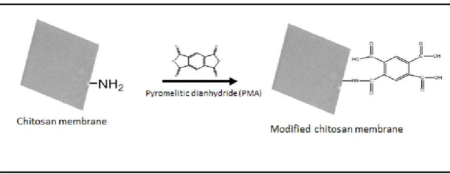
reaction scheme of PMA with chitosan amines is shown in Figure 2.
Figure 2: Reaction scheme for modification of chitosan membrane in order to introduce carboxyl groups and facilitate adsorption of the highly basic photosensitiser-TMPyP.
2.8 Midland blotting of PMA modified chitosan membrane
To detect free amine groups, PMA modified membranes and non-modified controls were
incubated in the presence of NHS-biotin (4 mg/ml in PBS containing 20% (v/v) DMSO) for 30
min in order to attach biotin to the free amine groups. After three washes in dH2O followed
by drying in argon, the membranes were then incubated with HRP- ml in
PBS) for 30 minutes. Then, the membranes were incubated in the presence of an appropriate
conjugated secondary antibody (1:1000 in PBS) for 1 hour to detect bound
HRP-streptavidin. After the addition of the HRP-conjugated reagent, membranes were washed
[image:10.595.77.522.243.415.2]10
removal of non-specifically bound HRP-conjugated secondary antibody, with a final wash in
PBS. The membranes were dried in argon in between incubations and after washing steps.
Finally, ECL reagent was pipetted carefully onto the membranes and chemiluminescence
detected after 1 minute using a G-BOX Gel Imaging System.
2.9 Functionalising PMA modified chitosan membrane with TMPyP
Modified membranes and non-modified membranes controls were stained in the dark with
200 µM of TMPyP placed on 3D rocking platform set at 30 rpm at room temperature. After
staining, the membranes were thoroughly washed in dH2O under mechanical agitation until
zero absorbance from TMPyP was seen in the washed solution. Absorbance measurements
at 421 nm (peak absorption of TMPyP) of reaction solution before and after immobilisation
and wash solutions were determined. The amount bound of TMPyP per cm2 of membranewill
be the difference between the concentrations (µg/ml) of TMPyP before immobilisation and
concentrations (µg/ml) of TMPyP contained in reaction solution and wash solutions after
immobilisation.
2.10 PDI of bacteriophage MS2 using TMPyP functionalised chitosan membrane
PDI of MS2 was investigated using TMPyP-functionalised chitosan membrane (CM-T)
illuminated at 32 mW cm-2. Both stationary and flow water models were used. For the
stationary model, CM-T was immersed in 10 ml 1x PBS containing MS2 at 109 PFU/ml while
being illuminated. For the flowing water model, 10 ml 1x PBS containing MS2 at 108 PFU/ml
was passed over the surface of CM-T using a peristaltic pump at a flow rate of 0.33 ml/min
while being illuminated. Treatments were performed in the light with TMPyP loaded
11
dark (D). All experiments were done in triplicate and a double layer agar plaque assay was
used to evaluate MS2 viability after treatment.
2.11PDI of E.coli BL21 using TMPyP functionalised chitosan membrane
PDI of E.coli BL21 was investigated using CM-T illuminated for 90 min at 32 mW cm-2. A
stationary model was used for the PDI. Prior to PDI, 50 ml of 2 h log phase bacterial broth
culture (about 108 CFU/ml) in TSB was washed three times by centrifugation at 1500 x g rpm
for 10 min, supernatant removed and re-suspended in 50 ml PBS. From this, 10 ml each was
used to set up 4 samples for PDI treatment using CM-T. Treatments were performed in the
light with TMPyP loaded membranes (L), no TMPyP sensitiser membranes (NS) and with
TMPyP membranes but in the dark (D). After PDI experiments, serial dilution of each sample
was performed and dilutions plated for bacterial enumeration. Experiments were repeated in
12
3. Results and discussion
3.1 PMA Modified and TMPyP functionalised chitosan membranes
Although the ultimate aim of this work was to get the TMPyP attached onto chitosan
membrane before photodynamic inactivation of microbial pathogens in water, testing the
photoinactivation capacity and efficiency of TMPyP in solution was the first step. To do this,
we employed the bacteriophage MS2 as a model virus. Our data from preliminary data shows
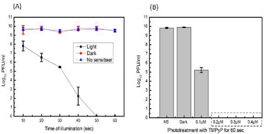
that TMPyP (at least 0.2 µM in solution) can achieve complete inactivation of MS2 within 60
sec when illuminated at 32 W cm-2 (Figure 3). At 10 seconds of illumination there were 1.5 log
reductions in PFU/ml and at 30 seconds of illumination there were 4 log reductions in PFU/ml
of MS2 (Figure 3). TMPyP alone in the dark (Dark experiment) or light alone (No sensitiser) do
not cause any detectable reduction in log PFU/ml of MS2 (Figure 3). Previous work has shown
that tetra-porphyrins like TMPyP can efficiently inactivate bacteriophages such as T4 in
solution (Costa et al., 2008, Costa et al., 2010). It was reported that complete inactivation
(>99.99% of inactivation, reduction of 7.2 log PFU/ml) of bacteriophage T4 from sewage was
achieved only at highest TMPyP concentration of 5 µM and illuminated at 40 Wm-2 for 270
minutes. Complete inactivation within 1 minute was also reported but at higher
concentrations of 1 mM and 0.01 mM of TMPyP illuminated at 2.2 mW cm-2 with a UV lamp
(Casteel et al., 2004). In this work, we avoided using UV lamp as source of light and used cold
visible light source instead, as UV light at certain wavelengths could inactivate
13
Figure 3: MS2 PDI in solution, illuminated at 32 mW cm-2. (A), PDI using 0.5 µM TMPyP in
solution from 10 to 60 sec. At 10 sec of illumination there were 1.5 log reductions in PFU/ml, at 30 sec of illumination there were 4 log reductions in PFU/ml and at 50 sec complete inactivation were observed. (B), PDI using different concentrations (0.1 µM to 0.4 µM) of TMPyP. Even 0.1 µM of TMPyP have caused 4 log reductions in PFU/ml after 60 seconds of illumination. Dark experiment was treated with 0.5 µM of TMPyP but was not exposed to light while NS (no sensitiser) was not treated with TMPyP but was exposed to light. Each value represents mean ± standard deviation of three independent experiments. Error bars show ± SD.
It has been reported that photosensitiser-5,10,15,20-tetrakishydroxyphenyl) porphyrin
(p-TAPP) could be immobilised onto polymeric chitosan membrane for possible water
disinfection application (Bonnett et al., 2006). In that study, p-TAPP was attached onto
chitosan membrane by adsorption without prior modification of the membrane and the
p-TAPP-chitosan composite have shown photomicrobicidal activity against E.coli (Bonnett et al.,
2006). However, in our case, direct adsorption of TMPyP onto polymeric chitosan membrane
was not possible because the net charge of chitosan membrane is positive and is not optimal
for adsorption of photosensitiser-TMPyP, which is a tetra cationic porphyrin (Figure 1).
Therefore, the chitosan membranes were first modified by pyromellitic dianhydride (PMA) in
[image:14.595.81.515.73.293.2]14
PMA modifies primary amine groups on the membranes and generates three free carboxyl
groups (Figure 2). The fourth carboxyl group becomes linked in a peptide bond to the amine
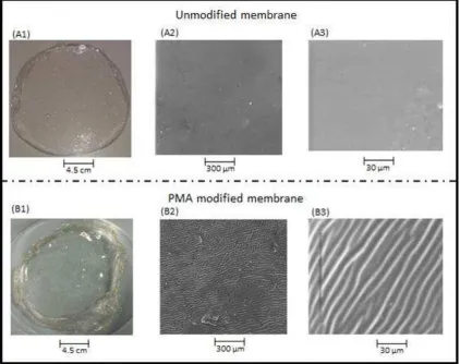
(Figure 2). Scanning electron microscopy was undertaken to analyse the physical properties
of PMA modified chitosan membranes as compared to unmodified chitosan membranes
(Figure 4). The scanning electron microscopy images of dried modified chitosan membrane
showed uniform contraction, compactness and folding of the membrane (Figure 4). The
membranes modified with succinic anhydride became soluble in water while being washed in
dH2O (result not shown). Control experiments using succinic anhydride to modify the
membrane amine group drastically affected its solubility and the whole membrane dissolved
in water during washing steps in H2O. In contrast, membranes modified with PMA remained
intact while in water and became denser and more compact compared to unmodified
membranes under both wet and dried condition as shown using SEM (Figure 4). It was
expected that chitosan modification with acid anhydride derivatives might increase solubility
in water as observed with succinic anhydride to modify the chitosan membrane. This was in
agreement with a previous study (Tangpasuthadol et al., 2003) which reported that such
modification with succinic anhydride could increase the hydrophilicity and hygroscopic
property of the chitosan in film or powdered forms. However, unlike succinic anhydride PMA
is a bis-anhydride with anhydride groups either side of an aromatic ring. Typically a PMA
anhydride group will react with one amine group, with the other anhydride then being
subjected to water attack. It is also likely that carboxyl-anhydride PMA molecules were able
to form peptide bonds between two or more units of chitosan polymer, thereby crosslinking
the chitosan units together. This could account for the compactness and insolubility in water
of chitosan membranes modified with PMA (Figure 4). Chitosan membranes prepared as
15
studies have reinforced the chitosan membranes with nylon to overcome brittleness before
functionalisation with photosensitisers (Bonnett et al., 2006). However, modification with
PMA improved the mechanical strength of the membranes and therefore, there was no need
for reinforcement of the membranes.
Figure 4: Images of PMA modified and unmodified chitosan membranes: (A1), unmodified chitosan membrane; (A2), (A3), SEM images of unmodified chitosan membrane; (B1), PMA modified chitosan membrane; (B2), (B3), SEM images of PMA modified chitosan membrane.
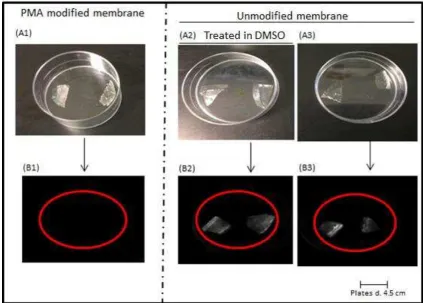
Midland blotting (Rushworth et al., 2014) was used to detect free amine (NH2) groups in order
[image:16.595.87.510.232.566.2]16
performed to analyse the chemical properties and success of PMA modified chitosan
membranes as compared to unmodified chitosan membranes. Unmodified chitosan
membranes have free amino groups at position 6 of the chitosan monomer while modified
membranes have free carboxyl groups. Chemiluminescence was detected for unmodified
membranes as they possess amino groups (and therefore became tagged by NHS-biotin,
allowing subsequent labelling with streptavidin-peroxidase) while it was not detected for
modified membrane as they have free carboxyl groups (Figure 5). The two controls;
unmodified chitosan membrane but treated in DMSO and unmodified chitosan membrane
showed chemiluminescence after Midland blotting thereby confirming the presence of free
amino groups whilst PMA modified chitosan membrane did not show chemiluminescence
after Midland blotting (Figure 5). The absence of chemiluminescence for the modified
17
Figure 5: Images of chitosan membranes to confirm amine modification: (A1), PMA modified chitosan membrane; (A2), unmodified chitosan membrane but treated in DMSO; (A3), unmodified chitosan membrane; (B1), chemiluminescence not detected for PMA modified chitosan membrane; (B2), (B3), chemiluminescence detected for unmodified chitosan membranes.
PMA modified membrane and the 2 control samples (DMSO treated but unmodified
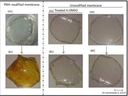
membrane and unmodified membrane) were stained in TMPyP. However, only PMA modified
membrane retained the TMPyP after vigorous washing in dH2O (Figure 6). It is clear that most
amino groups which gave net positive charge to the chitosan membranes had been modified
allowing efficient adsorption of the cationic TMPyP. It is important to note that TMPyP
attachment onto the PMA treated chitosan membranes is electrostatic and the dye could be
released into PDI medium of very high ionic strength. We carried out all our investigations in
18
the solution as monitored by spectroscopy. In practice, most potential drinking water from
the environment, freshwater streams or lakes will have low ionic strength. Within our study,
monitoring the release of TMPyP from the chitosan membrane and washing to zero
absorbance after staining of the membranes with TMPyP is very important as even 1 µM of
TMPyP can achieve complete inactivation of MS2 phage in solution within few minutes. This
[image:19.595.74.524.301.641.2]is shown from preliminary PDI investigations using TMPyP in solution (Figure 3).
Figure 6: Chitosan membranes functionalised with TMPyP. (A1), PMA modified membrane; (A2), DMSO treated but unmodified membrane; (A3), unmodified membrane. The corresponding samples after staining with TMPyP and washing in H2O are shown in B1-B3
19
3.2 PDI of bacteriophage MS2 and E.coli BL21 using TMPyP functionalised chitosan membrane
TMPyP functionalised chitosan membranes (CM-T) were used to investigate microbial
inactivation of MS2 and E. coli BL21 by PDI and for both, use of CM-T resulted in photodynamic
inactivation (Figure 7 and Figure 8). For the MS2 PDI stationary model, complete inactivation
(>99.99% of inactivation, reduction of 9.6 log PFU/ml) of MS2 was observed with CM-T after
90 min illumination with the light at 32 mW cm-2 (Figure 7). For the flowing water model,
complete inactivation of MS2 (>99.9% of inactivation, reduction of 8.8 log PFU/ml) was
observed for sample passed twice over the surface of CM-T at 0.33 ml/min while being
illuminated. Reduction of approximately 3 log PFU/ml were observed for sample passed once
under the same conditions (Figure 7). The same light intensity at 32 mW cm-2 was chosen and
used as in our PDI studies in solution. This was to allow genuine comparison between rate
and extent of PDI with TMPyP in solution and while attached onto chitosan membrane. This
light intensity is low and it is just about 3% of bright midday sunlight under clear sky conditions
in sub-Saharan Africa. And subsequent PDI investigation will be employ much higher light
intensities. However, use of real world light intensities would just give complete inactivation
and reveal little detail of the inactivation rates. It is also clear that the efficient PDI capacities
of CM-T at low light intensity mean it could also be used in the UK and other northern
European countries; in these nations the key advantage would be reduction in energy use for
water treatment. The rate of PDI with TMPyP attached onto chitosan membrane is slower as
compared to TMPyP in solution (Figures 3, 7). This was expected as proximity of unattached
TMPyP in solution to the microorganism will be greater as compared with attached TMPyP.
However, the ultimate aim was achieved as CM-T can cause PDI of MS2 phage under flow
20
Figure 7: PDI of MS2 using TMPyP functionalised chitosan membrane, illuminated at32 mW cm-2. (A), stationary water model, It took 90 min for complete inactivation of MS2 (>99.9% of
inactivation, reduction of 9.7 log PFU/ml); (B), flowing water model, CM-T: Light (A) was a sample passed once over the surface of CM-T at 0.33 ml/min while being illuminated, CM-T: Light (B) was a sample passed twice over the surface of CM-T at 0.33 ml/min while being illuminated. Complete inactivation of MS2 (>99.9% of inactivation, reduction of 8.8 log PFU/mL) was observed for CM-T: Light (B) while reduction of about 3 log PFU/ml were observed for CM-T: Light (A). CM-T: Dark was not exposed to light while CM only: Light (chitosan membrane not functionalised with TMPyP) was exposed to light. Each data point mean ± SD of three independent experiments. Error bars show ± SD.
For the photodynamic inactivation of E.coli using CM-T under the same stationary model used
for MS2 phage, reduction of only 3 logCFU/ml of the bacteria was observed (Figure 8). Our
data have also shown that washing of the cells lead to reduction of about 1 log CFU/ml of the
bacteria and the actual photodynamic inactivation was about 2 log reductions under that
conditions. It is important to state that more time of illumination and higher light intensity
can increase the log reductions of the bacteria as these factors are proportional related to the
21
(stationary model) conditions for the E.coli experiment allowed direct comparison. TMPyP in
solution can bind to bacteria cell membranes, partition between lipid bilayer and even move
into the cells to activate killing process in addition to ROS generated in solution. However the
only possibility of PDI with TMPyP attached unto chitosan membrane will be direct cell
[image:22.595.103.488.230.535.2]damage by singlet oxygen (or other ROS) generated close to the support surface.
Figure 8: PDI of E. coli BL21 using TMPyP functionalised chitosan membrane, illuminated at 32 mW cm-2. CM-T: Light, reduction of about 3 log CFU/ml was observed after 90 min of
22
4. Conclusions
No successful disinfection of water using the photodynamic effect of a photosensitiser during
wastewater treatment has been reported in either developed or developing countries.
However, there is ongoing worldwide research into the possibility of using photodynamic
disinfection during wastewater treatment. Trending and emerging issues in this area have
been finding the right solid supports, coupling chemistries and photosensitisers of
appropriate properties and qualities suitable for sustainable usage in water disinfection. The
driving force behind the research includes potential drastic cutting down of energy and
chemical usage to the barest minimum as sunlight could be used as the source of light for the
photodynamic effect and photosensitisers could be attached on to solid supports so that after
phototreatment of water the supported photosensitiser is not released into the water.
Photosensitiser-functionalised solid support could be re-used for water disinfection several
times thereby making it cheap and environmentally friendlier compared to conventional
methods of water disinfection during wastewater treatment.
This work has shown the possibility of making a simple, environmentally friendly and
zero-man made energy input device for water disinfection. The CM-T has a number of merits:
It employed the use of chitosan membrane which is non-toxic, biodegradable, wettable,
cheap and readily available, and of good mechanical strength to withstand the rigorous
process of water disinfection without tear and wear. We described a method of modifying
chitosan membrane without affecting its solubility in water using PMA. PMA modified
chitosan membrane could also probably be used to adsorb positively charged dyes and heavy
metals from contaminated water thereby remediating the water totally although this effect
is not its primary function. CM-T was used and has shown that it has photodynamic
23
detectable decline in its PDI capacity and efficiency. This will ultimately reduce cost.
Furthermore, the flowing water model for PDI mimics something close to the process in water
treatment plants.
The fact that CM-T was able to photodynamic inactivate MS2 and E. coli while using light
intensity of 32 mW cm-2 which is low compared to daytime sun brightness, is an indication
that this research can lead way to simple sunlight driven water disinfection system that could
be used as a zero man-made energy input systems to produce clean and safe drinking water
in both developed and developing countries.
Acknowledgement
This work was carried out at the University of Leeds and sponsored by Petroleum Technology
Development Fund (PTDF), Nigeria.
24 References
ALVES, E., SANTOS, N., MELO, T., MACIEL, E., DORIA, M. L., FAUSTINO, M. A. F., TOME, J. P. C., NEVES, M. G. P. M. S., CAVALEIRO, J. A. S., CUNHA, A., HELGUERO, L. A., DOMINGUES, P., ALMEIDA, A. & DOMINGUES, M. R. M. 2013. Photodynamic oxidation of Escherichia coli membrane
phospholipids: new insights based on lipidomics. Rapid Communications in Mass Spectrometry, 27, 2717-2728.
AMORNCHAI, W., HOVEN, V. P. & TANGPASUTHADOL, V. 2004. Surface
modification of chitosan films-grafting ethylene glycol oligomer and its effect on protein adsorption. Macromolecular Symposia, 216, 99-107.
BAUMLER, W. & MAISCH, T. 2012. Fast and Effective: Intense Pulse Light
Photodynamic Inactivation of Bacteria. Lasers in Surgery and Medicine, 44, 43-43.
BONNETT, R., KRYSTEVA, M. A., LALOV, I. G. & ARTARSKY, S. V. 2006. Water disinfection using photosensitizers immobilized on chitosan. Water Research, 40, 1269-1275.
BOURRE, L., GIUNTINI, F., EGGLESTON, I. M., MOSSE, C. A., MACROBERT, A. J. & WILSON, M. 2010. Effective photoinactivation of Gram-positive and Gram-negative bacterial strains using an HIV-1 Tat peptide-porphyrin conjugate. Photochemical & Photobiological Sciences, 9, 1613-1620.
CARVALHO, C. M. B., GOMES, A. T. P. C., FERNANDES, S. C. D., PRATA, A. C. B., ALMEIDA, M. A., CUNHA, M. A., TOME, J. P. C., FAUSTINO, M. A. F., NEVES, M. G. P. M. S., TOME, A. C., CAVALEIRO, J. A. S., LIN, Z., RAINHO, J. P. & ROCHA, J. 2007. Photoinactivation of bacteria in
wastewater by porphyrins: Bacterial beta-galactosidase activity and leucine-uptake as methods to monitor the process. Journal of Photochemistry and Photobiology B-Biology, 88, 112-118.
CASTEEL, M. J., JAYARAJ, K., GOLD, A., BALL, L. M. & SOBSEY, M. D. 2004. Photoinactivation of hepatitis A virus by synthetic porphyrins. Photochemistry and Photobiology, 80, 294-300.
CHO, M., LEE, J., MACKEYEV, Y., WILSON, L. J., ALVAREZ, P. J. J., HUGHES, J. B. & KIM, J. H. 2010. Visible Light Sensitized Inactivation of MS-2
Bacteriophage by a Cationic Amine-Functionalized C-60 Derivative. Environmental Science & Technology, 44, 6685-6691.
COSTA, D. C. S., GOMES, M. C., FAUSTINO, M. A. F., NEVES, M. G. P. M. S., CUNHA, A., CAVALEIRO, J. A. S., ALMEIDA, A. & TOME, J. P. C. 2012. Comparative photodynamic inactivation of antibiotic resistant bacteria by first and second generation cationic photosensitizers. Photochemical &
Photobiological Sciences, 11, 1905-1913.
25
COSTA, L., CARVALHO, C. M. B., FAUSTINO, M. A. F., NEVES, M. G. P. M. S., TOME, J. P. C., TOME, A. C., CAVALEIRO, J. A. S., CUNHA, A. & ALMEIDA, A. 2010. Sewage bacteriophage inactivation by cationic porphyrins: influence of light parameters. Photochemical & Photobiological Sciences, 9, 1126-1133.
CRINI, G. & BADOT, P. M. 2008. Application of chitosan, a natural
aminopolysaccharide, for dye removal from aqueous solutions by adsorption processes using batch studies: A review of recent literature. Progress in Polymer Science, 33, 399-447.
DAVIES, M. J. 2003. Singlet oxygen-mediated damage to proteins and its
consequences. Biochemical and Biophysical Research Communications, 305, 761-770.
GRACANIN, M., HAWKINS, C. L., PATTISON, D. I. & DAVIES, M. J. 2009. Singlet-oxygen-mediated amino acid and protein oxidation: Formation of tryptophan peroxides and decomposition products. Free Radical Biology and Medicine, 47, 92-102.
GRACANIN, M., PATTISON, D. I. & DAVIES, M. J. 2007. Singlet oxygen-mediated tryptophan oxidation: Characterization of potential markers of protein
oxidation. Free Radical Research, 41, S31-S31.
KOMAGOE, K., KATO, H., INOUE, T. & KATSU, T. 2011. Continuous real-time monitoring of cationic porphyrin-induced photodynamic inactivation of
bacterial membrane functions using electrochemical sensors. Photochemical & Photobiological Sciences, 10, 1181-1188.
KRAJEWSKA, B., LESZKO, M. & ZABORSKA, W. 1990. Urease Immobilized on Chitosan Membrane - Preparation and Properties. Journal of Chemical Technology and Biotechnology, 48, 337-350.
KROPINSKI, A. M., MAZZOCCO, A., WADDELL, T. E., LINGOHR, E. & JOHNSON, R. P. 2009. Enumeration of bacteriophages by double agar overlay plaque assay. Bacteriophages: Methods and Protocols, Volume 1: Isolation, Characterization, and Interactions, 69-76.
LOEB, S., HOFMANN, R. & KIM, J.-H. 2016. Beyond the Pipeline: Assessing the Efficiency Limits of Advanced Technologies for Solar Water Disinfection. Environmental Science & Technology Letters, 3, 73-80.
MAISCH, T., KOWALEWSKI, E., HOLZMANN, T., SCHNEIDER-BRACHERT, W. & BAUMLER, W. 2012a. Photodynamic killing of enterohaemorrhagic
Escherichia coli (EHEC) for the first time using TMPyP. Experimental Dermatology, 21, e40-e40.
MAISCH, T., SPANNBERGER, F., REGENSBURGER, J., FELGENTRAGER, A. & BAUMLER, W. 2012b. Fast and effective: intense pulse light photodynamic inactivation of bacteria. Journal of Industrial Microbiology & Biotechnology, 39, 1013-1021.
MARTEL, B., DEVASSINE, M., CRINI, G., WELTROWSKI, M., BOURDONNEAU, M. & MORCELLET, M. 2001. Preparation and sorption properties of a
26
MEEBUNGPRAW, J., WIARACHAI, O., VILAIVAN, T., KIATKAMJORNWONG, S. & HOVEN, V. P. 2015. Quaternized chitosan particles as ion exchange supports for label-free DNA detection using PNA probe and MALDI-TOF mass
spectrometry. Carbohydrate Polymers, 131, 80-89.
RUSHWORTH, J. V., AHMED, A. & MILLNER, P. A. 2014. Midland Blotting: A Rapid, Semi-Quantitative Method for Biosensor Surface Characterization. Journal of Biosensors & Bioelectronics, 2013.
SHANNON, M. A., BOHN, P. W., ELIMELECH, M., GEORGIADIS, J. G., MARINAS, B. J. & MAYES, A. M. 2008. Science and technology for water purification in the coming decades. Nature, 452, 301-310.
SILVERMAN, A. I., PETERSON, B. M., BOEHM, A. B., MCNEILL, K. & NELSON, K. L. 2013. Sunlight Inactivation of Human Viruses and Bacteriophages in
Coastal Waters Containing Natural Photosensitizers. Environmental Science & Technology, 47, 1870-1878.
SPANNBERGER, F., REGENSBURGER, J., FELGENTRAGER, A., BAUMLER, W. & MAISCH, T. 2012. Fast and effective: intense pulse light photodynamic inactivation of bacteria. Experimental Dermatology, 21, e40-e40.
TANGPASUTHADOL, V., PONGCHAISIRIKUL, N. & HOVEN, V. P. 2003. Surface modification of chitosan films. Effects of hydrophobicity on protein adsorption. Carbohydrate Research, 338, 937-942.