Int. J. Electrochem. Sci., 9 (2014) 5776 - 5787
International Journal of
ELECTROCHEMICAL
SCIENCE
www.electrochemsci.org
Novel Planar Chip Biosensors for Potentiometric Immunoassay
of Acid Phosphatase Activity Based on the Use of Ion
Association Complexes as Novel Electroactive Materials
Ayman H. Kamel1,2,*, Hoda R. Galal3, Adly A. Hanna3
1
Chemistry Department, Faculty of Science, Ain Shams University, Cairo, Egypt.
2
Chemistry Department, College of Science, Qassim University, Buraidah 6644, KSA.
3
National Research Center (NRC), Dokki, Giza,14211 Egypt
*
E-mail: [email protected]
Received: 13 May 2014 / Accepted: 28 June 2014 / Published: 16 July 2014
The construction and general performance characteristics of two poly (vinyl chloride) matrix chemical sensors for α-naphthyl acid phosphate (1-NAP) substrate were described. These sensors were based on the use of ion association complexes of (1-NAP) with cetylpyridinium chloride (CP) [sensor 1] and iron (II) - 4,7-bathophenanthroline [Fe(bphen)3]2+ [sensor 2] as novel electroactive materials dispersed
in o-nitrophenyloctyl ether (o-NPOE) plasticizer for ionometric sensor controls. The sensing membrane (3 x 5 mm) is immobilized on a wafer polyimide chip (size 13.5 x 3.5 mm) to offer a planar miniaturized design. The sensors displayed good selectivity and stability and demonstrates a near-Nernstian response for 1-NAP over the concentration range 1.0x10⁻²- 1.0x10⁻⁵ and 1.0x10⁻²- 9.0x10⁻⁶ mol L-1 with an anionic slope of -57.3±0.9 and -56.6±1.1 mV decade-1 and a detection limit of 9x10⁻⁶ and 8x10⁻⁶ mol L-1 over the pH range 2.8 – 5.5 for sensors 1 and 2, respectively. The sensors were used to follow up the decrease of a fixed concentration of 1-NAP substrate as a function of acid phosphatase enzyme activity under optimized conditions of pH and temperature. A linear relationship between the initial rate of 1-NAP substrate hydrolysis and enzyme activity hold 0.01- 4.3 IU L-1 of ACP enzyme. The sensors were used for the determination of ACP enzyme activities in biological fluids of some patients suffering from prostatic cancer. The results obtained agree fairly well with data obtained by the standard spectrophotometric methods.
Keywords: Acid phosphatase; Miniaturized sensors; 1-Naphthyl acid phosphate (1-NAP); Enzyme assay; PVC membrane, Potentiometry.
1. INTRODUCTION
Phosphomonoesterases (phosphoric monoester hydrolases categorized with the EC number 3.1.3) belong to the group of indicative enzymes mostly assayed in the clinical practice. These nonspecific enzymes are responsible for dephosphorylation. From the diagnostic point of view the most important are determinations of alkaline phosphatase (ALP. EC.3.1.3.1) and acid phosphatase (ACP, EC.3.1.3.2) levels.
Acid phosphatase enzyme (ACP) is located primarily in the lysosomal compartment of the cells, and it has been detected in the antigen-presenting dendritic cells in the hypertrophic pharyngeal tonsils in children [1]. Prostatic acid phosphatase (PAP) is an enzyme found in men, primarily in the prostate gland and semen which determine the health of the prostate gland. Prostate dysfunction results in the release of PAP into the blood. Prostate cancer that is located only in the prostate gland may not produce high enough levels to indicate a problem but it is of considerable biomedical interest as a diagnostic indicator of the development and metastasis of prostatic cancer [2,3]. The rapid and convenient analysis of ACP in blood is important since the level of ACP activity is used as a pre-preliminary early diagnosis for many diseases such as human prostatic diseases, prostatic cancer [4,5], and hypophosphatasia [6]. Usually, the determination of the activity of acid phosphatase is based on the spectrophotometric methods and utilizes p-nitrophenylphosphate (PNPP) as substrate. This, when dephosphorylated, forms the colored product p-nitrophenol (PNP), which is monitored by its absorbance at 405 nm [7–9]. ACP is also determined by electrochemical oxidation on graphite screen-printed electrode [10], square-wave voltammetry [11], cyclic voltammetry [12], differential puls voltammetry [13], amperometry [14], high performance liquid chromatography equipped with a cathodic detector [15], electrochemical impedance spectroscopy [16], phosphorescence [17], chemiluminescence [18] and fluorescence [19]. Most of these instrumental methods involved the use of sophisticated equipment, exhibit low selectivity, require careful control of the reaction and entail several time consuming manipulation steps.
and polyanion [25].
The present work describes the construction and characterization of planar chip biosensor for potentiometric immunoassay of acid phosphatase enzyme activity. The sensors are used to follow up the decrease of a fixed concentration of 1-NAP substrate as a function of ACP enzyme activity under optimized conditions of pH and temperature.
2. MATERIALS AND METHODS
2.1. Equipments
All potentiometric measurements were made at 250.10
C with a Cole-Parmer pH/mV meter (USA model 59003-05) connected to a PC through the interface ADC 16 (Pico Tech, UK) and Pico Log for windows (version 5.07) software and 1-NAP PVC membrane sensors in conjunction with a Sentek, Ag/AgCl double junction reference electrode (UK model R2/2MM) filled with 1.0 mol L-1 KNO3 in the outer compartment. A combination glass pH electrode (Schott blue line 25, Germany) was
used for all pH measurements. A computer-controlled spectrophotometer (Shimadzu, Model 1601 Japan) was used for the spectrophotometric measurements. Elemental analyses were carried out on Elementar Vario EL cube, Germany. Fe-content was determined by Perkin Elmer Atomic Absorption Spectrometer (Model 3100 USA).
2.2. Reagents and chemicals
All solutions were prepared using Millipore Milli-Q water. All chemicals were of analytical reagent grade and were used without further purification. 4,7-Diphenyl-1,10-phenanthroline (bphen) with a purity ≥ 97%, o-nitrophenyloctyl ether (o-NPOE), tetrahydrofuran (THF), cetyl pyridinium chloride (CP), and poly(vinyl) chloride powder (PVC) were obtained from Merck Inc. (Darmstadt, Germany). α-Naphthyl acid phosphate mono sodium salt (1-NAP), sodium acetate, and acetic acid were purchased from Fluka Inc.(Buchs, Switherland). Acid phosphatase from wheat germ [EC 3.1.3.2] lot No 2326309 (0.1-0.6 U mg-1) was purchased from Fluka (AG Chemische Fabrik, CH-9470 Buchs, Switzerland). Colorimetric diagnostic kit for acid phosphatase was obtained from Labkit Chemelex, (Barcelona ,Spain).
2.3. Sensors construction and calibration
impurities adsorbed on the surface of the precipitate, dried at room temperature for 24 h, and ground to a fine powder. A membrane cocktail was prepared by mixing 5.1 mg portion of the precipitate with 350.3 mg of o-nitrophenyloctyl ether (o-NPOE) and 191.2 mg PVC. The mixture was dissolved in 4 mL THF and thoroughly mixed to obtain a homogeneous transparent mixture.
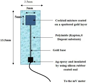
[image:4.596.150.452.215.501.2]A planar gold base electrode (3 x 5 mm) was sputtered on a (13.5 x 3.5 mm) flexible polyimide (Kapton, ® DuPont) substrate (125 μm thick), as shown in Fig.1; single site electrode (area = 0.06 cm2) (used for all the optimization and characterization studies), and used as previously described [26,27].
Figure 1. Planar-chip sensor
An electrical wire was connected to the electrode by means of Ag-epoxy (Epoxy Technology).Insulation of the electrical contact was made using silicon rubber coating seal (Dow Corning 3140 RTV). The membrane cocktail mixture was directly coated to the sputtered gold layer using micro syringe to apply few micro liters of the sensing solution (Typically 10 µL of membrane cocktail is dispersed), left to dry in the air for one minute before repeating further addition (i.e. four times of the sensing solution).
2.4. Potentiometric assay of acid phosphatase (ACP) enzyme
A calibration plot for ACP enzyme was made by transferring a 100 µL of 5.0x10⁻²
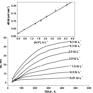
mol L-1 1-NAP to 10-mL volumetric flask, and diluted to the mark with 1.0x10⁻² mol L-1 acetate buffer of pH 4.7. The planar sensor, in conjunction with a double junction Ag/AgCl reference electrode was immersed in the solution. After a constant potential reading was obtained, 100 µL aliquots containing 0.01- 4.3 IU L-1 of ACP enzyme was added. The rate curves (potential / time) were plotted, and the maximum initial rate of potential change expressed as (∆E/∆t) was graphically obtained using the rate portion of the curve [23, 28]. The initial rate was plotted as a function of the enzyme activity. The calibration curve was used for subsequent measurements of unknown enzyme activity. A blank experiment was carried out under similar conditions without the enzyme.
For assay of ACP in human serum, the above procedure was utilized but using 100µL aliquot of the serum sample (obtained from healthy volunteers and patients with prostate carcinoma). The rate curve was recorded, and the maximum initial rate of the potential change expressed as (∆E/∆t) was graphically determined and compared with the calibration plot.
3. RESULTS AND DISCUSION
Acid phosphatase is a nonspecific esterase that catalyzes hydrolysis of many monoesters of phosphoric acid. It has been frequently used as a label in immunoassays because of its relatively small molecular size, high turnover rate, low cost, high stability, and variety of substrates available. Although a large number of substrates for ACP can be used, substrates should comply with several criteria that make them well suited for potentiometric measurements [29]. It has been shown that lipophilicity, ionizable form, stability and availability of substrates should be taken into consideration [30]. In this work, 1-naphthyl phosphate was selected based on the difference between its lipophilicity and that of its product ion after the hydrolysis by ACP enzyme. As shown in scheme 1, both the substrate (pKa = 5.85) and the product ions (pKa = 9.38) are anions at pH > 7 [31].
This reaction was used for determination of ACP activity by monitoring the decrease in the concentration of the un-hydrolyzed 1-NAP substrate. Attempts were made to develop a suitable membrane sensor for potentiometric monitoring of 1-NAP substrate.
O P O
OH O
-EC.3.1.3.2
OH
+ HPO4
2-pH 4.7
Scheme 1. Hydrolysis of 1-naphthyl phosphate (pKa1 = 1.24; pKa2 = 5.85) by acid phosphatase (ACP)
[image:6.596.102.497.98.489.2]
3.1. Characterization of α-naphthyl acid phosphate (1-NAP) ion–pair complexes
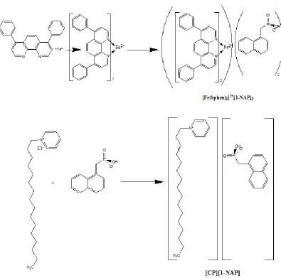
Figure 2. The chemical structure of the ion association complexes
1-NAP was reacted with cetylpyridinium chloride (CP) and iron (II)-4,7-bathophenanthroline [Fe(bphen)3]2+ at pH 4.0 forming cetylpyridinium-naphthyl acid phosphate [CP][1-NAP] and tris
bathophenanthroline Iron (II)-naphthyl acid phosphate [Fe(bphen)3]2+[1-NAP]2 ion associate
complexes. The precipitates were isolated and characterized by elemental analysis.
The elemental analysis obtained for the solid complex agree fairly well with the formula 2:1 of [Fe(bphen)3]2+:1-NAP (Calculated, C: 73.693%, H: 4.311%, N: 5.606%, P:4.131%, Fe: 3.724% found,
3.2. Response characteristics of the sensors
-6 -5 -4 -3 -2 -350
-300 -250 -200 -150
E
M
F
,m
V
log [2-NP], mol L-1
[image:7.596.228.373.104.308.2]sensor 1 sensor 2
Figure 3. Response characteristics of 1-NAP substrate PVC membrane sensor in 0.05 mol L-1 acetate buffer of pH 4.7.
Performance characteristics of two poly (vinyl chloride) matrix chemical sensors for α-naphthyl acid phosphate (1-NAP) substrate were systematically evaluated. These sensors were based on the use of ion association complexes of (1-NAP) with cetylpyridinium chloride [CP][NAP] [sensor 1] and iron (II)-4,7-bathophenanthroline [Fe(bphen)3][NAP]2 [sensor 2] as novel electroactive
materials dispersed in o-nitrophenyloctyl ether (o-NPOE) plasticizer for ionometric sensor controls. The membrane composition was 33% PVC the plastic matrix, 66% o,NPOE the solvent mediator and 2% of the sensing element.
As shown in Fig. 3, the calibration graphs were liner over the concentration rang 1.0x10⁻²- 1.0x10⁻⁵ and 1.0x10⁻²- 9.0x10⁻⁶ mol L-1 with an anionic slope of -57.3±0.9 and -56.6±1.1 mV decade-1 and a detection limit of 8x10⁻⁶ and 9x10⁻⁶ mol L-1 for sensors 1 and 2, respectively. The potentiometric response characteristics, based on data collected over a period of 8 weeks for six sensors were shown in Table 1.
Table 1. Potentiometric response characteristics of 1-NAP PVC membrane sensors based on ([CP)(NAP)]and [(Fe (bphen)3)(NAP)2]
Parameter [CP][1-NAP]
(Sensor 1)
[Fe (bphen)3][1-NAP]2
(sensor 2)
Slope, mV decade-1 -57.3±0.9 -56.6±1.1
Correlation coefficient, (r) -0.999 -0.999
Linear range, mol L-1 1.0 x 10⁻⁵-1.0x10⁻² 9.0 x 10⁻⁶-1.0x10⁻²
Detection limit, mol L-1 9.0 x10⁻⁶ 8.0 x10⁻⁶
[image:7.596.52.548.646.760.2]
Response time, s < 10 < 10
Precision, % 0.9 0.8
Accuracy, % 0.9 0.6
Within -day reproducibility, (CVw ), % 1.1 1.2
Between–day-variability (Cvb %) 0.8 0.7
The response time of the sensors was determined by recording the time required to achieve a steady-state potential within ±0.7mV. The potential readings at time intervals of 10 s over 2 min were recorded. The relation between potential reading and response time was plotted for 1.0x10⁻⁵–1.0x10⁻³ mol L-1 standard 1-NAP solutions. The time required to attain 95% of the equilibrium based on the two sensors was less than 10 s. The stability of the sensor response remained constant for ~ 10 min (drift < 1.5 mV). The potential readings of the sensors for 6 identical measurements over a period of 2 months showed variations of not more than 1.8 mV. The reproducibility of the calibration slope was within 2.3 mV decade-1 over a period of 2 months (n=20) indicating a long term potential stability. The sensors were stored in 10−3mole L-1 1-NAP solution between measurements and when not in use. They had no significant effect on the slope of the calibration plot up to 8 weeks, which remained within ±2.5 mV per [1-NAP] decade−1 of its original value.
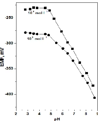
The robustness of the method was also examined by testing the influence of pH variation on the accuracy of the results Fig. 4.
Figure 4. Influence of the solution pH on the response of the 1-NAP membrane based sensor
[image:8.596.219.380.409.608.2]
suggests that the membrane may be deteriorated due to the presence of high hydrogen ion concentration. All subsequent measurements were done in 0.05 mol L-1 acetate buffer at pH 4.7.
The ruggedness of the potentiometric method was also evaluated by carrying out the analysis using four different sensors and two different instruments on different days. A relative standard deviation (RSD) of less than 1.0 % was observed for repetitive measurements during three different days (n=10). The results indicated that the proposed method is capable of producing results with high precision and stability.
To evaluate the possibility for using the proposed sensor for monitoring the rate of 1-NAP hydrolysis using ACP enzyme, the potentiometric selectivity ratio of 1-NAP over 1-naphthol was measured by the separate solutions method [32], with 1.0x10⁻⁴ mol L-1 solutions of both NAP and 1-naphthol at pH 4.7. No effect was detected up to ~1000-fold excess of 1-1-naphthol over 1-NAP. Phospahte ions have no effect at levels as high as 3000-fold excess. Since the initial rate method was used for monitoring the consumption of 1-NAP substrate, the interference from the 1-naphthol reaction product was significantly minimal. The selectivity coefficients (Kpot NAP,J) of the sensor were also measured for different diverse species commonly present in real samples. The effect of bovine serum albumin, lactate, casein, creatinine and uric acid was examined. Lactate, creatinine and urate were tolerated when present at concentration levels up to 1000-fold excess over 1-NAP. Up to 5 and 10% solutions of both casein and bovine serum albumin can be tolerated, respectively. These data confirm reliable determination of ACP activity in real blood serum without any significant interference.
3.3. Effect of 4-NPP substrate concentration
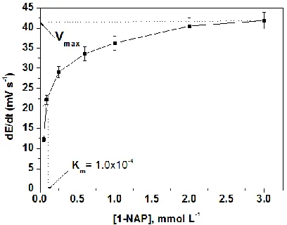
Figure 5. Initial reaction rates obtained with 1-NAP membrane sensor with several 1-NAP initial concentrations fitted to the Michaelis-Menten equation
The determination of Km and Vmax of the enzymatic reaction was carried out by 1-NAP sensor
[image:9.596.197.401.453.613.2]
measurements. This concentration level offered a measurable change in the reaction rate at low enzyme activity, a better linearity of calibration plot, and a fast response of the sensor. Fig. 5 presents the initial rate of substrate consumption measured from the initial slope of the potential - time graph (mV min-1), generated in enzymatic reaction, as a function of substrate concentration, showing a typical Michaelis–Menten compatible curve. From Fig. 5, a Km value of 1.01 mmol L-1 and a Vmax
equal to 41.4 mV min-1 were obtained. This value of Km is the same order of magnitude as that obtained with the spectrophotometric method, 1.6 mmol L−1 at pH 4.7 [31].
3.4. Potentiometric monitoring of Acid phosphatase-catalyzed hydrolysis of 1-NAP
Figure 6. Initial rate of hydrolysis of 1-NAP substrate at 25 ◦C, pH 4.7 as a function of ACP activities
1-NAP PVC potentiometric membrane sensor was used for determining acid phosphatase (ACP) enzyme activity. Since the optimal pH and temperature for ACP are about 3.8-5.7 and 25 oC [31], the enzymatic hydrolysis of 1-NAP was carried out using a pH 4.7 acetate buffer solution 0.05 mol L-1 and at a constant temperature of 25 oC. A typical kinetic-potentiometric plot of the ACP-catalyzed hydrolysis of 1-NAP obtained as described under Materials and methods is shown in Fig. 6. Linear dependence of the initial reaction rate (∆E/∆t) on the enzyme activity was observed with 0.01– 4.3 IU L-1 for ACP enzyme.
[image:10.596.147.461.241.557.2]
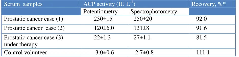
for analysis. The results obtained for the potentiometric assay of ACP in serum samples of some patient are much higher than that of the control values (Table 2).
Table 2. Potentiomeric determination of acid phosphatase (ACP) enzyme in blood serum of some patients using 1-NAP PVC membrane sensor
Serum samples ACP activity (IU L-1) Recovery, %*
Potentiometry Spectrophotometry
Prostatic cancer case (1) 230±15 250±20 92.0 Prostatic cancer case (2) 120±6.0 131±8 91.6 Prostatic cancer case (3)
under therapy
22±1.3 27±1.1 81.5
Control volunteer 3.0±0.6 2.7±0.8 111.1 * Average of Six measurements
The data agree fairly well with results obtained using the standard spectrophotometric method.
4. CONCLUSIONS
Planar chip biosensors for potentiometric immunoassay of acid phosphatase activity were prepared, characterized and used for the clinical assay of acid (ACP) phosphatase enzyme. The sensors were used to follow up the decrease of a fixed concentration of α-naphthyl acid phosphate (1-NAP) substrate as a function of acid phosphatase enzyme activity at optimized conditions of pH and temperature. The sensors displayed a wide linear response range (0.01–4.3 IU L-1 for ACP enzyme, low detection limit (0.01enzyme IU L-1), high accuracy and precision (~98.3±0.6%), good potential response stability (±3 mV), and long life-span (6–8 weeks). The sensors were applicable to biological fluids of some healthy volunteers and patients with carcinoma of prostate. The results obtained in this work enable to conclude that this methodology can be applied to the analysis of ACP enzyme in biological fluids rapidly and accurately.
ACKNOWLEDGEMENTS
The authors acknowledge the financial support from Deanship of Scientific Research, Qassim University, by means of project 2042/SR-D-013.
References
1. W. E. Bowers and C. de Duve, J. Cell Biol., 32 (1967) 349. 2. O. Bodansky, Adv. Clin. Chem., 15 (1972) 43.
3. V. Hoilander, in: P. D. Boyer (Ed.), Acid Phosphatase. In the Enzymes, 4, Academic Press, New York, NY, 1971, p. 450.
4. R. Wang, Q. Cai, D. Tong, L. Nie and S. Yao, Enzyme Microbiol. Technol., 22 (1998) 36. 5. P. B. Hudson, K. K. Tsuboi and A. Mittelman, Am. J. Med. 19 (1955) 895.
[image:11.596.64.537.176.290.2]
7. D. N. Gray, M. H. Keyes and B. Watson, Anal. Chem., 49 (1977) 1067.
8. I. M. Christie, P. H. Treloar, P. M. Koochaki, P. M. Vadgama and G. N. Smith, Anal. Chim. Acta, 257 (1992) 21.
9. N. W. Tietz, A. D. Rinker and L. M. Shaw, J. Clin. Chem. Clin. Biol., 21 (1983) 731.
10.D. Sazydlowska, M. Campas, J. L. Marty and M. Trojanowicz, Sens. Actu. B, 113 (2006) 787. 11.H. J. Kim and J. Kwak, J. Electroanal. Chem., 577 (2005) 243.
12.E. J. Moore, M. Pravda, M. P. Kreuzer and G. G. Guilbault, Anal. Lett., 36 (2003) 303.
13.P. Calvo-Marzal, S. S. Rosatto, P. A. Granjeiro, H Aoyama and L. T. Kubota, Anal. Chim. Acta, 441 (2001) 207.
14.M. S. Wilson and R. D. Rauh, Biosen. Bioelectron., 20 (2004) 276. 15.Y. Yamauchi, M. Ido and H. Maeda, J. Chromatogr. A, 1066 (2005) 127.
16.A. Radi, J. M. Montornes and C. K. O’Sullivan, J. Electroanal. Chem., 587 (2006) 140.
17.F. Watanabe, M. Takano, F. Tanaka, N. Amino, C. Hayashi and K. Miyai, Clin. Chim. Acta, 91 (1979) 273.
18.I. Bronstein, C. J. Voyta, G. H. Thorpe, L. J. Kricka and G. Armstrong, Clin. Chem., 35 (1989) 1441.
19.R. A. Evangelista, A. Pollak and E. F. Temleton, Anal. Biochem., 197 (1991) 213. 20.T. G. Henares, F. Mizutani and H. Hisamoto, Anal Chim Acta, 611 (2008) 17.
21.K. Konopińska, M. Pietrzak and E. Malinowska, J. Solid State Electrochem., 17 (2013) 1665.
22.T. Katsu, M. Kanamitsu and Y. Fujita, Chem. Pharm. Bull., 35 (1987) 3922.
23.S. S. M. Hassan, H. E. M. Sayour and A. H. Kamel, Anal. Chim. Acta, 640 (2009) 75. 24.D. Ogonczyk and R. Koncki, Anal. Chim. Acta, 600 (2007) 194.
25.J. M. Esson and M. E. Meyerhoff, Electroanalysis, 9 (1997) 1335. 26.A. H. Kamel, J. Pharm. & Biomed. Anal., 45 (2007) 341.
27.A. H. Kamel, Electroanalysis, 19 (2007) 2419.
28.S. S. M. Hassan and G. A. Rechnitz, Anal. Chim. Acta, 151 (1983) 473.
29.A. Preechaworapuna, Z. Dai, Y. Xiang, O. Chailapakul and J. Wang, Talanta, 76 (2008) 424. 30. . Szucs, E. Pretsch and . E. Gyurcs nyi, Analyst, 134 (2009) 1601.
31.E. Luchter-Wasylewska, Acta Biochimica Polonica, 44 (1997) 853.
32.Y. Umezawa, P. Buhlmann, K. Umezawa, K. Tohda and S. Amemiya, Pure Appl. Chem., 72 (2000) 1851.
![Table 1. Potentiometric response characteristics of 1-NAP PVC membrane sensors based on ([CP)(NAP)]and [(Fe (bphen)3)(NAP)2]](https://thumb-us.123doks.com/thumbv2/123dok_us/1900679.148123/7.596.228.373.104.308/table-potentiometric-response-characteristics-membrane-sensors-based-bphen.webp)