Vol.58, No. 2 JOURNALOF VIROLOGY, May 1986, p.704-708
0022-538X/86/050704-05$02.00/0
CopyrightC) 1986, American Society for Microbiology
Enhanced Rate of Conversion
or
Recombination
of Markers within
a
Region of Unique Sequence in the
Herpes
Simplex Virus Genome
KAY L. POGUE-GEILEt ANDPATRICIA G. SPEAR*
Departmentof MolecularGenetics andCell Biology andCommittee on Virology, The Universityof Chicago,
Chicago, Illinois 60637
Received 30 September 1985/Accepted 15 January 1986
Insertion mutants ofherpes simplex virus type 1, containing a second copy of the sequences of BamHI
fragmentL (map coordinates0.706to0.744) inserted in inverted orientation into the thymidine kinasegene(at
map coordinate 0.315), have been further characterized. We -reported previously that, as a result of intramolecularorintermolecular recombinationbetweencopies ofthe BamHI-Lsequenceatthe normal locus and insertedlocus, ahighproportion ofprogeny genomesexhibited either inversions oftheuniquesequence flanked bythese inverted repeats orother rearrangements. Now we reportthat a genetic marker (syn-l or syn-1+)originallypresentonlyinthe insertedcopyof BamHIfragmentLappearsinprogenyatboth the normal and inserted loci,and viceversa,athigh frequency. Becausethese phenomena havenotbeen observed with other insertion mutants containing duplications of other sequences from unique regions of the genome, we conclude thatBamHIfragmentLcontainsanelementthatenhancestherateof homologousrecombination in adjacentsequences,resulting ingenome rearrangementsandgene conversion-likeevents.
The genome of herpes simplex virus (HSV) (Fig. 1) is a linear duplex DNA molecule of approximately 100 x 106
daltons and is composed of a long (L) and a short (S)
component. Eachcomponentis flanked byinverted repeats,
the ends of which(a sequencedesignated a)arecommonto
both segments and are repeated in direct orientation at the endsofthe genome (22, 27, 28). Recombination apparently occurs between the inverted repeats, resulting in inversion
oftheunique segments(ULand Us)flankedbythe repeats.
Populations ofHSV genomesusually contain fourisomersof
viralDNA, because UL and Us can invert independently of
each other(6, 23, 29).
Evidence hasbeenpresented thatanelement within thea
sequence promotesthe recombinations that result in
inver-sions ofthe ULand Us components of the genome.
Specif-ically, insertion ofan additional a sequence into the
thymi-dine kinase(tk)gene results in novel rearrangements ofthe
genome, consistentwith inversion ofany genome segment boundedby inverted copies oftheasequence(2, 15-17,24)
anddeletion ofgenome segmentsboundedby directrepeats
(24).Inaddition, deletionofsequencescontainingafrom the
junction between the L and S components prevents
inver-sions ofthe L and S components(18).
With one exception,
duplication
of other regions of thegenome by insertion of an extra copy into the tk gene or elsewhere did not result in recombination between the repeated sequences at a frequency high enough to detect
genomic rearrangements (deletions or inversions fordirect
or inverted repeats, respectively) thatwould have resulted (5, 12, 15). Duplication of sequences from BamHI fragment L (map coordinates 0.706 to 0.744) in inverted orientation, however, resulted in inversions of unique sequences be-tween the duplications and possibly other genomic rear-rangements, apparently due to a high frequency of
* Correspondingauthor.
tPresent address:DepartmentofBiological Sciences,University ofPittsburgh, Pittsburgh, PA 15217.
intramolecular orintermolecularrecombination between the
repeated sequences present at the normal and inserted loci
(20).
The interactions between two copies ofthe a sequence
that can result in recombination and genomic rearrange-ments can also result in elimination of any differences in
nucleotide sequence in homologous sequences adjacent to
thea sequences (3, 7, 10, 11,26). Thesubjectof thisreport is the finding that differences in nucleotide sequence be-tween twocopies ofBamHIfragmentL, oneinserted inthe tk gene and the otheratthenormallocus, areeliminatedat
high frequency, presumablybyrecombinationsbetween the
two copies of the BamHI-L sequence that also result in
genomic rearrangements.
The cellfusion-inducing or syncytial (Syn) mutant, HSV
type 1 strain MP [HSV-1(MP)], differs from the related wild-type strain HSV-l(mP) at severalloci, oneofwhich is
containedwithin BamHIfragmentL.Mapping studies (1, 19)
and nucleotide sequence analysis
(Pogue-Geile
and Spear,submitted forpublication) have shown thatthe Syn
pheno-type ofHSV-1(MP)resultsfroma single nucleotide
substi-tution whichalsoeliminatesaThaIrestriction endonuclease
site present inHSV-1(mP) atmap coordinate 0.737
(Fig.
1). Thus, the presence or absenceofthis ThaI site identifiesthewild-type ormutantalleles, respectively, ofthe
syn-J
genesin HSV-1(mP) andHSV-1(MP). Theevidencefor this
iden-tification is that, within the 710-base-pair (bp) fragment to
which the Syn mutationwasmapped, nucleotide sequences for thetwovirusstrains differedonlyattheThaI
site,
andin recombinants of HSV-1(mP) and HSV-1(MP), all isolatesexpressing theSyn+ phenotype had the ThaIsite, whereas
all isolates
expressing
the Syn phenotype lacked this site(Pogue-Geile andSpear, submitted).
[Debroy et al. (4) recently reported the nucleotide
se-quences, betweenthe two rightmostPstI sites indicated in Fig. 1, forHSV-1(MP)and, between mapcoordinates0.732
and 0.745, for an unrelated wild-type strain
designated
HSV-1(KOS).Their sequence forHSV-1(MP)differs
slightly
704
on November 10, 2019 by guest
http://jvi.asm.org/
~~~~~~-I
Ba b . l' l S ca
/7
I
Th Th Th
/-o
233- 979CG
C(MP)
T CG TG(MP)
I
~~~~1212
FIG. 1. Location of the mutation in HSV-1(MP) that causes the Syn phenotype and eliminates a ThaI site (Pogue-Geile and Spear, submitted).TheBamHlLfragmentin theHSV-1 genome isrepresentedas acrosshatched box andexpandedbelow thegenome,wherethe blackbarrepresentsthe710-bp fragmentthatcontains theSyn mutation(x). InThaI digestsof mPDNA, twofragments(233 and 979bp) would be detectedbyaprobe preparedfrom thePstl-Hincllfragmentinwhich the mutation is located. InThaIdigestsof MPDNA,onlyone
fragment(1,212bp) would be detected. Ba, BamHl; Ps, Pstl; Hn, Hincll; Th, ThaI.
from ours, but not in the
vicinity
of the ThaI site(Pogue-Geile and
Spear,
submitted).They
detected ninedifferences in sequence between HSV-1(MP) and HSV-1(KOS) within part of theregion
to which theSyn
mutation wasmapped.
One difference is the same as described above for
HSV-l(MP) and HSV-l(mP), and the others appears to reflect
strain
variability.]
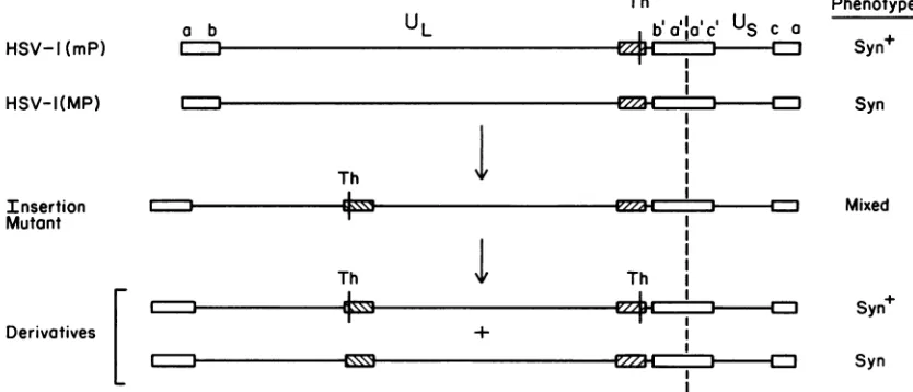
We isolated mutants that contained BamHI
fragment
L fromHSV-l(mP)
DNA inserted in inverted orientation into the tk gene of theHSV-1(MP)
genome(Fig.
2) and viceversa. Construction of these insertion mutants was
previ-ously
describedalong
withevidencethat,amongprogenyof these mutants, alarge
fraction hadgenomic
rearrangements such as inversions of theunique
squence between the inverted repeats of the BamHI-L sequence (20).Multiple
phenotypes
with respect toplaque morphology
wereob-HSV-lI(mP) a b
UL
served amongprogeny of the insertion mutants. For
exam-ple,
most of the mutants derived from HSV-1(MP)by
insertion of BamHI
fragment
L from HSV-l(mP) DNAinitially
formedplaques
of mixedphenotype
(partially Syn,
partially
Syn')
as showninFig.
3A. BothSyn (Fig.
3B) andSyn'
(Fig.
3C) variants, however,could be detectedamong the progeny of these mutants. Viruses isolated from theplaques
of mixedphenotype (Fig.
3A) wereheterogeneous
and included both the
Syn
andSyn'
variants, as well asviruses with mixed
phenotype,
whereas viruses isolated from theSyn
orSyn'
plaques
werehomogeneous
and bred true.An
explanation
for themultiple plaque phenotypes
emerged
from Southern blotanalyses
of ThaIdigests
donetodetermine which allelesof thesyn-J genewerepresent in the insertion mutants. The mutants as constructed should have
Th
IIa
US
HSV-I(MP)
Th Insertion
Mutant
Th
Jl-,
Derivatives
'I,
.1.
1
1
1 1 1 Th I I
I
I
I
--cmaczzzzl----m
I
I
+
Plaque Phenotype
Syn+
Syn
Mixed
Syn+
Syn
FIG. 2. Location and orientation of BamHI-Lsequences(crosshatched boxes)in thegenomesofHSV-l(mP), HSV-1(MP),and insertion mutantsobtainedbyinsertingtheBamHlL fragmentofHSV-l(mP)DNAinto the tkgeneofHSV-1(MP) DNA(20). TheThaI site (Th) is amarker for thewild-typeallele of the
syn-J
gene. This ThaIsite iseliminatedbythemutationresponsiblefor theSyn plaque phenotypeofHSV-1(MP).
:I
on November 10, 2019 by guest
http://jvi.asm.org/
[image:2.612.109.490.74.242.2] [image:2.612.99.516.504.683.2]706 NOTES
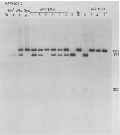
the mutant allele at one position (normal or inserted locus) and the wild-type allele at the other; ThaI digests of their DNAs should contain three fragments capable of hybridizing to thePstI-HincIIprobe described in the legend to Fig. 1 (the 1,212-bp fragment characteristic of the mutant allele and the 979- and 233-bp fragments characteristic of the wild-type allele). If the differences in nucleotide sequences between the mutant and wild-type alleles had been eliminated in progeny of the insertion mutants, then only the 1,212-bp fragment or two smaller fragments (979 and 233 bp) would be detected in the ThaI digests. The results (Fig. 4) demon-strated that DNAs of all the Syn variants isolated had only the ThaI fragment characteristic of the mutant allele (1,212 bp), DNAs of the Syn+ variants had only the fragments characteristic of the wild-type allele (979 and 233 bp), and DNAs from the viruses of mixed phenotype had all three fragments.
Several points about the results presented in Fig. 3 and 4 should be emphasized. First, with one exception, all the mutants analyzed in Fig. 4 actually contain insertions of the BamHI-L sequence in the tk gene, as determined by South-ern blot analyses published elsewhere (20) or not shown in the case of mP-TK133.5. Therefore, those mutants having only one kind of allele of the syn-J gene must have two copies of this allele. Second, some of the insertion mutants isolated contained two identical alleles of the syn-J gene (probably due to marker transfer at the normal or inserted locus having occurred prior to isolation of the mutant). However, most of the insertion mutants as originally isolated contained two different alleles of the syn-I gene. These mutants exhibited mixed plaque phenotype and, through several rounds of plaque purification, continued to throw off both Syn and Syn+ variants. Third, because both Syn and Syn+ variants are produced during development of the plaques of mixed phenotype, it is not possible to determine the plaque phenotype of the original insertion mutant. Fourth, in plaques producing both Syn and Syn+ variants of the insertion mutants derived from strain MP, the mutant and wild-type phenotypes appear to be codominant, in contrast to the Syn+ phenotype of infectious centers pro-ducing both HSV-1(mP) and HSV-1(MP) (9, 13, 14, 21). Possibly some of the other genetic differences between strains MP and mP (19) influence the results obtained in the latter case.
We conclude that, duringpropagation of insertion mutants of the kind described above, differences in nucleotide se-quences between the inserted and normal copies of the duplicated sequence are eliminated at high frequency. The genomicrearrangements described earlier for these insertion mutants (20) provided evidence that intermolecular or intramolecular recombination between inserted and normal copies of theBamHI-L sequence occurs at high frequency. Theelimination ofnucleotide sequence differences could be anotherconsequence of these recombinations, provided that multiple intermolecular recombinations occurred within the BamHI-L sequence or that gene conversion occurred.
We conclude further that an element within theBamHI L fragment must enhance the rate of homologous recombina-tion (and perhaps also gene conversion) in adjacent se-quences. Although a number of HSV-1 insertion mutants containing duplications of other regions of the genome have been analyzed (5, 12, 15), none except those having addi-tional copies of the BamHI-L sequence (20) or the a se-quence (2, 15-17, 24) have given evidence of high frequen-cies ofrecombination between the repeated sequences. The recombination-enhancing elements proposed to exist within
FIG. 3. PhotomicrographsofplaquesproducedonVerocellsby insertion mutants of the kind depicted in Fig. 2. Viruses isolated fromplaques of thekindshown inpanelAwereof threetypeswith respect to plaque phenotype: mixed (A); Syn (B), for derivative MP-TK106.2H; or Syn+ (C), for MP-TK106.2A. Both of these derivativeswereisolated from stocksofMP-TK106.2. The
Syn
and Syn+ derivatives breed true with respectto plaque phenotype.J. VIROL.
on November 10, 2019 by guest
http://jvi.asm.org/
[image:3.612.319.555.96.629.2]MP-TK 106.2
+ 6 PTK13
Syn Mix Syn
MP-TK-a6.
mP-TK133.B A F H I1 8 7 6 3 2 E E 6 5 4 2
mechanismsaredifferent.Onlyasmallportionof the BamHI
L fragment has been sequenced, and, within this region,
there is noevidenthomology with the asequence.
We thank RichardAspellfor technical assistance.
This work was supported by grants from the National Cancer Institute(CA21776 andCA19264) and the American Cancer Society. K.L.P.-G. was atraineeonNational Research Service award T32
I'HS
GM07281.Xh
E@i_
-1212_ so_amso dB
as
-979-233
FIG. 4. Southern blot analyses of ThaI-digested DNAs from insertionmutants and theparental viruses, 1(mP) and HSV-1(MP). TheDNAfragmentswerefractionated by electrophoresison a2% agarose gel and transferred to Nytran. The single-stranded radioactive probewasproduced by synthesis in vitro, by usingas templatetheopposite strandof thePstI-HincII fragment indicatedin the legend to Fig. 1. The insertion mutants TK106.2, MP-TK106.3, MP-TK106.6, MP-TK106.7, MP-TK106.8, and MP-TK106.11wereproduced by cotransfection of HSV-1(MP)genomic DNA with a plasmid containing theBamHI L fragment of HSV-1(mP)DNAinserted intoaclonedcopyof theHSV-1tkgene. The derivativesdesignatedB, A, F,andH wereallplaquepurified from MP-TK106.2. The insertion mutants mP-TK133.2, mP-TK133.4, and mP-TK133.5 were produced by cotransfection ofHSV-1(mP) genomicDNAwithaplasmid containingtheBamHI Lfragment of HSV-1(MP)DNAinserted intoaclonedcopyof the HSV-1 tkgene. Themutantdesignated 6 wasalso isolated fromprogeny obtained after thiscotransfection; it provedtobeaspontaneous TK- mutant, however, and not an insertion mutant. The insertion mutants designated MP-TK106.2, MP-TK106.5, MP-TK106.8, and MP-TK106.11andthederivativedesignated MP-TK106.2Fallproduced thethree typesofplaques shown in Fig. 3. The insertion mutants mP-TK133.2, mP-TK133.4, mP-TK133.5,and MP-TK106.7 and the derivative MP-TK106.2Hproduced only Syn plaques (Fig.3B).The insertionmutant derivatives MP-TK106.2B and MP-TK106.2A, as wellasthespontaneous TK- mutant 6,producedonly Syn+ plaques (Fig. 3C). Sizes of DNA fragments detected by the radiolabeled probearegiven in base pairs. The 979- and 233-bp fragmentswere consistently found together. Overexposure of the autoradiogram (notshown) wasnecessary to detect the 233-bpfragment inevery instance.
theBamHILfragment and thea sequence could be in some waysanalogous to other elements shown to enhance rates of recombination in their vicinity, in both procaryotic and
eucaryotic systems(see, forexample, references 8 and 25).
The question arises whether the BamHI L fragment of
HSV-1 DNA contains the same element causing enhanced rates of recombination as does the a sequence. Lack of
recombination between theaand BamHI-L sequences may
signify only that homology is too limited and not that the
LITERATURECITED
1. Bond,V.C.,and S. Person. 1984. Finestructure physicalmap locationsof alterations that affect cell fusion in herpes simplex virustype 1. Virology 132:368-376.
2. Chou, J., and B. Roizman. 1985. Isomerization of herpes simplex virus 1 genome: identification of the cis-acting and recombination sites within the domain of the asequence. Cell 41:803-811.
3. Davidson, A., and N. Wilkie. 1983. Inversion of the two seg-mentsoftheherpes simplex virusgenome inintertypic recom-binants.J. Gen. Virol. 64:1-18.
4. Debroy, C., N. Pederson, and S. Person. 1985. Nucleotide sequenceofaherpes simplexvirus type 1 gene thatcauses cell fusion. Virology 145:36-48.
5. Gibson, M. G., and P. G. Spear. 1983. Insertion mutants of herpes simplex virus haveaduplication of the glycoprotein D gene andexpress twodifferent forms ofglycoproteinD.J.Virol. 48:396-404.
6. Hayward,G.S.,R.J. Jacob,S. C.Wadsworth,and B. Roizman. 1975.Anatomyof herpessimplex virus DNA:evidence for four populations of molecules that differ in the relative orientations of their long and shortsegments. Proc. Natl. Acad. Sci. USA 72:4243-4247.
7. Hubenthal-Voss, J., and B. Roizman. 1985.Herpessimplex virus 1reiteratedS component sequences(cl)situated betweenthe a sequenceand a4geneare notessential for virusreplication. J. Virol.54:509-514.
8. Keil, R. L., and G. S. Roeder.1984. Cis-acting, recombination-stimulating activity inafragment of the ribosomal DNAof S. cerevisiae. Cell39:377-386.
9. Keller, J. M. 1976. Theexpression ofthe syn- geneofherpes simplex virustype 1. 1.Morphology of infected cells. Virology 69:490-499.
10. Knipe,D.M.,W. T.Ruyechan, R. W. Honess, and B. Roizman. 1979.Moleculargenetics of herpes simplex virus: the terminala
sequencesof theLand Scomponentsareobligatorily identical and constitute apart ofa structural gene mapping predomi-nantly in the S component. Proc. Natl. Acad. Sci. USA 76:4534-4538.
11. Knipe, D. M., W. T. Ruyechan, B. Roizman, and I. W. Hal-liburton. 1978. Molecular genetics of herpes simplex virus: demonstration ofregions of obligatory and nonobligatory iden-tity withindiploid regions of thegenome bysequence replace-mentandinsertion. Proc.Natl. Acad. Sci. USA75:3896-3900. 12. Lee,G.T.-Y.,K. L. Pogue-Geile, L.Pereira, andP. G. Spear. 1982.Expression of herpes simplex virus glycoprotein C froma DNAfragment inserted intothethymidine kinasegeneof this virus.Proc. Natl.Acad. Sci. USA79:6612-6616.
13. Lee, G. T.-Y., and P. G.Spear. 1980.Viral andcellular factors that influence cell fusion induced by herpes simplex virus. Virology 107:402-414.
14. Manservigi,R., P. G. Spear, and A. Buchan. 1977. Cell fusion inducedby herpessimplexvirus ispromotedandsuppressed by different viral glycoproteins. Proc. Natl. Acad. Sci. USA 74:3913-3917.
15. Mocarski, E. S.,L. E. Post, and B. Roizman. 1980. Molecular engineering oftheherpes simplex virus genome: insertion ofa
second L-Sjunction intothe genomecausesadditional genome inversions. Cell 22:243-255.
16. Mocarski, E. S.,and B.Roizman. 1981. Site-specific inversion sequence of the herpes simplex virus genome: domain and structural features. Proc. Natl.Acad. Sci. USA78:7047-7051.
on November 10, 2019 by guest
http://jvi.asm.org/
[image:4.612.61.298.65.330.2]708 NOTES
17. Mocarski, E. S.,and B.Roizman.1982.Structureand role of the herpessimplexvirus DNAterminiininversion, circularization andgeneration of virionDNA. Cell 31:89-97.
18. Poffenberger, K. L., E. Tabares, and B. Roizman.1983. Char-acterization of a viable, noninverting herpes simplex virus 1 genome derived by insertion and deletion ofsequences atthe junction ofcomponentsLand S. Proc. Natl.Acad. Sci. USA
80:2690-2694.
19. Pogue-Geile, K. L., G. T.-Y. Lee, S. K. Shapira, and P. G. Spear. 1984. Fine mapping of mutations inthefusion-inducing MPstrain ofherpes simplexvirustype1.Virology136:100-109. 20. Pogue-Geile, K. L., G. T.-Y. Lee, and P. G.Spear. 1985. Novel
rearrangementsof herpessimplex virusDNAsequences
result-ing fromduplication ofasequence withintheunique region of the Lcomponent.J. Virol. 53:456-461.
21. Roizman, B.1962.Polykaryocytosis. ColdSpringHarborSymp. Quant. Biol.27:327-334.
22. Sheldrick, P., andN.Berthelot. 1974. Inverted repetitions inthe chromosome of herpes simplex virus. Cold Spring Harbor Symp. Quant.Biol. 39:667-678.
23. Skare, J., and W. C. Summers. 1977. Structure and functionof herpesvirusgenomes. II.EcoRI, XbaIand Hind III
endonucle-asecleavage sitesonherpes simplex virustype1 DNA. Virol-ogy76:581-595.
24. Smiley,J. R., B. S.Fong,andW.-C.Leung. 1981.Construction of a double-jointed herpes simplex viral DNA molecule:
in-verted repeats are required for segment inversion and direct repeats promotedeletions. Virology113:345-362.
25. Stahl, F. W., J. M. Crasemann, and M. M. Stahl. 1975. Rec-mediated recombinational hot spot activity in bacterio-phage lambda.III. Chi mutationsaresite mutationsstimulating rec-mediated recombination. J. Mol. Biol. 94:203-212. 26. Varmuza,S. L.,andJ. R.Smiley. 1984. Unstableheterozygosity
in a diploid region of herpes simplex virus DNA. J. Virol.
49:356-362.
27. Wadsworth, S., G. S. Hayward, and B. Roizman. 1976.Anatomy ofherpes simplex virus DNA. V. Terminal reiteration. J. Virol. 17:503-512.
28. Wadsworth,S.,R.J. Jacob, and B. Roizman. 1975.Anatomy of herpes simplex virusDNA.II.Size, composition,and
arrange-mentof invertedterminalrepetitions. J. Virol. 15:1487-1497. 29. Wilkie, N. M. 1976.Physicalmapsforherpes simplexvirustype
1 DNAfor restriction endonucleasesHind III,Hpa-1, andX.
bad. J.Virol. 20:222-233.
J. VIROL.