Copyright © 1991,AmericanSocietyforMicrobiology
In
Vitro Mutagenesis of
a
Full-Length cDNA
Clone of Semliki
Forest
Virus: the Small
6,000-Molecular-Weight
Membrane Protein
Modulates Virus Release
PETER
LILJESTROM,l*
SARILUSA,1
DANNYHUYLEBROECK,2t AND HENRIKGAROFF'DepartmentofMolecular Biology, Karolinska Institute, Novum, S-14157Huddinge, Sweden,' and Cell Biology Program,
EuropeanMolecularBiologyLaboratory, D-6900Heidelberg, Germany2 Received11February 1991/Accepted20April1991
Wereport onthe construction ofa full-length cDNAcloneof Semliki Forestvirus (SFV). By placing the
cDNA under theSP6promoter,infectious RNAcanbeproduced in vitro and usedtotransfect cellstoinitiate virusinfection. Toachieveefficienttransfections, anewprotocol for electroporationof RNA wasdeveloped. This method gave up to 500-fold improvement over the traditional DEAE-dextran transfection procedure.
Sincevirtually 100% ofthe cellscanbe transfectedby electroporation, this method isausefultool for detailed biochemical studies of nullmutations of SFV that abolishproductionof infectious virusparticles.We usedthe cDNA clone of SFV to study what effects a deletion of the6,000-molecular-weight membrane protein (6K
membraneprotein) hadonvirus replication.Thesmall6Kproteinispartofthe structuralprecursormolecule (C-p62-6K-E1) of the virus. Our results conclusively show that the 6K protein is not needed for the heterodimerization of the p62 and El spike membrane proteins in the endoplasmic reticulum,norisit needed
for their transportouttothecell surface. The absenceofthe 6K protein did, however,result inadramatic
reduction in virus release, suggesting that the proteinexertsits functionlate in the assembly pathway, possibly during virus budding.
Enveloped viruses provide useful model systems with whichtostudy membrane assembly,structure,andfunction. Recentstudies with variousretroviruses have demonstrated that the capsid protein alone candirect membrane budding without the involvement of spike glycoproteins (14, 23, 44, 45). However, inthe case ofalphaviruses, it appears to be thevirustransmembranespike proteins which, through their interaction with the nucleocapsid, form the membrane around the particle (48, 50). Our studies focus on the assembly mechanism of the Semliki Forestvirus(SFV). This alphavirus has a single-stranded RNA genome of positive polarity, which therefore functions directly as an mRNA. Replication of this 11,442-nucleotide-long capped and poly-adenylated RNA starts soon after infection upon the initial
translation ofthe 5' two-thirds ofthe genomic RNA, pro-ducing a polyprotein which is posttranslationally cleaved into fournonstructural proteins (nsPl to nsP4). These
pro-teins are responsible for the replication of the plus-strand genomeinto full-length minus strands, which laterin infec-tionare copied into newcapped plus-strand genomes. The minus strands alsoserve astemplates for the synthesis ofa 4,073-nucleotide long subgenomic RNA species which is colinear with the last one-third of the plus-strand genome.
All structural proteins of the virus are encoded by this subgenomic RNA(53).
The structural proteins are synthesized as a polyprotein precursor in the order C (capsid)-E3-E2-6K (6,000-molecu-lar-weightprotein)-E1 (20, 49). OncetheC protein has been synthesized, itfolds to act as aprotease, cleaving itselfoff thenascentchain(28, 40). Thecleavagerevealsan N-termi-nalsignalsequenceinthenascentchain which isrecognized by the signal recognition particle, targeting the nascent
chain-ribosomecomplex totheendoplasmic reticulum(ER)
* Correspondingauthor.
tPresent address: Innogenetics, B-9710Ghent, Belgium.
membrane (5, 22), where it iscotranslationally translocated and cleaved by signal peptidase to the three membrane proteins p62 (precursorform ofE3/E2), 6K,and El (19, 34, 39). Via its E3domain, the p62 protein formsaheterodimer with El (37). The p62-El oligomers are transported out to theplasma membrane, where via thecytoplasmicdomain of p62, they initiate virus budding (21, 41, 56). At a very late (post-Golgi) stageoftransport, thep62 proteiniscleavedto E3 and E2(13), theforms that arefound inmature virions. Thiscleavage exposesthe entry functions ofthe spike and allows the membrane fusion potentialof El tobe activated duringvirusentryinto a newcell(30, 35, 36, 43, 57, 61).
While themajorrole ofp62appearstobeinassemblyand that ofEl in entry, the function ofthe 6K protein is still muchofanenigma.6Kwasoriginallyfound associated with microsomalfractions of infectedcells (58, 59), andwe have recently shown that 6K is anchored in the membrane as a
type I membrane protein. Its signal sequence is located in the cytoplasmic C-terminal region of the p62 polypeptide (34). One function for this small membrane protein is to providethesignalsequenceforEltranslocation. However, recentresultssuggestadditionalroles for 6K.First,although the El signal sequence is located within the C-terminal residuesof the6K, thisfunctioncanalso beprovided bythe 6K signal sequence at the C terminus of the p62 protein as well(34).Therefore, evolutionhashadnoreasontomaintain the6Kgene onthesole basis of El signal sequence
require-ment. Second, the 6Kprotein ispalmitylated, and mutation leadingtoitsunderacylationmodulate virusbudding,leading tothe formation of multinucleated virusparticles (16).
In this work we have studied the role of 6K. We first
describe the construction of a full-length cDNA clone of SFV, whichcanbe usedas atemplatefortheproductionof infectious runoff transcripts, and present a method for an extremelyefficientRNAtransfection system basedon elec-troporation. Usingthissystem, weshow that removal of6K
4107
on November 10, 2019 by guest
http://jvi.asm.org/
from the SFV genome gene region does not interfere with the synthesis or cell surface transport of the other structural proteins but does severely reduce the release of new virus particles on the infected cell surface.
MATERIALS ANDMETHODS
Virus growth and purification.BHK-21cells were grown in BHK medium (GIBCO) supplemented with 5% fetal calf serum, 10% tryptose phosphate broth, 10 mM HEPES (N-2-hydroxyethylpiperazine-N'-2-ethanesulfonic acid) and 2mMglutamine. Ninety percent confluent monolayers were
washed once with phosphate-buffered saline (PBS) and
infected with SFV in minimal essential medium (MEM)
containing 0.2% bovine serum albumin (BSA), 10 mM HEPES, and 2 mM glutamine at a multiplicity of 0.1.
Twenty-four hourspostinfection, themediumwascollected
and cell debriswas removed by centrifugationat8,000 x g
for20 minat4°C.The viruswaspelletedfrom the mediumby centrifugation at 26,000 rpm for 1.5 h in an SW28 rotor at
4°C. The virus was resuspended in Tris-HCl (pH 7.4)-100
mMNaCl-0.5 mM EDTA.
Metabolic labeling and immunoprecipitation. Confluent monolayers of BHK cells grown inMEMsupplemented with 10 mMHEPES, 2 mMglutamine, 0.2% BSA, pencillin (100
IU/ml), and streptomycin (100
Kg/ml)
were infected at amultiplicity of 50 at 37°C. At 1hpostinfection,the medium wasreplced with freshmedium and growth continued for 3.5 h. The medium was removed, and the cells were washed once with PBS and overlaid with methionine-free MEM
containing10 mMHEPES and 2mMglutamine.After 30 min at 37°C, the medium was replaced with the same medium
containing
[35S]methionine
(100,uCi/ml;
Amersham),and the plates were incubated for 10 min at 37°C. The cells were washed twice with labeling medium containing a 10-fold excessof methionine and then incubated insamemediumforvarious times. The plates were put on ice, the cells were washed once with ice-cold PBS, andfinally lysisbuffer(1%
Nonidet P-40, 50 mM Tris-HCl [pH7.6], 150 mM NaCl, 2 mMEDTA, 10
pKg
ofphenylmethylsulfonyl fluoride perml)was added. Cells were scraped off the plates, and nuclei wereremoved bycentrifugationat6,000 rpm for 5 minat4°C
inanEppendorf centrifuge. Immunoprecipitationofproteins
with monoclonal antibodies (4)was performed asdescribed
previously(57) except thatPansorbin(Calbiochem,LaJolla, Calif.) wasused instead ofprotein A-Sepharose.
Surface labeling of protein. Infected cells were
pulse-labeled and chased for the time periods indicated. Cell monolayerswerethenwashed with ice-cold PBS and
subse-quently incubated with PBS containing sulfosuccinimidyl 6-(biotinamido)hexonate (0.5 mg/ml; Pierce-Tecator Inc., Sollentuna, Sweden)for 30 minat4°C(6). The reactionwas stopped byachase-wash with PBScontaining 50mMlysine.
Cells were collected in lysis buffer, and the biotinated moleculeswereprecipitated by streptavidin agarose (Sigma Chemical Co., St. Louis, Mo.) by incubation overnight at
4°C. The agarose pellet was first washed as normally was doneforimmunoprecipitatesand then once with 1%sodium dodecyl sulfate (SDS). Finally, the samples were prepared for SDS-polyacrylamide gel electrophoresis (PAGE) by
heating in SDS sample bufferto
95°C
for 10 min.Electron microscopy. Fornegative staining of virus parti-cles, viruswas collected from the growth medium by sedi-mentation through a 3-ml 20% sucrose cushion inTNE (50 mMTris-HCI [pH 7.4], 100mM NaCl, 1 mM EDTA) in an
SW28rotor at26,000rpmfor 90 minat
4°C.
The supernatantwas removed, and the virus was resuspended in a small volume of TNEovernightonice. The virus
suspension
was stained on carbon-coated grids for 10 s with 1% uranylacetate before
analysis by
electronmicroscopy.
General DNAprocedures.Allbasic DNA
procedures
were doneessentially
as describedpreviously
(47). DNAfrag-ments were isolated from agarose gels by the freeze-thaw
method(3),withinclusion of 3 volumes ofphenolduringthe
freezing
step to increaseyield
andpurity.
Fragments werepurified by benzoylnaphthoyl-DEAE-cellulose chromatogra-phy (52). Plasmids used for
production
of infectious RNA were purified by sedimentation through1 M NaClfollowedby
banding
in CsCl(47).Construction ofafull-lengthSFV cDNA clone.Our strategy for
constructing
theSFV clonewastoprime
cDNAsynthe-sisonseveral
positions along
thetemplate
RNAdownstreamof suitable restriction endonuclease sites defined
by
theknown nucleotide sequence
(18, 55).
Virus RNA was iso-lated byphenol-chloroform
extraction frompurified
virus and used as atemplate
for cDNAsynthesis
essentially
aspreviously
described(27).
Afull-length
cDNA clone of the SFV genome was then constructedby
combining
indepen-dent
overlapping subclones,
using
these sites. Aftercomple-tion of second-strand
synthesis,
the cDNA wastrimmed,
thedouble-strandedadaptor
5'-sticky-EcoRI-HindIII-NotI-XmaIII-SpeI-blunt-3'
wasadded,
and the cDNAwasclonedintoEcoRI-cleaved
pTZ18R
(Pharmacia, Uppsala, Sweden)
as described earlier
(29).
The 5'-endregion
was clonedby
inserting
the cDNA into Smal-HindlIl-cutpGEM1
(Promega
Biotec,
Madison, Wis.).
Todrive in vitro
synthesis
of SFVRNA,
weused the SP6promoter carried on a derivative of
pGEM1.
The firstfull-length
clonecandidate, pSP6-SFV3,
contained the proper SFV sequence5'-ATGG,
with two additional C residues in front. For the 3'-end oftheclone,
the cDNAfragment
containing
69 A residues was selected.By
inclu-sion ofa
unique SpeI
site atthe 3' end of thecDNA,
theplasmid
canbelinearizedtoallowforrunofftranscription
in vitro.Site-directedoligonucleotide
mutagenesis.
For oligonucleo-tidemutagenesis,
relevantfragments
of the SFV cDNA clone were subcloned intoM13mpl8
orM13mpl9
(60)
and transformed(10)
intoDH5oaF'IQ
(GIBCO BRL,
Eggenstein,
Germany).
Replicative-form
DNA from these constructs was transformed intoRZ1032,
and virus was grown in the presence of uridine toincorporate
uracil residues into the viral genome.Single-stranded
DNAwas isolatedby phenol
extraction from
polyethylene
glycol-precipitated phage.
Phosphorylated
oligonucleotides
were used inmutagenesis
withSequenase (United
States BiochemicalCorp.,
Cleve-land, Ohio)
as described earlier(32, 54).
In vitro-madereplicative
formsweretransformed intoDH5atF'IQ,
and theresulting
phage isolates wereanalyzed
for the presence of correct mutationsby dideoxy
sequencing
according
to the U.S. Biochemicalprotocol
forusing Sequenase.
Deletion of the6Kregion
has beendescribed elsewhere(34).
In vitro transcription.
Spel-linearized plasmid
DNA wasused as a
template
for in vitrotranscription.
RNA wassynthesized
at37°C
for1 hin 10- to50-,ul
reaction mixturescontaining
40 mM Tris-HCl(pH 7.6),
6 mMMgCl2,
2 mMspermidine-HCl,
5 mMdithiothreitol,
100 ,ug of nuclease-free BSA perml,
1 mMeachATP,
CTP,
andUTP,
500,uM
GTP,
1 U of RNasin perRI,
and 100to 500 UofSP6 RNApolymerase (Promega)
per ml. Forproduction
ofcapped
transcripts (31),
theanalog
m7G(5')ppp(5')G
orm7G
(5')ppp(5')Awas included in thereaction mixture at 1 mM.
on November 10, 2019 by guest
http://jvi.asm.org/
For quantitation of RNA production, trace amounts of [a-32P]UTP(Amersham) wereincluded in the reaction
mix-tures, and incorporation was measured from trichloroacetic
acid precipitates.
Typically, about 50 ,ug of RNA per ,ug of template was
obtained withuseof20 Uofpolymerase, but the yield could be increasedconsiderably by the useofmore enzyme. The
conditions differ from those reported earlier forthe produc-tion of infectious transcripts of alphaviruses (12, 46). We found that maximumproduction of RNAwasobtained with ribonucleosidetriphosphate concentrations at1 mM.
How-ever,since infectivity also is dependentonthepresenceofa
5' cap structure (see Table 1), optimal infectivity was
ob-tained when the GTP concentration in the transcription reactionwashalved.This drop had onlyamarginal effecton
theamounts ofRNAproduced but raised the specific infec-tivity by a factor of 3 (data not shown). We also tested whetheranincrease intheconcentration ofcapanalog in the
transcription reaction (up to 10 mM) would give better specific infectivity, but thiswas notthe case.
RNA transfection. Transfection ofBHK monolayer cells by the DEAE-dextran methodwasdoneasdescribed previ-ously (46). For transfection by electroporation, RNA was
eitheradded directly from the in vitrotranscription reaction mixtureordiluted with transcription buffercontaining5 mM dithiothreitol and 1 Unit of RNasin per ml. Cells were trypsinized, washed oncewith complete BHKcell medium
andoncewithice-cold PBS (without MgCl2 and CaCl2), and
finally resuspended in PBS to give 107 cells per ml. Cells wereeitheruseddirectlyorstored (inBHK medium)onice
overnight. For electroporation, 0.5 ml of cells was
trans-ferred toa0.2-cmcuvette (Bio-Rad, Richmond, Calif.), and 10to50 ,ul of RNAsolutionwasadded.Electroporationwas performedatroomtemperaturebytwoconsecutive pulsesat
1.5 kV and 25 ,uF, using a Bio-Rad Gene Pulser apparatus with the pulse controller unit set at maximum resistance. The cellswere diluted 1:20 incomplete BHK cell medium
and transferredontotissue cultureplates.Forplaqueassays, theelectroporated cellswereplated together with about 3 x
105 fresh cellsperml, incubatedat37°C for1.5 h, and then overlaid with 1.8% low-melting-point agarose in complete BHKcell medium. After incubationat37°Cfor48h, plaques were visualized bystaining with neutral red.
Gel electrophoresis. Samples for SDS-PAGE were
pre-paredand run aspreviouslydescribed (9, 11, 34).
RESULTS
In vitro production ofinfectious RNA. In ourfirst test of pSP6-SFV3 infectivity, in vitro-made RNA (cRNA) was
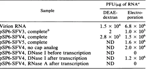
transfected into BHK cells by the DEAE-dextran method. However, although the pSP6-SFV3 RNA was ofapparent fulllengthandproducedvirus(datanotshown), it had much lower infectivitythan did RNA isolated from purified wild-typevirus(vRNA)(Table 1).Because of thispoorefficiency, we considered the possibility that the three additional
non-viral nucleotides GCC,expectedtobeincorporated into the 5' end of the RNA during transcription, could have a
deleterious effect (the Gcomes from initiation of
transcrip-tiononthevectorpart). Therefore,weused oligonucleotide-directedmutagenesistoremoveeither both Cresiduesorall three GCC residues. The changed fragments were subse-quently recloned into pSP6-SFV3, giving pSP6-SFV4 and pSP6-SFV5, respectively.
[image:3.612.312.555.90.205.2]The three clone derivatives were compared for their efficiencies of promoting RNA production in vitro. Since
TABLE 1. Infectivity of SFV RNA
PFU/ILg of RNA'
Sample DEAE- Electro-dextran poration
Virion RNA 1.5 x 10⁴ 6.8 x 10⁶
pSP6-SFV3, completeb 2 1.0 x 10³
pSP6-SFV4, complete 2.8 x 10³ 1.5 x 10⁶
pSP6-SFV5, complete ND 1.6 x 10⁶
pSP6-SFV4, no cap analog ND 2.0 x 10⁴
pSP6-SFV4,DNase Ibeforetranscription ND 0 pSP6-SFV4, DNase I after transcription ND 1.2 x 10⁶ pSP6-SFV4, RNase A after transcription ND 0
aLinearly extrapolated from transfections using10 to 100 ngofRNA.ND,
notdetermined.
b Complete,conditions oftranscriptionasoutlinedinMaterialsand Meth-ods.
pSP6-SFV5 lacked the G residue at the position of SP6
transcription initiation, transcription reactions with this
de-rivativeincluded the cap analogm7G(5')ppp(5')A. The RNA produced was analyzed by agarose gel electrophoresis and was full length in all cases (data not shown). However,
transcriptionof pSP6-SFV4 produced much more RNA than
didthatof the two other clone derivatives, andquantitation
byperformingsimilartranscriptionreactionsin the presence
of labeled UTP showed that the transcription levels of
pSP6-SFV3 and pSP6-SFV5 wereonly 16 and 2%, respec-tively, that ofpSP6-SFV4. Thus, the presence of the twoC residuesimmediatelydownstreamofthestartingGinhibited efficient transcriptionin pSP6-SFV3.
Optimization of transfectionefficiency. Sinceit isofutmost importance to be able to perform detailed biochemical analysis of null mutations (i.e., mutations that have abol-ished virusproduction),itwascrucialtodevelopatechnique
that would allow as high RNA transfection efficiencies as
possible. If all cells could be transfected, then the viral
replication machinery would ensure the same expression
levelfornullmutationasforwild-typeinfection.Clearly,the useof DEAE-dextran transfection wouldnotbeappropriate
for suchexperiments, sinceoptimally only 0.2%of thecells aretransfected, which would makeanalysisdifficult because ofhigh background levels from untransfected cells.
There-fore,wedecidedto usethemethodofelectroporation forcell transfection, and experimentswerecarriedout todetermine
theoptimal conditionsfor RNAtransfectionin BHKcellsby performingplaque titration assaysusingvRNAasdescribed in Materials and Methods. Optimal transfection close to
100%efficiencywasobtained by two consecutivepulses of 1.5 kV at 25 ,uF, under which negligible amounts of cells were killed. Wefound that itwasbettertokeepthecells at roomtemperature thanat0°C duringtheprocedure. Trans-fectionby electroporation was also measured as afunction
of input RNA, and about 2 ,ug of cRNA was needed to transfect all cells ofa sample containing 5 x 10⁶ cells. In
comparison, RNA isolated from purified virus was more
efficient, needing only 0.5 p,g to reach 100% transfection.
Since the 3' poly(A) tailof the cRNA carried four nonviral nucleotides (fromtheSpeI sticky end), thedifference could be due tothis fact.
Transfection as measured by plaque assays showed that therewere qualitativedifferences in the
specific
infectivities of the different cRNAs produced.pSP6-SFV4
andpSP6-SFV5wereaboutequally infectious, whereasthe
infectivity
of pSP6-SFV3 was1,000-fold
lower(Table
1). Since theon November 10, 2019 by guest
http://jvi.asm.org/
V C A V C AV c -A V C A V C A V C A C A
107kDa-D
ad
p62- -
*
r-I'll~~~~~~~~~~~~~~~~~~~~~~~~~~~~~~~~
us b@-O W
"me
E2E
.
C-1 2 3 4 5 6 7 B 9 10 11 12 13 14 15 16 17 18 U n
FIG. 1. Proteins from SFV-infected cells analyzed by 12% SDS-PAGE. Wild-type genomic RNA (V) or invitro-made RNA of the wild-type clone (C) or its 6Kdeletion derivative(A)waselectroporated intoBHKcells andpulse-labeled for 10min 7 hpostelectroporation. Lanes: 1 to 3, lysates after a10-minchase period;4to 6, lysates aftera2-h chase period;7 to9, immunoprecipitation with an anti-El monoclonal antibodyof10-min-chaselysates; 10 to12, immunoprecipitation withananti-E2 monoclonalantibody of10-min-chaselysates; 13 to15,immunoprecipitation with anti-El of 2-h-chaselysates;16to18,immunoprecipitation withanti-E2 of 2-h-chaselysates;19 and20, virusparticles pelleted from growthmedium aftera2-h chase (note that these lanes have beenexposed foralongerperiodoftimetobetter showtheproduction ofthe 6Kdeletionvirus). Positions ofthe107-kDa, p62, E2,El, and Cproteinsareindicatedonthe left.
infectivities of pSP6-SFV4 and pSP6-SFV5 RNAs were equal but the former allowed much more RNA to be pro-duced in vitro, pSP6-SFV4 was chosen for all subsequent experiments.
Synthesis and assembly of viral proteins when expressed from pSP6-SFV4. To analyze the structural proteins
pro-duced,cRNAwaselectroporated intocells andpulse-chase analysis was performed. vRNA was used as a control. In both cases, transfection resulted in the production of iden-tical structuralprotein species that had similar transport and assembly properties. Afterashortpulse and chase, the p62,
El,C, and 6K proteins as well as the 107-kDa species (which represents anuncleaved and unglycosylated protein of p62 plus 6K plus El)were produced(Fig. 1, lanes 1 and 2;Fig.
2, lanes 1 and 2), a pattern which typically can be seen in
SFV-infected cells. Upon a prolonged chase period (120
min), theseheterodimershadbeen transportedoutofthe ER through the trans-Golgi compartment, as visualized by the
cleavage ofp62toE2(andE3,whichisnotvisibleonthegel)
andby the slight increase in the size of El as aresult ofits
sialylation(26) (Fig. 1, lanes 4 and 5). Immunoprecipitation using monoclonal antibodies directed against El (Fig. 1, lanes 7, 8, 13, and 14) and E2 (lanes 10, 11, 16, and 17) showed that thep62-E2 and El membrane proteins formed
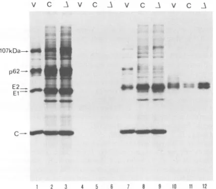
heterodimeric complexes. Biotinylation of cell surface pro-teins showed that pulse-labeled p62 andEl had reached the surface aftera60-min chase (Fig. 3, lanes 7, 8, 10, and 11) but notafter a 10-min chase (lanes 1, 2, 4, and 5). Analysis of the growth medium after a 2-h chase showed that efficient virus formation was taking place with use of cRNA (Fig. 1, lane 29). Thus, the cDNA clone of SFV appeared to function exactly like the vRNA.
6Kisnotneeded for correct production or heterodimeriza-tion ofp62 andEl. Using a 6K deletion mutation of the 26S
cDNA, we have earliershown that 6K is not needed for in
vitroproduction or membrane translocation of p62 and El (34). To determine how the deletion might affect virus assembly events, it was transferred into pSP6-SFV4, and RNA was produced in vitro and electroporated into BHK
cells. We first analyzed the protein synthesis and het-erodimerization ofp62 and El in the ER. Gel analysis of
pulse-labeled proteins showed that p62 and El were pro-duced, although signal peptidase cleavagebetween them was notquiteasefficientasfor the wild type,resultinginaslight accumulation of the 107-kDa species (Fig. 1, lane 3). A
portion of this protein had slower mobility, and the larger
V C A
E3-6K- _
1 2 3
FIG. 2. Tentotwenty percent gradient gel analysis ofproteins encodedbywild-type (V), clone(C),ordeletion6K(A)RNA upon transfectionintoBHKcells.Cellswerepulse-labeled for10min and then chased for 2 h. Totallysatesareshown.
E
on November 10, 2019 by guest
http://jvi.asm.org/
[image:4.612.118.517.77.252.2] [image:4.612.399.488.442.686.2]v c A v C A v c A v C A
..A.
107kDa- -s
p62-"91
E2 El
__O
A
41,01mw " .m
FIG. 4. Electron micrographs of negatively stained wild-type SFVderived from the cDNA clone (A) and 6K deletion virus (B).
1 2 3 4 5 6 7 8 9 10 11 12
FIG. 3. Twelve percent SDS-PAGE analysis of surface-labeled SFV membrane proteins. Cells were electroporated with genomic RNAfrom purified virus(V),cloneRNA(C),ordeletion6K RNA
(A),pulse-labeled with[35S]methionine for10min, and chased for10
(lanes 1to6)or60(lanes 7to12)min before biotinylation.Lanes: 1
to 3 and 7to9,total cell lysates;4to6and 10 to12, streptavidin precipitates of biotinylated proteins.
band probably represented molecules that had been partially translocated and thusglycosylated (34). 6Kwas missing,as expected (Fig. 2, lane 3). Immunoprecipitation of either El (Fig. 1, lane 9)orp62 (lane 12) with monoclonal antibodies
showed that the6K deletion hadno effect on heterodimer-ization, since almost quantitative coprecipitation was ob-tained.
6K isnotneededfortransportof p62-Elheterodimerstothe cell surface. When the cells expressingthe 6Kdeletionwere pulse-labeled and chased for 2 h, p62 was completely
cleavedtoE2(and E3)and theapparentsize ofElincreased (Fig. 1, lane 6), suggesting that both proteins had been transported from the ER through the Golgi compartment.
Coprecipitation ofElorE2showed that thecomplexeswere
intact and stable even after cleavage of p62 to E2 (Fig. 1, lanes 15 and18). Toprovethatthe heterodimer had reached
the cell surface, we performed cell surface labeling with
biotin. Cellswerepulse-labeledand thenchased foreither10 or60minbefore biotinylation. After the short chaseperiod, no labeled membraneproteins had reached the cell surface
andtherefore couldnot reactwithbiotin (Fig. 3,lanes3and 6). After the 60-min incubation, when most of the p62 species had been processed (lane 9), both El and E2 were efficiently labeled with biotin, showing that both proteins hadreached the cell surface (lane 12).
6K is not essential for virus production. To determine whether the 6K deletion construct would allow virus pro-duction, we analyzed media from transfected and pulse-labeled cells. After 2 h ofchase, virus particles could be detected in the growth medium, but only to about 2% (as quantified from the gel) of the wild-type amount (Fig. 1, lanes19 and 20). When electroporated cellswereassayed for plaque formation, wefound nodecrease in PFUper
micro-gram of RNA incomparison with the wild type. However, the plaques were clearly smaller, and subsequent virus growth experiments showed that the 6K deletion variant resulted in a marked reduction in virus growth (data not
shown). Figure4 showsan electron microscopic analysisof negatively stained virions. The virus particles derived from the 6K deletion clone derivative hadawild-typeappearance.
DISCUSSION
We constructedacDNA clone of SFV thatcanbe usedto
produce infectiousRNAtranscriptsinvitro by usingthe SP6 RNApolymerase.Thestructuresof the 5' and3'ends of the cDNA insertwerecrucialforbothefficient transcriptionand expression. Transcripts with three extra nonviral nucleo-tides at the 5' end were substantially lower in specific infectivity than those with only an extraG residue orthose without any additional 5' nucleotide. In the case of the closely related Sindbisvirus, the presence ofeightextra G residuesat the 5' endtotally abolishedinfectivity, whereas thepresenceof only oneextranucleotideat the end had no
apparenteffect(46). The SFV cRNAwasonlyabout 20%as infectious as RNA, probably because of the four nonviral nucleotides present atthe 3' end. This finding is consistent with the result with Sindbis virus showing that the 3' end contained nine extra residues, resulting in only 10% infec-tivity. These were apparently not replicated by the virus replicase andwere lostduring infection(46).
Themostcommonly used method of transfecting mamma-liancells with nucleic acids has traditionally been by calcium phosphate precipitation (24) ortreatment with
DEAE-dex-tran (38). However, these methods suffer from low effi-ciencies,and therefore newtransfection proceduressuchas
theuseofliposomes (42)orcationiclipids (2, 15)have been developed. Since most of these techniques are able to transfectonly a minor fraction of the cells ina sample, we
turned our focus to electroporation, a method that has gained increased popularity for the transfection of DNA, RNA, and proteins into both eukaryotic and prokaryotic cells (references 1, 7, 8, and 51 and referencestherein). By optimizingthetechnique, we wereabletotransfectvirtually
100% of the cells, an increase in transfection frequency which in our hands was 500-fold higher than with the
DEAE-dextran procedure, which has been used for the transfection of other infectious RNAs ofalphaviruses (33, 46). Most important,this method nowallowsustoperform biochemicalexperimentswith nullmutantsof the virus(i.e., variants that are unable to produce infectious particles because ofablock in theassemblyorentrypathways), using the same high expression level as that ofwild-type virus. This high expression level and concomitant host shut-off obviate the need for antibody-mediated concentration of viralproteinsbefore SDS-gel analysis.
B
on November 10, 2019 by guest
http://jvi.asm.org/
[image:5.612.65.284.77.269.2] [image:5.612.320.543.78.198.2]Also,ourexpressionsystemisverybeneficial for studying viable mutants of SFV since it allows phenotypic analysis afteroneround of replication. BeinganRNAvirus, SFV has a high frequency of mutation, andaspointed out byothers (25), it is crucial that analysis of mutated genomes be performed very early after transfection without extensive passaging of the virus; otherwise, revertants or second-site mutations mayarise, thus interferingwith theinterpretation
of the alleged genotypeunder study.
As a first application of the cDNA clone to study the assembly of SFV,weintroduceda6K deletion mutation into plasmid pSP6-SFV4. The 6K deletion construction allows the membrane translocation ofEl and internalcleavagefrom p62 via the remaining signal sequence which resides in the cytoplasmic domain ofp62 (34). Our results show thatp62 and El are synthesized normally and that they complex efficiently with each otheraswellasbecometransportedto the cell surface. However, amajorreduction could be seen
inthe release ofnewvirusparticles,and thusweassumethat
6K plays a role at a very late stage ofassembly, possibly during the budding process. Recently, Gaedigk-Nitschko
and coworkers (16, 17) showed that point mutations of 6K leadingtoits underpalmitylation also resulted in a substan-tialreduction in virus release.Incontrast toourresults, they also showed that the released viruses consisted mainly of aberrantly enveloped particles containing multiple nucleo-capsids. One explanation for this phenotype could be that the mutated 6K notonly reducedefficiency ofbuddingbut also caused interferencewith the normalenvelopmentof the nucleocapsid. The same group also showed that small amounts of 6K (8 to 10%) are incorporated into the virus particle. However, the datapresented here indicate that 6K is essentialfor neitherthestructurenortheinfectivityof the virion, sincethe specific infectivityof the 6K deletion virus equalsthatof thewild-type virus(datanotshown). Work is
nowdirected towardunderstandingthe mechanismbywhich 6Kexertsitsfunctiononvirus release. Weplantodetermine thepossibleassociations of 6K with the other virus subcom-ponents duringthe maturation process.
ACKNOWLEDGMENTS
Weacknowledge Paivi Nieminen andRaijaJonsson for excellent
technical assistance. WeareindebtedtoCarl-HenrikvonBonsdorff forexpertadviceonelectronmicroscopy.
The work was supported by the Swedish Medical Research Council(grant B88-12X-0872-O1A),the Swedish National Board for
TechnicalDevelopment (grant 87-02750P),and the Swedish Natural ScienceResearch Council(grantB-BU9353-301).
REFERENCES
1. Andreason, G. L., and G. A. Evans. 1989. Optimization of
electroporation for transfection of mammalian cell lines. Anal. Biochem. 180:269-275.
2. Behr, J.-P., B. Demeneix, J.-P. Loeffler, and J. Perez-Mutul. 1989. Efficientgenetransfer into mammalianprimaryendocrine cells with lipopolyamine-coated DNA. Proc. Natl. Acad. Sci. USA86:6982-6986.
3. Benson, S. A. 1984. A rapid procedure for isolation of DNA
fragments fromagarosegels. BioTechniques 2:66-68.
4. Boere,W. A.M., T.Harmsen, J.Vinje,B.J. Benaissa-Trouw,
C. A. Kraaijeeveld, and H. Snippe. 1984. Identification of
distinctantigenicdeterminantsonSemliki Forest virusby using
monoclonal antibodies with different antiviral activities. J. Vi-rol. 52:575-582.
5. Bonatti, S., G. Migliaccio, G. Blobel,and P. Walter. 1984.Role of thesignal recognition particle in themembraneassemblyof Sindbis viralglycoprotein. Eur. J. Biochem. 140:499-502.
6. Brandli, A.,R.G.Parton,and K. Simons. 1990.Transcytosisin
MDCK cells:identification ofglycoproteinstransported bidirec-tionallybetween bothplasmamembrane domains. J.Cell Biol. 111:2909-2921.
7. Callis, J., M. Fromm, and V. Walbot. 1987. Expression of mRNA electroporated into plant and animal cells. Nucleic Acids Res. 15:5823-5831.
8. Chakrabarti, R.,D. E.Wylie,andS. M.Schuster. 1989.Transfer of monoclonalantibodies intomammalian cellsby
electropora-tion.J. Biol. Chem. 264:15494-15500.
9. Chamberlain,J.P. 1979.Fluorographicdetectionof radioactiv-ity in polyacrylamide gels with water-soluble fluor, sodium salicylate. Anal.Biochem. 98:132-135.
10. Chung, C. T.,andR.T. Miller. 1988. Arapidand convenient method forthepreparationandstorageofcompetentbacterial cells. Nucleic Acids Res. 16:3580.
11. Cutler,D.F., and H. Garoff. 1986. Mutantsof the membrane-binding regionofSemlikiForest virus E2protein.I.Cellsurface transport andfusogenic activity. J.Cell Biol. 102:889-901. 12. Davis,N.L.,L.V.Willis, . F.Smith,andR.E.Johnston.1989.
In vitrosynthesisof infectious Venezuelanequine
encephalitis
virus RNA from acDNA clone: analysis ofaviable deletion
mutant. Virology171:189-204.
13. deCurtis, I.,andK.Simons.1988.Dissection of SemlikiForest virusglycoproteindeliveryfromthetrans-Golginetworktothe cellsurfaceinpermeabilizedBHKcells. Proc.Natl. Acad. Sci. USA85:8052-8056.
14. Delchambre, M.,D.Gheysen,D.Thines,C.Thiriart,E.Jacobs,
E. Verdin, M. Horth, A.Burny, and F. Bex. 1989. The GAG precursor of simian immunodeficiency virus assembles into virus-likeparticles. EMBO J. 8:2653-2660.
15. Felgner,P.L.,T. R.Gadek,M.Holm,R.Roman,H. W.Chan,
M. Wenz, J. P. Northrop, G. M. Ringold, and M. Danielsen. 1987.Lipofection:ahighlyefficient,lipid-mediated DNA-trans-fectionprocedure. Proc. Natl. Acad.Sci. USA84:7413-7417. 16. Gaedigk-Nitschko, K.,M.Ding,M. A.Levy,andM.Schlesinger.
1990. Site-directed mutations in the Sindbis virus 6K
protein
reveal sites for fatty acylation and theunderacylated proteinaffectsvirus releaseandvirionstructure.Virology175:282-291. 17. Gaedigk-Nitschko, K., and M. Schlesinger. 1990. The Sindbis virus 6Kproteincanbedetected in virions and isacylatedwith
fattyacids. Virology175:274-281.
18. Garoff, H., A.-M. Frischauf, K. Simons, H. Lehrach, and H.
Delius. 1980. Nucleotidesequence of cDNAcodingfor Semliki Forest virus membrane glycoproteins. Nature (London) 288: 236-241.
19. Garoff,H., D. Huylebroeck, A. Robinson, U. Tillman, and P. Liljestrom. 1990. The signal sequence of the p62
protein
of SemlikiForestvirus isinvolved in initiation butnotincomplet-ingchaintranslocation.J. Cell Biol. 111:867-876.
20. Garoff, H.,C.Kondor-Koch,and H.Riedel.1982.Structureand assembly of alphaviruses. Curr. Top. Microbiol. Immunol. 99:1-50.
21. Garoff, H.,and K.Simon.1974.Location ofthespike
glycopro-teins in theSemlikiForest virus membrane. Proc.Natl. Acad. Sci. USA71:3988-3992.
22. Garoff,H.,K. Simons,and B.Dobberstein. 1978. Assemblyof SemlikiForest virus membraneglycoproteinsinthemembrane of theendoplasmicreticulum invitro.J. Mol. Biol. 124:587-600. 23. Gheysen, D.,E.Jacobs,F. deForesta,C.Thiriart,M.Francotte,
D. Thines, and M. De Wilde. 1989. Assembly and release of HIV-1 precursor Pr55gagvirus-likeparticles fromrecombinant baculovirus-infectedcells. Cell59:103-112.
24. Graham,F.L.,andA.J.vander Eb.1973. Anew
technique
for theassay ofinfectivityof human adenovirus 5 DNA. Virology52:456-467.
25. Grakoui, A., R. Levis,R. Raju,H. V. Huang,and C.M. Rice. 1989. Acis-actingmutation in theSindbisvirusjunction
region
affecting subgenomicRNAsynthesis. J. Virol. 63:5216-5227. 26. Green, J., G.Griffiths,D. Louvard, P.Quinn,andG. Warren.1981. Passage of viral membrane proteins through the
Golgi
complex. J.Mol. Biol. 152:663-698.27. Gubler, U.,and B.J. Hoffman.1983. Asimpleand very efficient method forgeneratingcDNAlibraries. Gene 25:263-269.
on November 10, 2019 by guest
http://jvi.asm.org/
28. Hahn, C. S., and J. H. Strauss. 1990. Site-directed mutagenesis oftheproposedcatalytic amino acids oftheSindbis virus capsid protein autoprotease. J. Virol. 64:3069-3073.
29. Haymerle, H., J. Herz, G. M. Bressan, R. Frank, and K. K. Stanley. 1986. Efficientconstruction of cDNA libraries in plas-mid expressionvectorsusing an adaptor strategy. Nucleic Acids Res. 14:8615-8124.
30. Helenius, A., M. Kielian, I. Mellman, and S. Schmid. 1989. Entry ofenveloped viruses into their host cells, 145-161. In R. W.Compans,A.Helenius,andM. B. A. Oldstone (ed.), Cell biology of virus entry, replication, and pathogenesis. Alan R. Liss, Inc., NewYork.
31. Krieg, P. A., and D. A. Melton. 1987. In vitro RNA synthesis withSP6 RNA polymerase. Methods Enzymol. 155:397-415. 32. Kunkel, T. A., J. D. Roberts, and R. A. Zakour. 1987. Rapid and
efficient site-specific mutagenesiswithout phenotypic selection. MethodsEnzymol. 154:367-382.
33. Levinson, R. S., J. H. Strauss, and E. G. Strauss. 1990. Com-plete sequence ofthegenomic RNA of O'Nyong-nyong virus anditsuseintheconstruction ofalphavirus phylogenetic trees. Virology 175:110-123.
34. Liliestrom,P., and H. Garoff. 1991.Internally located cleavable signal sequencesdirect the formation of Semliki Forestvirus membrane proteins from a polyprotein precursor. J. Virol. 65:147-154.
35. Lobigs, M., and H. Garoff. 1990.Fusion function oftheSemliki Forest virus spike is activated by proteolytic cleavage of the envelopeglycoproteinp62. J. Virol. 64:1233-1240.
36. Lobigs, M., J. M. Wahlberg, and H. Garoff. 1990.Spike protein oligomerization control of SemlikiForestvirusfusion.J.Virol. 64:5214-5218.
37. Lobigs, M., H. Zhao, and H. Garoff. 1990.Function ofSemliki ForestvirusE3 peptide in virusassembly: replacement ofE3 with anartificial signalpeptide abolishes spike heterodimeriza-tionandsurface expression ofEl. J.Virol. 64:4346-4355. 38. McCutchan, J. H., and J. S. Pagano. 1968.Enhancement ofthe
infectivity of simian virus40deoxyribonucleicacid with dieth-ylaminoethyl-dextran.J. Natl.CancerInst.41:351-356. 39.
Melangon,
P., and H. Garoff. 1986.Reinitiation of translocationin theSemlikiForestvirusstructuralpolyprotein: identification of thesignal for the El glycoprotein. EMBOJ. 5:1551-1560. 40.
Melangon,
P., and H. Garoff. 1987. Processing ofthe SemlikiForestvirus structuralpolyprotein: role of the capsidprotease. J. Virol. 61:1301-1309.
41. Metsikko,K., and H. Garoff. 1990.Oligomers of the cytoplasmic domainof thep62/E2 membrane protein of SemlikiForestvirus bindtothenucleocapsid in vitro.J. Virol.64:4678-4683. 42. Nikolau, C., A. Legrand, and G. E. Grosse. 1987. Liposomesas
carriers for in vivo gene transfer and expression. Methods Enzymol. 149:157-176.
43. Omar, A., and H. Koblet. 1988. SemlikiForest virus particles containingonly the El envelope glycoproteinareinfectious and caninducecell-cell fusion. Virology166:17-23.
44. Rasmussen, L., J. K. Battles, W. H. Ennis,K. Nagashima, and M. A.Gonda. 1990. Characterization ofvirus-likeparticles pro-ducedbyarecombinant baculoviruscontainingthe gag geneof thebovineimmunodeficiency-like virus. Virology178:435-451.
45. Rhee,S.S.,H.Hui,and E.Hunter. 1990.Preassembledcapsids oftype Dretroviruses contain a signal sufficientfortargeting specificallytotheplasmamembrane. J. Virol. 64:3844-3852. 46. Rice, C. M., R. Levis, J. H. Strauss, and H. V. Huang. 1987.
Production ofinfectious RNA transcripts from Sindbis virus cDNAclones: mappingof lethalmutations,rescue of a temper-ature-sensitive marker, and in vitro mutagenesis to generate defined mutants. J.Virol. 61:3809-3819.
47. Sambrook,J., E. F. Fritsch, and T. Maniatis. 1989. Molecular cloning:alaboratorymanual, Cold Spring Harbor Laboratory, ColdSpring Harbor, N.Y.
48. Saraste, J., C.-H. von Bonsdorff, K. Hashimoto, L.Kaariainen, and. S. Keranen. 1980. Semliki Forest virus mutants with tem-perature-sensitivetransport defect ofenvelope proteins. Virol-ogy 100:229-245.
49. Schlesinger, S.S.,and M. J. Schlesinger. 1986. Formationand assembly of alphavirus glycoproteins, p. 121-148. In S. S. Schlesinger and M. J. Schlesinger (ed.), The Togaviridae and Flaviviridae. Plenum Press, New York.
50. Schlesinger, S. S., and M. J. Schlesinger (ed.). 1986. The TogaviridaeandFlaviviridae,p.453. PlenumPress,NewYork. 51. Shigekawa, K., and W. J. Dower. 1988. Electroporation of eukaryotes andprokaryotes: ageneral approach to the intro-duction of macromolecules into cells. BioTechniques6:742-751. 52. Silhavy, T. J., M. L. Berman, and L. W. Enquist. 1984. Experiments with gene fusions, p. 303. Cold Spring Harbor Laboratory,ColdSpring Harbor,N.Y.
53. Strauss,E.G.,andJ. H. Strauss.1986. Structure andreplication of the alphavirusgenome, p. 35-90. In S. S. Schlesinger and M. J.Schlesinger (ed.), TheTogaviridae and Flaviviridae. Ple-numPress,NewYork.
54. Su, T.-Z., and M. R. El-Gewely. 1988. A multisite-directed mutagenesisusing T7 DNApolymerase: application for recon-structing a mammalian gene. Gene 69:81-89.
55. Takkinen, K. 1986.Complete nucleotide sequenceofthe
non-structural proteingenesof Semliki Forest virus. NucleicAcids Res. 14:5667-5682.
56. Vaux, D. J. T., A. Helenius, and I. Mellman. 1988. Spike-nucleocapsid interaction in SemlikiForest virus reconstructed usingnetworkantibodies. Nature(London) 336:36-42. 57. Wahlberg, J. M., W. A. Boere, and H. Garoff. 1989. The
heterodimeric association between the membrane proteins of Semliki Forest viruschanges its sensitivity to mildly acidic pH duringvirus maturation.J. Virol. 63:4991-4997.
58. Welch, W.J., and B. M. Sefton. 1979. Two smallvirus-specific polypeptidesareproducedduring infectionwith Sindbis virus. J. Virol. 29:1186-1195.
59. Welch, W. J., and B. M. Sefton. 1980. Characterization ofa
small,nonstructural viralpolypeptidepresentlateduring infec-tion of BHKcellsby SemlikiForestvirus.J.Virol. 33:230-237. 60. Yanisch-Perron, C., J. Vieira,andJ. Messing. 1985. Improved M13 phage cloning vectors and host strains: nucleotide se-quencesoftheM13mpl8andpUC19vectors.Gene33:103-119. 61. Ziemiecki,A., H.Garoff,and K.Simons. 1980.Formationof the SemlikiForestvirus membraneglycoprotein complexes in the infected cell.J. Gen. Virol. 50:111-123.