0022-538X/91/062829-10$02.00/0
CopyrightC) 1991, American Society for Microbiology
A
269-Amino-Acid
Segment with
a
Pseudo-Leucine
Zipper and
aHelix-Turn-Helix Motif
Codes for the Sequence-Specific
DNA-Binding Domain of
Herpes
Simplex
Virus
Type
1Origin-Binding Protein
SUMITRA DEB* ANDSWATI PALIT DEB
Department of Microbiology, UniversityofTexasHealth Science Center,
7703Floyd Curl Drive, SanAntonio, Texas 78284-7758
Received 26 November 1990/Accepted 27 February 1991
The UL9geneof herpes simplex virus (HSV) codes foraDNA-binding protein (OBP) thatinteractssequence specificallywiththe originof replication. This protein is essential for HSV DNA replication incultured cells. TheUL9gene wasclonedintoaplasmidvectordownstream of theSP6 RNA polymerasepromoter. Byusing in vitro transcription and translationsystems, a full-length OBP was synthesized. This synthetic protein is recognized byanantiserumgeneratedagainsttheC-terminal decapeptide of OBP and is functionally active in
binding to
Oris
sequence specifically. The in vitro-synthesized protein has sequence specificity for bindingsimilartothat found for theinvivo-generated OBP. A totalof14in-framedeletion and insertionmutantsof
theUL9gene weregenerated and expressed in vitro. Using these deletion mutants, we determined that the 269-amino-acidstretchdefinedbyamino acids 564to832 localizes the
Oris-specific
DNA-binding domain. TheN-terminalboundaryisbetweenamino acids 565and596, while the C terminus lies between amino acids 833 and 805.This segment containsahelix-turn-helix moiety andapseudo-leucine zipper, neither ofwhich alone cansupportDNAbinding.The other leucinezipper fromaminoacids150to173 isnotrequiredforthe in vitro
sequence-specffic DNA-binding activityofOBP.
Sevenvirus-encoded genes havebeen identified that are
required and sufficient for origin-dependent replication of herpes simplex virus (HSV)DNA incultured cells (3-5, 18, 25, 28, 31). Thegene products include a DNA polymerase (UL30), asingle-stranded DNA-binding activity (UL29;
pre-viously termed ICP8),anabundant protein of 65 kDa(UL42)
that binds tightly to double-stranded DNA in a
sequence-independentmannerandinteracts with HSVDNA polymer-ase as an accessoryfactor (12, 13, 17), andanorigin-binding
protein (OBP; UL9)that sequence specifically bindsto the
originofreplication (10, 14-16, 20, 27, 35). Threeadditional
proteinsencoded by the UL5, UL8, and UL52genescanbe
isolated fromHSV-infectedcellsas acomplexand haveboth
primase and helicase functions (9, 13, 36).
OBP is theonly proteinknowntodate that bindssequence specifically to the origin of replication and hence may functionastheinitiator ofDNAreplication. Eliasetal. (16) initially detected OBP in HSV-infected nuclear extracts.
This OBP is theproduct ofthe UL9gene(27). We (10) and others (20, 35) have also identified its presence in HSV-1-infected Verocell nuclearextracts.OBP has beenpurifiedto apparent homogeneity from HSV-infected Vero cells (15)
andfrom insectcellsinfected withrecombinantbaculovirus expressing OBP (Sa, 20a, 25a). We have shown that the
binding of OBP is correlated to the origin function of
Oris
(10). An
Oris
mutant withpartial deletion in the sequence-specific binding sitecan neitherreplicate norbind toOBP.Competition experiments with a set of OBP-binding-site mutantsperformed byus(10)andothers (14), a
demonstra-tion of homology between origin sequences of different
herpesviruses(1, 33),andmethylationinterferenceanalysis
* Correspondingauthor.
(20) indicate that the sequence 5'-YGYTCGCACT-3' is
crucialfor binding. Apart from the origin-binding activity,
OBP has helicase and ATPase activities(2, 5a).Correlations amongstructureand functionsof thismultifunctionalprotein
areyet tobereportedindetail inthe literature. Weir etal.
(35)cloned approximatelytwo-thirds of theN-terminal por-tion of theUL9codingframe inafusionsystem.Extractsof Escherichia coli transformed with this clone showed
se-quence-specific binding.
To ourknowledge, the data in this report represent the first successful expression of OBP by using in vitro tran-scription and translationsystems. The syntheticgene prod-uct binds to the
Oris
sequence with the same sequence specificity as does OBP from HSV-infected Vero cells orthat synthesized by recombinantbaculovirus. Deletion
mu-tants of the UL9 gene were constructed to define the boundaries of the DNA-binding domain. The N-terminal
boundarylies between amino acids 565 and 596, while the
C-terminal boundary is betweenamino acids 833 and 805.
These results indicate thata stretch of 269 amino acidsnear
the C terminus is sufficient for sequence-specific
Oris-bind-ing activity. The Cterminus, the N terminus, the presump-tive leucine zipper from amino acids 150 to 173, and the presumptive ATP-binding site are not requiredfor in vitro
DNAbinding.However,the datapresentedherein show that
a pseudo-leucine zipper from amino acids 570 to 591 is required for binding. The 269-amino-acid-long region also
encodes ahelix-turn-helix motif, but neither this motifnor
the pseudo-leucine zipper alone canfunction as the
DNA-binding domain. Insertions of amino acids between these motifsdrasticallyreduce DNAbinding.Thisfindingsuggests
thatintegrityof the entiresegmentisnecessaryfor success-ful DNAbinding.
2829
on November 10, 2019 by guest
http://jvi.asm.org/
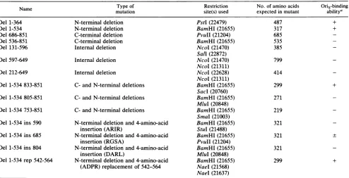
TABLE 1. UL9 mutants
Name Typeof Restriction No. of amino acids
Oris-binding
mutation site(s)used expectedin mutant
abilitya
Del 1-364 N-terminaldeletion PstI(22479) 487 +
Del 1-534 N-terminal deletion BamHI(21655) 317 +
Del686-851 C-terminaldeletion PvuII(21204) 685
-Del536-851 C-terminal deletion BamHI(21655) 535
-Del 131-596 Internaldeletion NcoI(21470) 385
-Sal (22872)
Del 597-649 Internaldeletion NcoI(21470) 799
-NcoI(21311)
Del212-649 Internal deletion NcoI(22628) 414
-NcoI(21311)
Del1-534833-851 C- andN-terminal deletions BamHI(21655) 299 +
Sac (20760)
Del 1-534 805-851 C- and N-terminal deletions BamHI(21655) 271
MluI(20848)
Del1-534 753-851 C-and N-terminal deletions BamHI(21655) 219 SmaI (21003)
Del1-534ins 590 N-terminal deletionand4-amino-acid BamHI(21655) 321
insertion (ARIR) StuI (21488)
Del 1-534 ins685 N-terminal deletion and 4-amino-acid BamHI(21655) 321 +
insertion(RGSA) PvuII(21204)
Del1-534 ins 804 N-terminal deletionand4-amino-acid BamHI(21655) 321
insertion (DARL) MluI (20848)
Del 1-534 rep542-564 N-terminaldeletion and4-amino-acid BamHI(21655) 299 + (ADPR) replacement of 542-564 NaeI(21568)
NaeI(21637)
a +,binds toOris(90 to100% of wild-type binding); -, doesnot bind toOris
(<1%
ofwild-typebinding);+,
bindstosome extent(about10% ofwild-typebinding).
MATERIALS ANDMETHODS
Cloning ofthe UL9 gene in the polylinker ofthe pGEM3 vector. The entire UL9 gene from EcoRV (20461) to Narl
(23539) sites was cloned into the SmaI site of pGEM3
(Promega) afterfillinginofthe Narlsite withthehelp ofthe
Klenow fragment of E. coli DNA polymerase I. The frag-ment encoding UL9 has been isolated from plasmid clone
pKG(akind gift fromS.Weller), which contains the HSV-1
DNA segment KpnI (17793)-BglII (25149) cloned between
KpnI and BamHI sites in pUC19. The UL9 gene in the
pGEM3-UL9clonecanbetranscribedin vitrobyeither SP6
or T7 RNA polymerase, but the SP6 RNA
polymerase-driven transcript is the UL9 mRNA.
Construction of mutants of the UL9 gene cloned in the
pGEM3 vector. Deletion mutants were generated from the UL9 gene cloned in pGEM3. Restriction sites on the UL9
geneusedforconstructingthemutantswereBamHI(21655),
MluI (20848), NaeI (21568 and 21637), NcoI (21311 and 21470), PstI (22479), PvuII (21204), SacI (20760), Sall
(22872), and StuI (21488). The in-frame deletion mutants
generated and the restriction sites used are indicated in Table 1 and Fig. 1. The C-terminal deletion mutants were generatedby opening the plasmid at different restriction sites and inserting an NheI linker (CTAGCTAGCTAG) contain-ing stop codons in all the three reading frames. The NcoI sites were used to generateN-terminal deletions that would
usestartcodonsin the restriction sites themselves to initiate translation. These mutants were constructed by cutting the
pGEM3-UL9clone partially with Sall andNcoIin the UL9
coding sequence, filling in the recessed ends with the
Kle-nowfragmentof E. coli DNA polymeraseI,andself-ligating
thelinearplasmid by T4 DNA ligase and ATP. One
N-ter-minal mutant with a deletion from the N terminus up to the PstI site at22479wasused. This constructionwas made by
cleaving the polylinker and the UL9 coding sequence with PstI and self-ligating the linear plasmid after removal of the internal fragment. The NcoI sitesatpositions21311, 21470, and 22628 werealso used togenerate two internal deletion
mutants bydeleting sequences from 21470to22628orfrom 21311 to 21470. These two deletion mutants preserve the coding frame. An N-terminal deletion mutant wasgenerated by deleting up to the BamHI siteat 21655 and inserting an NcoI linkertorestorethereading frame rightattheBamHI site.
Threemutants were constructed, eachhaving N-terminal deletions up to the BamHI site (21655) but with different C-terminal deletions: oneuptotheSmaI site(21003),oneup
totheMluIsite (20848),and oneuptotheSaclsite(20760). In addition, three insertion mutants and one replacement
mutant in the contextoftheN-terminal deletionmutantdel 1-534 (Table 1) were generated. As shown in Fig. 1, four amino acidswere insertedatpositions 804, 685,and 590 by using restriction sites MluIat20848,PvuIIat21204,and StuI at21488. Syntheticoligonucleotide linkers(CAGATCTGfor insertionat804 andCGCGGATCCGCG for insertionsat685 and590)wereinsertedatthosesites afterrepairof ends with either theKlenowfragmentofE. coliDNApolymeraseI or
T4 DNA polymerase. NaeI sites were used at 21568 and 21637 to generate a replacement mutant, and the coding frame was maintained by substituting a synthetic linker CGCGGATCCGCG for DNA sequence between the two sites.Thus,theprotein would have amino acids encoded by thesequencefrom 542 to 564 replacedby Ala-Glu-Pro-Arg.
In vitrotranscriptionandtranslation of the UL9 genecloned in the pGEM3 vector. After linearization of the ethidium bromide-cesium chloride density gradient-purified plasmid
DNAbyEcoRI,the DNAwasphenol-chloroform extracted andethanol precipitated. Cappedtranscriptscorresponding
on November 10, 2019 by guest
http://jvi.asm.org/
A.
NucleotideSequence No.
Amino AcidPoskion
RestrictionEnzyme Site
B.
Wild-type OBP
Del1-364 Del 1-534
Del 686-851
Del 536-851
Del 131-596
Del 597-649
Del 212-649
Del1-534833-851 Del1-534805-851
Del1-534 753-851
C.
Wild-typeDel 1-534
Del1-534 Ins 590
Del1-534 Ins 685
Del1-534 Ins 804
Del1-534 Rep 542-564
IN C*O 0
IN N- 03N
N NCY N em
_ N a3 a I e a 2 a i
S N P B N NPv Sm Ml Sa 1 131 212 364 534 686 804 8651
1 364
1 634
686 851
536 851
131 696 597 649 212 649
1 534 833 851 1 534 805 851 534 753 851
1 534 851
1 1 634 86153 ~~~~~~ARIR~ 651 5690
1 534 651
6865
1 634 86 1
604 861 542 564
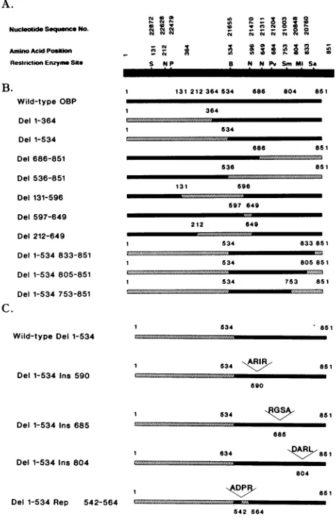
FIG. 1. (A) Schematic representation of the UL9 gene.
Land-markaminoacidpositionsandnucleotidepositionsforafew ofthe
restrictionenzymecleavagesitesare shown. S, Sall; N, NcoI; P, PstI; B, BamHI; Pv, PvuII; Sm, SmaI; Ml, MluI; Sa, Sacl.
Sequencenumbers aregiven accordingto reference 24. (B)
Sche-maticrepresentationof OBPanditsdeletion mutants. Positionsof
amino acids are indicated. The solid bar represents amino acid sequences remaining; the hatched bar represents amino acid
se-quences deleted. (C) Schematic representation of wild-type del
1-534 and its insertion and replacement mutants. Inserted amino
acids are indicated by standard one-letter code. The solid bar representsamino acidsequencesremaining;the hatched bar
repre-sents aminoacidsequencesdeleted.
to the UL9 gene were synthesized by SP6 polymerase as
described by Promega. The DNA template was then
re-moved by treatment with RNase-free RQ1 DNase (Promega), phenol-chloroform extracted, and ethanol
pre-cipitated. The transcripts were visualized, and the length
wascompared againstRNA molecularweightmarkers (Be-thesdaResearchLaboratories) by agarosegel electrophore-sis. The synthetic RNA was then translated in vitro, using rabbit reticulocyte extracts (Promega) in the presence
[35S]methionine to radiolabel the nascent protein as
sug-gestedbythe manufacturer'sprotocol.
SDS-polyacrylamide gel electrophoresis and autoradiogra-phy. Sodiumdodecyl sulfate(SDS)-polyacrylamide gel elec-trophoresis of the synthetic proteins and molecularweight
markers(Sigma/Pharmacia) wascarried out asdescribedby Laemmli(22).Afterelectrophoresis, the gelwasstained with Coomassie brilliantblue,destained, and dried. The driedgel wasexposed to XAR-5 or X-Omat-RP film (Kodak) with or without theuseofEn3Hance (New EnglandNuclear).
Filterbindingandgel retardation assays for the
determina-tion ofsequence-specific DNA binding by the synthetic OBP and mutant derivativestothe
Oris
sequence.DNAbindingof the synthetic OBP and its derivatives was analyzed asdescribedby Deb and Deb(10).
Oris
(wild typeormutant) is cloned within HindIII and NcoI sites into the pORvector(11). Thus, radioactive end labeling can be performed at
either the HindlIl or NcoI site. Sequences of individual origin derivatives, including the wild type, areshown in the figures. The in vitro translation mixture was incubatedwith a 32P-labeled wild-type or mutant
Oris
probe on ice for 30 min inabinding buffer (50 mMN-2-hydroxyethylpiperazine-N'-2-ethanesulfonic acid [HEPES; adjusted to pH 7.5 with
NaOH], 0.1 mM EDTA,0.5 mM dithiothreitol, 10%[wt/vol] glycerol, 100 mMNaCl) with orwithout competitor DNA. For these assays, different volumes of mutantOBP transla-tion mixtures were used for binding so that comparable
amountsofsyntheticproteins (as judged by polyacrylamide gelelectrophoresis) could be used for the binding assay.
In the filter binding assay, afterincubation, the mixtures
werepassed through nitrocellulose filter papers and washed with the buffer mentioned above. The bound DNA was
counted by Cerenkovcounting and then eluted from the filter with 100 ,lI of10 mMTris-borate (pH 8.3)-0.2% SDS-10% glycerol-1 mM EDTA for 4 h at 45°C and loaded onto a5% native polyacrylamidegel. DNA was visualized by autora-diography.
Forgel retention analysis, loading dye was added to a 10% volumeoftheincubated sample and the mixture was loaded
ontoanative 6%polyacrylamide gel as described by Preston
etal. (30), usinga0.5x Tris-borate-EDTA gel and
perform-ing electrophoresisat roomtemperatureand at 30 mA. The
gelwasthen dried and autoradiographed.
Production of antiseraagainst the C-terminal decapeptide of
the UL9 coding sequence. Antisera were prepared as de-scribedby Olivoetal. (27, 28). The C-terminaldecapeptide
CQGAVNFSTLwassynthesizedandconjugated to keyhole
limpet hemocyanin byMultiple Peptide Systems. This
con-jugated peptidewasused toraise antibody in New Zealand
rabbits. Thereactivityof the antiserumagainst the UL9 gene
product wastested by Westernimmunoblot analysis, using
purified UL9 (agenerous gift of M. Challberg; 25a) and a
Western blot kit from Promegaaccordingtothe
manufactur-er'sprotocol. Western blot analysisrevealed aband in the
lane containingpure OBP atthe same position where pure
OBP could be seen by staining the acrylamide gel with Coomassie brilliant blue(not shown).
ImmunoprecipitationofOBPand its derivatives.
Immuno-precipitations of synthetic OBP and its derivatives were
carriedoutby usingthe rabbitantiserumdeveloped against the C-terminal decapeptide of the UL9 codingframe and
Staphylococcus aureus (Enzyme Center) slurry (prepared
according
tothe manufacturer's protocol). A protocol pro-vided by Kathy Partin of DukeUniversity, Durham, N.C.,was used as follows. After in vitro translation, the rabbit
reticulocyte
lysateswerediluted 10-fold in 10mM Tris HCl(pH 7.4)-S50 mM NaCI-0.1% SDS-10 mM dithiothreitol,
boiled for 2 min, and again diluted 10 times in a buffer
containing 10 mM Tris HCI(pH 7.4), 50 mM NaCl, 0.5%
Triton X-100
(Sigma
Chemical Co.), and 0.5% sodiumde-oxycholate.
Thesamples
werethenincubated with S. aureuson November 10, 2019 by guest
http://jvi.asm.org/
[image:3.612.65.306.76.453.2]A. BMV UL-9
-205 Cl
-97.4 n
-45
-29
FIG. 2. (A) Invitrosynthesisof OBP(35S labeled)(seeMaterials and Methods). The autoradiogram ofan SDS-polyacrylamide gel demonstrates synthesis ofaprotein ofasizeidenticaltothatofpure OBP.The arrowheadindicates theposition ofpureOBPobtained by recombinant baculovirus expression. Acontrolreactionwascarried
out with brome mosaic virus RNA (BMV); UL-9, product from synthetic UL9 mRNA. Sizes are indicated in kilodaltons on the right. (B) Filter binding competition experiments showing Oris-specific binding by the invitro-synthesized UL9geneproduct. The in vitro translation mixture was incubated with 32P-labeled Oris
probeasdescribedinMaterialsandMethods. Afterincubation, the
mixtures were passed through nitrocellulose filter papers and
washed. The bound DNAwas counted byCerenkov counting and
then eluted from the filter and loaded onto a5% native
polyacryl-amide gel. DNA was visualized by autoradiography. Each lane
contained 5 p1l of in vitro-translated product, labeled Oris,andthe indicatedamountofcompetitorDNA.
slurry andcentrifugedtoremovethe bacteria.Antiserawere
added, themixture was incubated for5 min at370C, and S. aureus slurry was again added. The suspension was
incu-bated for 30 min at 4°C and centrifuged. The pellet was
washed successively with (i) 10 mM Tris HCl (pH 7.4)-50
mM NaCl-0.5% Triton X-100-0.5% sodium deoxycholate, (ii) 10 mMTris HCl (pH7.4)-i MNaCl, and(iii)10 mMTris
HCl (pH 7.4)-50mM NaCl, suspended in Laemmli loading buffer (22), boiled for 2 min, and loaded onto a 10%
poly-acrylamide gel containing SDS.
RESULTS
Invitrotranscription andtranslation ofthe UL9 gene. An
EcoRV (20461)-to-Narl (23539) fragment containing the en-tire UL9 coding sequence was cloned into the SmaI site of the pGEM3 vector (Promega). SP6-driven transcript from
this plasmid should produce a UL9 mRNA sense product.
Afterlinearizationof the plasmidwithEcoRIonthe3'endof
the insert, an in vitro transcript corresponding to the full-lengthinsertwas synthesized. This transcript migrated as a
single band as assessed by 1% agarose gel electrophoresis (data not shown). This transcript was used to synthesize
OBP in vitro, using commercial rabbit reticulocyte lysates (Promega) in thepresence of[35S]methioninefor labeling the
nascent protein. Figure 2A shows that the most prominent
protein (lane UL-9) detected by SDS-polyacrylamide gel
electrophoresisin the rabbitreticulocyte extractafter
trans-lation corresponds to a molecular mass of about 82 to 83 kDa. The
migration
of this protein band matchesexactly
with the size of the purified OBP from a recombinant
baculovirus expression system(27). The migration ofOBP
(arrowhead in Fig. 2A) from the recombinant baculovirus was visualized by staining the polyacrylamide gel with
Coomassie blue.
Although
the predicted molecular massfrom the
coding
frameof UL9 should be about 94kDa,boththe synthetic OBP and the purified form from insect cells
infected withanOBP-expressingbaculovirus show apparent molecularmasses of about 82to83 kDa. Thereasonfor the
discrepancy between predicted and observed molecular
masses is unknown. Figure 2Aalso shows the analysis ofa
control reaction in which in vitro translationwascarriedout
with brome mosaic virus RNA. This resulted in numerous
prominent bands asopposedto one major band in the UL9
lane.
Oris-binding
activity ofthe synthetic OBP. To determine the specificity of binding of the in vitro-translated products tothe HSVOris
region,theDNA-binding ability ofsynthetic OBPin the presence and absence of competitor DNAswasanalyzed by filter binding experiments. After incubation of an extract with radiolabeled
Oris,
the mixture was filtered through nitrocellulose filter paper, the radioactivity wasdetermined, and the labeled
Oris
was eluted from the filtersand electrophoresed into a native polyacrylamide gel. The
presenceof a labeled fragment indicated binding of
Oris
toafactor(s) in the extract (Fig. 2B). To determine the sequence specificity of binding, competition experiments were per-formed in which incubation of the extract with the radiola-beled
Oris
probe was carried out in the presence of cold pBR322 (nonspecific competitor) or pOR-S (11)(self-com-petitor). Clearly, thetranslated mixture binds specificallyto
Oris
because this binding was inhibited by cold plasmid containingOris
morestrongly (4- to 10-fold) than bypBR322 DNA (0- to 2-fold), as determined by comparing counts retained on the filters when the competitor was present at50-fold molar excess over the probe. The extract itself, which did not have synthetic OBP, did not bind the probe (see Fig. 5A). These data show that the synthetic OBP is functionally active in binding to
Oris
sequence specifically. Later it will be shown that the synthetic protein is recog-nized by an antiserum generated against the C-terminal decapeptide OBP.OBP binding domain on
Oris
recognized by purebaculo-virus-expressed OBP and invitro-synthesized OBP.To deter-mine whether synthetic OBP (in vitro-synthesized UL9) is similar innucleotiderequirements to the protein synthesized in vivo, binding-sitemutantconstructs described previously
(10) were analyzed. Six of the binding-site mutants were
substitution mutants in which three consecutive base pairs
were substituted. In these mutants, T's were substituted by G's, A's were substituted with C's, and vice versa. Two
othermutants, del-31 and bs-1', whose sequencesare shown in Fig. 3, were also examined. To determine the relative importance of the nucleotides in binding, the end-labeled origin insert ofpOR-Sl was incubated with either the rabbit reticulocyte extract containing the synthetic OBP or the purified baculovirus-expressed OBP (25a) in the presence of individual cold plasmid DNAs mutated at the binding site in 50-fold molar excess over the probe. Mutants bs-9, bs-10, bs-11, and bs-12 were the weakest competitors of the end-labeled origin insert ofpOR-Sl for complex formation with the synthetic (Fig. 3A) as well as the pure (Fig. 3B) protein. This finding suggests that bases altered in these mutants are
on November 10, 2019 by guest
http://jvi.asm.org/
[image:4.612.56.300.75.275.2]A.
4-
14-A. (j
-f
0 0) 00 U, U.0 .0 .0 .0 .0 -c .0 .0
., ..:.
am
0
hD~ ~~~~~~~3o.'bt,Ss3"';,,i b%l
[image:5.612.84.278.73.295.2]IL"eII, 11 > :
FIG. 3. Oris-bindingactivity of in vitro-synthesized OBP (A) and purified baculovirus-expressed OBP (B) in thepresenceof different
Oris mutants that have mutations in the OBP-binding site. The procedure for this experimentwassimilarto thatdescribed in the legendtoFig. 2exceptthat 200ngof each of cold competitor DNA withamodified OBP-binding sitewasadded during incubation with
radioactive Oris. For thesynthetic protein, eachsystemhad about 0.4pmol of protein; forpureOBP, eachsystemhad about 0.5,ug of protein. The OBP-binding site is overlined on the wild-type se-quence. Sequences of the binding sitemutants usedfor the DNA-binding competition experiments are shown at the top. Del 31 is deleted insequencesthatarenotdepicted.
crucial forbinding. These results indicate that the in vitro-synthesized protein is similar in sequence specificity for
bindingtopure OBPorcrude OBPinHSV-1-infected Vero
cell extracts (10, 20) and that the sequence 5'-GTTCG
CACTT-3' is essential for efficient binding. Thus, the in
vitro-synthesized OBP defines thesame binding siteasdoes
the in vivo-synthesized OBP.
Construction of deletionmutantsof the UL9geneproduct. To determine the DNA-binding domain of OBP, deletion mutantsof the UL9gene wereconstructed by using
conve-nient restriction sites in and around the UL9 coding
se-quence.ThemutantsareshowndiagrammaticallyinFig. 1B,
and their constructions are explained in Table 1. Two
C-terminal deletion mutants, two N-terminal deletion
mu-tants, three N- aswell as C-terminaldeletion mutants, and three internal deletionmutantsweregenerated.The
produc-tion of proteins was measured by the appearance of
35S-labeled proteins in SDS-polyacrylamide gels. Figure 4A shows protein bands detected by SDS-polyacrylamide gel
electrophoresis and autoradiography. The majorbands are
slightly smaller in molecular weight than predicted from amino acid numbers shown in Table 1. This was expected
because of the observed faster mobility of OBP in an
SDS-polyacrylamide gel (27). In the case of mutant del
1-364, a deletionfrom the N terminus uptothe PstI site at
22479wasmade. This deletiongeneratedtworeadingframes thatcanstarteitheratthe Met in theoriginal codingframeat nucleotide position 22169 (codon 365) or at a Met in an
alternate reading frame six nucleotides earlier. The
corre-FIG. 4. (A) SDS-polyacrylamide gel analysis of in vitro-synthe-sized OBPand its deletionmutants.Theprocedure for productionof UL9 and its derivatives is described in thetext.Each lane contained 5 ,ul ofa sample. (B) Immunoprecipitation of in vitro-synthesized OBP and its deletionmutantscarriedoutasdescribed in Materials and Methods. Only those mutants with an intact C terminus are shown.wt, wild type.
sponding band in Fig. 4A is slightly diffused, probably
because ofprotein synthesis being initiated atboth sites. Allof the C-terminal deletionsweregenerated by inserting
NheI linkers with stop codons in all three possible coding
framesafter cuttingwith indicated restriction enzymes
(Ta-ble1). In thecaseof C-terminal deletionmutantdel1-534,an
NcoI linkerwasusedattherestrictionsitetoregeneratethe
correctcodingframe. This mutantDNAwasusedto gener-atethreemoredeletion mutants, del 1-534833-851,del 1-534
804-851,and del 1-534 753-851. Thesemutantsweredeleted
at both the C and N termini. The three internal deletion
mutants were generated by using internal NcoI and Sall
sites, preservingthe codingframe.
Construction of insertion and replacement mutants of del
1-534. To investigate the internal structure of the
DNA-binding domain,three insertionmutantswereconstructed in
which four amino acids were inserted after positions 590
(Ala-Arg-Ile-Arg),685(Arg-Gly-Ser-Ala), and 804 (Asp-Ala-Arg-Leu).Thesemutantsweregenerated by cuttingthegene of del 1-534atMluI (20848), atStuI (21488), and atPvuIII
(21204), repairingthe termini, and inserting synthetic
oligo-nucleotide linkers. One replacementmutant wasgenerated
by replacingamino acids242to264bythe four-amino-acid segment Ala-Asp-Pro-Arg, usingthe NaeI sites (21568 and 21637).
Immunoprecipitation of thesynthetic OBP and its deletion mutants. To determine whether the synthetic OBP and its
f
CL E 0
3. Z: N - c .m = "
on November 10, 2019 by guest
http://jvi.asm.org/
[image:5.612.342.532.74.365.2]_ __
.b
-,zZN
sir
o c 0 cr 0cc
= - -i = A
ClC: O e:
-1 IO G
I9 c4 I toFN L
a, a:
c
7
._
:.
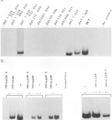
.-FIG. 5. (A) Filter binding experiments to determine the
Oris-bindingspecificity of synthetic OBP anddeletion mutants. Condi-tionswere similartothose described in the legendtoFig. 2. Each binding reaction had approximately equal amounts of synthetic protein. The volume ofextracts added was determined after gel
electrophoreticanalysis.Noprotein,extractalonewithout synthetic OBPorits derivatives. (B) Filter bindingcompetitionexperiments
showingOris-specificbindingby thein vitro-synthesized mutantsof OBP. The in vitro translation mixture was incubated with 32p_
labeledOris probe with or without indicated competitors and
processedasdescribedfor Fig. 2B. In eachcase,the self-competitor
inhibitedmorethan didthe nonselfone.WT, wildtype.
derivatives retain the wild-type coding frame, immunopre-cipitationwithan antiserum reactive against wild-type OBP
was performed. An antiserum against the C-terminal deca-peptide was generated according to Olivo and coworkers (27, 28). This antiserum can react specifically with purified OBP (25a) in immunodiffusion, immunoprecipitation, and Western blotting assays. It was reacted with the synthetic
OBP and its derivatives. The complexes were then
precip-itated by S. aureus slurry as described in Materials and
Methods. The immunoprecipitates were analyzed in an
SDS-polyacrylamidegeland then subjectedto autoradiogra-phy. Figure 4B shows bands in all lanes with synthetic proteins having an intact C terminus. C-terminal deletion
mutants of OBP were not immunoprecipitated (data not
shown). Since an antiserum wasraised against the
C-termi-nal protein, synthetic proteins with C-terminal deletions
were thereforenotexpectedtobe immunoprecipitated. The
datapresented in Figure4B demonstrateintactness ofthe C
terminus, suggestingthat thecorrectreading frames forOBP and its derivatives were retained. Because the N terminus
wasnotalteredinanywayduring construction ofC-terminal
deletion mutants, no change in the reading frame was anticipated. That the N-terminal deletion mutant del 1-364 (Fig. 1B)couldbe immunoprecipitated(Fig. 4B) showed that thereading framewasintact, and the migration of the band
inthe SDS-polyacrylamidegel suggested that thetranslation systemutilizestheMet at365 as theinitiation codon.
Oris-binding
activityofthe synthetic OBP deletionmutantsasobservedby ifiterbindingassay.Results ofafilter binding
experiment with in vitro-prepared synthetic proteins (Fig. SA) show thatonlyproteins encoded bytheintactUL9gene,
the N-terminal deletion mutantsdel 1-364 anddel 1-534, and the double-deletion mutantdel 1-534833-851 bind efficiently to the origin sequence. The internal deletion mutant del 131-596, which extends beyond amino acid 534, failed to
bind to
Oris,
suggesting the N-terminal boundary to be between amino acids 535 and 596. Near the C terminus, although del 1-534 833-851 could bind toOris,
del 1-534 804-851 could not. This result indicated that the C-terminal boundary is between aminoacids 833 and 805.The amounts of synthetic proteins used in the binding reactions were normalized afteranalysis by SDS-polyacryl-amide gel electrophoresis. In a standard assay, about 0.4 pmol of the synthetic proteins was used, as determined by trichloroacetic acid-precipitable counts. Quantitative analy-sis of the filter binding data did not show any difference in binding ability between proteins that were able to bind to
Oris
(data not shown).We examined the sequence specificity of binding of the wild type and three mutants of OBP that could bind to
Oris
(Fig.SB).
As described earlier, competition binding assays were carried out by using specific (pOR-S) and nonspecific (pBR322) DNAs as competitors. Again, the binding was found to be competed for more efficiently by pOR-S than by pBR322 under a 50-fold molar excess of competitor over the probe. This result suggests that the observed DNAbinding is highly sequence specific.Insertion and replacement mutations in the DNA-binding domain of OBP. To investigate the internal structure of the DNA-binding domain, three insertion mutants (del 1-534 ins 590, del 1-534 ins 685, and del 1-534 ins 804) and one replacement mutant (del 1-534 rep 542-564) were generated (Fig. 6C). Figure 6 shows SDS-polyacrylamide gel analysis of these mutants and their immunoprecipitates. The sizes of the prominent bands correspond to those expected for them. Figure 7 shows a filter binding analysis of the Oris-binding ability of these mutant proteins. Only rep 542-564 shows binding ability. This binding is inhibited more by the pres-ence of pGEM3z(-) Oris
(self-competitor)
than by pGEM3z(-). Although not shown in this figure, del 1-534 ins 685 bound toOris;
this binding, however, was considerably less than that of the wild type (about 10% of the wild-type level; data not shown). Thus, this analysis suggests that replacement of amino acids 542 to 564 by four foreign amino acids has no significant effect onOris binding.Gel retardation assay to determine the DNA-binding do-main. Oris-binding capacities of del 1-534 and its mutants were also determined by agelretardation analysis (Fig. 8). Figure 8A shows the schematics of mutants tested in this assay, and Fig. 8B depicts an autoradiogram in which the
different mutants were analyzed in 6% native polyacryl-amide gels after incubation of the in vitro-synthesized pro-tein with wild-type and mutant origins from
pOR-Sl
(withOBP-binding site I) (11). As shown in the figure, only del 1-534 wild-type, del 1-534 833-851, and del 1-534 rep
542-564
formed aretardedcomplex, while all other mutants, includ-ing insertion mutants del 1-534 ins 590, del 1-534 ins 685, and del 1-534 ins 804, did not show complex formation. Mutant del 1-534 ins 685 formed a complex that was detected after overexposure of the dried gel. Thus, this insertion mutant
retained some binding activity even after the four-amino-acid insertion at position 685. Interestingly, although both
del 1-534 833-851 and del 1-534 rep 542-564 have 299 amino acids, the complex formed with the former migrates faster than that formed with the latter. Those proteins that could form the retarded complex were also tested for sequence specificity by performing binding reactions with mutant
on November 10, 2019 by guest
http://jvi.asm.org/
[image:6.612.83.273.78.283.2]B ;O 1 534 851
Del 1-534
833
Del1-534 833-851 -::...
805
Del 1-534 805-851 :.
753
Del 1-534 753-851 :::::
ADPR
Rep 542-564
Del 1-534Ins 590
27.8
Del 1-534 ins685
534 EGSA 851
Del1-534 Irs5 85:85-:--:
585
(c)
De: 1-534 1Is804
Del1-534Rep 542-564
ARIR
590
8DARL
ADPR 8s
542 564
Del 1-534 Ins 804
. ... I---
--543 564
ARIR
590 RGSA
I...
685 DARL
EN04
13.
FIG. 6. (A)SDS-polyacrylamide gel analysis of in
vitro-synthe-sized insertion andreplacementmutantsof del 1-534 and its deletion mutants. Theprocedureforproduction of themutantsisdescribed
in the text. (B) Immunoprecipitation ofin vitro-synthesized prod-ucts shown in panel A, using an antiserum directed against the C-terminaldecapeptide ofOBPandS.aureusslurry. (C)Schematic
representationofwild-type del 1-534 and its insertion and replace-ment mutants. Inserted amino acids are indicated by standard
one-letter code. The solid bar represents amino acid sequences
remaining; the hatched bar represents amino acid sequences
de-leted.
Ori-Slsequences. None of them could bindtoOri-Sl bs-9, -10 or -11 (10). These mutant origins are mutated in the
primary binding site of OBP and failedtobind appreciablyto
acrude OBPpreparation (10). Thus,origin-specific binding
, 0c
~~~\
_
no_
[image:7.612.320.557.76.422.2]A B
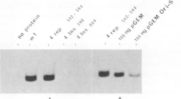
FIG. 7. (A) Filterbinding experimentstodetermineOris-binding
specificity of synthetic insertion and replacement mutants ofdel 1-534. Conditions were similartothose describedin the legendto Fig. 5. (B) Filter binding competition experiments showing
Oris-specific binding by the invitro-synthesized del 1-534 rep 542-564. The in vitrotranslation mixturewasincubated with 32P-labeledOris
probewithorwithout the indicatedcompetitors andprocessed as describedforFig.SB. Inthiscasealso,theself-competitorinhibited
morethandid thenonselfone.
A
F- .---7
7 = .! 7
I . :.
0 =
J, I.
6-FIG. 8. (A) Schematicrepresentation of wild-type del 1-534and
its deletion, insertion, and replacement mutants. Inserted amino acids are indicatedby the standard one-letter code. The solid bar
represents amino acid sequencesremaining; the hatched bar
repre-sents amino acid sequences deleted. (B) Gel retardation assay to
determine Oris-bindingcapacity of the C-terminal317amino acids ofOBP (del 1-534) and theirmutants. The procedure for the gel retardationanalysis is described in text. Thetype oforigin probe used(either Ori-Sl orits mutant,bs-9, -10, or11) andthe nameof the synthetic proteins used are indicatedat the top of each lane. Arrowheadspointtothe retardedbandswhereprotein-boundDNA
migrates.Freeprobesareatthe bottomof thegel. Only theportion ofautoradiographs showingthefree probe and retarded bandsare
presented. Rabbit reticulocyte extracts alone do not form any significant retarded band. Wild-type del 1-534andthe mutants del
1-534833-851 and del 1-534 rep 542-564 can bind successfully to
wild-type Ori-Slbut nottoitsmutantderivatives with substitutions
in theprimaryOBP-binding site. All other mutants failed toforma significant protein-DNA complex. Sequences of Ori-Sl and its
mutantsaredepictedatthe top.
wasobservedinthosecasesofmutantproteinsthat retained
the capacityto bindto
Oris.
DISCUSSION
To determine the structural motif of the protein that
dictates thesequence-specific DNA-binding activityofOBP, advantage was taken of the powerful tools of in vitro transcription and translation. Using synthetic mutant
pro-teinsand filterbindingandgelretardationassays,aminoacid
.A'
45
-S51~
Kd
105.7
71.0-44.2 .
AAAA GAA0-,; A,; AA,:,; 7 'r ATA-TA.
on November 10, 2019 by guest
http://jvi.asm.org/
[image:7.612.52.310.77.335.2] [image:7.612.91.275.543.644.2]Wild-type Del 1-534 1NPFVGGAZSGDPLGAGRPIGDDECZQYTSSV8LMHNLYGGDLAZWVPRVHPKTTIERQQHGPVTFPNASAP
Del1-534Replac 542-564
Del 1-534 Ins 590
Del 1-534 Ins 685
Del 1-534 Ins804
534 851
AminoAcidPositions
FIG. 9. Hydrophobicity plots of thewild-type OBP C-terminal
317 amino acids (del 1-534) and its insertion and replacement mutants. Hydrophobicity plots were generated by the method of Kyteand Doolittle (21). Positions above the x axis represent positive hydrophobicity; positions below indicate hydrophilicity. Arrow-headsindicate the positions at which mutations were introduced.
sequences required for sequence-specific DNA-binding
ac-tivityof OBP were determined. These data(Table 1) indicate
that the 269amino acidsfrom564 to 832definethe
sequence-specific DNA-binding domain ofOBP.
Noneoftheinsertion mutants(del1-534 ins590, del 1-534
ins 685,and del 1-534ins 804)could formasequence-specific
complex efficiently in the gel retardation assay (Fig. 8A), although del 1-534 ins 685 formed the complex thatbecame visible only after prolonged exposure of the dried gel. Figure 9 represents plotsfor hydrophobicity (21)ofsynthetic pro-teins derived fromthe differentinsertionmutants analyzed. Thepolypeptide encoded by mutant del 1-534 ins 590 has the greatest structural alteration at the site ofinsertion, andit does notbindtotheorigin. Mutantsdel 1-534ins685 anddel 1-534 ins 804 have less effect on hydrophobicity. Of these two,del 1-534 ins 685 can bind to
Oris
to some extentwhile del1-534ins804 cannot. It ispossible thatinsertionof aminoacids in del 1-534 ins 590 caused loss in DNA-binding
activity because of structural alteration. Loss of binding
capacity may not be related to possible changes in the
positions ofcontactresidues. The insertionsat 685 and 804
have moderate effects on hydrophobicity but havea
signifi-cant effect on binding. This might be indicative of more
directroles for theresiduesnear685 and 804 in DNAbinding
thanfor those near amino acid 590. More subtle mutational analysis, including amino acid substitution (both conserva-tive and nonconservative) at these sites, is necessary to resolve thisissue.
Sequence analysis by the Chou-Fasman method (7)
pre-dictedahelix-turn-helixmotif atamino acids 698 to 733 (Fig.
10) in the DNA-bindingdomain. Ahelix-turn-helixmotif has
been implicated in DNA binding and dimerization of a number of sequence-specific DNA-binding proteins (29). However, deletion mutant del 130-596, which does not
disturbthemotif, cannot bind to
Oris.
Thus, thishelix-turn-helix motif, even if required for sequence-specific binding, is
notsufficient alone for binding.
OBP is an interesting protein with 851 amino acids, of which only 269 or fewer are required for sequence-specific
recognitionof the origin of replication. Direct evidence is not
yetavailable to relate function to sequences outside of the
DNA-binding domain of 269 amino acids (positions 564 to
832). Several helicase motifs have been predicted for the
OBP sequence on the basis of homology (19). Sequence
information suggests the existence of a presumptive
ATP-72 "ATP Binding Site"
TARCvT_wvRPNGSGKTTALIRWLRZAIHSPDTSVLVVBCRRSFTQTLATRFAE8GLvDFvTYFSBTNYIN
143 * * * *
NDRPIHRLIVQVZ8LHRVGPNLLNNYDVLVLDZVXBTLGQLYSPTfQQLGRVDALNLRLLRICPRIIAIDA "Leucine Zipper"
214
TANAQLVDFLCGLRGEKNVHVVVGYYAXPGF8ARRCLLPRLGTZLLQAALRPPGPPSGPSPDA8PEARGA
285
TFFGELEARLGGGDNICIF88TVSFAEIVARFCRQFTDRVLLLHSLTPLGDVTTWGQYRVVIYTTVVTVGL
356
SFDPLHFDGNFAYVXPNNYGPDNVSVYQSLGRVRTLRKGZLLIYXDGSGARBEPVFTPNLLNNWVSCGQw
427
PAQFSQVTNLLCRRFKGRCDASACDTSLGRGSRIYNKFRYKYFEYRCTLACL8DSLNILHNLLTLNCIRVR
498 565
FWGHDDTLTPKDFCLFLRGVHFDALRAQRDLRELRCRDPEASLPAQAAETEZVGLFVEKYLRBDVAPAEIV
569 N-terminal boundary 596
ALKRNLN8LMGRTRPIYYLALLEACLRVPNATRBSAIFRRIYDHYATGVIPTINVTGELELVALPPTLNVTP
"L-ucine Zipper"
640 Helix
VWELLCLCSTHAARLHWDSAAGGSGRTFGPDDVLDLLTPHYDRYNQLvFELGHCNVTDGLLL8EEAVKRVA
711 Helix
DALSGCPPRGSVSETDHAVALFKIIWGELFGVQNAKSTQTFPGAGRVKNLTKQTIVGLLDAHHIDN8ACRT
Turn
782 C-terminalBoundary
HRQLYALLHANKRBFAGARPKLRVPAWGRCLRTHSSSANPNADIILEAALSELPTEAWPNQGAVNFSTL
804 833 851
FIG. 10. Amino acid sequenceofwild-type OBP. Positions of presumptive leucine zippers, the ATP-binding domain, and the helix-turn-helix motif are indicated. Alsoindicatedarethe N- and C-terminal boundaries of the sequence-specific DNA-binding do-mainidentified inthis report.
binding site VRAPMGSKRT (25, 32) which may be involved in the ATPase/helicase function. Sequence analysis also reveals a leucinezipper from amino acids 150 to 173 which may be involved in homo- or heterodimer formation (23,34). Since thisleucine zipperisnotrequiredfor in vitro binding totheorigin, it may take part in the formation of a dimer for replication function of the OBP or it could function in heterodimer formation withotherreplicationproteins.
Examination of sequences for other replication protein genesreveals thefollowing: in UL5, aleucine stretch from amino acids 160 to 237 and anATP-binding site (ITGNAGS GKSTCVQTI); for UL30 (DNA polymerase), a leucine stretch from amino acids 427to540; and for UL29(ICP8),a
leucine stretch from amino acids 1076 to 1109. The implica-tion of the presence of leucine stretches in these coding frames is not yetclear. One distinct possibility is that they
imply homo- or heterodimerization/DNA-binding domains
important for their biological functions, e.g., in the forma-tion ofa specialized nucleoprotein structure, a hypothesis that needs to be tested. The ATP-binding site in the UL5 coding sequence may be indicative of the helicase function of theUL5-UL8-UL52 complex (5a, 9, 13, 25,36).From the results described here and elsewhere (35), it appears the leucine stretch frompositions 150to 179 isnotrequiredfor
DNAbinding.Thus, eitherdimerizationis not critical for the
sequence-specific DNA-binding activity or the leucine
stretch is not involved in this dimerization. Interestingly,
however, there is a sequencefrom amino acids 570 to 591,
LMRNLNSLMGRTRFIYLALLEA, which is somewhat
similartothe classicalleucinezipper. It should be noted that therearereports ofleucinezippermotifs that have
replace-ments in some leucine positions (6). Although a deletion
on November 10, 2019 by guest
http://jvi.asm.org/
[image:8.612.71.288.77.203.2] [image:8.612.318.554.80.355.2]upstreamof thissegment toamino acid 563 (in del 1-534 rep 542-564) does not affect
Ons
binding significantly, a deletion up to amino acid 596 (in del 131-596) destroys the binding activity. These data signify that sequences between amino acids 563 to 596, a region that includes the pseudo-leucine zipper, are crucial for DNA binding. This result, however, does not directly imply the motif's involvement in dimeriza-tion. It is still possible, however, that dimerization is not required for DNA binding or that del 1-534 dimerizes in a mannerthat is different from that of intact OBP. Neverthe-less, the fact that a pseudo-leucine zipper sequence is required for sequence-specific DNA binding is interesting and requires further study. The four-amino-acid insertion (Arg-Ile-Arg-Ala) at the junction of this pseudo-leucine zipper and the rest of the molecule toward the C terminus destroyed DNA binding. This observation strengthens our hypothesis that the pseudo-leucine zipper is crucialforDNA binding. Thus, our results predict that the DNA-binding domain of OBP is composed of two known DNA-binding/ dimerizationdomains, apseudo-leucine zipper, and ahelix-turn-helix motif. Insertions of four amino acids are not toleratedin the DNA-binding domain, suggesting that
integ-ntyof the 269-amino-acid stretch is necessary forsuccessful
Oris
binding.ACKNOWLEDGMENTS
We thank Charles Gauntt and David Martin forcriticallyreading the manuscript. We are indebted to Joel Baseman andAnnaLazzell for their generous help in preparingthe antisera. We thank Jane Tate
for excellent typing assistance.
This work was supported by grants from the American Cancer Society (MV-420) andthe Elsa U. Pardee Foundation and by a Basil
O'Connor Starter Scholar Research Award from the March of
Dimes to Sumitra Deb. This work was also supported by an
American Cancer Society institutional grantto Sumitra Deb and an
institutional research grant to Swati Palit Deb.
REFERENCES
1. Baumann,R. P., V. R. R.Yalamanchili,and D. J. O'Callaghan.
1989. Functional mapping and DNA sequence of an equine
herpesvirus 1 origin of replication. J. Virol.63:1275-1283. 2. Bruckner, R. C., J. J. Crute, M. S.Dodson, andI.R.Lehman.
1991. The herpes simplexvirus 1 origin binding protein: a DNA
helicase. J. Biol. Chem. 266:2669-2674.
3. Carmichael, E. P., M. J. Kosovsky, and S. K. Weller. 1988.
Isolation and characterization ofherpes simplex virus type 1
host rangemutants defective in viral DNA synthesis. J. Virol. 62:91-99.
4. Carmichael,E. P., and S. K. Weller. 1989. Herpes simplex virus
type 1 DNA synthesisrequires theproduct ofthe UL-8 gene: isolation andcharacterizationof theICP6::1acZ insertion
muta-tion. J. Virol. 63:591-599.
5. ChalHberg,M. D. 1986. A method for identifying the viral genes required forherpesvirus DNA replication. Proc. Natl. Acad.
Sci. USA83:9094-9098.
Sa.Challberg,M. D. Personalcommunication.
6. Chang, Y.-N., D. L.-Y., Dong, G. S. Hayward, and S. D.
Hayward. 1990. The Epstein-Barr virus Zta transactivator: a member of the bZIP family with uniqueDNA-bindingspecificity and a dimerization domain that lacks the characteristic heptad
leucine zipper motif. J. Virol. 64:3358-3369.
7. Chou, P.Y.,andG. D.Fasman. 1978. Prediction ofsecondary structure of proteins from their amino acid sequences. Adv. Enzymol. 47:45-50.
8. Crute, J.J., and I. R. Lehman. 1989. Herpes simplex-1 DNA polymerase.Identificationofan intrinsic 5'-3'exonucleasewith ribonuclease Hactivity. J.Biol. Chem. 264:19266-19270.
9. Crute, J. J., T. Tsurumi, L. Zhu, S. K. Weller, P. D. Olivo,
M. D. Challberg, E. S. Mocarski, and I. R. Lehman. 1989.
Herpes simplex virus 1 helicase-primase: a complex of three herpes-encoded gene products. Proc. Natl. Acad. Sci. USA
86:2186-2189.
10. Deb, S., and S. P. Deb. 1989. Analysis of Ori-S sequenceof
HSV-1: identification ofone functionalDNAbinding domain. Nucleic AcidsRes. 17:2733-2745.
11. Deb, S., and M. Doelberg. 1988. A67-base-pairsegmentfrom
theOri-Sregion of herpessimplex virustype 1encodesorigin
function.J.Virol. 62:2516-2519.
12. Digard, P., and D. M.Coen.1990. Anovelfunctional domain of
analpha-like DNA polymerase. The DNAbinding siteonthe
herpessimplex viruspolymerase for the viral UL42protein. J. Biol. Chem.265:17393-17396.
13. Dodson, M.S., J. J.Crute,R.C.Bruckner,and I. R. Lehman.
1990.Overexpression andassembly oftheherpessimplexvirus
type 1 helicase-primase in insect cells. J. Biol. Chem. 264:
20835-20838.
14. Elias, P., C. M. Gustafsson, and 0. Hammarsten. 1990. The
originbinding protein ofherpessimplexvirus1binds
coopera-tively to the viral origin of replication
ori..
J. Biol. Chem. 265:17167-17173.15. Elias, P., andI.R. Lehman.1988. Interactionoforiginbinding
protein withanorigin ofreplication ofherpessimplexvirus1. Proc. Natl. Acad. Sci. USA85:2959-2963.
16. Elias, P., M. E.O'Donnell,E. S.Mocarski,and I. R. Lehman.
1986.ADNAbinding proteinspecificforanoriginofreplication
of herpes simplex virus type 1. Proc. Natl. Acad. Sci. USA 83:6322-6326.
17. Gallo,M.L., D. I.Dorsky,C. S.Crumpacker,and D.S. Parris.
1989. Theessential 65-kilodalton DNA-bindingprotein of her-pessimplex virus stimulates the virus encodedDNA polymer-ase. J.Virol. 63:5023-5029.
18. Goldstein, D. J., and S. K. Weller. 1988. AnICP6::IacZ
inser-tional mutagen is used todemonstrate that the UL52gene of herpes simplex virus type 1 is required for virus growthand
DNAsynthesis. J.Virol. 62:2970-2977.
19. Gorbalenya, A. E.,E. V. Konin, A. P. Donchenko, andV. M. Blinov. 1989. Two related superfamilies of putative helicases
involved inreplication,recombination, repairandexpressionof
DNA and RNAgenomes. Nucleic Acids Res. 17:4713-4730.
20. Koff, A., and P. Tegtmeyer. 1988. Characterization of
major
recognitionsequencesforaherpes simplexvirus type 1
origin-bindingprotein. J. Virol. 62:4096-4103.
20a.Koff, A., and P.Tegtmeyer. Personal communication.
21. Kyte, J., and R. F. Doolittle. 1982. A simple method for displaying thehydrophobiccharacter ofaprotein. J. Mol. Biol.
157:105-132.
22. Laenmli, U. K. 1970. Cleavageof structuralproteinsduringthe assembly of the head of bacteriophage T4. Nature (London)
224:680-685.
23. Landschulz, W.H.,P. F.Johnson, andS. McKnight. 1989. The DNAbinding domain oftheratliver nuclearproteinC/EBP is bipartite. Nature (London)243:1681-1688.
24. McGeoch, D. J., M. A. Dalrymple, A. J. Davidson, A. Dolan, M.C.Frame,D.McNab,L.J.Perry, J.E.Scott,and P.Taylor.
1988. Thecomplete sequence of the longunique
region
in the genomeofherpessimplexvirus type 1.J. Gen. Virol. 69:1531-1574.25. McGeoch, D.J., M. A. Dalrymple, A. Dolan, D. McNab, L. J.
Perry, P.Taylor, andM. D. Challberg. 1988. Structures of the herpes simplex virus type 1 genes required for replication of virus DNA. J.Virol. 62:444 453.
25a.Natajaran, M.,and S. Deb. Unpublished data.
26. Olivo, P.D.,and M. D. Challberg. 1988. Herpes simplexvirus DNAreplication: identification of theessential genes and their
products. Cancer Cells 6:43-51.
27. Olivo, P.D., N. J. Nelson, andM. D. Challberg. 1988. Herpes
simplexvirus DNAreplication: theUL9 gene encodesanorigin
binding protein. Proc. Natl. Acad. Sci. USA 85:5414-5418.
28. Olivo,P.D., N. J.Nelson,and M. D. Chaliberg. 1989. Herpes
simplexvirus type 1 gene products required for DNA
replica-tion: identification andoverexpression. J. Virol. 63:196-204.29. Pabo, C.O., andR.T. Sauer. 1984. Protein-DNA
recognition.
on November 10, 2019 by guest
http://jvi.asm.org/
Annu. Rev.Biochem. 53:293-321.
30. Preston, C.M.,C. M. Frame, and M. E. M. Campbell. 1988. A complexformed between cell components and anHSV struc-turalpolypeptide binds toaviral immediateearlygene
regula-tory DNAsequence. Cell 52:425-434.
31. Roizman,B.,and A. E.Sears. 1990. Herpessimplex viruses and theirreplication. In B. N. Fieldset al. (ed.), Virology, vol. 2. Raven Press, New York.
32. Sekimizu, K., D. Bramhill, andA. Kornberg. 1987. ATP acti-vatesdnaAprotein in initiating replication of plasmids bearing theorigin of the E. coli chromosome. Cell 50:259-265.
33. Stow, N. D., and A. J. Davison. 1986. Identification of a
varicella-zoster virus origin of DNA replication and its activa-tionby herpes simplexvirustype1geneproducts.J. Gen. Virol. 67:1613-1623.
34. Turner, R., and R.Tjian.1989. Leucinerepeatsandanadjacent DNA binding domain mediate theformation of functional cFos-cJun heterodimers. Nature(London) 243:1689-1694.
35. Weir, H. M., J. M. Calder, and N. D.Stow. 1989. Binding of the herpes simplex virustype 1 UL9gene producttoan origin of viral DNA replication. NucleicAcids Res. 17:1409-1425. 36. Weller, S. Personal communication.