Vol.65,No. 1 JOURNALOFVIROLOGY, Jan. 1991,p. 170-179
0022-538X/91/010170-10$02.00/0
Copyright ©) 1991,American Society forMicrobiology
Structural Elements That
Regulate
pp59c-fl` Catalytic Activity,
Transforming Potential, and Ability To Associate with
Polyomavirus Middle-T Antigen
SENG H.CHENG,1* PEARLC. ESPINO,1JOHN MARSHALL,' ROBERTHARVEY,' JANETMERRILL,2 ANDALAN E. SMITH'
Laboratory of Cellular Regulation' and Laboratory ofDNA Chemistry,2 Genzyme Corporation, One Mountain
Road,
Framingham,
Massachusetts 01701Received 19 July 1990/Accepted 1 October 1990
Except for its unique amino-terminal region (residues 1 through 83), which possibly dictates substrate recognition,pp59c-fY- bearsahighdegreeof homology with other members of thesrcfamilyoftyrosine kinases. Hereweshowthat the carboxyterminus ofpp59c-fynisnecessaryfor stablemiddle-T-antigen association,that
pp59C-fyfis
normally phosphorylated onboth serine and tyrosine residues,and thatTyr-531 andTyr-420 arephosphorylation sites in vivo and in vitro,respectively. Analysis ofaspontaneously generated mutantencoding atruncatedform ofpp59c-fyn and of variantsspecificallymutatedattheTyr-531andTyr-420 phosphorylation sites indicates thatpp59c-fyn hasregulatory elements analogoustothose that havealreadybeen identifiedfor othersrc-liketyrosinekinases.However,further examination of thepp59clYfy variants suggests the likelihood ofadditionalmeansbywhich its activitiesmightberegulated. Althoughalteration ofTyr-531tophenylalanine (531F) inpp59'-fy'resultsinaprotein which ismoreactiveenzymaticallythatthe wildtype,the enhancement is much less than that for the analogous variant of pp6o-src. Furthermore, contrary to results of similar experimentsonother src-likeproto-oncogene products, 531F didnotinducetransformationof NIH 3T3 cells. Studies involving
pp59c`fy-pp60csrc
chimeras in which the unique amino-terminal sequences (residues 1 through 83) of the two kinases were precisely interchanged implied that the inability of 531F to induce transformationisprobablynotcaused by the absenceofsubstrates forpp59cfYnin NIH3T3 cells but ratherby the insufficient enhancement of pp59c-fyn kinase activity. It is therefore probable that the kinase and transforming activities of pp59C-fyf are repressed by additional regulatory elements possibly located in the amino-terminalhalf of the molecule.There is increasing evidence that members of the src
family ofprotein tyrosine kinases participate either as
me-diatorsoramplifiers ofcellularsignal-transducingpathways
(forreviews, seereferences 9 and 32). This conceptwasfirst
formulatedbecauseof theabilityofv-srcfamilymembersto regulate cellular growth and was recently highlighted by the demonstration that
p56ck,
a member of this family, is associated with the T-lymphocyte surface glycoproteins CD4 and CD8 (35, 39). The notion that these cellular enzymes have an ascribed role in cellular proliferation is further corroborated by the observation that mutants ofpp6Ocsrc (and its viral homolog), pp62c-Yes,
pp59Cfn,
andpp55c-fgr
are capable ofcausing deregulated cellular prolif-eration and malignant transformation (for a review, see reference 9).The
fyn
gene, amember of thisfamily, encodes a 59-kDa src-like protein tyrosine kinase that is myristylated and phosphorylated (5, 18, 24, 28, 36). Originally identified in fibroblasts and endothelial cells,pp59C-Ify
has since been detected in a variety of other cells and at especially high levels in the brain and hematopoietic cells (8, 25, 36). Two distinctfyn-encoded
transcripts that result fromalternative splicing ofa single exon and that exhibit mutually exclusive patterns of expression have been reported (8). One of theisoforms has been shown to accumulate principally in the
brain, while the other is located in thymocytes and certain
hematolymphoid cell lines. Although the normal
physiolog-*Correspondingauthor.
ical functions of the
fyn
gene products have not beenun-equivocally established, acausalrole between the
pp59C-hf
kinase and T-cell development and differentiation has been demonstrated (20, 21).Regulationof thesrcfamilyof cellular kinases is achieved in part by control ofphosphorylationatthetyrosines homol-ogous to Tyr-527 and Tyr-416 ofpp6Oc-sr. Preventing the
phosphorylation of Tyr-527 of pp6Oc-src by mutation to
phenylalanine (3, 26, 33), by association with middle-T
antigenofpolyomavirus(4, 12),orbyphosphatasetreatment (11) results in activation of the kinase and transforming
activities ofpp6Ocsrc. Similarly, the oncogenicpotential of
pp56lck(1, 30) andp59hck(40) is unmaskedbymutationof the homologous residue corresponding to Tyr-527 ofpp6Ocsrc. Concomitant with the activation observed with the 527F mutantofpp6Oc-src isphosphorylationatTyr-416, the site of autophosphorylation. Phosphorylation of Tyr-416 was deemedimportant,sincea mutantwherein bothTyr-416 and Tyr-527weremutagenized to phenylalanine is nontransform-ing and exhibitsalower kinase activity than the 527Fmutant (16, 26, 33).
The viral oncogene product of polyomavirus, middle-T
antigen, iscapableofforming stablecomplexes with
pp6OC-sr, (13), pp62c-Yes (27), and
pp59C-if
(5, 18, 29). Complex formation withpp60-src and pp62c-Yes results in activation of their catalytic activities to severalfold over those of their unassociated counterparts (2, 12, 27). However, incontrast tothec-srcand c-yes geneproducts, associationofpp59c-hf
withmiddle-T antigen does not result in similar activation (5, 18, 29),implyingthatthere maybe significant differences in
170
on November 10, 2019 by guest
http://jvi.asm.org/
MATERIALS AND METHODS
I I
If-;
-P-Ser
-P-Thr
-P-Tyr
1
Origin (-)FIG. 1. Phosphoamino acid analysis of wild-typepp59Cf1tl. The 59-kDa protein obtainedfollowing immunoprecipitation of c-fyn/3T3 cells with theCFN-2antibodywashydrolyzed invacuo,appliedto cellulose thin-layer chromatography plates, and then separated by electrophoresis at pH 3.5. The origin is marked withan X. Spots
corresponding to ninhydrin-stained amino acid standards are
cir-cled. Autoradiographywasfor 3 weeksat -70°C.
themechanisms bywhich the activities of these kinasesare
normally regulated.Toinvestigate thecauseof this anomaly, wesoughttoidentify and characterize the regulatorysites of phosphorylationon pp59c-Jfy and thesequences involved in
mediating the interaction between pp59C-fy and middle-T antigen.
A
c E.
N.
~ t.-L
LL l
E- ° u. (J LL
r,-0O CM >- >- Cj
l) 1 uZZ I In
Mutagenesisofhumanc-fynandplasmid constructions. For mutagenesis experiments, a human c-fyn cDNA isolated from human embryo fibroblasts was used (36). All the mutationswere introduced into theplasmid pSP65c-fyn (5), which contains the entire cDNA for human pp59C-fy. Site-directed mutagenesis was performed by using the gapped heteroduplex approach essentially as described previously (5-7, 15, 16).Thegapped heteroduplexusedformakingthe 420Fmutantwasformed by annealing pSP65c-fynlinearized at its BamHI site with the 3.6-kb NdeI-BglI fragment of pSP65c-fyn. For the531Fmutant, thegapped heteroduplex was made byannealing pSP65c-fynlinearized with BamHI with the 3.6-kb Scal-Sall fragment of pSP65 c-fyn. The synthetic oligonucleotide usedtogenerate the420F mutant was 5'-GACAATGAGTTCACAGCAAG-3', and that used for the 531F mutant was 5'-GCCCCAGTTCCAACCTG GTG-3'. The combined 420F-531F mutant (420/531F) was assembled by replacingthe 1.4-kb BamHI-BstEII fragment of 531F with that of 420F.
Construction of chimeras HY F/S527 and HY SIF531. To construct the chimeras HY F/S527 and HY S/F531, the codons forGly-82, Val-83, and Thr-84 of pp6Oc-src and those forGly-83, Val-84, and Thr-85 ofpp59C-fy werechangedto
5'-GGGGTGACC-3', thus creatingaBstEII recognition site in each cDNA without altering the coded amino acid se-quence of either protein. The BstEII restriction enzyme recognition site was engineered into pSP65 531F (36) and pSP65 527F (33) by using the oligonucleotides 5'-GAA CAGGGGTGACCCTCTTTG-3' and 5'-GCTGGCGGGGT GACCACTTTC-3', respectively. Theresultant recombinant plasmids were referred toas531F-BstEII and 527F-BstEII. HY F/S527 was assembled by replacing the 1.4-kb BstEII-Sall fragmentof531F-BstEII with thatof 527F-BstEII. The chimera HY S/F531 wasgenerated by annealingthe 1.2-kb
B
vo
Ff
Q'
)Ulh-
Un
v>->-VI u
PU-CMJ
92.5
-
69-.t.. 4- 4*m
46-0*
pp60c-src
_
\PP59c-
Yni. _v -enolase
[image:2.612.122.238.82.299.2]-
~~sw
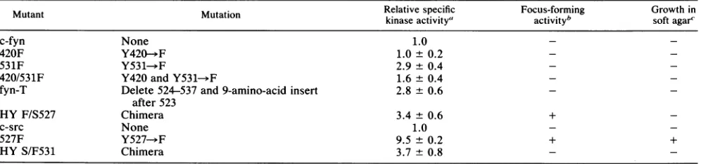
FIG. 2. Relative specific tyrosine kinase activities of the pp59C-fyl variants. Quantitative kinase assays were performed as described
previously (7, 15, 16) andasindicated inMaterials andMethods. Except for wild-type pp60c-src, 527F, and HY S/F531, forwhichassayswere
performedonEC10-derived immunoprecipitates, the kinase activities of pp59C-hf anditsderivativeswereassayedwith theantibodyCFN-2. The relative specific kinase activities were calculated by dividing the amount of 32p incorporated into enolase (B) by the amount of
[35S]methionine incorporated intothetyrosine kinases (A). Values for themutantpp59C-fr" werenormalized relativetothose forwild-type pp59C-fyf, and values for 527F and HY S/F531 were normalized to those for wild-type pp6Oc-rc. Average specific activities from three experimentsof this typearegivenin Table 1. The[35S]methionine gelwasfluorographedandexposedfor 4days. Autoradiographytime for the32p gelwas2 hat-70°C. Molecular size markersare onthe leftinkilodaltons.
(+)
on November 10, 2019 by guest
http://jvi.asm.org/
[image:2.612.124.497.481.657.2]172 CHENG ET AL.
BstEII-BstEII and the 0.2-kb BstEII-SalI fragmentsof 531F-BstEII to527F-BstEII digested withBstEII and SalI.
Biological and biochemical assays. The procedures for monitoring transformingactivity by focus formation and by growth in softagar were asdescribed by Piwnica-Worms et al. (33) and Cheng et al. (7). All of the procedures for metabolic labeling of cells; preparation of cell lysates; im-munoprecipitation of proteins using antibodies forpp59c-`n, pp60c-src, and middle-T antigen; tyrosine kinase assays; complexformationassay;phosphoamino acid analysis;
two-dimensional tryptic phosphopeptide analysis; one-dimen-sional peptide mapping using Staphylococcus aureus V8
protease; andin vitro transcription and translation ofc-fyn
andc-srccDNAs have been fully described elsewhere (5-7, 14-16, 33).
Rescue of proviral fyn-T DNA from c-fyn/3T3 cells. The fyn-T DNAwasrescuedfrom c-fyn/3T3 cells by the rescue-fusion protocol (5). Briefly, c-fyn/3T3 cellsweretrypsinized and plated at a ratio of 1:1 with cos-1 cells such that confluency was attained following approximately 48 h in culture.Cellswererinsed with serum-free medium andthen fused by the addition of 50% polyethylene glycol for1 min. The fused cellswereallowedtogrowfor 3days, after which Hirt DNA (17) was prepared and usedto transform DH5aL bacteria. TheDNAs from kanamycin-resistantbacterial col-onies were digested with BamHI and Sall and ligated to
pUC18 predigested with the same enzymes. Two clones (pUCfyn-T1 and pUCfyn-T2) which exhibited anomalous restriction enzyme digestion patterns compared with those ofwild-type c-fynwere selected for furtheranalysis.
Western blotting. The method for preparing samples for Westernblot (immunoblot) analyses and the conditions for transfer ofproteinstonitrocellulosefilterswere asdescribed by Kampsand Sefton (19). Typically, approximately 100 ,ug of protein from each lysate was loaded onto the gels. Following transfer to nitrocellulose, the filters were
pre-blockedwithblockingbuffer(5%bovineserumalbumin and 1% ovalbumin in TNA buffer [10 mM Tris, pH 7.2; 0.9% NaCl; 0.01% NaN3]) for 18 h at room temperature. The filterswere thenprobed withpolyclonalantibodies to phos-photyrosine (gift of B. M. Sefton) at a concentration of 2 ,ug/ml in blocking buffer for 2 h at room temperature.
Following washing with TNA and TNAsupplemented with
0.05% NonidetP-40, the filterswereincubated with 10 ,uCi of125I-labeled protein A in 40 ml of blocking buffer for 30 min at room temperature. The filters were washed
exten-sively with TNA and TNA containing 0.05% Nonidet P-40 andthen allowed todry.
RESULTS
Sites of phosphorylationonthehuman pp59ciYykinase. To determine the sites phosphorylated in vivo, the engineered cellline c-fyn/3T3(5), whichoverexpresseshumanppS9c-`n,
was metabolicallylabeled with32Pi.The lysates were incu-bated with CFN-2serum, ananti-pp59cfnserum(5), and the immunoprecipitates derived thereinweresubjectedto phos-phoamino acid analysis. Inunsynchronized cells, pp59c-fn wasdetermined tobe phosphorylated primarily ontyrosine and serine residues (Fig. 1). The stoichiometry of serine phosphorylation to that of tyrosine was determined to be approximately 2:1.
Judged by the homology between the amino acid
se-quences ofpp59c-fn and the closely related pp60c-src, Tyr-420 and Tyr-531 are candidate sites oftyrosine phosphory-lation. To localize the sites ofphosphorylation onpp59c
)5n
andtoascertain the effect ofthese posttranslational modifi-cations on the biochemical and biological activities of thecellular kinase, mutant variants of the human c-fyn gene product containing lesions atthepresumptive phosphoryla-tion sitesweregenerated.Two pointmutants(Tyr-420---Phe [420F] and Tyr-531-*Phe [531F])andadouble mutant(420/ 531F) combining the lesions in 420F and 531Fwere
gener-ated. These mutantgeneswere engineered intoaretroviral expression vector(33)whichwas then used to transfectthe variantgenesintoNIH3T3 cells. StableNIH 3T3cell lines expressing themutant proteinswere identifiedby immuno-precipitation using CFN-2 serum(Fig. 2A).
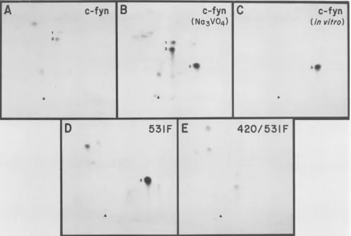
To localize the sites of phosphorylation on pp59c-f", two-dimensionaltrypticphosphopeptide analysisof32p
met-abolically labeled mutant proteins was performed (Fig. 3). Analysis of the resultant phosphotryptic maps suggested
Tyr-420asthe site ofautophosphorylation andTyr-531 asa residue normally phosphorylated in wild-type pp59c-fYn. Theseassignments were deducedfromthe following obser-vations. Spots 1 and 2 were present in the tryptic
phos-TABLE 1. Biochemicaland biologicalpropertiesofpp59C"-1'"mutants
Relativespecific Focus-forming Growth in
Mutant Mutation kinaseactivity' activityb softagar'
c-fyn None 1.0 -
-420F Y420-+F 1.0± 0.2 -
-531F Y531-.F 2.9 ± 0.4 -
-420/531F Y420andY531- F 1.6± 0.4
fyn-T Delete 524-537and9-amino-acid insert 2.8 ± 0.6 after523
HYF/S527 Chimera 3.4 0.6 +
c-src None 1.0
527F Y527-*F 9.5±0.2 + +
HY S/F531 Chimera 3.7 ± 0.8
aDeterminationswereperformed with enolaseasdescribed inthelegendto Fig. 2. Thevalues for wild-typepp60c-" and forthe527F and HYS/F531mutants
weredeterminedbyusingthemonoclonal antibodyEC10(31) and werecalculated in relation to the value for wild-typepp60c"c,which was taken as 1.0. All other mutantswereassayedwith theanti-pp59c1fyl antibody CFN-2. Thevaluesfor theserepresentfold activationrelative to that of normalpp59C-hf,which was
assignedavalue of 1.0. The data are expressed as thearithmetic averagefrom threeseparatetrials + theaveragedeviationfromthe mean.
bTransforming activitywasmonitoredby focusformationover amonolayerof NIH3T3cells followinginfectionwith thevarious recombinantretroviruses
encodingthepp59c-hf andpp603-srvvariants. +,Mutantsdemonstratingfocus-forming activity;-, mutantsdemonstratingno focus-formingactivity. cTransforming activitywas alsoassessedbymonitoringthe ability ofNIH3T3 cellsexpressingthevariousmutants to grow in a 0.3% agarsuspension.Cells
abletoproliferateto adiameterof1 mmafter3 weeks were counted. +,Cellscapable of growthinsuspension;-, cellswhich failedtoproliferate.
J. VIROL.
on November 10, 2019 by guest
http://jvi.asm.org/
[image:3.612.61.559.549.666.2]REGULATION OF THE pp59C-fi KINASE 173
c-fyn
B
c-fyn
(No3VO4)
I.1
...X.I
...
-c-fyn
(
in
vitro)
23
to
U a U U U
D
531F IE
*
3a
a
420/531
F
FIG. 3. Two-dimensional tryptic phosphopeptide maps of the
pp59C-f!'I
mutant proteins. 32P-labeled pp59C-if proteins obtained by immunoprecipitation with CFN-2antibody were gel purified, digested with trypsin, resolved by electrophoresis and chromatography in two dimensions, and then analyzed by autoradiography.Electrophoresis at pH 8.9 was first performed in the horizontal dimension with the anode ontherightand wasfollowedbyascendingchromatography. The originsare marked with arrowheads. Spots 1 and 2, peptides containing Tyr-531; spot 3, peptide containing Tyr-420. Exposure time was 4 weeks at -70°C.phopeptide map of wild-type
pp59c-"f
(Fig. 3A) but absent in the maps ofmutants531F(Fig. 3D) and 420/531F (Fig. 3E) and were therefore deduced to correspond to peptidescontaining Tyr-531. Spot 3 was deduced to be thepeptide harboringTyr-420, since this phosphopeptide was observed in maps of in vitro-phosphorylated
pp59C-fy
(Fig. 3C) and the 531Fmutant (Fig. 3D) butabsent in the double mutant420/531F (Fig. 3E). The observation of increased
phosphor-ylationonTyr-420 with the 531Fmutantandwhen wild-type
pp59C-Yfy
was labeled in the presence of the phosphataseinhibitor sodium vanadate (Fig. 3B) is analogous to data obtained withpp6Oc-src (15).
Biological activities ofNIH 3T3 cells expressing wild-type and mutant
pp59C'Yfy.
To investigate whether thephos-phorylation sites identified above are involved in the regu-lation ofthe
pp59C-fy
kinase, the mutants were tested for their abilities to transform cells in culture. Like wild-typepp59C-&fy
(5), noneof thepp59c-Jfy
point mutants were able to induce focus growth when assayed on NIH 3T3 cells or to sustain anchorage-independent growth in soft-agar suspen-sion (Table 1).Duringthecourseof thiswork,wealsogenerated another mutantof
c-fyn
whichwerefer toasfyn-T. Themutantfyn-T was rescued from an NIH 3T3 cell line overexpressing wild-type humanpp59C-hf
which had been continuouslypassaged for over 20generations. Following repeated
pas-saging, a proportion of the cells expressed a spontaneous truncated mutant of c-fyn whose gene product displayed aberrant enzymatic activity (see below). The cells
express-ingthe
mutant_fyn
proteinwerephenotypically normal. The mutantfyn
DNA(fyn-T) was molecularly cloned from this cell line and upon DNA sequencing of the entire rescued cDNAclone wasshown toharboradeletion of nucleotides 2149 to 2153 (nucleotide numbering according to Semba et al. [36]).Asaconsequenceof the frameshiftmutation, fyn-Twaspredicted toencodeatruncatedprotein of 532 residues
(Fig. 4A). The carboxy-terminal truncated protein would contain residues1through523 ofpp59c-:fyV andanadditional nine unrelated residues resulting from translation of an alternativereadingframe (Fig. 4A).
Analysis of
[35S]methionine-labeled,
in vitro-translated fyn-Ton polyacrylamidegels showed that the gene product migrated morerapidly thanwild-typepp59c-Jf,
a factcon-sistent with the presence of the expected deletion(Fig. 4B;
compare lanes3 and4). Immunoprecipitation analysis
con-firmed that the deletion resided atthe carboxy terminus of
fyn-T.Invitro-synthesizedwild-type
pp59c-Jfl
(Fig. 4B,lane 5) butnotthefyn-T product(lane 2)wasimmunoprecipitated by cst-1, a peptide antibody raised against thecarboxy-terminal seven residues common to both
pp59C`-Y?
and VOL.65, 1991A
I *
2V.
3
It
C
on November 10, 2019 by guest
http://jvi.asm.org/
[image:4.612.58.555.87.421.2]174 CHENG ET AL.
A
Wild Type c-fyn
523 531
l
D Y F T A T E P Y P G E N L
GAC TAC TTT ACC GCG ACA GAG CCC CAG TAC CAA COT CGT GAA AAC CTG TAA
fyn-T: GAC TAC DY
523
C GCG ACA GAG CCC CAG TAC CAA CCT CGT GAA AAC CTGTAA
R D R A P V P T W
ck~- cfr- C
>% I >% I >
B
Lysate
7g
7 a 7C\M
Antibody:
Ec)
Peptide:
+
I
Ij
o 0 Cu
C
c-fyn
fyn-T
F---r----t-Il
92.5-
69-46-;
69-p
59c-fyn
--f
yn-T
30-V,
S_
[image:5.612.112.510.81.511.2]M 2 3 4 5 M 1 2 3 4 5 6M
FIG. 4. Immunoprecipitation and one-dimensional peptide analysis of the fyn-Tmutant.(A) Nucleotidesequenceof the 3'coding regions
presentinwild-type human c-fynandfyn-T. Termination codonsareunderlined. Aminoacid sequencespredicted for human
pp59'-ft"
and fyn-Tarealso shown by one-letterdesignations. (B) pSP65c-fJnand pSP65 fyn-Tweretranscribed with SP6 RNA polymerase. Followingpurification, the RNA transcriptsweretranslated in vitro inarabbitreticulocytelysatein thepresenceof[35S]methionine. Sampleswerethen immunoprecipitated with peptide antibodies raised againstsequencespresentwithin the amino terminus of pp59Cf-rf (CFN-2)orthe carboxy
terminus of pp59C-fyn (cst-1).Immunoprecipitation reactionswereperformed in thepresenceof thecognatepeptide (+)orinthe absence of
added peptides (-). (C) Wild-type
pp59'-f""
(lanes 1 to 3) and fyn-T mutant (lanes 4 to 6) were purified from 32P-labeled cells byimmunoprecipitation with CFN-2 antibody and sodium dodecyl sulfate-polyacrylamide gel electrophoresis, digested with S. aureus V8
protease(0.0017 p.g/g±l [lanes 2 and 5]or0.017 p.g/p.l [lanes 3 and41),andthenelectrophoresedon a15%polyacrylamide gel. Lanes 1 and 6
wereuntreated controls. Lanes Mcontain molecular size markers in kilodaltons. The V fragmentscorrespondingtothecarboxy-terminal portions of wild-type pp59c-fynand fyn-T
(VO-T)
areindicated byarrows. Exposuretimewas1week.pp60c-src (14). Finally,thedeletionwithin thefyn-T carboxy terminus was verified by one-dimensional peptide analysis using S.aureusV8protease(Fig. 4C). The carboxy-terminal V, fragment ofthe fyn-T mutant protein
(V,3&T)
migrated withgreatermobilityonthe gel,whichis consistent with the expected deletion.Thefyn-Tgeneproduct which lacksTyr-531,like the 531F mutant,wasalso judgedtobe nontransformingwhen
exam-inedby the focus-forming assay (Table 1). Hence,asis not thecasewith the othersrc-liketyrosine kinases, preventing
phosphorylation at Tyr-531 does notresult in activation of thetransforming activity ofpp59C-fyf.
Enzymaticactivitiesofpp59C-fyf variants. To determinethe effectof the lesionsontheprotein tyrosine kinase activityof pp59C-fYfl, therelativespecifickinaseactivitiesofthemutant
proteins were assessedby monitoring theirability to phos-phorylate enolase in vitro. Briefly, lysates prepared from [35S]methionine-labeled cells were first normalized to con-tain approximately equivalent amountsof
pp59c-fyn.
Immu-noprecipitates derived by using CFN-2 serum were then
46-S'
J. VIROL.
on November 10, 2019 by guest
http://jvi.asm.org/
REGULATION OF THE pp59c-fn KINASE 175
82 84
I V 299 420.£--.... 531[f(Y-F)537
HY
F/S527
,,...
...I...t...I...83 835 295 416 527(Y-F)
C) LJL
c (*L .
.%- £h >- >
ou
2:x
i) Li-Ucj
Lysote:
z z
iL) U)to f)IC) ID)
U- LcnLL LL L
%% N. . N. N. N
Peptide:
- - - -+
- -+
Antibody:
ECIO
CFN-
2
cst-I
m
.
X_l
5
67
8
9
1011
12
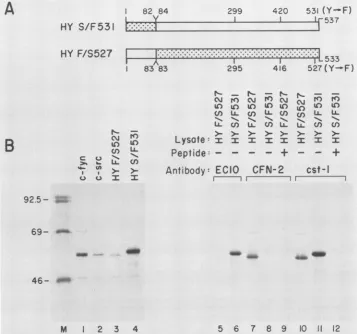
FIG. 5. Immunoprecipitationanalysis of the pp59cfv"-pp6fc-src chimeras. (A) Predicted amino acidsequencesof the chimeras. Theprotein
sequencecoded for byc-srcisshaded, while that for c-fyn isunshaded. The numbers correspondtoamino acids in the wild-type proteins. (B) pSP65 HY S/F531 and pSP65 HYF/S527weretranscribed, and the productsweretranslated in vitro in rabbit reticulocyte lysates. Samples wereeither analyzed directlyonpolyacrylamide gels (lanes 1to4)orimmunoprecipitatedwithEC10 monoclonal antibody(lanes 5and6),
CFN-2antibody(lanes 7to9),orcst-1 peptide antibody (lanes 10to12).Immunoprecipitationswereperformed in thepresenceof thepeptide
immunogen(+)orinthe absence ofadded peptide (-). Thepresenceofanapproximately 75-kDa band from in vitro translates ofc-fynhas beendescribedpreviously (5). Interestingly, thesame75-kDaband(which is fyn related)wasalsoobserved with the chimera HYF/S527but notwithHYS/F531. Fluorographywasfor24 hat-70°C.
either resolvedon gels (Fig. 2A) to confirm the amount of pp59C-fY orincubatedwith[-y-32P]ATPandenolase (Fig. 2B) toevaluate theabilitiesof the variantproteins to phosphor-ylate enolase.
AlterationofTyr-531 tophenylalanine in the531F mutant or deletion of Tyr-531 in the fyn-T mutant enhanced the ability of pp59C'-- to phosphorylate enolase in vitro by approximately threefold (Table 1). Interestingly, although the modification ofTyr-420 to phenylalanine alone didnot
detectablyalter the kinaseactivityof themutant, combining themutations atresidues420 and 531(420/531F) resulted in activating the kinase activity, albeit toonly approximately 50% of that observed with 531F. This observation is
analo-gous to that obtainedwith pp6Oc-src (3, 16, 26, 33). Hence, preventing phosphorylation atTyr-531 results in activation of the pp59c`tfl tyrosine kinase activity but to a level insufficientto transform NIH 3T3 cells in culture.
Properties ofchimeras HYF/S527 and HY S/F531. It has beenproposedthat thedivergenceofsequencesobserved in theamino-terminal 80or soresidues of the various members of the src family oftyrosine kinases may confer different substrate specificity (for a review, see reference 9). The
inability of the 531F mutant ofpp59C-fy to transform NIH 3T3 cells despite a detectable enhancement ofits tyrosine kinaseactivity could thereforehave beenaconsequenceofa lack ofappropriate substrates in the test cells. To examine this possibility,two chimeric constructs were generated by the reciprocal exchange of the amino-terminal sequences
uniquetothe531Fmutantofpp59C-tfy and thetransforming
mutant(527F) ofpp6Oc-rc (7, 33).The resultantchimera,HY F/S527, encoded the amino-terminal 83 amino acids of
pp59C-fyf and residues 83 to 533 of the 527F mutant of
pp60csrc, while thechimera HYS/F531 encodedthefirst82 aminoacids ofpp6Oc-src and residues 84to537 ofthe 531F mutantofpp59C-Yfy (Fig. 5A).
Thestructuresof the recombinant chimericplasmidswere verified by restriction mapping, and those of theirencoded
productswere verified by usingdomain-specificantibodies. Hence, both in vitro-synthesized chimeric proteins were immunoprecipitable with cst-1 peptide antibodies (14), whose epitopes are within thecarboxy-terminal seven resi-duescommontothetwotyrosinekinases(Fig. SB,lanes 10 and 11). The chimera HY S/F531 but not HY F/S527 was reactive with EC10antibody (31), whoseepitope islocated
A
HY
S/F531
B
92-
-
69--_-_
46-
-0
M 1 2 3 4
VOL.65,1991
on November 10, 2019 by guest
http://jvi.asm.org/
[image:6.612.129.486.85.419.2]176 CHENG ET AL.
B
n
0n
U) LLII.
, NJrO
C Lo
l--0) Ni
LL c)
n c LL LL I Xfi) >
4.
PP6Oc
src}-F
59C- flchimaeras
[image:7.612.129.500.79.334.2]l 2 3 4 5 6 1 2 3 4 5 6 7
FIG. 6. Analysis of phosphotyrosine-containing proteins in cells expressing wild-typeand variant
pp59C-f-"'
andpp60c-sr.(A)Clonal NIH3T3 cells expressingapproximately equivalent amounts of eitherwild-type ormutant pp59C-fPn orpp6Oc-sr' asjudged by [35S]methionine
labelingwereselected for analysis. Wild-type pp6Oc-r(, 527F, and HY S/F531wereimmunoprecipitatedwithEC10,whilewild-typepp59C-hf, 531F, and HYF/S527wereimmunoprecipitated with CFN-2. Fluorography wasfor 4days. (B)Anequalamountofproteinfrom eachcell
line waslysed in hot sample buffer, and the proteins were separated by gelelectrophoresis. Following transfertoanitrocellulose filter,
phosphotyrosine-containing proteins were detectedby incubation with anantiphosphotyrosine antibody and 125I-protein Aessentiallyas
describedby Kamps and Sefton (19). The autoradiogramwasexposed for18 h.
withintheamino terminus of pp6Oc-sr (Fig. SB, lanes 5and 6), which is consistent with HY S/F531 having the amino-terminalportion ofpp6oc-sr.Conversely,HYF/S527butnot HYS/F531wasrecognized by CFN-2serum(5),anantibody raised againstsequences located within theaminoterminus of pp59C-ftf, confirming that HY F/S527 containedfyn-like
amino-terminal sequences (Fig. SB, lanes7and 8).
Examination by the focus formationassay demonstrated that HYF/S527 butnotHYS/F531 wastransforming (Table 1). Foci appeared around 40 days postinfection compared with 14days for 527F. Moreover, the number of foci induced was approximately 10% of the total number of G418-resis-tant colonies (average of 40 G418-resistant colonies per 90-mm dishwhen the transientviralsupernatantswereused) compared with 70% for 527F. Furthermore, unlike cells expressing 527F, stable NIH 3T3 cells expressing the HY F/S527 mutant were also unable to proliferate in soft-agar suspension (Table 1). The kinase activities of the chimeras werealso determined by monitoring thephosphorylation of enolase invitro (Fig. 2). ChimeraHYF/S527wasjudgedto have a 3.4-fold-increased kinase activity relative to that of wild-type
pp59C-fyf
(Table 1), while HY S/F531 wasdeter-minedtobe activated 3.7-foldoverwild-type pp60csrc. The findingthat HYS/F531 isnontransformingis consistent with the kinase activity measurements, since the fold elevation observed for this chimera is below the threshold we
ob-servedtobe necessaryformutantsof pp6ocsrc totransform NIH3T3cells (15,16, 33). However, HYF/S527wasjudged
to be transforming, although its enolase-phosphorylating activitywasshowntobe lessthan thatofHYS/F531.Totest whether this result was misleading, for example, reflecting
differences between the abilities ofpp59C`-fl andpp6Oc-srcto
phosphorylateenolase rather than intrinsic enzymatic activ-ity, the kinase activities of the variantproteinsin vivowere compared by usingantiphosphotyrosine antibodies.
Tyrosinekinase activities ofmutantsassayedinvivo. Thein
vivo kinaseactivities of the various mutantswere assessed by monitoringthephosphorylation ofcellularproteinsin an
immunoblot analysis using antiphosphotyrosine antibodies (19). Lysates from cell linesexpressing approximately sim-ilaramountsofwild-type andmutanttyrosinekinases were used (Fig. 6A). The highest level ofimmunoreactivity was reproduciblyobserved with thetransforming527F variant of pp60c-sr followedbyHY F/S527, HYS/F531,and the 531F mutant
pp59C-if"'
(Fig. 6B). Furthermore,it is clear from thisassaythat HYF/S527ismorekinaseactive in cells than the HY S/F531 counterpart, with the former displaying both quantitatively and qualitatively increased immunoreactive bands.
Carboxy terminus of pp59C-fV is necessary for complex formation with middle-T antigen. To address whether the carboxy-terminal regioninpp59c-Jf wasinvolved inbinding middle-Tantigen, the ability ofthetruncated fyn-T protein
to associate with the viralantigen was assayed. Two addi-tional cell lines, one coexpressing wild-type pp59Ci-fy and middle-T antigen (c-fyn/mt) and the other coexpressing fyn-T and middle-T antigen (fyn-T/mt), were generated. Figure 7,lanes 2and3, shows the results ofinvitrokinase
assaysofimmunoprecipitatesobtainedby usingHK3serum,
anantiserumspecificfor theearly regionofpolyomavirus (6, 14). The signal observed represents the total kinase-active fraction of middle-T antigen present in these cells. To
A
rl)
to)
LL-b-r- U)
U) C\ >
oL LL I
N-cn
C L 11L
I4.
t4 I>-a.
92.5
-69
-46
-J. VIROL.
on November 10, 2019 by guest
http://jvi.asm.org/
HK3
CFN-2
n E E E
69- ~ ~ ~ ~
c c c c
> >% >
4-pc 59c f
46-~~~~~~~~~~~b
925-69- -~
2.X.)p59c-fY
W "middle-T
46- _'
30
M 2 3 4
5
6FIG. 7. The fyn-T mutant is unable to associate stably with
middle-T antigen. Lysates were prepared from NIH 3T3 cells expressing wild-typepp59C-fy1 (c-fyn/3T3), thefyn-Tmutant(fyn-T/
3T3), both wild-type
pp59c-Jf
and middle-T antigen(c-fyn/mt), andboth fyn-T and middle-T antigen (fyn-T/mt). The lysates were
normalized for protein content, immunoprecipitated with HK3
(anti-middle-T-antigen serum raised in hamsters) or CFN-2
(anti-pp59Ci-fy serum) antibody, and phosphorylated in vitro by using [_y-32P]ATP. The positions of
pp59C-nfl
and middle-T antigen areindicatedonthe right. Autoradiography wasfor 18 h.
determine whether fyn-T is capable of associating with middle-T antigen, in vitro kinase assays were repeated on
CFN-2-derived immunoprecipitates. Phosphorylation of middle-Tantigen inthisassayis indicative of
coimmunopre-cipitation and hence of complex formation with the viral protein. Middle-T antigen was phosphorylated only in
ly-sates containing wild-type
pp59c-f-y
but not detectably in thosecontainingfyn-T(Fig. 7, lanes 5 and 6), indicating that fyn-T is unable to associate stably with middle-T antigen. We were also unable to detect coimmunoprecipitation of middle-T antigen from [35S]methionine-labeled fyn-T/mt cells (datanotshown).DISCUSSION
pp59C-Vfy kinase isregulated byphosphorylationattyrosines 420 and531. PreventionofphosphorylationatTyr-527in the
case of pp60csrc by mutagenesis to phenylalanine or by
middle-T-antigen binding activates both the tyrosine kinase and transforming activities ofthe enzyme (3, 4, 12, 26, 33).
Although middle-T-antigen binding also activates the kinase activity of complexed pp62c-Yes, in the case of complexed
pp59c-fyf very little or no activation is observed (5, 18, 29).
Therefore, either the pp59C-fl kinase is regulated by mech-anisms other than phosphorylation at Tyr-531 (the residue analogous toTyr-527 ofpp6Oc-src) ormiddle-T-antigen
bind-ing doesnotdirectly affectthe abilityof cellular proteins to regulate phosphorylation at this site. When two-dimensional tryptic phosphopeptide mapping was used, the residue Tyr-531 was identified as asite normally phosphorylated in vivo and Tyr-420 was determined to be the site of autophosphor-ylation in vitro. These assignments are consistent with previous mapping data on the related pp60c-src (10, 38),
pp56Ick
(1, 30), andp59hck
(40)kinases. However, unlike thesituation with pp6csrc
(3,
26, 33),
ppS6Ick(1, 30),
orp59hck(40) mutated at the equivalent site, mutagenesis of the Tyr-531 of
pp59Cf-f
to phenylalanine as in the 531F mutant orits absence in thetruncated fyn-Tmutant did not result inoncogenic conversion of the
pp59C-f".
The tyrosine kinase activities of the 531F and fyn-T mutants were, however, judged to be elevated approximately threefold above that ofthe normal counterpart. Hence the
pp59C-hf
kinase, like the other members of the src family, is at least partially sup-pressed by tyrosine phosphorylation atTyr-531. Thisfinding is consistent with the report by Kypta et al. (28) indicatingthat
pp59C-hf
is negatively regulated by tyrosinephosphor-ylation. Dephosphorylation results in activation of the cata-lytic activity, albeit to a low level previously determined to beinsufficient for neoplastic transformation. Derepression of kinase activity in the 531F mutant isadditionally associated with increased phosphorylation at Tyr-420, a site not nor-mally phosphorylated in wild-type pp59Ci5-lf. Stable phos-phorylation of Tyr-420 is observed only in overexpressed
pp59C-rfy
in NIH 3T3 cells in the presence of the phosphataseinhibitor sodium orthovanadate. The phosphorylation of Tyr-420in the 531F mutant is important, since the additional introduction of a Tyr-420->Phe mutation suppresses the kinase activity to approximatelyhalf themaximum observed with the Tyr-531---Phe mutation alone.
The finding that neither the 531F nor the fyn-T mutant is transforming in NIH 3T3 cells contrasts with the report by Kawakami et al. (23), who demonstrated that oncogenic forms
offyn
selectedfor their abilities to transform NIH 3T3 cells lacked sequences including Tyr-531 at the carboxy terminus. We do not have a satisfactory explanation for this discrepancy. Differences in levels of expression or the choice of target cells may be responsible. However, inter-estingly, the site of mutation of the spontaneous transform-ing mutants described byKawakami et al. (23) and ourfyn-Tmutant all impinge on codon 524.
The inability of 531F or fyn-T to transform NIH 3T3cells
suggests that pp59C-fy may be additionally suppressed by mechanisms which include events other than or in addition to phosphorylation at theregulatory Tyr-531. Thesuggestion that differentmembers of the src family have widelydifferent
kinase activities in NIH 3T3 cells isillustrated by thefinding
thatp59hckmutated at the correspondingTyr-501 is 100-fold more potent as atransforming agent than
pp56Ick
orpp60-src mutated at the homologous Tyr-505 and Tyr-527, respec-tively (40). Regulation of the transforming potential may be effected by restricting either the kinase activity or the substrate specificity of the cellular enzymes or both. This differential restriction may be imposed by the structural variability resulting from the unique amino terminusof each tyrosine kinase.Inability of 531F totransform NIH 3T3 cellsis due tolimited enhancement of its kinase activity. To ascertain whether the inability of the 531F mutant to elicit neoplastic transforma-tion resulted from theabsence of
pp59C--fy
substrates in NIH 3T3 cells or was related to insufficient activation of thepp59C-Vfy
tyrosine kinase activity, weconstructed thechime-ras HY S/F531 and HY F/S527. We rationalized that if the
on November 10, 2019 by guest
http://jvi.asm.org/
[image:8.612.64.284.83.355.2]178 CHENG ET AL.
lack of
pp59cfyn-specific
substrateswas the cause, then the chimera HYS/F531,
becauseitcontainstheamino-terminaldomain of
pp6Oc-src'
believedtoconfer substratespecificity,
should be able to transform NIH 3T3 cells.
However,
HYF/S527 but not HY S/F531 was determined to be
weakly
transforming.
Provided sufficient sequences weretrans-ferredto alter substrate
specificity,
this result suggeststhat theinability
of531F to transform wasprobably
not due tolack of
appropriate
substrates in NIH 3T3 cells but moreprobably
wasa consequence of insufficient activation of itskinase
activity.
Consistentwith thesuggestion
that 531Fwasinsufficiently
activated wasthe observationthat the in vivokinase
activity
of the 531Fmutant was less than thatofthetransforming
HY F/S527. Results from studies of othermutants of
pp6Oc-src
indicate that it is necessary for thekinase
activity
of thecorresponding
mutants to reach athreshold in orderto transform
(7, 15, 16, 26, 33).
Elements within the amino terminus of
pp59cfYn
down-regulateits kinaseactivity.ThechimeraHYF/S527was
only
weakly transforming
compared with the 527F mutant ofpp6Oc-src.
HYF/S527,
unlike527F,
induced focusgrowth
only
afteran extendedperiod,
and cellsexpressing
the HYF/S527 mutant were unable to
proliferate
in soft agar.Perhaps
the structure of the chimera was altereddeleteri-ously; alternatively,
replacement
oftheunique portion
oftheamino terminus of 527F
(residues
1 to82)
with that ofpp59c-fyl
could suppress thetransforming activity
of the 527F mutant. If this is the case, there may be elements within thisunique
portion of the amino-terminal region ofpp59c-fyl
which are absent inpp6Oc-sc
that are additionally involved insuppressing
thetyrosine
kinaseactivity
ofpp59c-ffy.
The involvement of the amino-terminal variable domain of these kinases inregulating
kinaseactivity
orinteractionwith other
proteins
has been documented earlier(15, 22, 29, 34, 37).
If the
unique
amino-terminal domain ofpp59c-fl
issolely
responsible
forrepressing
thekinaseactivity
of the enzyme,thenthereversechimeraHYS/F531 should exhibitakinase
activity
which is greater than that of the 531F mutant.However,
we detectedonly
a very modest increasecom-pared
with that detected with531F,
which weinterpret
tomean that there are other
repressive
elements within theamino-terminalhalf of the
pp59c-fr1
molecule. Such elements may be located within the conservednoncatalytic
domain(residues
83 to 260) of the srcfamily
of kinases. Thissuggestion
issupported by
the demonstration that afusionprotein
encodedby
5' sequences forv-fgr
(the oncogenepresentinGardner-Rasheedfelinesarcoma
virus)
and the 3'two-thirds of the
fyn
gene(residues
220 to 537) is highlyoncogenic (24).
This fusionincludes,
inaddition,
theTyr-531residue of
pp59c-fyl.
Takentogether,
these results suggest that theinability
of 531Ftotransform may beaconsequence of the presence of the aforementionedrepressive
elements within the amino terminus ofpp59C-:fl
which act to restrict its kinaseactivity.
Complex
formation with middle-T antigen requires theintegrity
ofthe carboxy-terminal sequencesofpp59c-fyn.
Thefyn-T
mutant which lacks carboxy-terminal sequences was unable to complex with middle-T antigen. This finding is consistent withprevious
mapping data withpp6Ocsrc.
A domaininfluencing middle-T-antigen
bindinghasbeenlocal-izedto sequences
proximal
totheregulatoryTyr-527 of the enzyme(3, 8).
Presumably, as withpp6O"cs',
middle-T-antigen binding disrupts
theregulationofphosphorylationatTyr-531
ofpp59c-fyl
andtherebyactivates its kinaseactivity.However,
thedata fromthe531Fmutantpredict
thatmiddle-T-antigen binding might not activate the kinase to greater than threefold. Indeed, the kinase activity of pp59C-fy? in complex with middle-T antigenwasjudged not tobe
signif-icantly elevated over that of the unassociated form (5, 19, 29). Although we cannot exclude the possibility of achange in substrate specificity resulting from binding middle-T anti-gen,the data can beinterpreted to indicate thatpp59c-fl has alesser role than pp60csrcorpp62c-Yes in middle-T-antigen-mediated transformation of fibroblasts. It is also unclear whether the separatetyrosine kinases havespecialized func-tions in different tissues. Hence, in certain tissues and at particular developmental stages,
pp59C-fy
may become an important substrate for middle-Tantigen.ACKNOWLEDGMENTS
WethankS. Parsonsforproviding EC10 and GD11 monoclonal antibodies; B. Sefton for antiphosphotyrosine antibodies and advice onWesternimmunoblotting techniques; K. Semba, K. Toyoshima, and T. Yamamotofor the humanc-fyncDNA; H. Piwnica-Worms and T. Roberts for the avian c-srccDNA; and G. White forsynthesis of theoligonucleotides.
This work was supported by Public Health Service grants CA 43186and CA 50661 from the National Cancer Institute.
REFERENCES
1. Amrein, K. E., and B. M. Sefton. 1988. Mutation of a site of tyrosine phosphorylation in the lymphocyte-specific tyrosine protein kinase,
p56Ick,
reveals its oncogenic potential in fibro-blasts.Proc. Natl. Acad. Sci. USA 85:4247-4251.2. Bolen, J. B., C. J. Thiele, M. Israel, W. Yonemoto, L. A. Lipsich, and J. S. Brugge. 1984. Enhancement of cellular src gene product associated tyrosyl kinase activity following polyoma virus infection andtransformation. Cell 38:767-777.
3. Cartwright, C. A., W. Eckhart, S. Simon, and P. L. Kaplan. 1987. Cell transformationby pp6Ocsrc mutated in the carboxy-terminalregulatorydomain.Cell49:83-91.
4. Cartwright, C.A., P. L.Kaplan, J.A.Cooper,T.Hunter,and W.Eckhart. 1986. Altered sites oftyrosine phosphorylation in pp6Oc-src associated with polyomavirus middle tumor antigen. Mol. Cell. Biol. 6:1562-1570.
5. Cheng,S.H.,R.Harvey,P.C.Espino,K.Semba,T.Yamamoto, K.Toyoshima, and A. E. Smith. 1988.Peptide antibodiestothe humanc-fyngene product demonstrate pp59C-f"t iscapable of complexformation with the middle-Tantigen ofpolyomavirus. EMBO J. 7:3845-3855.
6. Cheng, S. H., W. Markland, A. F. Markham, and A. E. Smith. 1986. Mutations around the NG59 lesion indicate an active association ofpolyomavirus middle-T antigen with pp6Oc-src is requiredfor celltransformation. EMBOJ.5:325-334.
7. Cheng,S.H.,H.Piwnica-Worms, R. W. Harvey, T. M. Roberts, and A. E. Smith. 1988. The carboxy terminus ofpp6OC-src is a regulatory domain and is involved in complex formation with themiddle-Tantigen ofpolyomavirus. Mol. Cell. Biol. 8:1736-1747.
8. Cooke, M. P., and R. M. Perlmutter. 1989. Expression ofa novel form of thefyn proto-oncogene in hematopoietic cells. NewBiol. 1:66-74.
9. Cooper, J. A. 1990. Thesrcfamilyofprotein-tyrosinekinases, p. 85-113. B. E.Kemp(ed.),InPeptidesandprotein phosphor-ylation.CRCPress, Inc., Boca Raton, Fla.
10. Cooper, J. A., K. L. Gould, C. A. Cartwright, and T. Hunter. 1986. Tyr527 is phosphorylated in pp60csrc implications for regulation. Science 231:1431-1434.
11. Cooper, J. A., and C. S. King. 1986. Dephosphorylation or antibody bindingto thecarboxy terminusstimulatespp6Ocsrc. Mol. Cell.Biol. 6:4467-4477.
12. Courtneidge, S. A. 1985. Activationofthe pp6O -src kinase by middle-T antigen and by dephosphorylation. EMBO J. 4:1471-1477.
13. Courtneidge, S. A., and A. E. Smith. 1983. Polyoma virus transforming protein associateswith the product of the cellular J. VIROL.
on November 10, 2019 by guest
http://jvi.asm.org/
srcgene. Nature(London)303:435-439.
14. Courtneidge, S. A., and A. E. Smith. 1984. The complex of polyomavirusmiddle-T antigenand pp6Osrc.EMBOJ.
3:585-591.
15. Espino, P. C., R. Harvey, R. L. Schweickhardt, G. A. White,
A. E.Smith, and S. H. Cheng. 1990. Theamino-terminal region ofpp6oc-srchasamodulatory domain and contains multiplesites
oftyrosinephosphorylation. Oncogene5:283-293.
16. Harvey,R., K. M.Hehir, A. E. Smith, and S. H. Cheng. 1989. pp6Oc-srcvariantscontaining lesions that affect phosphorylation
attyrosines416and 527. Mol.Cell. Biol. 9:3647-3656. 17. Hirt, D. 1967. Selective extraction of polyoma DNA from
infectedmousecell cultures. J. Mol. Biol. 26:365-369.
18. Horak, I. D., T. Kawakami, F. Gregory, K. C. Robbins, and
J. B. Bolen. 1989. Association of
p6O'W-"
with middle tumorantigeninmurinepolyomavirus-transformed ratcells.J. Virol. 63:2343-2347.
19. Kamps,M. P., and B. M. Sefton. 1988.Identificationof multiple novel polypeptide substrates ofthe v-src, v-yes, v-fps, v-ros,
and v-erb-Boncogenic tyrosine proteinkinasesutilizing antisera againstphosphotyrosine. Oncogene2:305-315.
20. Katagiri, T., J. P. Y. Ting, D. Y. Ruth, C. Prokop,P.Cohen,and
H. S. Earp. 1989. Tyrosine phosphorylation of a c-src-like
protein is increased in membranes of CD4- CD8- T lympho-cytesfromIprllprmice. Mol.Cell. Biol. 9:4914-4922. 21. Katagiri, T., K. Urakawa, Y. Yamanashi, K. Semba,T.
Taka-hashi, K. Toyoshima, T. Yamamoto, and K. Kano. 1989. Over-expressionofsrcfamilygenefortyrosine-kinase p59fe in CD4-CD8- Tcells of mice withalymphoproliferative disorder.Proc. Natl. Acad. Sci. USA 86:10064-10068.
22. Kato, J. Y., A. Takeya, C. Grandori, H. Iba,J.B.Levy, andH. Hanafusa. 1986. Amino acid substitutions sufficientto convert thenon-transforming pp60f-src proteintoatransforming protein.
Mol.Cell. Biol.6:4195-4201.
23. Kawakami, T., Y. Kawakami, S. A. Aaronson, and K. C. Robbins.1988. Acquisitionoftransformingpropertiesbyfyn,a
normal src-related human gene. Proc. Natl. Acad. Sci. USA 85:3870-3874.
24. Kawakami, T., C. Y. Pennington, and K. C. Robbins. 1986. Isolation and oncogenic potential ofa novel human src-like
gene. Mol.Cell. Biol. 6:4195-4201.
25. Kawakami, Y., M. Furue, and T. Kawakami. 1989. Identifica-tion offyn-encoded proteins in normal human blood cells. Oncogene 4:389-391.
26. Kmiecik, T. E., and D.Shalloway. 1987.Activation and suppres-sion ofpp6-src transforming ability bymutationofitsprimary sites oftyrosine phosphorylation. Cell 49:65-73.
27. Kornbluth,S., M. Sudol, and H. Hanafusa. 1987. Association of polyomavirusmiddle-Tantigenwithc-yesprotein.Nature (Lon-don)325:171-173.
28. Kypta, R. M., A. Hemming, and S. A. Courtneidge. 1988. Identification and characterization of p59/f (asrc-likeprotein
tyrosine kinase) in normal andpolyoma virus transformed cells. EMBO J. 7:3837-3844.
29. Louie, R. R., C. S. King, A. MacAuley, J. D. Marth, R. M. Perlmutter, W. Eckhart, and J. A. Cooper. 1988.p561ck protein-tyrosine kinase is cytoskeletal and does not bind to polyomavi-rus middle-Tantigen. J. Virol. 62:4673-4679.
30. Marth, J. D., J. A. Cooper, C. S. King, S. F. Ziegler, D. A. Tinker, R. W. Overell, E. G.Krebs, and R. M. Perlmutter. 1988. Neoplastictransformationinduced by an activated lymphocyte-specific proteintyrosine kinase(pp56lck). Mol. Cell. Biol. 8:540-550.
31. Parsons,S.J., D. J. McCarley, C.M.Ely,D.C.Benjamin,and J. T. Parsons. 1984. Monoclonal antibodies to Rous sarcoma viruspp6Osrc react with enzymatically active cellularpp6lsrc of avian and mammalianorigin.J. Virol. 51:272-282.
32. Perlmutter,R.M., J. D. Marth, S.F. Ziegler,A. M.Garvin,S. Pawar, M. P. Cooke, andK. A. Abraham. 1988. Specialized protein tyrosine kinases in hematopoietic cells. Biochim. Biophys. Acta948:245-262.
33. Piwnica-Worms, H., K. B. Saunders, T. M. Roberts, A. E. Smith,and S. H. Cheng. 1987.Tyrosine phosphorylation regu-lates thebiochemical andbiological properties ofpp60-src.Cell 49:75-82.
34. Potts,W.M.,A. B.Reynolds,T.J. Lansing,andJ.T. Parsons. 1988. Activation ofpp6Ocsrc transforming potential by muta-tions altering the structureof an amino terminal domain con-taining residues 90-95.OncogeneRes.3:343-355.
35. Rudd,C.E., J.M.Trevillyan,L.L.Wong, J.D.Dasgupta,and S. F. Schlossman. 1988. The CD4 receptor is complexed in detergentlysatestoprotein tyrosine kinase (ppS8) fromhuman T-lymphocytes. Proc.Nati. Acad. Sci. USA 85:5190-5194. 36. Semba, K., M. Nishizawa, N. Miyajima, M. C. Yoshida, J.
Sukegawa, Y. Yamanashi, M. Sasaki, T. Yamamoto, and K. Toyoshima. 1986. yes-Related protooncogene, syn, belongs to theprotein-tyrosine kinasefamily.Proc.Natl. Acad. Sci. USA 83:5459-5463.
37. Shenoy, S., J. K. Choi, S. Bagrodia, T. D. Copeland, J. L. Mailer,and D. Shalloway. 1989. Purified maturationpromoting factorphosphorylatespp6Oc-scatthesitesphosphorylated dur-ing fibroblast mitosis. Cell 57:763-774.
38. Smart, J. E.,H.Oppermann,A. P.Czernilofsky,A. F. Purchio,
R. L.Erikson,andJ.M.Bishop. 1981.Characterization of sites fortyrosinephosphorylationin thetransformingproteinofRous sarcoma virus (pp6ovsrc) and its normal cellular homologue
(pp60-src). Proc. Natl. Acad. Sci. USA 78:6013-6017. 39. Veillette, A.,M. A. Bookman, E. M. Horak,and J. B. Bolen.
1988.TheCD4and CD8Tcell surfaceantigensareassociated with theinternal membrane tyrosine kinase
p56"ck.
Cell 55:301-308.40. Ziegler, S. F., S. D. Levin, and R. M. Perlmutter. 1989. Transformation ofNIH 3T3fibroblastsbyanactivated form of


![FIG.7.pp59Ci-fyindicatednormalized(anti-middle-T-antigenbothexpressing3T3),[_y-32P]ATP.middle-T The fyn-T mutantis unableto associatestably with antigen.Lysateswere prepared from NIH 3T3cells wild-type pp59C-fy1(c-fyn/3T3), the fyn-T mutant (fyn-T/ both wi](https://thumb-us.123doks.com/thumbv2/123dok_us/1317450.85274/8.612.64.284.83.355/fyindicatednormalized-antigenbothexpressing-mutantis-unableto-associatestably-antigen-lysateswere-prepared.webp)
