JOURNALOFVIROLOGY, June 1973,p.840-847
Copyright 0 1973 American Society for Microbiology Vol. 11, No. 6 Printed inU.S.A.
Function
of
T4D
Structural
Dihydrofolate
Reductase in
Bacteriophage
Infection
CAROLYN J. MALE ANDLLOYD M. KOZLOFF
Department ofMicrobiology, University of Colorado MedicalSchool, Denver, Colorado80220
Received for publication 15January1973
Various properties of the bacteriophage structural dihydrofolate reductase (DFR) have beenexamined todetermine its function during phage infection. It hasbeenfound thatabinding site for reduced nicotinamide adenine dinucleotide
phosphate (NADPH), mostlikelyonthe DFR presentinthe phage tail plate, is required for phage viability. Attachment of adenosine diphosphoribose, an
analogue of NADPH, tothis site prevents phage adsorption and injection. This
adenosine diphosphoribose inhibition can be competitively reversed by the
addition of NADPH or oxidized nicotinamide adenine dinucleotide
phosphate. It is suggested that, during phage infection, the host bacterial cell might leak compounds functionally similar to the pyridine nucleotides. These compounds have been shown tononenzymatically change the conformation of thephage tail plate DFR which isapparentlynecessaryforsuccessfulinjection.
Kozloff et al. reported that bacteriophage
T4D-induced dihydrofolate reductase (DFR) was an integral partofthe phage particle and that its inclusion in the phage tail plate was essential forphage assembly andviability (6). Mathewsconfirmedthe presenceofphage DFR as a structural component byshowing that the heat sensitivities ofwhole phage particles were due tothe particularDFR genethey contained
(7).
ThepresenceofDFR inthe
phage
tailplate,
along with its substrate
dihydropteroylhexa-glutamate, is most unexpected. Immediately
one can ask what role(s) phage DFRis playing
as astructural component. Knowingthat
phage
are inactivated by reduced nicotinamide
ade-nine dinucleotide phosphate
(NADPH;
6), asubstrate for DFR, and that NADPH attach-ment can alter DFR conformation
(1),
Kozloffet al. proposed that the phage structural DFR
could be involved in phage adsorption or
in-jection, or both. It was also suggested that NADPH, presumably fromcell leakage, bound totail plateDFR,reducingthe
dihydropteroyl-hexaglutamate to the tetrahydro-compound
and further that accompanying the reduction there was a conformational change of the en-zyme which was reflected by a
morphological
alteration of the tail plate (6). In view ofthe observations of Greenfield et al. (2) that
con-formational changes of Escherichia coli DFR need not involve enzymic reduction, reduction
to the tetrahydro-compound may not be
re-quired.
The above facts and conjectures lend them-selves to two predictions. (i) If a NADPH-induced conformational change of DFR is essen-tialforphage attachment or injection, orboth, then the DFR of every potentially infectious particle must be able to react with NADPH. The ability ofNADPH to bind to DFR can be determined by measuring the inactivation of whole phage particles. This NADPH inactiva-tion isprobablynot physiologicallysignificant, but it is a convenient method to measure the ability ofphage DFRtobind NADPH. Thus,if binding ofNADPH is essentialforphage infec-tivity, every infective phage article must con-tain DFR which can bind NADPH, and one should not be able to isolate phage particles resistant to NADPH inactivation. (ii) A struc-tural analogue of NADPH might bind to DFR reversibly, or irreversibly, and effect an altera-tion ofphageadsorptionorinjection,orboth. If an alteration was found and the analogue was reversiblybound to DFR, then added NADPH should compete with the analogue to restore normal phageinfection.
Examination of the above predictions indi-cates that phage T4D structural DFR partici-pates nonenzymatically in phage infection, probably by virtue of its ability to undergo a conformational changeas aresult of interaction withpyridine and adenine nucleotides.
MATERIALS AND METHODS
Bacteriophage and bacteria. These have been described in aprevious paper(5).
840
on November 10, 2019 by guest
http://jvi.asm.org/
Determination of adsorption and injection rates.
Bacteria were grown overnight at 37 C in tryptone
broth (Difco) plus 0.5% NaCl, Fresh logphase cells (2 x 10⁸ to 4 x 10⁸/ml) were sedimented once and suspended in tryptone broth with the aid ofaVortex mixer.Mostexperimentsweredoneat room tempera-ture(20-22C),sincethisslowed the infection process and facilitated accurate measurements. Phage were
addedtobacteriaat amultiplicityof infectionof0.1. The total volume of the contents of the adsorption
tuberangedfrom0.2to0.5ml in variousexperiments.
Within a singleexperiment, theamount of bacteria andthe total volume were kept constant. At
desig-nated times, 0.02-mlsampleswereremovedtotubes in an ice bath which contained 2.0 ml of tryptone broth without NaCl(SFB;salt-freebroth). One-milli-liter samples were blended in the microattachment container ofa Sorvall Omnimixer. Cell viability is unaffectedbytheblendingtreatment.The containers were prechilled onice and were immersed in an ice bath during blending. Cell-phage mixtures were blended for 1 min at asetting of5 (9,000rpm), and 0.05ml of theblended mixture wasplacedinatube containing0.45ml of T4D antiserum. A separate tube containing 0.45 ml of antiserum received 0.05 ml of theice-cold unblendedadsorptionmixture.T4D anti-serawith different K valueswereusedthroughoutthe courseoftheseexperiments.Preliminary experiments
established the dilution of antisera (in SFB) which would inactivate about5 x 104T4D particles perml within5min at roomtemperature.Antisera potency was checked periodically. Blended and unblended samples were incubated in antiserum for 10 min, diluted 10-fold into SFB and plated in the usual manner. Adsorption rates were obtained from the plaque counts (infected cells) ofunblended samples.
The fraction ofadsorbed phage which had injected
their DNA was calculated for a given sample by dividing the plaquecountoftheunblendedsample by
theplaquecountofthe blended sample.
The pH ofunadjustedtryptone brothwasslightly
below neutrality(pH6.8-6.9)and is referredto aspH 7inthis report. Whenadsorption and injectionrates weredone at pH 5, tryptone brothadjusted topH 5 with HClwasused for resuspensionofthelog phase cells used in theadsorption tube.
Competitive inhibition of adsorption and
injection. Solutions of adenosine diphosphoribose (ADPR) and other compounds were prepared at a concentration of 10 times the value desired in the
adsorption tube and diluted 10-fold into the
adsorp-tiontube mixture (i.e., 0.025 ml of 0.05 M ADPR into a final volume of 0.25 ml to give a final ADPR concentration of5 mM). The pH of all solutions was determined, and the pH was adjusted with HCl or NaOH when necessary.Allcompoundsbeing testedin aparticular adsorption mixture were added to cells before additionofphage.
Inactivation of T4D particles. Inactivation of T4Dat pH5with NADPHwasdonebyadding0.03 mlof0.03 M NADPH in0.5 M ammonium acetate buffer (pH 5) to 0.27mlof aphage suspension which had beendialyzedfor18hagainst 0.05 M ammonium acetate buffer (pH 5). The incubation mixture was overlaid with mineral oil to prevent air oxidation of the NADPH.
Heatinactivation of T4D in the presence of ADPR orNADPH was done in 0.1 Mpotassiumphosphate buffer(pH 7.0) at 60 C. Phage were diluted so that the initial phage concentration in the reaction mixture wasabout 107 particles per milliliter.
Other methods (chemicals). The hydroxylamine mutagenesis method of Hall and Tessman was used (4). Isolation of soluble DFR and determination of enzymatic activity by a spectrophotofluorometric method were as described earlier (6). All pyridine nucleotides and ADPR were products of P. L. Bio-chemicals. Adenosine diphosphoglucose (ADPG), adenosine diphosphomannose (ADPM), and nicotina-mide mononucleotide (NMN) were purchased from Sigma Chemical Co.
RESULTS
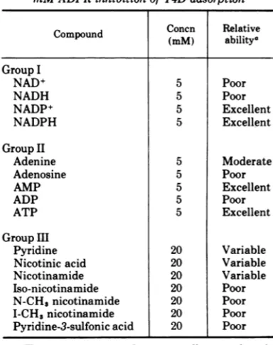
Attempts to isolate aT4D NADPH resist-ant mutresist-ant. Large numbers of phage were treated with NADPHatpH5.In the absenceof NADPH the phage are stable for atleast 24h. Phage were inactivated with NADPH at pH5 ratherthan atpH 7 because at pH5 inactiva-tion was fasterand exponential (no initial lag, reference 6). All of the particles were inac-tivated exponentially within 24 h, and 1012 particles were completely inactivated in other experiments(Fig. 1). Onecanconclude thatif a mutation to NADPH resistance can occur, (i) the frequency of such a mutation is less than 10-12or, (ii) sucha mutation is lethal.
One hundred T4D DFR (wh) mutants have been isolated following hydroxylamine muta-genesis. The mutants were classified as DFR mutants according to their distinctive plaque morphology (3). Itwasfelt thatthe frequencyof NADPH resistantmutants mightbe enrichedin this collection of 100DFR mutants. However, when the inactivationof each mutantby3 mM NADPHinphosphate buffer(pH 7.2, 37 C) was examined, noresistant mutants werefound.
The sensitivity of T4D particles to NADPH plus the lack of NADPH resistant mutants indicated that the abilityofphageDFR to react withNADPH wasessential for viability.
The effect of ADPR on phage adsorption and injection rates. The first structural ana-logue chosenforstudy was ADPR. ADPR lacks the nicotinamide portion of the pyridine nu-cleotidestructure. At a concentration of 5 mM, ADPR inhibited phage adsorption, but at 1 mM ADPR there was no effect on the rates of phage adsorption orDNAinjection. Intermedi-ate concentrations of ADPR inhibited both adsorption and injection rates (Table 1). Whereas ADPR decreases the rate of adsorp-tion at 1 min (2.5 mM ADPR, Table 1; 3 mM ADPR in other experiments), the amount of adsorption is equal to that of the control (no ADPR)at6min.Typically the injection process
on November 10, 2019 by guest
http://jvi.asm.org/
MALEANDKOZLOFF
-
I-HOURS
FIG. 1. Inactivation ofT4DbyNADPH.
Inactiva-tion by 3 mM NADPH was carried out in 0.05 M acetate buffer (pH 5)underoil.
TABLE 1. Effect ofADPRonadsorptionand
injectionratesa
Experi- ADPR Adsorption(%) Injection(%)
concn
ment (mM) 1min 6mi 1min 6min
1 0 43 91 45 77
1 55 97 41 92
2.5 20 88 33 56
5.0 0 0 0 0
2 0 33 66 45 81
2.5 20 74 35 67
5.0 0 0 0 0
aAdsorptionbasedonphage inputof 4.65x 10⁷per
ml. Percent injection = (infected cells, blended/
infectedcells,unblended) 100.
was still impairedat 6 min relative to control
values.
In subsequent experiments ADPR was used at 5 mM because this concentration was the
minimal amount needed to completely inhibit
phage adsorption in 90% of the experiments, andonecouldbe assuredthat therewas nolarge
excess ofADPR in the system. In 10% of the
experiments, 5 mM ADPR did not give
com-plete
inhibition, and a few plaques appeared(about
1-3% of control values at any timepoint).
This variability was of some importanceonly
with respect to data presented in Table 2 and will bediscussedinthat section.In vitro enzyme assays strongly suggested that the ADPR effecton adsorption and
injec-tionwasrelatedtothe specific binding of ADPR
to
phage
structural DFR. Phage-induced DFRwaspartiallypurified, and the ability of ADPR
to bind and inhibit enzymatic activity ofthe
soluble enzymewastested. The results in Fig. 2 confirm that ADPR does bind and inhibit enzymatic activity. Comparison of Fig. 2 with
Table 1 shows a good correlation between the
concentration of ADPR needed to inhibit
en-zyme activity (5 mM inhibited 95%) and the
concentration of ADPR needed to inhibit the
adsorption
ofwhole phage particles containingDFR (5mM inhibited adsorption about 98%).
Assured that ADPR was binding to DFR,
probably
atthe NADPH binding site, NADPHwasadded with ADPRtocell-phagemixturesto
determinewhether NADPH couldcompetewith
ADPRfor thebindingsite. AtNADPH concen-trations from 1to10mM andatpH7,phageare
[image:3.493.54.248.52.375.2]notinactivatedforseveral hours.The resultsof
TABLE 2. Ability ofvarious compoundsto relieve 5 mMADPRinhibitionof T4Dadsorption
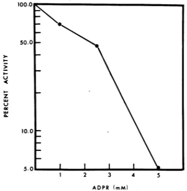
Concn Relative
Compound (mM) abilitya
GroupI
NAD+ 5 Poor
NADH 5 Poor
NADP+ 5 Excellent
NADPH 5 Excellent
GroupII
Adenine 5 Moderate
Adenosine 5 Poor
AMP 5 Excellent
ADP 5 Poor
ATP 5 Excellent
GroupIII
Pyridine 20 Variable
Nicotinic acid 20 Variable
Nicotinamide 20 Variable
Iso-nicotinamide 20 Poor
N-CH3nicotinamide 20 Poor
I-CH3nicotinamide 20 Poor
Pyridine-3-sulfonicacid 20 Poor
aThe terms poor, moderate, excellent, and varia-blearedefined in the text.
842 J.VIROL.
on November 10, 2019 by guest
http://jvi.asm.org/
[image:3.493.257.449.397.640.2]a typical expei
These experin although ideni C. NADPH (t
slightly at6m
only complete but, infact, st
mannerwhich
3). Although it addition of5 r
rates to those Addition of 3 restoringthe ir the amount of
I.-z
uL
rimentareshowninFig. 3and 4. control value (Fig. 4). Addition of 5 mM aents were carried out at 18 C, NADPH (not shown) or 10 mM NADPH fully
tical results were obtained at 22 restored injection rates to those seen with the 3 mM) restores adsorption only control. The results shown inFigures 2, 3, and 4
in, whereas 10mM NADPH not strongly suggest that ADPR is a competitive
ly relieves the ADPR inhibition inhibitor of NADPH and that binding of imulates the adsorptionrateina NADPH tothephage particle maybe essential
isnotpresentlyunderstood (Fig. for phage infectiontooccur.
isnotshown in thisexperiment, Ability of various compounds to inhibit nM NADPH restored adsorption phage adsorptionorrelieveADPRinhibition
of the control (no additions). of phageadsorption. To gainsomeinsightinto
mM NADPH was effective in the bindingspecificities,theabilityofavariety
njection of phage DNA. At6min, ofcompounds structurally relatedtoNADPHto
injection was about 80% ofthe either inhibit phage adsorptionorrelieve ADPR
*_________________________ inhibition of adsorption was studied. The
re-sults are shown in Table 2. None of the
com-pounds listed in Table 2 inhibited phage ad-sorption. Thetermspoor, moderate, and
excel-lent refer to a compound's ability to relieve ADPR inhibition relative to a control
adsorp-tion (no additions) donewith thesamecellson
thesameday. Thetermpoor indicates that the
compound relieved the ADPR inhibition of adsorption to about 10% of the level of the control; the term moderate indicates that the compound relievedADPR inhibition of adsorp-tionto about50% ofthe control; and theterm
excellent indicates that the compound
com-pletely relieved the 5 mM ADPR inhibitionof phage adsorption (i.e., was equal to the
con-trol; noadditions).
Inthe firstgroupofcompounds, the pyridine
1 2 3 4 5 nucleotides, it significant that oxidized
ADPR (mM)
FIG. 2. ADPR inhibition of dihydrofolate
reduc-tase activity. The spectrophotofluorometric method used hasbeen described (6).
-0
LIA -v
z
MINUTES
FIG. 3. Effect of ADPR and NADPH on T4D
adsorption. Adsorption was carried out in pH 7 tryptone brothat18C.
MINUTESS
FIG. 4. Effect of ADPR and NADPH on T4D
injection. The experimentwasdone in pH7tryptone brothat 18C.
z
u z
z
LI
AT 180
6_5mM+lOmM ADPR
NADPh /
4
NOAr'DITIONS
2
5mMADPR+3mM NADPH
0 a
5mMADPR
I I I I I I
1 2 3 4 S d
on November 10, 2019 by guest
http://jvi.asm.org/
[image:4.493.46.239.216.413.2] [image:4.493.250.442.415.625.2] [image:4.493.51.240.446.633.2]MALE ANDKOZLOFF
nicotinamide adenine dinucleotide phosphate
(NADP+) was as effective as NADPH on an
equimillimolar basis in competing with ADPR to restore adsorption. Although it is notshown
inTable 2, NADP+ alsorestored injectionrates to those obtained with a control culture. The
ability of NADP+ to effectively restore both
adsorption and injection clearly indicates that
phage structural DFR is functioning
nonenzy-matically in both processes since the oxidized
form, NADP+, cannot reduce dihydropteroyl-hexaglutamate to
tetrahydropteroylhexagluta-mate. The observation of Greenfield et al. that conformational changes of DFRcanoccur
with-out enzymatic reduction of dihydrofolate sup-ports this conclusion (2).
Of the adenine-related compounds in Group II, AMPand ATP effectively competed with 5
mM ADPRto restore phage adsorption. Thethird group of compounds, analogues of
pyridine, were ineffective in competing with
ADPR. When ADPR inhibition was complete, as determined in a control experiment, pyri-dine, nicotinamide,and nicotinic acidwerepoor
competitors. When ADPR inhibition was in-complete,their abilities tocompetewithADPR
ranged from moderatetoexcellent. These
varia-ble compounds were less effective than
NADPH, NADP+, AMP, and ATP in compet-ing with ADPRsinceGroupIIIcompoundswere
tested at a concentration of 20 mM. This variable response was not seen with the other fourcompounds in Group IIIorwithany other
compounds in Table2.
Also tested were the ADPR related
com-pounds, ADPM and ADPG. Neithercompound inhibited phage adsorption when tested at a
concentration of 5 mM, whereas 5 mM ADPR
completely inhibited phage attachment. Also, ADPM and ADPG didnot bind tophage DFR
in vitroas evidencedby unimpaired enzymatic
activity of the DFR. The fact that neither ADPG nor ADPM inhibit phage adsorption or
DFR enzymatic activitydemonstratesthe spec-ificityof the terminal riboseportionof ADPRas
beingsacrucial determinentinbindingtoDFR. NMN (nicotinamide with anattachedribose
and phosphate) was the only other compound tested besides ADPR which inhibited phage adsorption. Initial experiments show that, like ADPR, NMN inhibits enzymatic activity of DFRin vitro. Sinceaddition of nicotinamide to
aphage-cellmixture does not inhibitadsorption
and NMN does, the importance of the ribose portion of NMN (structurally the terminal ribose ofADPR) is againdemonstrated.
These effects of NMN added to those of ADPR clearly support the conclusion that a
binding site exists on the phage structure for compounds containing the components nico-tinamide-ribose-P-P-ribose-adenine. A com-pound containing a specific ribose (ribose at-tached toNMN, terminal ribose of ADPR) but notthe rest of the pyridine nucleotide structure inhibits phage adsorption and injection. Competition of these inhibitors by thecomplete pyridinecoenzymes, NADPHand NADP+, and other analoguesstructurally related to NADPH and NADP butnot containing the critical ribose moiety support the above conclusion regarding a pyridine nucleotide binding site. DFR is the most likely and probable component of the tail platetohavesuch a specific site.
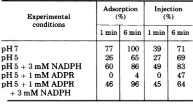
Effect of pH 5 broth on phage adsorption and injection. Since it is known: (i) that isolated phage tail plates are sensitive to acid
(C.J.Male, V.A.Chapman,S. S. DeLong, and L. M. Kozloff, Bacteriol.Proc. p. 183, 1970); (ii) that therate ofphage inactivation by NADPH isfourtimes faster at pH 5 than at pH 7 (6); and (iii) that the rate of phage inactivation by antiseratophage DFRis twice as fast at pH 5 as at pH7.2(unpublishedexperiments), it seemed reasonable to assumethat the structure of the phage tail plateisalteredatpH 5. Incubation at pH5,eitherby structural alterationof proteins in the tail plate or by a direct conformational change of DFR, changes the reactivity of the phage DFR with respect to substrate binding and antisera inactivation. It thus seemed plaus-ible that at pH 5 phage infection might be inhibited. Comparisonof lines 1 and 2 of Table 3shows that both adsorption and injectionare inhibited at 1 min at pH 5. The amounts at 6 min are close to those of the pH 7 control sample. The pH 5-mediated decrease of ad-sorptionand injectionrates canbe abolishedby the additionof3mM NADPH(line3). Addition of 1 mM ADPR to a phage-cell suspension at
TABLE 3. Effect of incubation at pH 5 on adsorption andinjectiona
Adsorption Injection
Experimental (%) (%)
conditions
1min 6 min 1 min 6min
pH7 77 100 39 71
pH5 26 65 27 69
pH5+3mM NADPH 60 86 49 83
pH5+1mMADPR 0 4 0 47
pH5+1mMADPR 46 96 45 64
+3mM NADPH
aAdsorption basedonphage inputof 4.65 x 10⁷ per ml. Percent injection = (infected cells, blended/
infectedcells, unblended) 100.
844 J.VIROL.
on November 10, 2019 by guest
http://jvi.asm.org/
[image:5.493.256.450.528.634.2]pH 5 completely inhibited phage attachment (line 4). One mM ADPR did not affect adsorp-tion or injection at pH 7 (Table 1). The com-bined inhibitions due topH5 broth and 1 mM ADPRare relievedby 3mM NADPHasshown in line 5 of Table 3. Presumably, binding of NADPH to DFR "protects" the DFR or asso-ciated proteins from the conformational change induced bytreatment ofpH 5.
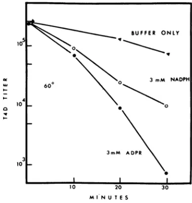
EffectofNADPH and ADPR on heat inac-tivationofT4Dphageparticles.The evidence presented above that a conformational change
of DFR is associated with phage infection is circumstantial but attractive. It is difficult to demonstrate the conformational change of a structural protein in situ, but heat inactivation experiments below offer independent evidence that binding of either ADPR or NADPH to structural DFRisaccompanied by a conforma-tional change.
It is well known that the heat sensitivity of a protein is related to its tertiary structure. With respect to structural DFR, Kozloff et al. first
demonstrated a correlation between the heat
sensitivity of a whole phage particle and the
structural DFR it contained (6). Thisdiscovery
wasexpanded and confirmed by the genetic and
biochemical studies of Mathews (7). Thus one
might expect to find differences in the heat sensitivity of particles treated with ADPR or NADPH if their binding to DFR induced a conformational change. Phage particles in the presence of 3 mM NADPH are moresensitive to heat inactivation than in the absence of NADPH (Fig. 5). Similarly, phage particles heated in the presence of 3 mM ADPR were 100-fold more sensitive than phage heated in
the absenceofADPR (measured at 30 min).
Ability of ADPR to inhibit phage
adsorp-tion as a function of time of addition. ADPR (2.5 mM) decreased the rates of both phage
adsorption and injection (Table 1). But upon
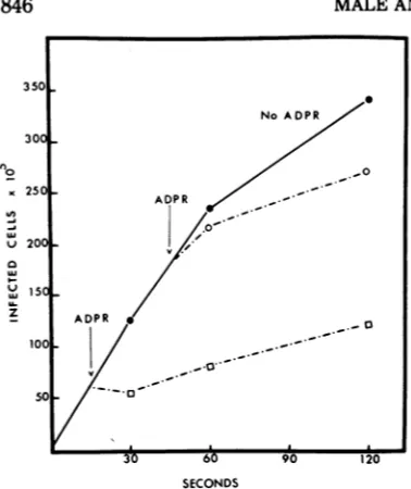
continuedincubation, adsorption and injection rates approached those seen in the absence of ADPR. Restoration of these rates to normal, as afunction of time, could reflect the increasing concentration of a compound(s) which com-petes with ADPR for the NADPH binding site onphage DFR. Thus, one could predict that the sensitivity of a phage-cell suspension to ADPR inhibition of adsorption might decrease with time.The experiment shown in Fig. 6 supports this hypothesis. In this experiment cells and phage were mixed, and either ADPR or broth was added at 15 or 45 s. Adsorption was identical in the twobroth controls. If ADPR was added at 15 s, therewas an immediate inhibi-tionofphage attachment, whereas if ADPR was
added at 45 s the phage were much more resistant to ADPR inhibition. Identical results were obtained in two separate experiments.
Phage attachment in the absence of ADPRwas linear through 1 min, which makes the differ-ence in ADPR inhibition valid.The data inFig. 6suggeststhatthere is an increased concentra-tion of a compound(s) present between 45 and 60 s ascomparedtobetween 15 and30s, which competeswith ADPR for the DFR bindingsite. After these 15-s intervals, the adsorption rates are approximately the same and show the inhibiting action of the ADPR.
Of the compounds tested, NADPH, NADP+,
AMP, and ATP, all normal bacterial cell con-stituents, were effective in competing with ADPR. Thus itappears that thephysiologically active compound(s) needed to bind to phage DFR and induce the conformational change necessary for successful phage infection might bea product of cell leakage.
DISCUSSION
Several lines of evidence indicate that the NADPH and ADPR bindingsite ofthe phage particle is the phage structuralDFR. (i) Phage-induced DFR is a structural component ofthe phage particle (6, 7), and no other protein (enzyme)with a NADPHbinding site is known
to be aphagestructural component. (ii) ADPR
and NMN inhibit phage infection, and both compounds inhibit enzymatic activity of solu-ble DFRinvitro. (iii) Heatsensitivity of phage particles is correlated with the product of the DFR gene (7), and both ADPR and NADPH
BUFFER ONLY
10 6
'U 6O~~~~~~0 3mM NADPH
4
100 0
MINU T E S
FIG. 5. Effect of ADPR and NADPH on heat inactivation of T4D. Inactivation was carried out in 0.1M phosphate buffer (pH 7) at 60 C.
VOL.11, 1973
on November 10, 2019 by guest
http://jvi.asm.org/
[image:6.493.254.444.430.631.2]MALE AND KOZLOFF
0
-j
L)i u
z
[image:7.493.53.242.51.276.2]SECONDS
FIG. 6. Effect oftimeofadditionofADPRonT4D
adsorption. Four adsorption tubes were prepared
(phageatamultiplicity ofinfection of 0.1). ADPR (5
mM)wasaddedtoonetubeat15safter mixingcells and phage, and00.02-ml samples wereremovedat30, 60, and 120 sto 2 mlofcold SFB andprocessedas
described in MaterialsandMethods. ADPR (5 mM)
wasaddedtoasecond tubeat45s,andsampleswere
removed at 60 and 120s. The results ofthecontrol (broth addedat 15s or45s)areshownasone curve
since the separate control curves were
superimposable.
treatment alter heat sensitivity bypresumably inducing a conformational changeof the
struc-tural DFR.
ADPR appears to be useful as a probe to
investigate the role of the phage tail plate structural DFR in infection. By virtue of its
bindingtoDFR, it blocksinfection and creates an artificial requirement for molecules which
both can displace ADPR by competition and
which can function physiologically in allowing infection toproceed', normally. Clearly, ADPR
inhibits bothadsorption andinjection, and the
inhibition ofboth processes can be relievedby
the additionofvarious compounds.
The data suggeststhat ADPR actsdifferently with respect to inhibition of adsorption and
injection. It was shown (Table 1) that after 6
min of incubation with ADPR, the amount of
adsorption was equal to that reached in the
absence ofADPR,whereas the amount of injec-tion still remained low. Thus injection is more
sensitive totheADPR-induced conformational
changeof DFR thanisadsorption.Inaddition,3
mM NADPH (Fig. 3 and 4) was virtually
ineffective in restoring the amount of adsorp-tion after 6 min, whereas the same
concentra-tionof NADPH restored injection to 80% of the normal value. Thus the injection process is much more sensitive to the NADPH-induced conformational change of DFR than is the adsorption process, which is what would be predicted if indeed a NADPH (NADP+, etc.)-induced conformational change was obligatory for injection but not for adsorption.
It is quite possible that the ADPR-induced conformational change of DFR blocks adsorp-tionby not allowing for proper orientation of tail fibers. Since all of the input phage were recov-ered when the reaction was not stopped by dilution and antisera treatment, it is clear that ADPR does not cause an abortive adsorption, but rather that adsorption does not occur. Thus, we interpret the effectiveness of NADPH, NAD+, etc., to indicate not that these mole-cules are required for adsorption but rather
that by displacing ADPR they allow DFR to
participate normally in tail fiber orientation. This "normal" participation of DFR in
fi-ber orientation does not require a NADPH
(NADP+, etc.)-induced conformational change. It occurs as a result of the plasticity of the DFRmolecule.
Infection of a bacterium by phage T4D in-volves numerous complex steps (11, 12). The precise sequence of events is not clearly estab-lished, but it is apparent that an exact temporal sequence of events exists to ensure successful phage infection. It is also generally accepted
that phage infection alters host membrane
permeability and that leakage ofcell contents
occurs(8, 9. 10).Recent evidenceofWeintraub
and Frankel showed that phage-induced pro-teins became associated with the host
mem-braneinas little as 4min after infection. It was
implied that these new phage proteins may be involved in sealing the membrane and restoring its permeability functions (13). No systematic studies on the appearance and nature of early
leakage products hasbeen done.
The next question to be dealt with is the identity of cell leakage product(s) which might participate in the injection process. Kozloff et al.proposed that NADPH might be the physio-logically active cell leakage product. Since it wasshownhere that the oxidized form, NADP+, isas an effective ADPR competitor as NADPH, enzymatic activity ofDFR is not essential for adsorption orinjection. It has been found that adsorption and injection of phage T4Dwhl, a DFRmutant whichinfects "normally" and has
enzymaticallyinactiveDFRinitsbaseplate (6),
is also inhibited byADPR and the inhibition is relievedby NADPH.
Ofthe compounds tested (Table 2) for their
846 J.VIROL.
on November 10, 2019 by guest
http://jvi.asm.org/
REDUCTASE
ability of relieve ADPR inhibition of T4D ad-sorption, NADPH, NADP+, AMP, and ATP
weremosteffective. Allof these compoundsare presentin E. coli, and Sekiguchi (10) andPuck and Lee (8, 9) showed that the pyridine and adenine nucleotides are cell-leakage products
resulting from phage infection.These
cell-leak-ageproducts presumably canallreactwith the phage DER and initiate thetailplate conforma-tional change which leads to successful infec-tion.
ACKNOWLEDGMENTS
We thank Louise K. Crosby forisolating solubleDFR and performingthe in vitroenzyme assays.
This workwassupported by PublicHealthServicegrant AI-06336 from the National Institute of Allergy and Infectious Diseases.
LITERATURE CITED
1. Burchall, J. 1969. Comparative studies of dihydrofolate reductase.Postgrad. Med. J. 45:29-32.
2. Greenfield, N. J., M. N. Williams, M. Poe, and K. Hoogsteen. 1972. Circular dichroism studies of dihy-drofolate reductase from a methotrexate-resistant strain ofEscherichia coli.Biochemistry 11:4706-4711. 3. Hall, D. H. 1967. Mutants of bacteriophage T4 unableto
induce dihydrofolate reductase activity. Proc. Nat. Acad. Sci. U.S.A. 58:584-591.
4. Hall,D.H.,andI.Tessman.1966. T4mutantsunableto
induce deoxycytidylate deaminase activity. Virology 29:339-345.
5. Kozloff, L. M., M. Lute, L. K. Crosby, N. Rao, V. A. Chapman, and S. S. DeLong. 1970. Bacteriophage tail components.I.Pteroyl polyglutamates in T-even bac-teriophages. J.Virol. 5:726-739.
6. Kozloff, L. M., C. Verses, M. Lute, and L. K. Crosby. 1970.Bacteriophage tailcomponents.II.Dihydrofolate
reductase in T4Dbacteriophage. J. Virol.5:740-753. 7. Mathews, C. K.1971.Identity ofgenescoding for soluble
and structural dihydrofolate reductases in bacterio-phage T4. J. Virol. 7:531-533.
8. Puck, T. T., and H. Lee. 1954.Mechanism of cell wall penetration by viruses. I. An increase in host cell permeability induced by bacteriophage infection. J. Exp. Med. 99:481-494.
9. Puck, T. T., and L. H. Lee.1955.Mechanism of cell wall penetration of viruses. II. Demonstration of cyclic permeability change accompanying virus infectionof Escherichiacoli B cells. J.Exp. Med. 101:151-175. 10. Sekiguchi, M. 1966. Studiesonthe physiological defectin
rII mutants of bacteriophage T4. J. Mol. Biol. 16:503-522.
11. Simon, L. D., and T. F. Anderson.1967.Theinfection of Escherichia coli by T2 and T4 bacteriophagesas seenin
the electronmicroscope. I. Attachment and penetra-tion. Virology32:279-297.
12. Simon, L. D., and T.F.Anderson.1967.The infection of Escherichia coliby T2 and T4bacteriophagesas seenin
the electronmicroscope.II.Structureand functionof
thebaseplate. Virology 32:298-305.
13. Weintraub, S. B., and I. R. Frankel.1972.Identification ofthe T4rIIBgeneproductas amembrane protein. J.
Mol. Biol.70:f589-615.
on November 10, 2019 by guest
http://jvi.asm.org/