Title: The thalamic reticular nucleus: A functional hub for thalamocortical network dysfunction in schizophrenia and a target for drug discovery
Authors: Professor Judith A Pratt 1,2 and Professor Brian J Morris 2,3
1. Strathclyde Institute of Pharmacy and Biomedical Sciences, University of
Strathclyde, Glasgow, G4 ONR, UK; 2. Psychiatric Research Institute of
Neuroscience in Glasgow (PsyRING), University of Glasgow, Glasgow, G12 8QQ,
UK; 3. Institute of Neuroscience and Psychology, School of Medical, Veterinary and
Life Sciences, University of Glasgow, Glasgow, G12 8QQ, UK
Abstract
The thalamus is composed of many distinct nuclei that have reciprocal topographically organised connections with sensory, motor, limbic and cognitive regions of the cortex. Although it was traditionally considered to be a simple relay station, it is now clear that the thalamus plays a key role in the processing and integration of information (Jones, 2007).A thalamic nucleus of particular importance in regulating thalamocortical function is the TRN.
The TRN is a thin sheet of GABAergic neurones that surrounds other thalamic nuclei (Houser et al., 1980; Mitrofanis and Guillery, 1993; Steriade et al., 1997). Glutamatergic projections from the cortex pass through the TRN en route to the thalamus, innervating TRN cells via collateral fibres. Thalamocortical glutamatergic projections also provide a collateral innervation of TRN neurones. Unlike other thalamic nuclei, the TRN does not send direct projections to the cerebral cortex, but rather sends dense, GABAergic inhibitory projections to the other thalamic nuclei (Mitrofanis and Guillery, 1993; Steriade et al., 1997; Pinault and Deschenes, 1998; Deng and Elberger, 2003; Ferrarelli and Tononi, 2011). Hence the TRN is in an anatomically strategic position to modulate the flow of information between the cortex and thalamus.
Thirty years ago Crick proposed the ‘searchlight hypothesis’ for the TRN (Crick, 1984) noting that if the thalamus acted as a gateway to the cortex then the TRN acts as a guardian of that gateway (reviewed in (Pinault, 2004; Ferrarelli and Tononi, 2011).In other words,consistent with the idea thatthe TRN enables the focussing of information to facilitate salient stimuli and suppress irrelevant stimuli. Since that time, a body of evidence has accumulated to support an involvement of the TRN in sensory gating, attentional modulation, sleep spindles, γ oscillations, emotional salience and cognitive flexibility. These aspects of neural function are all notably disrupted in patients with schizophrenia, consistent with the TRN playing a key role in the disease. Nevertheless, the study of the direct involvement of the TRN in pathological and physiological processes has been hampered by the limitations in technology available to date to precisely probe the function of this thin layer of cells
. .
1) TRN: functional anatomy, neurotransmitter receptors and physiology
provides a powerful means to modulate this circuitry via inhibitory GABAergic projections to thalamic nuclei. The inhibitory output of TRN neurones is largely dependent on the level of excitatory drive to these cells from the cortex and thalamus, and in part by synaptic mechanisms within the TRN. It is notable that collateral projections from many thalamocortical neurones and corticothalamic neurones innervate a single TRN GABAergic neurone which in turn sends projections to many thalamocortical neurones. The large and convergent receptive fields for the TRN inhibitory cells has led to the idea that the TRN has an integrative role (Pinault, 2004)
The TRN was at one time thought to have non-specific diffuse projections within the thalamocortical system. However, there is now strong evidence that TRN projections to sensory-related ‘first-order’ thalamic nuclei are topographically organised although projections to ‘higher-order’ thalamic nuclei tend to be diffuse. (Guillery et al., 1998; Ferrarelli and Tononi, 2011). Discrete sectors in the TRN are considered to represent distinct modalities. These comprise sensory (auditory, visual, gustatory, visceral and somatosensory), motor, limbic and cognitive modalities. Whilst there is some anatomical overlap, the anterior ventral regions of the TRN are associated with the cognition-related mediodorsal nucleus of thalamus (MD) whereas limbic regions are associated with the anterior dorsal TRN. Visual and auditory projections are located more caudally, in dorsal and medial positions respectively (see (Guillery et al., 1998); (Pinault, 2004).
Each sector of the TRN receives projections from corresponding cortical and thalamic neurones and projects back to the thalamic nucleus which innervates it. The thalamic nuclei have been classed as ‘first’ or ‘higher’ order (Sherman, 2001).Afferents to these nuclei arise from a range of sources and are classed as ‘drivers’ (Class 1) or ‘modulators’ (Class 2). The driving inputs, convey the message that is to be transmitted to the cortex whereas the modulators (which constitute the majority of the inputs onto thalamic relay cells) modify the way the message is to be communicated or whether it is conveyed at all (Sherman and Guillery, 2002; 2011)
The ‘higher-order’ (HO) thalamic nuclei (e.g MD thalamus, midline and intralaminar nuclei) receive driving inputs from collaterals of layer V cortical neurones (which are en route to motor centres in the brain stem and spinal cord). and convey information back to the cortex (typically layer IV) where information is also transmitted to other cortical regions (corticocortical connections). The HO nuclei also receive inputs from layer IV of the cortex (as do the first order thalamic nuclei and TRN) and, as noted above, projections from the TRN which tend to be diffusely organised (Lam and Sherman, 2007). This pattern of connectivity has led to the proposal that the HO thalamic nuclei are not only able to transmit relevant information to the cortex but also have the capacity to inform other cortical areas about instructions being sent to subcortical regions (Guillery and Sherman 2002)(Guillery and Sherman, 2011; Sherman and Guillery, 2011). The potential mechanisms through which HO nuclei may regulate transmission in cortical circuits in the context of cognitive processing is the subject of a recent review (Saalmann, 2014). Importantly, the widespread projections of the TRN to the HO thalamic nuclei suggest that the TRN could play a key role in regulating transthalamic corticocortical communication through its ability to modify the mode of firing of thalamocortical cells (Guillery and Sherman, 2002)(Vukadinovic, 2011)).
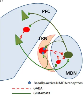
Collaterals from the prefrontal cortex (PFC) and MD (which provides the major thalamic innervation of the PFC) terminate more widely in the TRN than collaterals from other cortical and thalamic areas (Zikopoulos and Barbas, 2007), suggesting a particularly effective gating of thalamic relay activity by PFC circuits (Fig 1). In addition, recently discovered TRN afferents from the amygdala (Zikopoulos and Barbas, 2012) implicate the TRN in imparting emotional salience to thalamic filtering of sensory information.
A key feature of the TRN neurones is that they can fire in two different modes: short lasting high frequency bursts and a tonic firing mode. Low voltage-activated T type calcium channels play a key role in enabling TRN neurones to generate rhythmic bursts of action potentials, which in turn are thought to be important in the generation of thalamocortical oscillations. Indeed the TRN has been shown to be important in a range of different rhythms including sleep spindles, slow oscillations and delta and gamma rhythms (Macdonald et al., 1998a; Huguenard and McCormick, 2007).
for fuller explanation). Pinault (2011) argues that this process enables the TRN cells to modulate thalamocortical cells efficiently in order to amplify relevant information that should be conveyed to the cortical areas, and to delete/reduce non-relevant or distracting information.
Whilst the TRN neurones are GABAergic (Houser et al., 1980; Mitrofanis and Guillery, 1993; Steriade et al., 1997), they also contain high levels of the calcium-binding protein parvalbumin, along with certain neuropeptides such as TRH, VIP and NPY (Morris, 1989; Burgunder et al., 1999) and the endocannabinoid 2-AG (Sun et al., 2011). Calcium binding proteins such as parvalbumin play an important role in regulating intracellular availability of Ca+, and hence modulation of membrane potential, synaptic transmission and firing patterns
of TRN neurones.
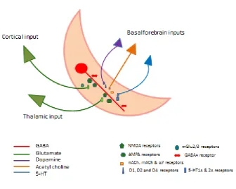
The TRN is rich in glutamate and GABA receptors, and expresses a range of other receptors including acetylcholine (M2, α4β2, α7) (Rubboli et al., 1994; Sun et al., 2013b), serotonin (5-HT1A and 5-HT2A) (Rodriguez et al., 2011), dopamine (D1 D2 and D4) (Zhang et al., 2009; Govindaiah et al., 2010), cannabinoid (CB1) (Sun et al., 2011) and melatonin (MT2) (Comai and Gobbi, 2014) receptors (Fig 2). This neurochemical signature supports a regulatory influence of a range of neuromodulatory systems on TRN activity (see below).
The TRN GABAergic neurones communicate with each other via both electrical and chemical synapses, the former clearly playing an important role in synchronisation of TRN output, while the latter potentially restrict the numbers of TRN neurones contributing at that time to rhythmic oscillations (Sohal and Huguenard, 2003). A reduction in intra-TRN communication can hence over-synchronise output, and potentially lead to seizure-like activity (Huntsman et al., 1999).
Within the TRN, neurones are interconnected by a network of GABAergic synapses. GABA(A) receptor evoked depolarisations can interact with T-type Ca(2+) channels to
powerfully control burst firing in TRN neurones, which results in feed forward inhibition in thalamic relay cells. This information is transmitted back to RTN cells through axon collaterals of projections (Llinas and Jahnsen, 1982; Huguenard, 1996; Sun et al., 2012). These events are mediated by activation of predominantly postsynaptic GABAA receptors
with an additional contribution of GABAB receptors during intense TRN output (Huguenard
Glutamate released from glutamatergic inputs to the TRN from the cortex and the thalamus acts upon a range of glutamate receptors in the TRN, with postsynaptic NMDA receptors considered to play an important role (Jones et al., 1998; Zhang et al., 2009). It has been argued that thalamic circuitry is potentially vulnerable to excessive glutamatergic cortical drive to the RTN or intensive GABAergic inhibition of thalamocortical relay neurones by neurones of the reticular thalamus. Hence physiological mechanisms that enable fine tuning of this circuit activity are required to protect against prolonged activity in the network. A range of presynaptic receptors may fulfil this role. For example, metabotropic receptors may play a protective role by suppressing synaptic release. Activation of mGluR3 and mGluR4/8 receptors modulates the activity of glutamatergic cortical synapses onto RT neurones, and GABAergic synapses onto thalamocortical neurones in the somatosensory thalamus, with the overall effect of suppressing thalamic oscillations (Gu et al., 2008; Kyuyoung and Huguenard, 2014). Similarly the endocannabinoid system may play a critical role in maintaining thalamic synchrony. Under conditions of increased levels of intra TRN synaptic activity, a presynaptic CB1 receptor dependent suppression of inhibitory synaptic strength has been shown which is potentially mediated through alterations in the release of 2-AG from TRN neurones (Sun et al., 2012).
Furthermore, there is in vitro evidence that tonically active presynaptic NMDA receptors and mGlu2Rs act in concert with postsynaptic NMDA and GABA receptors (Crabtree et al., 2013). It is argued that when TRN neurones are relatively hyperpolarised their ability to fire in burst mode will be enhanced, whereas when these neurones are relatively depolarised their firing in single-spike tonic mode will be reduced (Crabtree et al., 2013).
trigger action potentials in TRN neurones. Moreover, short trains of cholinergic synaptic inputs can powerfully entrain ongoing TRN neuronal activity. The M2 mAChRs appear to play a key role in controlling nAChR-mediated excitation through their ability to inhibit ACh release (via presynaptic M2 autoreceptors) and via M2 mediated postsynaptic hyperpolarisation (Sun et al., 2013b; Beierlein, 2014).
Agonists at the melatonin 2 receptor have also been shown to increase the neural activity of the reticular thalamus, enhancing the rhythmic burst activity and promoting non REM sleep (Ochoa-Sanchez et al., 2011).
2) The role of the TRN in relation to oscillatory activity, cognition and behaviour
Oscillatory activity,
The constant activity of the cerebral cortex as recorded by the EEG or within brain regions as local field potentials (LFPs) largely results from the interacting oscillations (rhythms) of neuronal networks. The frequencies of these oscillations, which are preserved across mammals, span 0.05-500HZ and include sleep spindles (12-18HZ), theta- (4-10HZ) and gamma-band oscillations (30-90HZ) (Buzsaki and Draguhn, 2004). Most forms of oscillations are inhibition based and in this respect the GABAergic neurones of the TRN play an important role.
The TRN is involved in the generation of ‘sleep spindles’ characteristic of stage 11 sleep and gamma oscillations, where repetitive TRN bursting produces rhythmic inhibition of the thalamocortical pathways (Steriade et al., 1985; Pinault and Deschênes, 1992; Macdonald et al., 1998b), Oscillations in the gamma range are linked to cognitive processes in the adult (Osipova et al., 2006; Colgin et al., 2009), and underpin plasticity at thalamocortical synapses during development (Minlebaev et al., 2011). Theta rhythms also play a role in memory processes (Osipova et al 2006). Operationally gamma and theta rhythms are proposed to work together to form a neural code that optimises cognitive processing. (for review see (Lisman and Buzsaki, 2008).
recordings from microelectrode arrays in behaving mice, the same group discovered functional diversity amongst TRN microcircuits. Two TRN subpopulations were discovered, one that correlated with spindles and the other that correlated with arousal (Halassa et al., 2014)
Cognition and behaviour
The basis of the Crick hypothesis (Crick, 1984) is that the TRN acts as an ‘attentional searchlight’. Some support for this hypothesis has arisen from lesion and histological studies in rodents. In an attentional orienting task, lesions of the visual TRN produced deficits in behaviour, supporting a link between the TRN and the attentional effects of visual cues (Weese et al., 1999). Another study showed that there was an increase in c-Fos expression in a sector of the TRN that correlated with the modality of an attended stimulus (McAlonan and Brown, 2002). However given that the TRN is a thin layer of cells, lesioning approaches may impact on the function of surrounding thalamic nuclei, thereby limiting interpretation of the data.. Changes in immediate early gene expression in relation to behaviour are also limited in the sense that they are correlative in nature.
Important investigations in primates have produced more direct evidence to support the Crick hypothesis. McAlonan et al. (2006) recorded visual TRN neurones in awake monkeys, and the modulation of the response as the monkeys shifted attention between visual and auditory stimuli. They demonstrated that TRN activity is modified by shifts of visual attention, and that these changes in attention could affect visual processing in the lateral geniculate thalamic nucleus via the inhibitory connections back to the thalamus. They conclude that the TRN contributes to attentional processing, whereby a relevant sensory input is selected for additional processing at the expense of an irrelevant input. Whilst the sensory regions of the TRN have been investigated in attentional tasks, the contributions of the ‘cognitive’ related TRN inputs, from the PFC and via the MD, have not been explored. The PFC-MD-TRN circuitry is likely to play a prominent role in cognitive flexibility and working memory, and hence future studies that investigate the role of the TRN in tasks that reflect these cognitive domains will be important.
visual or anterior limbic thalamus of VGAT (vesicular GABA transporter)-Cre mice. Clear differences were found in the functionality of these TRN projection neurones, with visually-tagged neurones showing correlations to cortical spindle power during slow wave sleep whereas limbic-tagged neurones were negatively correlated. Importantly, during a visual detection task that required attention there was a reduction in firing rate for visual-projection neurones but not limbic-projection TRN neurones. Since the reduction in activity of vision associated TRN neurones occurred after the onset of a trial but just before the stimulus presentation, the authors suggest that these neurones are involved in attentional states. This conclusion was further supported using an alternative optogenetic strategy to reduce the firing rate of sensory TRN neurones further (using eNpHR3.0, a light activated Cl- pump
that hyperpolarises cells and inhibits spiking) in response to which the mice showed the predicted increase in behavioural performance. The authors raise the possibility that the demonstrated distinct subgroups of TRN neurones can facilitate the switching from attention to external stimuli to an internal focus. Specifically, they suggest that TRN engagement may contribute not only to “offline” consolidation of memory processes, but also to dynamic conversion between default mode and cognitive-control networks. This is intriguing, given the abnormal default mode network connectivity in schizophrenia (Whitfield-Gabrieli et al., 2009).
.
The TRN and schizophrenia risk factors: preclinical evidence
Converging evidence from preclinical studies in rodents strongly supports the hypothesis that TRN dysfunction may play an important role in the disease process. NMDA receptor (NMDA-R) glutamatergic antagonists such as ketamine and phencyclidine are able to induce a variety of effects in humans and rodents that resemble the positive, negative and cognitive symptoms of schizophrenia The effects are observed with acute administration, but are consolidated and exacerbated by repeated administration (Krystal et al., 1994; Tamminga, 1998; Morris et al., 2005; Pratt et al., 2012).
et al., 1992; Hill et al., 2004; Molina et al., 2005; Potkin et al., 2009) and PFC GABAergic deficits – may occur secondary to TRN dysfunction. This speculative idea highlights the importance of understanding the neurobiological processes through which the TRN may impact on cortical function.
Acute administration of NMDA-R antagonists in rats also induces various effects on thalamocortical circuitry: a) elevated glutamate release in the PFC, although the site of action of the NMDA-R antagonists appears to be outwith the PFC (Lorrain et al., 2003; Lopez-Gil et al., 2007); b) c-fos induction in the PFC and medial thalamus but not in the TRN, consistent with a disinhibition of thalamocortical circuits (Kargieman et al., 2007; Santana et al., 2011); c) increased metabolic activity in the PFC (hyperfrontality), but reduced metabolism in the TRN (Dawson et al., 2013). It was previously thought that NMDA-Rs on PFC PVALB-positive interneurones were the locus of action for the hyperfrontality and increased activity of prefrontal cortex pyramidal cells (Homayoun and Moghaddam, 2007) following acute ketamine/PCP, but opinion is now shifting away from this idea (Rotaru et al., 2011; Povysheva and Johnson, 2012). Thalamic neurones have also been implicated in the actions of NMDA receptor antagonists since PCP alters the activitiy of thalamic relay neurones of the MD and centromedial (CM) thalamus along with mPFC neurones in anaesthetised rats (Santana et al., 2011). Furthermore microinjections of MK801 into the MD thalamus, but not the PFC, modulates cortical delta band activity in anaesthetised animals similar to that observed following systemic administration (Kiss et al., 2011). Because the TRN provides tonic feed forward inhibition to thalamic nuclei, changes in MD/CM activity could result from NMDA receptor blockade in the TRN. NMDA-Rs in the TRN are active under resting conditions (Jacobsen et al., 2001; Gentet and Ulrich, 2003; Zhang et al., 2009), probably because TRN NMDA-Rs contain the NR2C subunit, and hence show little voltage dependent block at resting potentials. Metabolic hyperfrontality can be induced by elevated activity in PFC afferents (Canals et al., 2009), and so the increased metabolism/activity in the PFC induced by ketamine/PCP may reflect increased thalamocortical activity, due to disinhibition, caused by blockade of NMDA-R drive of GABAergic TRN neurones. Indeed functional connectivity analysis following acute ketamine treatment in rodents provides evidence to support this hypothesis (Dawson et al., 2013) as do recent in vivo electrophysiological studies which showed inhibition of TRN activity after PCP (Troyano-Rodriguez et al., 2014).
induced by PCP provide some translation to schizophrenia where deficits occur in the analogous components of the CANTAB task.
In summary, accumulating evidence is consistent with altered PFC function induced by NMDA-R antagonist treatment being secondary to a primary site of action in the TRN.
In terms of genetic risk factors for schizophrenia, DISC1 has attracted considerable attention. The DISC1 gene is located on chromosome 1q42.1, and an abnormal translocation in this gene increases the risk of developing schizophrenia, bipolar disorder, or major depression by ~50 fold in comparison to the general population (Millar et al., 2000; Brandon et al., 2009) DISC1 expression is apparent in schizophrenia-relevant regions such as the prefrontal cortex, hippocampus and TRN. Of particular relevance are the findings that DISC1 is highly expressed in the TRN during periods of development when corticothalamic connections are forming, and that during the developmental period the TRN is relatively larger than the adult size (Mitrofanis and Guillery, 1993; Austin et al., 2004). Taken together these findings suggest that DISC1 in the TRN could play a critical role in the formation of the thalamocortical system, and provide an explanation for the vulnerability to schizophrenia arising from the impact of genetic abnormalities on this circuitry during the neurodevelopmental period. In order to assess the impact of the DISC1 translocation upon adult brain function we have determined the brain imaging signatures of mice with modifications in the DISC1 gene. We have demonstrated that the TRN is a key brain region affected in these mice (Dawson et al., 2014) adding further weight to the concept that TRN dysfunction may be a key factor in the aetiology of schizophrenia.
generally suggestive of reduced activity (Andreasen et al., 1994; Buchsbaum et al., 1996; Hazlett et al., 2004; Hazlett et al., 2008; Minzenberg et al., 2009). Hence abnormal function of corticothalamic and thalamocortical pathways is clearly a component of the network dysfunction in schizophrenia (Andreasen et al., 1995; Andreasen et al., 1996; Buchsbaum and Hazlett, 1998; Anticevic et al., 2013).
On the thalamic side of this disrupted network, there appears to be a regional selectivity (Andreasen, 1997; Jones, 1997) that parallels disease-specific changes in cortical structure, activity, connectivity and neurochemistry. Thus cortical abnormalities are centred on prefrontal, cingulate and temporal areas (Hill et al., 2004; Cole et al., 2011; Shepherd et al., 2012; Haijma et al., 2013), with thalamic pathology and connectivity deficits most evident in thalamic nuclei connected with the most-affected areas of cortex. Thus abnormalities in MD and anteroventral thalamus have been frequently reported (Danos et al., 1998; Young et al., 2000; Kemether et al., 2003; Hazlett et al., 2004; Andrews et al., 2006; Shimizu et al., 2008; Byne et al., 2009), while evidence for dysfunction in other parts of the thalamus, such as the lateral geniculate nucleus for example, is sparse. Despite its well-known role in modulating and synchronising activity in thalamocortical loops, and especially in limbic thalamocortical loops, the TRN has been relatively neglected in relation to schizophrenia. As noted earlier in this review, the widespread projections of the TRN to the higher order thalamic nuclei suggest that the TRN could play a key role in regulating transthalamic cortico-cortical communication through its ability to modify the mode of firing of thalamocortical cells (Byne et al., 2009)(Guillery and Sherman, 2002{Vukadinovic, 2011 #4103). The consequences of a disrupted pattern of connectivity in thalamic-TRN-cortical circuitry leading to impaired transthalamic corticocortical communication could result in the incorrect assignation of the relevance of a stimulus, the cortex processing information with only limited information about its context and potentially the inability to determine whether a stimulus is externally or internally derived. Such dysfunction could result in many of the symptoms of schizophrenia (see reviews by Byne et al 2009; Vukadinovic 2011)
Although limited, evidence from a variety of different sources is now focussing attention on the TRN as a key area of dysfunction in patients.
require synchronised activity between cortical and sub-cortical areas (Fries et al., 2007; Womelsdorf et al., 2007). It is now appreciated that the TRN plays a major role in the synchronisation of thalamocortical and corticothalamic activity (Pinault, 2004), and hence in the control of gamma (high frequency) oscillations (Huguenard and McCormick, 2007; Jones, 2009). Gamma oscillations can be maintained locally in cortical networks, but evidence suggests that these are likely to be regulated by the TRN influence on thalamocortical loops (Pinault, 2004). The literature on disturbances in gamma oscillations in schizophrenia is not always consistent (Uhlhaas and Singer, 2010). Nevertheless, the general picture is that this high-frequency oscillatory activity is compromised in schizophrenia. Reduced levels of spontaneous and evoked cortical gamma oscillations are typically detected in patients, in association with sensory processing or performance of executive tasks (Rutter et al., 2009; Uhlhaas and Singer, 2010; Sun et al., 2013a). The abnormal gamma oscillatory activity in schizophrenia may therefore be sending a strong signal that TRN dysfunction is a fundamental component of the disease.
Considering the relationship between oscillatory activity and cognition, the characteristic cognitive deficits of schizophrenia may be directly connected with these impairments. Gamma band oscillations are linked with performance of working memory tasks (Yamamoto et al.; Howard et al., 2003; Roux and Uhlhaas, 2014), where patients show impairment (Perlstein et al., 2001; Bor et al., 2011). Indeed, the deficits in gamma oscillatory activity in patients are correlated with working memory deficits (Cho et al., 2006). The TRN has been particularly strongly linked with attentional mechanisms (Weese et al., 1999; Zikopoulos and Barbas, 2012) – and deficits in attentional processes are characteristic of schizophrenia (Andreasen et al., 1995; Buchanan et al., 1997; Birkett et al., 2007). Equally, the positive symptoms of schizophrenia can also be readily equated with deficient thalamic filtering of sensory information, and thus with impaired modulation of thalamocortical feedback by the TRN.
sleep pattern deficits. These deficits are not drug-induced effects, since they are observed in antipsychotic-naive patients. There is increasing acceptance that sleep problems are in fact a core symptom of schizophrenia (Wilson and Argyropoulos, 2012), also present in ultra-high risk subjects prior to the onset of psychosis (Ruhrmann et al., 2010; Lunsford-Avery et al., 2013). This in itself implicates TRN dysfunction in the development of the disease. Potentially NMDA receptor hypofunction on TRN cells could result in impaired transthalamic cortico-cortico communication leading to sleep spindle deficits (Vukadinovic, 2011).
There is a paucity of data from imaging or postmortem pathology studies to assess the degree of TRN dysfunction in schizophrenia. Unfortunately the TRN is too narrow to be resolved by current in vivo imaging methodologies such as PET or fMRI. However, a recent functional imaging combined with transcranial magnetic stimulation showed thalamic dysfunction in schizophrenia (Guller et al., 2012), which may also implicate altered TRN function given the strong functional connectivity between the TRN and thalamic nuclei. In
addition, altered TRN expression of nicotinic cholinergic 7 receptors and glutamate transporters (EAAT1-3) has been reported in post-mortem tissue from patients (Court et al., 1999; Smith et al., 2001), but otherwise there is little information on possible neurochemical or neuropathological changes in the TRN. Future studies should address this issue as a matter of urgency.
Implications for drug discovery
Based upon the accumulating evidence that the TRN plays a role in thalamocortical and transthalamic cortico-cortico communication dysfunction in schizophrenia, it is reasonable to suggest that drugs which target receptors localised in the TRN could have the ability to restore circuitry dysfunction resulting in improvements in a range of symptoms. Both postsynaptic and presynaptic receptors are important in modulating TRN activity (see section 1 and Fig 2). Presynaptic receptors that offer potential as drug targets include the mGlu2/3 agonists. Indeed initial clinical trials proved promising with a Glu2/3 agonist although more recent studies have proved inconclusive (Patil et al., 2007; Adams et al., 2013). Another interesting target is the postsynaptic nicotinic alpha 7 receptor and clinical trials suggest potential beneficial effects of alpha 7 receptor agonists on negative symptoms and cognitive deficits (Freedman et al., 2008). Restoration of GABAergic dysfunction in the TRN with GABAA receptor positive allosteric modulators could be another option. Indeed this
concept has been proposed for modulation of PFC parvalbumin containing GABAergic neurones known to be deficient in schizophrenia (Volk and Lewis, 2005). Arguably if the TRN is the driver of PFC-induced changes then the action of drugs in the TRN could be the fundamental basis for treatment. Of the 5-HT receptors localised in the TRN, 5-HT2A receptors are of interest given that many current antipsychotic drugs show affinity for these receptors. Understanding how these receptors modulate thalamocortical function at the level of the TRN and the relationship with specific symptom domains is an important question. Similarly, understanding how other receptors such as CB1 receptors can modify TRN activity at the level of specific modalities will provide greater insight into the potential of compounds to restore particular symptom domains. Ultimately the orchestration of TRN activity depends on a complex interplay of a range of neurotransmitters that modulate pre- and post- synaptic activity. Gaining greater insight into the precise role of these receptors in the topographical organisation of the TRN, their relationship with particular physiological processes and how these may be dysfunctional in schizophrenia is important not only from a disease mechanism perspective but informative for drug discovery.
In summary, despite the strong indications from both preclinical and clinical areas for disease-related TRN dysfunction, much more research is required to understand the neurobiology of the TRN in relation to thalamic and cortico-cortical communication and to demonstrate unequivocally that there is a casual relationship with TRN dysfunction and the aetiology of schizophrenia. Ultimately this improved knowledge may lead to more rational approaches for drug discovery in schizophrenia.
This research received no specific grant from any funding agency in the public, commercial, or not-for-profit sectors.
Figure Legends
Fig 1. Schematic diagram of the connections between the Prefrontal cortex (PFC), Mediodorsal nucleus of the thalamus(MDN) and the Thalamic reticular nucleus (TRN)
Fig 2. Schematic diagram showing afferent inputs to a dendrite of a GABAergic TRN neurone together with neurotransmitter receptors. Cell bodies from cortical, thalamic and basal forebrain inputs are represented as triangles and coloured according to the
neurotransmitter system utilised. Note that NMDA, AMPA and GABAA receptors are located
postsynaptically (although presynaptic NMDA receptors also exist). GABAA receptors are
activated by locally released GABA from nearby TRN GABAergic neurones (not shown). mGlu2/3 are located presynaptically.on glutamatergic inputs from the cortex and presynaptic CB1 receptors may modify intra TRN GABAergic transmission. ACh, DA and 5-HT
References