Bacteria and Functional Genes in a Complex Microbial
Community
Yun Wang1, Yin Chen2, Qian Zhou3, Shi Huang3, Kang Ning3, Jian Xu3, Robert M. Kalin4, Stephen Rolfe5, Wei E. Huang1*
1Kroto Research Institute, University of Sheffield, Sheffield, England, United Kingdom,2School of Life Sciences, University of Warwick, Coventry, England, United Kingdom,3BioEnergy Genome Centre, Chinese Academy of Sciences Key Laboratory of Biofuels and Shandong Key Laboratory of Energy Genetics, Qingdao Institute of BioEnergy and Bioprocess Technology, Chinese Academy of Sciences, Qingdao, China,4David Livingstone Centre for Sustainability, Strathclyde University, Glasgow, Scotland, United Kingdom,5Department of Animal and Plant Sciences, Alfred Denny Building, University of Sheffield, Sheffield, England, United Kingdom
Abstract
Most microorganisms in nature are uncultured with unknown functionality. Sequence-based metagenomics alone answers ‘who/what are there?’but not ‘what are they doing and who is doing it and how?’. Function-based metagenomics reveals gene function but is usually limited by the specificity and sensitivity of screening strategies, especially the identification of clones whose functional gene expression has no distinguishable activity or phenotypes. A ‘biosensor-based genetic transducer’ (BGT) technique, which employs a whole-cell biosensor to quantitatively detect expression of inserted genes encoding designated functions, is able to screen for functionality of unknown genes from uncultured microorganisms. In this study, BGT was integrated with Stable isotope probing (SIP)-enabled Metagenomics to form a culture-independent SMB toolbox. The utility of this approach was demonstrated in the discovery of a novel functional gene cluster in naphthalene contaminated groundwater. Specifically, metagenomic sequencing of the13C-DNA fraction obtained by SIP indicated that an unculturedAcidovoraxsp. was the dominant key naphthalene degraderin-situ, although three culturablePseudomonas
sp. degraders were also present in the same groundwater. BGT verified the functionality of a newnag2operon which co-existed with two other nag and two nah operons for naphthalene biodegradation in the same microbial community. Pyrosequencing analysis showed that thenag2 operon was the key functional operon in naphthalene degradationin-situ,
and shared homology with bothnagoperons inRalstoniasp. U2 andPolaromonas naphthalenivoransCJ2. The SMB toolbox will be useful in providing deep insights into uncultured microorganisms and unravelling their ecological roles in natural environments.
Citation:Wang Y, Chen Y, Zhou Q, Huang S, Ning K, et al. (2012) A Culture-Independent Approach to Unravel Uncultured Bacteria and Functional Genes in a Complex Microbial Community. PLoS ONE 7(10): e47530. doi:10.1371/journal.pone.0047530
Editor:Ying Xu, University of Georgia, United States of America
ReceivedJuly 24, 2012;AcceptedSeptember 12, 2012;PublishedOctober 17, 2012
Copyright:ß2012 Wang et al. This is an open-access article distributed under the terms of the Creative Commons Attribution License, which permits unrestricted use, distribution, and reproduction in any medium, provided the original author and source are credited.
Funding:The authors have no funding or support to report.
Competing Interests:The authors have declared that no competing interests exist.
* E-mail: w.huang@shef.ac.uk
Introduction
Bacteria account for approximately half of the total carbon of the global biomass [1] and play fundamental roles in bio-geochemical cycles (e.g. C and N) [2]. Bacteria provide a free service worth trillions of dollars to maintain and restore ecosystems - cleaning water and soil, and maintaining soil fertility [3]. However, the vast majority of bacteria (.99%) present in natural environments have not yet been cultured [4,5,6,7,8]. These uncultured bacteria are often referred to as a ‘black box’ containing a ‘hidden’ community that represents an untapped genetic resource encoding novel and valuable catalysts, enzymes and building blocks for industry and medicine [9,10,11,12]. In addition, whilst cultivation of pure isolates enables the detailed study of bacterial physiology, it is often more desirable to study microbial genetic function and ecological rolesin-situ. Therefore, the development of culture-independent approaches will provide a global view of the bacterial community, help predict ecosystem
functioning and lead to further understanding of bacterial evolution [13,14].
Metagenomics circumvents the cultivation issue by extracting total DNA from an environmental sample, followed either by direct sequencing (sequence-based metagenomics) or cloning into a culturable model organism for functional analysis (function-based metagenomics) [15,16,17]. Advances in next generation
DNA sequencing technologies (e.g. 454 pyrosequencing)
screening strategies, especially for the identification of functional genes whose expression has no detectable activity or distinguish-able phenotypes. Therefore, identification of gene function is one of the key challenges in the post-genomic era.
To address these challenges, we have developed a culture-independent SMB toolbox that integrates Stable isotope probing (SIP)-enabled Metagenomics with a Biosensor-based gene trans-ducer (BGT) technique. SIP provides the link between bacteria and the metabolism of stable isotope (e.g. 13C, 15N) labeled
substrates. The 13C-DNA fraction obtained by SIP provides
a significantly-reduced but relevant gene-pool for metagenomic analysis [25]. Sequencing of the 13C-DNA fraction specifically reveals microorganisms whose activities are involved in metabo-lism of the 13C-labelled substrate. BGT identifies and verifies functional genes involved in the metabolism of a specific target compound.
In this study, we chose a well-characterised naphthalene contaminated groundwater site [26,27] to demonstrate the use of the SMB toolbox. We employed both culture-dependent and culture-independent methods, as illustrated in Fig. 1A, to provide a holistic picture of the microbial community in the groundwater. For the cultured fraction, traditional methods including cultiva-tion, conjugation and plasmid extraction were used to isolate and identify putative naphthalene degraders and their functional genes. For the uncultured fraction, the SMB toolbox was applied to identify uncultured but active naphthalene degraders and to investigate their functional genesin-situ(Fig. 1A and 1B).
Results
Since the active members for naphthalene degradationin-situ are unknown, we employed both culture dependent and in-dependent approaches to investigate the microbial community by splitting a naphthalene-contaminated groundwater sample into cultured and uncultured fractions. For cultured fraction, we isolated threePseudomonasspp. that can grow by using naphthalene as a sole carbon source and identified two types ofnah operons located on plasmids in the hosts. For uncultured fraction, we applied SMB to study the active bacteria in the microbial
community. We found that an uncultured Acidovorax sp. was
responsible for naphthalene degradation in-situ. Metagenomic
sequencing and the BGT technique revealed that one mosaic patternnagand two othernagoperons were the active functional operons for naphthalene biodegradation in the groundwater.
Cultured Fraction: Naphthalene Degradation Genes were Located on Conjugative Plasmids of Isolated
Naphthalene Degraders
Conventional cultivation approaches were used for preliminary investigations of naphthalene-degrading microorganisms. Pseudo-monas fluorescens (WH2) and two Pseudomonas putida (WH1 and WH3) naphthalene degraders were isolated from the contaminat-ed groundwater. All three isolates could grow on the minimal medium (MM) [28] with naphthalene as a sole carbon source.
Conjugation between donor bacteria: P. putida WH1, WH3, P.
fluorescensWH2 andP. putidaNCIB9816 (used as a positive control) [29] and recipientP. putidaUWC1 [30] separately indicated that the naphthalene degradation genes were located on conjugative
plasmids in P. putida WH1, WH3, P. fluorescens WH2. The
conjugation frequencies for Pseudomonas WH1, WH2, WH3 and
NCIB9816 were respectively 2.0360.1261027, 2.3360.73 61027, 1.9360.1461027and 4.3361.261028transformants/ recipient.
Restriction enzyme digestion patterns indicated that theP. putida
WH1 and WH3 plasmids had the same structure whilst P.
fluorescensWH2 differed (Fig. S1). DNA sequence analysis showed that the genes encoding the salicylate hydroxylase (NahG) and the transcriptional regulator (NahR) on the plasmids ofP. putidaWH1
and WH3 were identical to those on pDTG1 inP. putidaNCIB
9816 [31] despite the different plasmid structures (Fig. S1). The nahGandnahRgenes inP. fluorescensWH2 were identical to those in the chromosome ofPseudomonas stutzeriAN10 [32].
Uncultured Fraction: SIP Enriched13C-DNA Revealed Acidovoraxsp. as a Key Naphthalene Degrader
The use of SIP requires an appropriate incubation period that sufficient for nucleic acids of active degraders to become isotopically enriched. However, the physical (e.g. temperature) and chemical (e.g. naphthalene concentration) conditions of this incubation may alter the microbial community structure. There-fore the impact of altering incubation conditions on the microbial community was investigated. Denaturing gradient gel electropho-resis (DGGE) of 16S rRNA genes was used to assess microbial community structure and diversity in the groundwater. The DGGE profiles of microcosms incubated in darkness at 14uC with
3.8mM naphthalene (ambient concentration) for 168 h were
similar to the profile from the native groundwater (Fig. S2). However, changing either the temperature of incubation (from 14
to 20uC) or naphthalene concentration (from 3.8 to 30mM)
significantly altered both bacterial diversity and relative abun-dance in the microbial community (Fig. S2). This demonstrates the importance of incubation replicating, as far as possible, field
conditions (dark, 14uC and 3.8mM naphthalene) in order to
identify thein-situdegraders.
The incubation period must also be as short as possible to
minimize 13C-cross-labeling of other community members with
13
C-metabolites released from the primary degraders. Aerobic
degradation of 13C-naphthalene produced salicylate in the
microcosms which was monitored using a salicylate biosensor ADPWH_luxas a rapid detection tool [28]. A peak of salicylate production was found to appear after 120 h in the microcosms which returned to the background level at 144 h (Fig. S3), suggesting that active aerobic naphthalene biodegradation had occurred during this period. Total nucleic acid containing12 C-and13C-DNA was extracted from the microcosms after 120 h and purified. After equilibrium density gradient ultracentrifugation, the
‘heavy’ 13C-DNA fractions were recovered, which contained
genomic DNA of the active naphthalene degradersin-situ.
DGGE analysis of the13C-DNA fraction showed that band B
(labeled as B1, B2, B3 respectively in the three13C-DNA fractions
in Fig. S4) was dominant in the 13C-DNA fraction (Fig. S4).
Sequence analysis of band B suggested that it was derived from Acidovoraxsp. (designated asAcidovoraxsp. WH), consistent with our previous reports [26]. The result suggested that an uncultured Acidovoraxsp., rather than the culturedPseudomonassp., played a key role in naphthalene biodegradation in-situeven though they co-existed in the same groundwater. It is not unusual that uncultured Acidovorax sp. was found to play an important role in aromatic hydrocarbon biodegradation, as it has also been found in other studies [33].
Strong PCR products were obtained with Comamonas-type
naphthalene dioxygenase (NDO) primers in 13C-DNA fractions
but no PCR products were observed usingPseudomonas-type NDO
Pseudomonasspp. The PCR products were cloned into a plasmid vector. Ten were chosen at random and sequenced. Of these sequences, 20% were identical toComamonas-type NDO in thenag
operon hosted by Ralstonia sp. U2, and 80% were hybrids of
Ralstoniasp. U2 andP. naphthalenivoransCJ2.
Pyrosequencing Analysis of Microorganisms and Functional Genes Involved in Naphthalene Degradation
Pyrosequencing of the12C- and13C-DNA fractions produced separately 269,001 and 797,526 reads, ranging from 30 to 530 bp
in length. Analysis of 16S- rRNA genes within the 13C-DNA
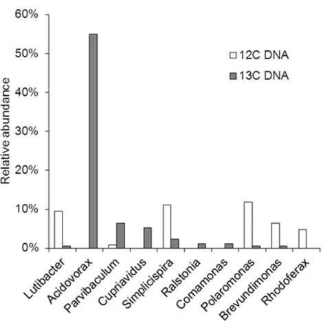
fraction pyrosequencing data showed that Acidovorax sp. (55%) were dominant andRalstonia sp. (1%) andPolaromonassp. (0.1%) were also present (Fig. 3 and Fig. S5). None of these bacteria have yet been isolated using traditional culture-dependent methods. The 16S- rRNA gene ofPseudomonassp. was not found in the13
C-DNA fraction. The 16S- rRNA gene sequences in the13C-DNA
fraction also included those with 100% identity to that ofRalstonia sp. U2 (1180–1450 bp in AF301897) and 99% identity to that of Polaromonas naphthalenivorans CJ2 (1014–1514 bp in NC_008781). These pyrosequencing reads include hypervariable regions of the 16S rRNA gene [34], suggesting that the naphthalene degraders
revealed by SIP are similar to Ralstonia sp. U2 and P.
naphthalenivorans CJ2 which have been reported previously to be naphthalene degraders.
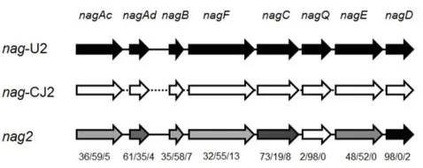
Pyrosequencing assembly (DNA assembly file) recovered three assemblies: one 21,173 bp and one 25,765 bp assemblies each have .99.9% identity to thenagoperon inRalstonia sp. U2 (nag-U2) [35] andPolaromonas naphthalenivorans CJ2 (nag-CJ2) [36]; the third and dominant one designated asnag2, was a hybrid ofnag-U2 andnag-CJ2. Thenag-CJ2,nag-U2 andnag2separately account for 18.0% and 17.7% and 64.3% of thenag-related reads in the13 C-DNA fraction (Fig. S6 and Table S1). Nonahoperon was found in the13C-DNA fraction. The nag2forms a mosaic pattern operon
sharing homology with both nag-CJ2 and nag-U2 (Fig. S6 and
Table S1).
Biosensor-based Gene Transducer (BGT) Technique Confirmed a New Operon Carrying a Naphthalene Degradation Function
ThenagFCQEDgene cluster was amplified from the13C-DNA
fraction using primers designed on the basis of the nag operon sequence (Table 1). This was then ligated into the plasmid pWH1274 and transformed into the salicylate biosensor strain ADPWH_lux(Huang et al., 2005) to produce a strain designated as ADPWH_Nag (Table 2). The well-characterised gene cluster nahFCQED was amplified by PCR from the naphthalene
degradation plasmid pDTG1 in P. putida NCIB9816 [31] and
inserted into pWH1274 in the same manner to produce ADPWH_Nah for the use as a positive control (Table 2). The plasmid pWH1274 is anE. coli - Acinetobacter shuttle [37] vector and thenagFCQEDornahFCQEDwas inserted into the BamHI site of pWH1274 under the control of a constitutive promoter Ptet.
ADPWH_lux containing the plasmid pWH1274 without inserts
(ADPWH_1274) was used as a negative control (Table 2). The enzymes encoded by the targeted gene cluster should convert the substrate 1,2-dihydroxynaphthalene (DHN) into salicylate, which
activates the Psal promoter and triggers the expression of
bioluminescence in ADPWH_lux (Fig. 1B). As shown in Fig. 4,
both ADPWH_Nag and ADPWH_Nah were induced to express
bioluminescence within 5 min in the presence of 50mM DHN,
indicating that thenagFCQEDgene cluster was functional and its
expression in ADPWH_lux was able to convert DHN into
salicylate. In contrast, ADPWH_Nag and ADPWH_Nah re-mained silent in the absence of DHN. The negative control ADPWH_1274 did not respond to DHN (Fig. 4).
The full-length sequence of the inserted gene clusternagFCQED
and NDO PCR products (containing nagAc and nagAd) were
consistent with the pyrosequencing reads ofnag2(Fig. 5 and S6), confirming that this functional cluster nag2 represented a new operon. A single pyrosequencing read in the13C-DNA fraction linkednagB andnagF (Fig. S7) in the nag2 operon suggesting that
a complete nag2 operon for naphthalene biodegradation was
Figure 1. A. Schematic of a toolbox to dissect microbial community structure and their functional genes in a complex community.
The culture-independent SMB toolbox comprises stable isotope probing, metagenomic sequencing and biosensor-based gene transducer.B. Schematic of the biosensor-based gene transducer.This approach can be used to screen for gene encoded functions for the metabolism of molecules with no distinguishable activity or phenotypes.
doi:10.1371/journal.pone.0047530.g001
Figure 2. Naphthalene dioxygenase (NDO) PCR products.Two pairs of degenerate primers, COM1_F&COM1_R (Comamonas-type), and PSE1_F&PSE1_R (Pseudomonas-type) (Table 1) and different DNA templates: 1, 2 -13C-DNA; 3, 4 -12C-DNA; 5– blank control were used. M - DNA molecular weight Ladder.
[image:4.612.60.411.524.685.2]present in the groundwater. Interestingly, comparisons of the individual gene sequencesnagAc, Ad, B, C, D, E, F, Qin thenag2 operon showed a mosaic-pattern. nagDand nagQwere similar to their counterpart genes inRalstoniasp. U2 andP. naphthalenivorans CJ2 respectively, whereasnagAc, nagAd, nagB nagF,C,andE,were partially similar to bothRalstoniasp. U2 andP. naphthalenivoransCJ2 (Fig. 5 and S6). A few unassigned DNA sequences were also
observed innagFCQEDandnagAc, nagAdandnagB,which may have been due to point mutations (Fig. 5 and S6).
Discussion
Although metagenomic analyses have a great potential to identify putative novel genes and gene clusters, a better un-derstanding of microbial community function requires functional analyses that can be linked to community members. In this report we integrated SIP, metagenomics and a biosensor-based gene transducer technique to investigate the role of microbial naph-thalene biodegradation and the corresponding functional gene clusters in contaminated groundwater.
BGT as a New Strategy For Function-based Metagenomics
A number of screening approaches have been developed previously for function-based metagenomics, including colony pigment identification [38], antibiotic and enzymatic activity selection [39,40], substrate-induced gene expression [41] and also in-vitro compartmentalization screening [42]. However, these strategies are difficult to apply for screens of metabolic pathways involving small molecules. A BGT system could provide an alternative and sensitive strategy to screen for gene clusters whose expression produce small molecules that activate biosensors (Fig. 1B) [43,44,45].
Although metagenomic analysis of 13C enriched DNA had
identified the nag2 operon, its functionality was unknown.
Moreover, it was not clear whether the nag2 cluster was from
the same operon or an artifact due to inappropriate assembly of short (,530 bp) pyrosequencing reads. Therefore the putative gene cluster was amplified directly from the13C-enriched DNA and cloned into the BGT system to confirm its function in naphthalene metabolism. Salicylate is a central metabolite found in all known aerobic naphthalene biodegradation pathways,
[image:5.612.61.556.79.324.2]including Gram-positive and negative- bacteria
Table 1.Primers used in this study.
Primers Sequence (59R39) Reference
NagF1_For TTCCCAGGAGACAACCCATG This study
Nag_F AGTTCATCACTGGCACCGTA This study
NagD_Rev TGAGGCGACAATGAACATGC This study
NagSeq1_F ATCGTTCTGGACGACGCTGAC This study
NagSeq2_F CAAGCCAGCAACTGTCATTG This study
NagSeq3_F1 CGAAGATTTGGGTTACACACACC This study
NagSeq3_F2 ACCTACAACCTGCCACAGATG This study
NagSeq4_F ATTCAAGGCGCATGGGTCATCA This study
NagSeq5_F GGATGCTGGACTTGGATCTGA This study
GC338F CGCCCGCCGCGCCCCCGCCCCGGCCCGCCGCCCCCGCCCACTCCTACGGGAGGCAGC [60]
530R GTATTACCGCGGCTGCTG [60]
COM1_F AAAAGAGTTGTACGGCGATG [61]
COM1_R ACGGTAGAATCCGCGATAGC [61]
PSE1_F AAAAGAGCTGTATGGCGAGT [61]
PSE1_R CCGATAGAAGCCACGATAACT [61]
pWH1274_P12 CATGATCGCGTAGTCGATAG This study
pWH1A1_1_P4R AGTGCCACCTGACGTCTAAG This study
doi:10.1371/journal.pone.0047530.t001
Figure 3. Taxonomic assignments of the microbial communi-ties in the12C- and13C-DNA fractions revealed by pyrosequen-cing.Relative abundance of the species as a % of total 16S-rRNA reads is shown.
[image:5.612.60.295.442.675.2]Table 2.Strains and plasmids used in this study.
Bacteria and Plasmids Description Reference
Acinetobacter baylyistrains
ADP1(BD413) Wild type [62]
ADPWH_lux A salicylate biosensor. The promoterlessluxCDABEfrom pSB417 were inserted between thesalAandsalRgenes in the chromosome of ADP1
[28]
ADPWH_1274 ADPWH_luxcontaining the plasmid pWH1274; AmpR
This study
ADPWH_Nag ADPWH_luxcontaining the plasmid pWH1274 inserted with a positive gene cluster capable of transforming 1,2-dihydroxynaphalene to salicylate. The tinserted gene cluster was from the13
C-DNA fraction.
This study
ADPWH_Nah ADPWH_luxcontaining plasmid pWH1274 withnahFCQEDfrom pDTG1 inPseudomonas putidaNCIB9816. The gene cluster was inserted into BamHI site of pWH1274
This study
Pseudomonasstrains
P. putidaNCIB9816 Naphthalene degrader [29]
P. putidaUWC1 Spontaneous rifampicin-resistant mutant ofP. putidaKT2440 [30]
P. putidaWH1 Naphthalene degrader This study
P. fluorescensWH2 Naphthalene degrader This study
P. putidaWH3 Naphthalene degrader This study
Plasmids
pWH1274 E. coli-A. baylyishuttle plasmid; AmpR
, TetR
[37]
pGEM-T Commercial TA cloning vector Promega
pDTG1 nahFCQEDsource plasmid, fromPseudomonas putidaNCIB9816 [31]
pWH_NagFCQED An unknownnagFCQEDgene cluster cloned into BamHI site of pWH1274 This study
pWH_NahFCQED nahFCQEDcloned into the BamHI site of pWH1274 This study
[image:6.612.59.416.406.678.2]doi:10.1371/journal.pone.0047530.t002
Figure 4. Bioluminescence of biosensor transducers incorporating DHN degradation genes.ADPWH_Nag containing anag2 gene cluster was rapidly activated to show bioluminescence after the addition of 50mM DHN, confirming the function of the gene cluster. No bioluminescence was detected in the absence DHN. The negative control ADPWH_1274 did not respond to DHN. ADPWH_Nah containing anahgene cluster was used as a positive control.
[35,36,46,47,48,49]. Hence, the salicylate biosensor A. baylyi
ADPWH_lux [28] was employed to ‘light up’ in response to
expression of the 4973-bpnagFCQEDgene cluster that converted DHN into salicylate (Fig. 1B and Fig. 4). Although in this instance the product was amplified by PCR and cloned directly into the expression system, this same approach could, in theory, be used to screen functional genes from a DNA clone library. A variety of biosensors [28,50,51,52,53] can be potentially tailored to search and screen for genes of interest.
Although functional gene screening using BGT is a powerful approach for gene discovery in uncultured fractions, there are a number of limitations. For screening of environmental samples, BGT would be performed better if it is combined with SIP, thus limits BGT to the study of carbon metabolism. In addition, BGT will be strongly influenced by the maximum size of fragment that is taken up by the host and library size. It would also be difficult to apply BGT to screen functional genes that are not clustered together.
Multiple Naphthalene Degradation Operons Co-existed in the Same Groundwater and Biased Horizontal Gene Transfer (HGT) Occurred in the Microbial Community
In the groundwater microbial community, at least five
naphthalene degradation operons were discovered: P. putida
NCBI9816-type nah (in P. putida WH1 and WH3), P. stutzeri
AN10-typenah (inP. fluorescens WH2),Ralstonia sp. U2-typenag, Polaromonas naphthalenivorans CJ2-type nag and mosaic-pattern nag2. The nag2, nag-CJ2 and nag-U2 operons were present in
active microbial community (the 13C-DNA enriched fraction),
whilst the nahoperon was not activein-situ. The DNA sequence
of the nag2 cluster cloned by PCR and expressed by BGT is
consistent with the assembled pyrosequencing sequence (Fig.
S6), precluding the possibility that the new discovered nag2
cluster was due to pyrosequencing bias, PCR or DNA assembly artefacts.
In the 13C-DNA fraction, Acidovorax sp. WH (55%) was the
dominant species while nag2 (64.3%) was the most abundant
functional gene cluster for naphthalene degradation, suggesting that Acidovorax sp. WH may be associated with nag2. In our previous report, we have already proven thatAcidovorax sp. WH was indeed a key degraders in-situ by using Raman-FISH (fluorescence in-situ hybridization) [26]. It is possible, however, that the correlation between the copies of genes and the abundance of species is not obvious, since gene copy number may vary (e.g. multi-copy gene in chromosome or gene located on plasmids), and high gene expression (at mRNA and protein levels) would made low-copy genes a major function contributor.
Ultimately, single cell genomics would provide a confirmation to link the functional genes and their associated species.
Although the nah and nag operons were present in the same
microbial community and thenahoperons could be disseminated
through conjugative plasmids, the nah and nag operons were
preferentially associated with different bacterial groups: Pseudomo-nadalesandBurkholderialesorders respectively. The sequence of the nag2 gene cluster inAcidovoraxsp. WH shares high similarity to the closely relatedBurkholderiales(Ralstoniasp. U2 andP. naphthalenivor-ansCJ2) rather than the distantly-relatedPseudomonadales. Thenah operons only disseminated withinPseudomonadales, whilst the nag operons transferred between theBurkholderiales. This suggests that a biased horizontal gene transfer (HGT) [54] could have occured, whereby bacteria preferentially exchange DNA with closely related species.
Sequence- and function-based metagenomics are like two sides of a coin. Sequence-based metagenomics ‘reads’ the life code of a microbial community and function metagenomics aims to ‘understand’ the meaning of the DNA code. One challenge for a function-based metagenomics approach is to develop screening strategies to identify the desired clones. BGT, as a part of the SMB toolbox, is able to characterize genes involving metabolism of molecules which contribute little distinguishable activity or phenotypes to the host clones. An additional advantage of BGT is that a biosensor-based transducer can indicate gene expression activity in a quantitative manner. Hence, BGT potentially enable screening and fine-tuning ‘trapped’ gene expression after muta-genesis (e.g. directed evolution). In this way, after the screening of user-customised ‘biobricks’, the discoveredbiobricks can be further optimised for gene expression in the same BGT system. The SMB toolbox will shed light on the ‘hidden’ world of uncultured microorganisms and reveal microbial genetics that culture-de-pendent approaches and conventional functional metagenomics cannot offer.
Materials and Methods
Bacteria Strains, Plasmids, Culture Media and Chemicals The bacterial strains and plasmids used in this study are listed
in Table 2. Pseudomonas putida NCIB9816 was obtained from
Professor Peter Williams’s laboratory (University of Wales,
Bangor, UK) and Pseudomonas putida UWC1 is a spontaneous
rifampicin resistant mutant ofPseudomonas putidaKT2440 (a gift from Dr. Andrew Lilly, CEH-Oxford, UK). Luria-Bertani broth (LB) and agar (LBA) (Fisher Scientific) or a minimum medium (MM) were used for bacterial cultivation. One litre MM contains 2.5 g Na2HPO4, 2.5 g KH2PO4, 1.0 g NH4Cl, 0.1 g
MgSO4N 7H2O, 10ml CaCl2 solution (745 g/l), 10ml FeSO4
solution (256 g/l) and 1 ml Bauchop & Elsden solution [55]. MM-succinate (MMS) was prepared by adding 20 mM succi-nate (final concentration) to MM. A final concentration of
300mg/ml ampicillin (Amp) or 10mg/ml kanamycin (Km) for
A. baylyi ADP1 and its derivatives were applied when required. A. baylyi strains were grown at 30uC.
All chemicals were purchased from Sigma-Aldrich Co., UK and were of analytical-grade unless otherwise stated. The uniformly
13
[image:7.612.59.298.57.152.2]C-labelled naphthalene (.99%13C) was purchased from Isotec Sigma-Aldrich (OH). Naphthalene and DHN were dissolved in dimethyl sulfoxide (DMSO) to make 3.8 mM and 100 mM stock solutions respectively, and sodium salicylate was dissolved in distilled water to make 10 mM stock solution. All of the stock solutions were filter-sterilised by passing through 0.22mm syringe filters (Millipore Inc.).
Contaminated Site Characterisation and Groundwater Sampling
The contaminated site, from which the groundwater for this study was acquired, is located in Southwest England and has been described previously [26,27]. Briefly, the groundwater and soil were contaminated by complex polycyclic aromatic hydrocarbons (PAHs). A sequential permeable reactive barrier (PRB) was installed to aerobically treat the mixed contaminants within the groundwater. The main contaminant in the plume across the site
is naphthalene with an average concentration of 3.8mM. The
groundwater temperature was consistently 1462uC [27]. The
groundwater was sampled from the inlet of the PRB where contaminated groundwater converged before entering the PRB. The samples were collected in pre-sterilised bottles and were immediately sealed and stored at 4uC in the dark for further analysis.
Cultured Fractions: Isolation of Naphthalene Degraders
Naphthalene degraders were isolated by spreading 200ml
groundwater on MM plates, which contains no carbon source. Naphthalene crystals were added on the lid of the inverted Petri dishes which vaporised to supply a volatile carbon source to the MM. Negative controls were performed by plating the ground-water on MM plates only. Plates were incubated at 14uC for 1 week to allow colonies to develop. Each experiment was carried out in triplicate.
Bacterial Conjugation
Conjugation was used to identify the location of the degradation genes. The plasmid-freeP. putidaUWC1 is rifampicin resistant, it cannot utilise naphthalene and was used as the recipient strain for the conjugation experiments. The isolated naphthalene degraders, P. putida (WH1 and WH3) and P. fluorescens (WH2) which were unable to grow on LB medium containing 100mg/ml rifampicin, were used as the donor strains.P. putidaNCIB 9816 was used as a positive control donor. Membrane conjugation mating was
performed between each naphthalene degrader and P. putida
UWC1. Overnight LB cultures of WH1, 2 3, NCIB9816 and UWC1 (5 ml each) were washed with phosphate buffered saline (PBS) three times. The donor cultures (0.5 ml each) were mixed with 0.5 ml UWC1 separately. Each mixture was added onto a cellulose nitrate membrane filter (25 mm diameter, 0.2mm pore size, Millipore Inc.). The filters, with the bacteria side up, were set
on a LBA plate and incubated at 28uC for 24 h. Cells were
removed from the filters by vortex-mixing and re-suspended in 10 ml PBS, diluted and plated on MM agar plates, which were supplemented with 100mg/ml rifampicin and naphthalene as the sole carbon source. The plates were incubated at 28uC for 48 h. All experiments were carried out in triplicate.
Large Plasmid Isolations
The isolated naphthalene degraders,P. putida(WH1 and WH3), P. fluorescens(WH2) andP. putidaNCIB9816, were inoculated in MM supplemented with 2 mM naphthalene. After growing at 28uC with 150 rpm shaking for 3 days, cells were harvested by centrifugation at 3500 rpm for 10 min. Cells were washed three times with PBS and then loaded in a bench-top Nucleoplex BAC Automated DNA Purification System (Nucleoplex, T1000, Tepnel Co., Manchester, UK). The plasmid extraction was carried out using Nucleoplex BAC DNA kit (Tepnel Co., Manchester, UK). After extraction, the plasmids were digested with EcoRI and electrophoresed in a 0.8% agarose gel to view the restriction fragment patterns.
Groundwater Incubated at Different Temperature and Naphthalene Concentrations
Two hundred and fifty millilitres of groundwater were in-cubated in the dark at 14uC (groundwater temperature in the field) and or 20uC with final concentrations of naphthalene of 0, 3.8 and 30mM. Three replicates were carried out for each treatment. After 168-h incubation, total DNA was extracted and used as the template for PCR-DGGE analyses of 16SrRNA genes.
In-situ13C-naphthalene Enrichment and Measurement of Naphthalene Degradation
An uniformly 13C-labelled naphthalene stock solution was
added to 250 ml groundwater at a final concentration of 3.8mM and incubated in the dark at 14uC. Two replicates were performed. As an intermediate metabolite of naphthalene catabolism, accumulation of salicylate in the system was chosen as an indicator of naphthalene degradation as demonstrated in previous studies [26]. Two microlitre aliquots were taken to monitor the naphthalene degradation at the following time points: 0, 24, 48, 54, 72, 96, 120, 144 and 168 h. Detection of salicylate concentration was implemented using the well-characterised
biosensor ADPWH_lux [28] which expresses bioluminescence in
the presence of salicylate.
Total Nucleic Acids Extraction
Two hundred and fifty millilitres of the labeled groundwater sample was passed through a 47 mm-diameter Sterifil aseptic system filter with a 0.22mm pore size (Millipore Inc.). The filter was subsequently packed into a BIO-101 tube (Q-biogene) and then 1 ml of DNA extraction buffer (100 mM Tris-Cl (pH 8.0), 100 mM sodium EDTA (pH 8.0), 100 mM phosphate buffer (pH 8.0), 1.5 M NaCl, 1% cetyl-tri-methyl ammonium bromide) was added to the tube, which was subsequently incubated in a water bath at 65uC for 30 min. The sample was subjected to agitation in a FastPrep FP120 bead-beating system (Bio-101, Vista, CA) for 30 s at a speed of 5.5 m/s. The aqueous phase was separated by centrifugation (14000 rpm, 5 min) and then the proteins were precipitated by adding an equal volume of chloroform: isoamylalcohol (24:1 v/v) and removed by centrifu-gation (14000 rpm, 5 min). Thereafter the nucleic acids were isolated by precipitation with isopropanol for 2 h, centrifuged at 14,000 rpm for 10 min, washed in 70% (v/v) ethanol, air dried, re-suspended in 50ml RNase-free water (Invitrogen) and stored at
220uC for later analysis.
Stable Isotope Probing Fractionation
Separation and recovery of the 13C-labelled and unlabeled
were precipitated by glycogen and PEG solution for 2 h, washed with 70% ethanol, air-dried and dissolved in 30ml DNase-free water. Control experiments with12C-napthalene were set up and
subsequent centrifugation, fractionation and DNA precipitation were also carried out.
General DNA Manipulation
Established methods were used for the DNA purification, digestion with restriction endonucleases, ligation and agarose gel electrophoresis (1% unless otherwise specified) [57]. All used
restriction endonucleases and modification enzymes were
purchased from New England Biolabs. Ligations were per-formed using Fast-Link DNA Ligation Kits (EPICENTRE) following the manufacturer’s instructions. Plasmid isolation from the Acinetobacter (50 ml overnight culture) was performed using the QIAprep Spin Miniprep Kit (QIAGEN). DNA fragments were purified using either a QIAquick PCR Purification Kit or a QIAquick Gel Extraction Kit (QIAGEN) as appropriate. Primers (Table 1) for PCR and sequencing were purchased from Eurofins MWG Operon. PCR amplifications were carried
out in a 50ml reaction containing 16 reaction PCR buffer
(Fermentas Co. UK), 200mM of each deoxynucleotide
tri-phosphate (Bioline), 0.5mM of each primer, 1–2 unit
Dream-Taq DNA polymerase (Fermentas Co. UK) and 50 ng template DNA. PCR was accomplished following the manufacturer’s instructions (Fermentas Co. UK).
PCR Amplification and DGGE Analysis of 16S rRNA Genes
The 12C- and 13C-DNA fractions were separately used as
templates for PCR amplification of NDO genes using degenerate primers ofComamonas-type andPseudomonas-type NDO (Table 1). The PCR products were cloned into the pGEM-T vector as the manufacturer’s instruction. Ten pGEM-T clones bearing NDO genes were chosen randomly, purified and sequenced. The
purified 12C- and 13C-DNA from each fraction was amplified
using the 16S rRNA gene primer pairs GC338F and 530 R, and the GC-clamped products were loaded on a 10% (w/v) poly-acrylamide gel with a 30–60% urea/formamide denaturing gradient [58]. The denaturing gradient gel was cast and then run using the Ingeny PhorU2 system at 60uC for 16 h. Gels were stained with SYBR gold nucleic acid stain and visualised by a VersaDoc Imaging system (MP4000, Bio-Rad Laboratories). Bands of interest were excised from the gel, re-amplified and sequenced to provide phylogenetic information.
Nucleotide Sequencing and Computational Analysis The13C-DNA fraction was amplified using a REPLI-g Mini Kit (Qiagen, UK) according to the manufacturer’s instruction prior to pyrosequencing. Metagenomic sequencing of total 13C and 12 C-DNA was carried out using a 454 sequencing platform (Genome Sequencer FLX system, Roche Applied Science). Assembly of the
sequence data was performed through GS De Novo Assembler
(Roche), (also calledNewbler Assembler). Taxonomic analysis based on 16S rRNA genes was accomplished using a recently developed software Parallel-META (http://www.computationalbioenergy. org/parallel-meta.html) and compared to the SILVA database [59] to investigate the relative abundance of different species present in the13C-incorporated community.
PCR products were sequenced using a 48-capillary 3730 DNA Analyzer (Applied Biosystems) and the sequence data was analysed using BioEdit (Tom Hall, North Carolina State University). The insertion of the DHN degradation gene cluster in pWH1274 was confirmed by sequencing and further alignment using the primers pWH1274_P12 and pWH1A1_1_P4R, and the sequence of this
whole gene cluster was determined by assembly of sequence data obtained using the primers Nag_F, NagSeq1_F, NagSeq2_F,
NagSeq3_F1, NagSeq3_F2, NagSeq4_F, NagSeq5_F and
NagD_R. Homology searches were performed by BLAST avail-able at the National Center for Biotechnology Information website (http://blast.ncbi.nlm.nih.gov/Blast.cgi).
Cloning of a Putative DHN Degradation Gene Cluster A putative DHN degradation gene cluster was identified by homology alignment to the known sequences in the NCBI database and obtained by PCR using the primers NagF1_For and NagD_Rev with 1ml13C-DNA as the template. The products were then ligated into the BamHI site of pWH1274, and the
mixture transferred into ADPWH_lux by electroporation. The
transformants were incubated in MM containing 50mM DHN
with bioluminescence as a selection marker for the positive clones.
Kinetic Detection of Bioluminescence
The bioluminescence signal of the Acinetobacter strains was detected using a Synergy 2 multimode microplate reader
(BioTek Instruments, Inc., USA) as described previously
[50,52]. Cells were cultured for 18 h at 30uC in MMS (with
appropriate antibiotics) and then 20ml of the cells were added into 180ml fresh MMS with the inducer as appropriate. Sodium
salicylate 50mM was used as an inducer for ADPWH_lux to
characterise the biosensor, 50mM DHN for the transformants,
and 50mM DMSO as a negative control. The cells were
transferred into the wells of a black clear-bottomed 96-well microplate (Fisher Scientific, UK) and incubated for 200 min at 30uC with 4 replicates for each treatment. During the incubation, the bioluminescence intensity and the optical density (OD600) were measured every 10 min. The relative
biolumines-cence was obtained by dividing the bioluminesbiolumines-cence intensity by cell density for each well.
Nucleotide Sequence Accession Number
The DNA sequences have been submitted to NCBI GenBank. The accession number of the E. coli-Acinetobacter shuttle plasmid
pWH1274 is JN381160. The accession number for thenag2gene
cluster is JN563842. The 16S rRNA genes of the isolated
naphthalene degraders, P. fluorescens WH2, P. putida WH1 and
WH3, were previously documented and the accession numbers are EF413073, EF413072, and EF413074 respectively.
Supporting Information
Figure S1 Restriction enzyme digests of plasmids.
Plasmids were extracted fromP. putidaWH1,P. fluorescensWH2, P. putidaWH3 andP. putidaNCIB9816 and digested by EcoRI. Samples were run on a 0.8% agarose gel.
(PDF)
Figure S2 The effect of temperature and naphthalene
concentration on the structure of the microbial commu-nity determined by 16S rRNA DGGE analysis.
(PDF)
Figure S3 Salicylate (an intermediate metabolite)
accu-mulation in groundwater indicating naphthalene
(3.8mM) degradation. Salicylate detection was performed
using the salicylate biosensor ADPWH_lux which showed
bio-luminescence in the presence of salicylate. Naphthalene catabo-lism releasing salicylate was evident after 120 h incubation. Results are the mean+/2SD of 4 replicate measurements.
Figure S4 16S-rRNA DGGE analysis of the microbial communities in13C- and12C-enriched DNA.Two duplicate experiments (1 & 2) were performed. F stands for different fractions after SIP separation. The most intense bands B, present in the13C-DNA but not in the12C-DNA (labelled B1, B2 and B3), were excised, re-amplified, and sequenced. The result revealed its affiliation withAcidovoraxsp. and was designated asAcidovorax sp. WH.
(PDF)
Figure S5 Phylogenetic tree of some classified 16S rRNA
[image:10.612.308.551.335.738.2]sequences in the 12C- and 13C-DNA fractions. Species relative abundance of the total 16S-rRNA reads is shown as %. (PDF)
Figure S6 Multi-sequence alignment of thenagoperons,
includingnag2,nag-U2 andnag-CJ2.Bases which were not
the same between nag2, nag-U2 and nag-CJ2 are colored. Grey
highlight represents homologous bases betweennag2andnag-U2, but different fromnag-CJ2; Red highlight represents homologous bases betweennag2andnag-CJ2, but different fromnag-U2; Blue highlight represents bases ofnag2with no homologies with either nag-CJ2 ornag-U2. The DNA sequence ofnagFCQEDis from the plasmid pWH_NagFCQED. Bases from single 454 sequencing read are underlined and marked alongside each gene. The
numbers on the bottom right are the codes of individual 454 sequence reads.
(PDF)
Figure S7 A single pyrosequencing read in the 13C-DNA
fraction linksnagB andnagF in nag2 operon. (PDF)
Table S1 Homology analysis of nag2 and nag-CJ2/nag-U2.
(XLSX)
Acknowledgments
We thank Chinese Scholarship Council for providing studentship to YW. We are indebted to Professor Wolfgang Hillen (Friedrich-Alexander-Universita¨t Erlangen Nu¨rnberg) for supplying the plasmid vector pWH1274 and Professor Colin Murrell (the University of Warwick) for advice on DNA-SIP. We also thank BBSRC-LINK grant (references BRM19108 and BRM19109) for site knowledge and samples. We are grateful to Prof Victor de Lorenzo for helpful and critical discussion.
Author Contributions
Conceived and designed the experiments: WEH. Performed the experi-ments: YW YC. Analyzed the data: YW QZ KN SH WEH. Contributed reagents/materials/analysis tools: RMK JX. Wrote the paper: YW YC SR WEH.
References
1. Whitman WB, Coleman DC, Wiebe WJ (1998) Prokaryotes: the unseen majority. P Natl Acad Sci USA 95: 6578–6583.
2. Falkowski PG, Fenchel T, Delong EF (2008) The microbial engines that drive Earth’s biogeochemical cycles. Science 320: 1034–1039.
3. Whitfield J (2005) Is Everything Everywhere? Science 310: 960–961. 4. Amann RI, Ludwig W, Schleifer KH (1995) Phylogenetic identifacation and
in-situ detection of individual microbial-cells without cultivation. Microbiol Rev 59: 143–169.
5. Head IM, Saunders JR, Pickup RW (1998) Microbial evolution, diversity, and ecology: A decade of ribosomal RNA analysis of uncultivated microorganisms. Microb Ecol 35: 1–21.
6. Rappe MS, Giovannoni SJ (2003) The uncultured microbial majority. Annu Rev Microbiol 57: 369–394.
7. Venter JC (2003) Unleashing the power of genomics: understanding the environment and biological diversity. Scientist 17: 8–8.
8. Venter JC, Remington K, Heidelberg JF, Halpern AL, Rusch D, et al. (2004) Environmental genome shotgun sequencing of the Sargasso Sea. Science 304: 66–74.
9. Lorenz P, Eck J (2005) Metagenomics and industrial applications. Nat Rev Microbiol 3: 510–516.
10. Bode HB, Muller R (2005) The impact of bacterial genomics on natural product research. Angew Chem Int Edit 44: 6828–6846.
11. Fortman JL, Sherman DH (2005) Utilizing the power of microbial genetics to bridge the gap between the promise and the application of marine natural products. Chembiochem 6: 960–978.
12. Galvao TC, Mohn WW, de Lorenzo V (2005) Exploring the microbial biodegradation and biotransformation gene pool. Trends Biotechnol 23: 497– 506.
13. Vieites JM, Guazzaroni ME, Beloqui A, Golyshin PN, Ferrer M (2009) Metagenomics approaches in systems microbiology. FEMS Microbiol Rev 33: 236–255.
14. Raes J, Bork P (2008) Systems microbiology - Timeline - Molecular eco-systems biology: towards an understanding of community function. Nat Rev Microbiol 6: 693–699.
15. Handelsman J (2004) Metagenomics: application of genomics to uncultured microorganisms. Microbiol Mol Biol Rev 68: 669–685.
16. Schloss PD, Handelsman J (2003) Biotechnological prospects from metage-nomics. Curr Opin Biotech 14: 303–310.
17. Verberkmoes NC, Russell AL, Shah M, Godzik A, Rosenquist M, et al. (2009) Shotgun metaproteomics of the human distal gut microbiota. ISME J 3: 179– 189.
18. MacLean D, Jones JDG, Studholme DJ (2009) Application of ‘next-generation’ sequencing technologies to microbial genetics. Nat Rev Microbiol 7: 287–296. 19. Rothberg JM, Leamon JH (2008) The development and impact of 454
sequencing. Nat Biotechnol 26: 1117–1124.
20. Shendure J, Ji HL (2008) Next-generation DNA sequencing. Nat Biotechnol 26: 1135–1145.
21. Cardenas E, Tiedje JM (2008) New tools for discovering and characterizing microbial diversity. Curr Opin Biotech 19: 544–549.
22. Ishoey T, Woyke T, Stepanauskas R, Novotny M, Lasken RS (2008) Genomic sequencing of single microbial cells from environmental samples. Curr Opin Microbiol 11: 198–204.
23. Stepanauskas R, Sieracki ME (2007) Matching phylogeny and metabolism in the uncultured marine bacteria, one cell at a time. P Natl Acad Sci USA 104: 9052– 9057.
24. Schnoes AM, Brown SD, Dodevski I, Babbitt PC (2009) Annotation Error in Public Databases: Misannotation of Molecular Function in Enzyme Super-families. PLoS Comput Biol 5: e1000605.
25. Chen Y, Murrell JC (2010) When metagenomics meets stable-isotope probing: progress and perspectives. Trends Microbiol 18: 157–163.
26. Huang WE, Ferguson A, Singer A, Lawson K, Thompson IP, et al. (2009) Resolving genetic functions within microbial populations: in situ analyses using rRNA and mRNA stable isotope probing coupled with single-cell raman-fluorescence in situ hybridization. Appl Environ Microbiol 75: 234–241. 27. Ferguson AS, Huang WE, Lawson KA, Doherty R, Gibert O, et al. (2007)
Microbial analysis of soil and groundwater from a gasworks site and comparison with a sequenced biological reactive barrier remediation process. J Appl Microbiol 102: 1227–1238.
28. Huang WE, Wang H, Huang LF, Zheng HJ, Singer AC, et al. (2005) Chromosomally located gene fusions constructed inAcinetobacter spADP1 for the detection of salicylate. Environ Microbiol 7: 1339–1348.
29. Cane PA, Williams PA (1982) The plasmid-coded metabolism of naphthalene and 2-methylnaphthalene inPseudomonasstrains: phenotypic changes correlated with structural modification of the plasmid pWW60-1. J Gen Microbiol 128: 2281–2290.
30. Cane PA, Williams PA (1982) The plasmid-coded metabolism of naphthalene and 2-methylnaphthalene inPseudomonasstrains: phenotypic changes correlated with structural modification of the plasmid pWW60-1. J Gen Microbiol 128: 2281–2290.
31. Dennis JJ, Zylstra GJ (2004) Complete sequence and genetic organization of pDTG1, the 83 kilobase naphthalene degradation plasmid fromPseudomonas putidastrain NCIB 9816–4. J Mol Biol 341: 753–768.
32. Bosch R, Garcia-Valdes E, Moore ERB (2000) Complete nucleotide sequence and evolutionary significance of a chromosomally encoded naphthalene-degradation lower pathway from Pseudomonas stutzeri AN10. Gene 245: 65– 74.
33. Murrell JC, S WA (2011) Stable Isotope Probing and Related Technologies. Washington DC: ASM Press. 1–351 p.
34. Fuchs BM, Wallner G, Beisker W, Schwippl I, Ludwig W, et al. (1998) Flow cytometric analysis of the in situ accessibility ofEscherichia coli16S rRNA for fluorescently labeled oligonucleotide probes. Appl Environ Microbiol 64: 4973– 4982.
35. Zhou NY, Fuenmayor SL, Williams PA (2001)naggenes ofRalstonia(formerly Pseudomonas)spstrain U2 encoding enzymes for gentisate catabolism. J Bacteriol 183: 700–708.
implica-tions for two gene clusters and novel regulatory control. Appl Environ Microbiol 72: 1086–1095.
37. Hunger M, Schmucker R, Kishan V, Hillen W (1990) Analysis and nucleotide-sequence of an origin of DNA-replication inAcinetobacter-calcoaceticusand its use forEscherichia-colishuttle plasmids. Gene 87: 45–51.
38. Gillespie DE, Brady SF, Bettermann AD, Cianciotto NP, Liles MR, et al. (2002) Isolation of antibiotics turbomycin A and B from a metagenomic library of soil microbial DNA. Appl Environ Microbiol 68: 4301–4306.
39. Torres-Cortes GT-CG, Millan V, Ramirez-Saad HC, Nisa-Martinez R, Toro N, et al. (2011) Characterization of novel antibiotic resistance genes identified by functional metagenomics on soil samples. Environ Microbiol 13: 1101–1114. 40. Uchiyama T, Miyazaki K (2010) Product-induced gene expression, a
product-responsive reporter assay used to screen metagenomic libraries for enzyme-encoding genes. Appl Environ Microbiol 76: 7029–7035.
41. Uchiyama T, Abe T, Ikemura T, Watanabe K (2005) Substrate-induced gene-expression screening of environmental metagenome libraries for isolation of catabolic genes. Nat Biotechnol 23: 88–93.
42. Ferrer M, Beloqui A, Vieites JM, Guazzaroni ME, Berger I, et al. (2009) Interplay of metagenomics and in vitro compartmentalization. Microb Biotechnol 2: 31–39.
43. Williamson LL, Borlee BR, Schloss PD, Guan CH, Allen HK, et al. (2005) Intracellular screen to identify metagenomic clones that induce or inhibit a quorum-sensing biosensor. Appl Environ Microbiol 71: 6335–6344. 44. Carren˜o CA, de Lorenzo D (2010) Genetic traps for surveying new catalysts in
(meta) genomic DNA. In: Timmis KN, editor. Handbook of Hydrocarbon and Lipid Microbiology. Berlin Heidelberg: Springer-Verlag. 4563–4579. 45. Dietrich JA, McKee AE, Keasling JD (2010) High-throughput metabolic
engineering: advances in small-molecule screening and selection. Annu Rev Biochem 79: 563–590.
46. Yen KM, Gunsalus IC (1982) Plasmid gene organization - naphthalene salicylate oxidation. P Natl Acad Sci USA 79: 874–878.
47. Jones RM, Britt-Compton B, Williams PA (2003) The naphthalene catabolic (nag) genes ofRalstonia spstrain U2 are an operon that is regulated by NagR, a LysR-type transcriptional regulator. J Bacteriol 185: 5847–5853.
48. Allen CCR, Boyd DR, Larkin MJ, Reid KA, Sharma ND, et al. (1997) Metabolism of naphthalene, 1-naphthol, indene, and indole byRhodococcus sp strain NCIMB 12038. Appl Environ Microbiol 63: 151–155.
49. Grund E, Denecke B, Eichenlaub R (1992) Naphthalene degradation via salicylate and gentisate byRhodococcus spstrain B4. Appl Environ Microbiol 58: 1874–1877.
50. Huang WE, Singer AC, Spiers AJ, Preston GM, Whiteley AS (2008) Characterizing the regulation of the Pu promoter inAcinetobacter baylyiADP1. Environ Microbiol 10: 1668–1680.
51. Song YZ, Li GH, Thornton SF, Thompson IP, Banwart SA, et al. (2009) Optimization of bacterial whole cell bioreporters for toxicity assay of environmental samples. Environ Sci Technol 43: 7931–7938.
52. Zhang D, Fakhrullin RF, O¨ zmen M, Wang H, Wang J, et al. (2011) Functionalization of whole-cell bacterial reporters with magnetic nanoparticles. Microbl Biotechnol 4: 89–97.
53. Zhang D, He Y, Wang Y, Wang H, Wu L, et al. (2012) Whole-cell bacterial bioreporter for actively searching and sensing of alkanes and oil spills. Microbl Biotechnol 5: 87–97.
54. Andam CP, Gogarten JP (2011) Biased gene transfer in microbial evolution. Nat Rev Microbiol 9: 543–555.
55. Bauchop T, Elsden SR (1960) The growth of micro-organisms in relation to their energy supply. J Gen Microbiol 23: 457–469.
56. Neufeld JD, Vohra J, Dumont MG, Lueders T, Manefield M, et al. (2007) DNA stable-isotope probing. Nat Protoc 2: 860–866.
57. Sambrook J, Fritsch EF, Maniatis T (1989) Molecular cloning: a laboratory manual: Cold Spring Harbor Laboratory, Cold Spring Harbor, N.Y. 58. Griffiths RI, Whiteley AS, O’Donnell AG, Bailey MJ (2000) Rapid method for
coextraction of DNA and RNA from natural environments for analysis of ribosomal DNA- and rRNA-based microbial community composition. Appl Environ Microbiol 66: 5488–5491.
59. Pruesse E, Quast C, Knittel K, Fuchs BM, Ludwig W, et al. (2007) SILVA: a comprehensive online resource for quality checked and aligned ribosomal RNA sequence data compatible with ARB. Nucleic Acids Res 35: 7188–7196. 60. Manefield M, Whiteley AS, Griffiths RI, Bailey MJ (2002) RNA stable isotope
probing, a novel means of linking microbial community function to Phylogeny. Appl Environ Microbiol 68: 5367–5373.
61. Moser R, Stahl U (2001) Insights into the genetic diversity of initial dioxygenases from PAH-degrading bacteria. Appl Microbiol Biotechnol 55: 609–618. 62. Juni E, Janik A (1969) Transformation of Acinetobacter calcoaceticus(Bacterium