Electrical and Computer Engineering Publications
Electrical and Computer Engineering
2012
Developments in deep brain stimulation using time
dependent magnetic fields
Lawrence J. Crowther
Iowa State University, [email protected]
Cajetan Ikenna Nlebedim
Iowa State University, [email protected]
David C. Jiles
Iowa State University, [email protected]
Follow this and additional works at:
http://lib.dr.iastate.edu/ece_pubs
Part of the
Electromagnetics and Photonics Commons
The complete bibliographic information for this item can be found at
http://lib.dr.iastate.edu/
ece_pubs/26
. For information on how to cite this item, please visit
http://lib.dr.iastate.edu/
howtocite.html
.
Developments in deep brain stimulation using time dependent magnetic
fields
Abstract
The effect of head model complexity upon the strength of field in different brain regions for transcranial magnetic stimulation (TMS) has been investigated. Experimental measurements were used to verify the validity of magnetic field calculations and induced electric field calculations for three 3D human head models of varying complexity. Results show the inability for simplified head models to accurately determine the site of high fields that lead to neuronal stimulation and highlight the necessity for realistic head modeling for TMS applications.
Keywords
Ames Laboratory, Coils, Electric fields, Brain, Magnetic field measurements, Magnetic fields
Disciplines
Electromagnetics and Photonics
Comments
The following article appeared inJournal of Applied Physics111 (2012): 07B325 and may be found at
http://dx.doi.org/10.1063/1.3676623.
Rights
Copyright 2012 American Institute of Physics. This article may be downloaded for personal use only. Any other use requires prior permission of the author and the American Institute of Physics.
Developments in deep brain stimulation using time dependent magnetic fields
L. J. Crowther, I. C. Nlebedim, and D. C. Jiles
Citation: Journal of Applied Physics 111, 07B325 (2012); doi: 10.1063/1.3676623
View online: http://dx.doi.org/10.1063/1.3676623
View Table of Contents: http://scitation.aip.org/content/aip/journal/jap/111/7?ver=pdfcov
Published by the AIP Publishing
Articles you may be interested in
Transcranial magnetic stimulation: Improved coil design for deep brain investigation
J. Appl. Phys. 109, 07B314 (2011); 10.1063/1.3563076
Effect of the stimulus frequency and pulse number of repetitive transcranial magnetic stimulation on the inter-reversal time of perceptual inter-reversal on the right superior parietal lobule
J. Appl. Phys. 107, 09B320 (2010); 10.1063/1.3357987
Measurements of evoked electroencephalograph by transcranial magnetic stimulation applied to motor cortex and posterior parietal cortex
J. Appl. Phys. 105, 07B321 (2009); 10.1063/1.3070623
A method for estimation of stimulated brain sites based on columnar structure of cerebral cortex in transcranial magnetic stimulation
J. Appl. Phys. 105, 07B303 (2009); 10.1063/1.3068631
Finite element method-based calculation of the theoretical limit of sensitivity for detecting weak magnetic fields in the human brain using magnetic-resonance imaging
J. Appl. Phys. 97, 10E109 (2005); 10.1063/1.1861553
Developments in deep brain stimulation using time dependent
magnetic fields
L. J. Crowther,1,a)I. C. Nlebedim,2and D. C. Jiles1 1
Electrical and Computer Engineering, Iowa State University, Ames, Iowa 50011, USA
2
Ames Laboratory of US Department of Energy, Iowa State University, Ames, Iowa 50011, USA
(Presented 3 November 2011; received 23 September 2011; accepted 14 November 2011; published online 7 March 2012)
The effect of head model complexity upon the strength of field in different brain regions for transcranial magnetic stimulation (TMS) has been investigated. Experimental measurements were used to verify the validity of magnetic field calculations and induced electric field calculations for three 3D human head models of varying complexity. Results show the inability for simplified head models to accurately determine the site of high fields that lead to neuronal stimulation and highlight the necessity for realistic head modeling for TMS applications. VC 2012 American
Institute of Physics. [doi:10.1063/1.3676623]
I. INTRODUCTION
Transcranial magnetic stimulation (TMS) is an estab-lished diagnostic and investigatory technique capable of activating neurons in the brain noninvasively.1Increasingly, TMS is showing promise for therapeutic purposes, but new applications are limited due to the rapid decrease of field in-tensity as a function of distance from the coils.2,3The ability to accurately target a specified brain region and to stimulate brain tissue at depth would allow new applications of the technique to be developed, which would replace invasive methods currently used to achieve these objectives. Attempts to improve the performance of TMS have been made by changing the coil design4 as well as modifying the pulse shape5 and current waveform.6 In order to improve the design of TMS stimulator coils, the ability to accurately pre-dict the site of stimulation within the human brain is necessary.
Previous investigations7have shown tissue heterogene-ity and anisotropy of electrical conductivheterogene-ity can have a significant effect upon induced electric field. By directly comparing homogeneous and heterogeneous head models, the extent to which tissue heterogeneity affects the induced electric field in the brain can be assessed and the importance of realistic head modeling evaluated.
II. DETAILS OF THEORETICAL AND EXPERIMENTAL ANALYSIS
Calculations of magnetic and electric field have been performed using SEMCAD X electromagnetic modeling software with three 3D human head models of varying com-plexity. The simplest model comprises of a homogeneous sphere with radius of 100 mm. Two further models are shown in Fig.1; the homogeneous standard anthropomorphic model (SAM) and a realistic, heterogeneous head model obtained from MRI data of a 34-year-old adult male. The
realistic model allows for values of relative permittivity and electrical conductivity to be applied independently for each structure within the head. This capability is important as grey matter has an electrical conductivity an order of magnitude less than the cerebral-spinal fluid which sur-rounds it, whereas a simplified head model would apply a single averaged value throughout. Values of dielectric properties used for the main tissue regions are shown in Table I.
The method for solving the magnetic and electric fields implements a low frequency solver based on a quasi-static model, assuming zero Neumann boundary conditions and requiring current sources to be external to the lossy computa-tional domain.
Each model incorporated a sinusoidal magnetic flux density of 2.5 kHz and a current of 5 kA in the modeled coil with a solution domain resolution of 0.1 mm. The spherical and SAM homogeneous human head models were specified as having electrical conductivity, relative permeability, and relative permittivity of 0.33 S/m, 1.0, and 11 000, respec-tively. In each case, the coil was modeled as being placed directly over the vertex of the head models.
[image:4.612.315.561.647.741.2]To ensure that valid calculations of electric field were obtained, magnetic field measurements were performed using a gaussmeter and axial probe with an active area of 0.46 mm2positioned by a multi-axis linear stage system with an accuracy of 0.6lm/lm. The magnetic field measurements were taken for a “figure-of-eight” type8 Magstim Double
TABLE I. Values of dielectric properties used for heterogeneous head model.
Tissue Permittivity Elec. Cond. (S/m)
Brain (grey matter) 7.81104 1.04101
Brain (white matter) 3.43104 6.45102
Cerebellum 7.84104 1.24101
Cerebrospinal fluid 1.09102 2.00
Skin 1.14103 2.00104
Skull 1.44103 2.03102
a)Author to whom correspondence should be addressed. Electronic mail:
0021-8979/2012/111(7)/07B325/3/$30.00 111, 07B325-1 VC2012 American Institute of Physics
70 mm remote control coil energized using a Magstim 2002 stimulator at 100% output.
III. RESULTS AND DISCUSSION
Axial magnetic field measurements were taken across the x-axis of the Magstim Double 70 mm remote control coil at a distance of 20 and 50 mm (z-axis) as shown in Fig. 2. The figure shows that the greatest field intensity is present at the center of both coil windings, reaching approximately 0.5 MA/m at a distance of 20 mm and 0.15 MA/m at a distance of 50 mm.
A typical distance between the scalp and the cortical surface is 14.3 mm.9In this investigation, we focus on field profiles at a distance of 20 mm from the plane of the coil, a distance at which neuronal activation is feasible and 50 mm, a depth at which it is unlikely that stimulation can currently be achieved non-invasively.
The geometry and number of turns in the Magstim Dou-ble 70 mm remote control coil was established and modeled for use in electromagnetic modeling software. Fig.3shows the calculated axial component of the magnetic field pro-duced by this modeled coil at a distance of 20 mm (z-axis) from the coil plane.
As the results of the measured and calculated magnetic field show good agreement, the calculated electric field val-ues can be assumed to be valid. The time dependent mag-netic field produced by the TMS stimulator coil induces an electric field inside the head. If the electric field is of suffi-cient magnitude, an action potential is created, and neuronal activation is achieved. For this reason, we use the calculated induced electric field to determine where neuronal activation is likely to occur.
FIG. 2. (Color online) Measured axial magnetic field in plane 20 mm and 50 mm from the Magstim Double 70 mm remote control coil at 100% output.
FIG. 3. (Color online) Calculated axial magnetic field in plane 20 mm from the coil surface.
FIG. 1. (Color online) (a) Homogeneous standard anthropomorphic model and (b) heterogeneous MRI-derived human head model of an adult male.
FIG. 4. (Color online) (a) Calculated electric field in homogeneous and (b) realistic human head model in plane 20 mm from the coil surface.
07B325-2 Crowther, Nlebedim, and Jiles J. Appl. Phys.111, 07B325 (2012)
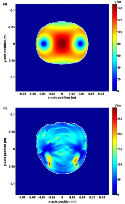
[image:5.612.60.289.66.220.2] [image:5.612.333.541.68.231.2] [image:5.612.330.545.391.740.2] [image:5.612.70.278.556.731.2]The electric field required to achieve neuronal stimula-tion typically occurs between 30 and 100 V/m,10 although this can vary depending on direction. The calculated electric field in the plane 20 mm from the 70 mm double coil for the homogeneous SAM and heterogeneous MRI-derived head model is shown in Fig.4.
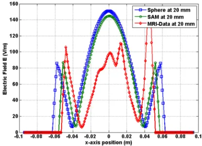
The calculated electric field in each human head model at a distance of 20 mm from the coil surface is shown in Fig.5. It is expected that the maximum field intensity will be directly beneath the center of the coil at x¼0. The magni-tude of electric field for the heterogeneous head model in this region is approximately 70% of that calculated for the two homogeneous models. Conversely, the peaks at650 mm are greater for the heterogeneous head model than the two homogeneous head models, indicating that field intensity closer to the surface of the head is greater than that deter-mined with a homogeneous head model.
Calculated electric field 50 mm from coil surface for each human head model is shown in Fig.6. Here it is demon-strated that use of the simplified homogeneous human head models may erroneously lead to the prediction of stimulation below the vertex at a depth of 50 mm using a 70 mm double coil, due to the magnitude of electric field exceeding 30 V/m. The realistic heterogeneous head model indicates the value of induced electric field in this region is likely to be well below the field required to generate an action potential. In contrast, the field seen closer to the surface of the head at this distance from the coil is greatly increased, giving rise to the possibility of having two stimulated sites with a spatial separation of 100 mm.
IV. CONCLUSION
Experimental measurements and theoretical calculations have been performed to assess the impact of human head
model complexity upon the calculation of electric field in the brain for neuronal activation. It has been shown that tissue heterogeneity has a significant effect on the distribution of electric field and that in general a simplified head model will underestimate the field intensity at the surface of the head and overestimates the field intensity at depth in the brain.
As the physical characteristics of the brain will vary for each TMS patient, the spatial variation of the induced elec-tric field will change. The consequence of this is that a TMS coil may not be able to accurately stimulate the same brain region for different patients. The necessity to implement re-alistic head modeling for assessing coil designs for noninva-sive deep brain stimulation has also been demonstrated.
ACKNOWLEDGMENTS
Research at the Ames Laboratory was supported by the Department of Energy-Basic Energy Sciences (Contract No: DE-AC02-07CH11358).
1
A. T. Barker, R. Jalinous, and I. L. Freeston,Lancet1, 1106 (1985).
2
D. Cohen and B. N. Cuffin,J. Clin. Neurophysiol.8, 102 (1991).
3L. G. Cohen, B. J. Roth, J. Nilsson, N. Dang, M. Panizza, S. Bandinelli, W.
Friauf, and M. Hallett,Electroencephalogr. Clin. Neurophysiol.75, 350 (1990).
4
L. J. Crowther, P. I. Williams, Y. Melikhov, and D. C. Jiles,J. Appl. Phys.
109, 07B314 (2011).
5A. V. Peterchev, R. Jalinous, and S. H. Lisanby,IEEE Trans. Biomed.
Eng.55, 257 (2008).
6
A. Hyodo, K. Iramina, and S. Ueno, in Proceedings of the 31st Annual International Conference of the IEEE EMBS, Minneapolis, Minnesota, USA, Sept. 2–6, 2009.
7P. C. Miranda, M. Hallett, and P. J. Basser,IEEE Trans. Biomed. Eng. 50, 1074 (2003).
8
S. Ueno, T. Tashiro, and K. Harada,J. Appl. Phys.64, 5862 (1988).
9F. A. Kozel, Z. Nahas, C. debrux, M. Molloy, J. P. Lorberbaum, D. Bohning,
S. C. Risch, and M. S. George,J Neuropsychiatr. Clin. Neurosci.12, 376 (2000).
10
T. Kammer, S. Beck, A. Thielscher, U. Laubis-Herrmann, and H. Topka, Clin. Neurophysiol.112, 250 (2001).
FIG. 6. (Color online) Calculated electric field profile 50 mm from coil surface for three human head models.
FIG. 5. (Color online) Calculated electric field profile 20 mm from coil surface for three human head models.
[image:6.612.69.277.62.213.2] [image:6.612.335.539.62.212.2]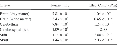
![Poly[[(μ4 1,3,5 triamino 1,3,5 trideoxy cis inositol)sodium] bromide]](data:image/gif;base64,R0lGODlhAQABAIAAAP///wAAACH5BAEAAAAALAAAAAABAAEAAAICRAEAOw==)