R E V I E W
Open Access
Ebola virus disease and critical illness
Aleksandra Leligdowicz
1, William A. Fischer II
2, Timothy M. Uyeki
3, Thomas E. Fletcher
4,5, Neill K. J. Adhikari
1,6,
Gina Portella
7, Francois Lamontagne
8, Christophe Clement
9, Shevin T. Jacob
10, Lewis Rubinson
11,
Abel Vanderschuren
12, Jan Hajek
13, Srinivas Murthy
14, Mauricio Ferri, Ian Crozier
15, Elhadj Ibrahima
16,
Marie-Claire Lamah
16, John S. Schieffelin
17, David Brett-Major
18, Daniel G. Bausch
19, Nikki Shindo
19,
Adrienne K. Chan
20, Tim O
’
Dempsey
21, Sharmistha Mishra
22, Michael Jacobs
23, Stuart Dickson
24,
G. Marshall Lyon III
25and Robert A. Fowler
1,6*Abstract
As of 20 May 2016 there have been 28,646 cases and 11,323 deaths resulting from the West African Ebola virus disease (EVD) outbreak reported to the World Health Organization. There continue to be sporadic flare-ups of EVD cases in West Africa.
EVD presentation is nonspecific and characterized initially by onset of fatigue, myalgias, arthralgias, headache, and fever; this is followed several days later by anorexia, nausea, vomiting, diarrhea, and abdominal pain. Anorexia and gastrointestinal losses lead to dehydration, electrolyte abnormalities, and metabolic acidosis, and, in some patients, acute kidney injury. Hypoxia and ventilation failure occurs most often with severe illness and may be exacerbated by substantial fluid requirements for intravascular volume repletion and some degree of systemic capillary leak. Although minor bleeding manifestations are common, hypovolemic and septic shock complicated by multisystem organ dysfunction appear the most frequent causes of death.
Males and females have been equally affected, with children (0–14 years of age) accounting for 19 %, young adults (15–44 years) 58 %, and older adults (≥45 years) 23 % of reported cases. While the current case fatality proportion in West Africa is approximately 40 %, it has varied substantially over time (highest near the outbreak onset)
according to available resources (40–90 % mortality in West Africa compared to under 20 % in Western Europe and the USA), by age (near universal among neonates and high among older adults), and by Ebola viral load at
admission.
While there is no Ebola virus-specific therapy proven to be effective in clinical trials, mortality has been dramatically lower among EVD patients managed with supportive intensive care in highly resourced settings, allowing for the avoidance of hypovolemia, correction of electrolyte and metabolic abnormalities, and the provision of oxygen, ventilation, vasopressors, and dialysis when indicated. This experience emphasizes that, in addition to evaluating specific medical treatments, improving the global capacity to provide supportive critical care to patients with EVD may be the greatest opportunity to improve patient outcomes.
Keywords:Ebola, Critical care
* Correspondence:rob.fowler@sunnybrook.ca
1Interdepartmental Division of Critical Care, University of Toronto, Toronto,
ON, Canada
6Department of Critical Care Medicine, Sunnybrook Health Sciences Centre,
Toronto, ON, Canada
Full list of author information is available at the end of the article
© 2016 Leligdowicz et al.Open AccessThis article is distributed under the terms of the Creative Commons Attribution 4.0 International License (http://creativecommons.org/licenses/by/4.0/), which permits unrestricted use, distribution, and reproduction in any medium, provided you give appropriate credit to the original author(s) and the source, provide a link to the Creative Commons license, and indicate if changes were made. The Creative Commons Public Domain Dedication waiver (http://creativecommons.org/publicdomain/zero/1.0/) applies to the data made available in this article, unless otherwise stated.
Background
In December 2013, transmission of Zaire ebolavirus (Ebola virus (EBOV)) to humans occurred in southeastern Guinea [1], spreading to Liberia and Sierra Leone and rap-idly surpassing the cumulative total of previous Ebola virus disease (EVD) outbreaks [2, 3]. Prior outbreaks oc-curred primarily in remote, resource-challenged settings, with case fatality proportions of 50–88 % [4]. This current outbreak, due to its size and spread in West Africa, in addition to exported and medically evacuated cases to Europe and North America, has engaged a much broader health worker community, including critical care clini-cians. While the clinical manifestations, duration of ill-ness, and transmissibility appear similar to previous EVD outbreaks [1, 5–9], with the availability and provision of advanced supportive care in Europe and North America, mortality was less than 20 %, emphasizing the potential importance of supportive and critical care in the manage-ment of EVD patients. This review provides an up-to-date examination of EVD using the knowledge gained during the 2013–2016 West African outbreak to highlight rele-vance for the critical care physician.
Viral hemorrhagic fevers
Viral hemorrhagic fever describes the syndrome of acute severe febrile illness caused by over 30 viruses from four different taxonomic families—Filoviridae, Arenaviridae, Bunyaviridae, and Flaviviridae. Although they differ in disease epidemiology, transmission, and pathogenesis, most of these RNA viruses are zoonotic and cause non-specific symptoms including fever, headache, weakness, vomiting, and diarrhea. Infection with filoviruses, includ-ing Marburg and Ebola viruses, can be associated with a rapid progression to hemodynamic instability, shock, and multiorgan dysfunction [4, 8].
Filoviruses were discovered in 1967 when 31 labora-tory workers became ill after coming into contact with green monkeys imported from Africa [10]. The newly discovered virus killed 23 % of infected workers in the German town of Marburg. Ebola virus was discovered in 1976 during simultaneous outbreaks in Zaire and Sudan in which 88 % and 53 % of patients died, respectively [2, 11, 12]. Since its discovery there have been approxi-mately 25 EVD outbreaks [3].
Epidemiology
The West African EVD outbreak was first recognized in March 2014 in the forested region of southeastern Guinea. However, the first EVD case may have happened as early as December 2013 with zoonotic transmission of EBOV from an animal to a human, and subsequent human-to-human spread [1]. Transmission was likely well underway throughout West Africa in the spring of 2014 [13]. By June 2014 there were a few hundred
confirmed or probable EVD cases, 3000 by the end of August—outstripping the ability of providers at existing Ebola treatment units to isolate and care for patients—and eventually over 20,000 cases by the end of December 2014 [14]. As of 20 May 2016 there have been 28,610 reported confirmed, probable, and suspected EVD cases and 11,308 deaths in Guinea, Sierra Leone and Liberia [15]. Another 36 cases have primarily received care in Mali, Senegal, and Nigeria as well as countries outside Africa including the USA, UK, Germany, Spain, France, Italy, the Netherlands, Norway, and Switzerland (Table 1) (Additional file 1). EVD cases in West Africa have been reported equally among males and females, with children (0–14 years of age) accounting for 19 %, young adults (15–44 years) 58 %, and older adults (≥45 years) 23 % of reported cases [5, 6, 16].
Characteristics of transmission
Person-to-person transmission of EBOV occurs through mucous membrane contact with bodily fluids (e.g., vomit, feces, and blood) from those who are infected and symptomatic or by touching the body of someone who died of EVD [17]. Droplet transmission is less likely to occur due to low prevalence of respiratory symptoms [16, 18]. While there are animal transmission models of aerosolized EBOV [19, 20], the clinical relevance of small particle droplet nuclei transmission is unclear and may only apply to the care of critically ill patients undergoing aerosol-generating procedures (intubation and ventilation, bronchoscopy) [18]. Percutaneous transmission with sharps (needle-stick, glass-related exposure) contaminated with infected bodily fluids is thought to be a very efficient mechanism of Ebola virus transmission [11].
Infection prevention and control practices
infection control recommendations or guidance from international technical organizations. This has often re-sulted in PPE that cannot be safely tolerated by healthcare personnel for more than 45–60 min due to excessive heat and humidity [21], risking syncope, potentially dangerous behaviour (e.g., adjusting fogged facial protection with soiled gloves), and inability to safely perform tasks such as insertion of intravenous catheters. In West Africa, Ebola virus transmission likely also occurs among healthcare personnel during informal healthcare provision in the com-munity to patients or colleagues, without appropriate infec-tion preveninfec-tion and control practice [22, 23]. Nosocomial EBOV transmission in Spain [24, 25] and the USA [26] similarly reinforces the importance of rigorous Ebola IPC practices and healthcare personnel training, irrespective of the healthcare system [27, 28].
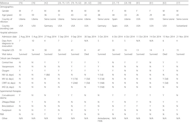
Table 1Chronological demographic description of 27 Ebola virus disease patients treated outside West Africa (August 2014–May 2015)
Age (years)
Occupation Country where Ebola
virus infection occurred
Case presentation Country of Hospitalisation Hospital LOS (days)
Outcome
1a 33 Health worker Liberia Medically evacuated USA 19 Survived
2a 59 Health worker Liberia Medically evacuated USA 14 Survived
3 75 Non health worker Liberia Medically evacuated Spain 5 Died
4 29 Health worker Sierra Leone Medically evacuated UK 10 Survived
5a 36 Health worker Sierra Leone Medically evacuated Germany 30 Survived
6a 51 Health worker Liberia Medically evacuated USA 20 Survived
7a 43 Health worker Sierra Leone Medically evacuated USA 41 Survived
8 N/A Health worker Liberia Medically evacuated France 16 Survived
9 70 Nonhealth worker Sierra Leone Medically evacuated Spain 3 Died
10a 45 Unknown Liberia Imported infection USA 8 Died
11a 38 Health worker Sierra Leone Medically evacuated Germany 47 Survived
12a 44 Health worker Spain Secondary infection Spain 30 Survived
13a 33 Nonhealth worker Liberia Medically evacuated USA 16 Survived
14 30 Health worker Sierra Leone Medically evacuated Norway 13 Survived
15 56 Health worker Liberia Medically evacuated Germany 6 Died
16a 26 Health worker USA Secondary infection USA 13 Survived
17a 29 Health worker USA Secondary infection USA 14 Survived
18 33 Health worker Guinea Imported infection USA 19 Survived
19 N/A Nonhealth worker Sierra Leone Medically evacuated France 21 Survived
20a 44 Health worker Sierra Leone Medically evacuated USA 2 Died
21a 43 Health worker Sierra Leone Medically evacuated Switzerland 15 Survived
22 50 Health worker Sierra Leone Medically evacuated Italy 38 Survived
23 N/A Nonhealth worker Liberia Medically evacuated Netherlands 13 Survived
24 39 Health worker Sierra Leone Imported infection UK 25 Survived
25 25 Health worker Sierra Leone Medically evacuated UK 15 Survived
26 N/A Health worker Sierra Leone Medically evacuated USA 27 Survived
27 N/A Health worker Sierra Leone Imported infection Italy 31 Survived
a
Medical management (including utilization of invasive therapies) is described in peer-reviewed format (Table3) and in reference [40]
LOSlength of stay (days),N/Anot available
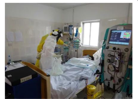
Fig. 1West African Ebola Treatment Facility—April 2014
[image:3.595.57.290.552.712.2]Pathophysiology
The pathogenesis of EVD in humans remains poorly understood but shows similarities with, and differences from, other causes of viral hemorrhagic fever or bacterial sepsis. End-organ dysfunction seems to result from a combination of a direct viral cytopathic effect, the host immune response, and from under-resuscitated hypovol-emic shock [5, 6]. EBOV binds to lectins and other surface receptors, with monocytes, macrophages, and dendritic cells as targets. These virus-containing cells spread through the lymphatic system, liver, and spleen, resulting in a widely disseminated viral infection [29, 30]. Endothe-lial cell infection and activation may lead to increased levels of soluble adhesion molecules, thombomodulin, and inflammatory mediators such as interferon-gamma and -alpha, interleukins (IL)-2, 6, and 10, interferon-inducible proteins, and tumor necrosis factor alpha [29, 30], result-ing in vascular injury.
Thrombocytopenia, consumption, and reduced pro-duction of clotting factors, in addition to increased concentrations of fibrin degradation products in pa-tients with severe EVD, may contribute to bleeding [29, 30]. Hepatocellular inflammation is common, and myositis with elevations of creatine kinase and pancreatitis (elevated blood amylase and lipase levels) occurs in severe cases [29, 30].
While acute kidney injury can often be explained by under-resuscitated hypovolemia, it might also arise from viral or secondary bacterial sepsis, acute tubular necrosis, myoglobinuria, and microvascular renal thrombi associated with sepsis or disseminated intra-vascular coagulation [29, 30]. Adrenal gland viral infec-tion has been shown in animal models and might contribute to hypotension, renal sodium loss, and hypovolemia [30].
Diagnostics
Diagnostic testing is recommended when a patient ex-hibits symptoms meeting the EVD case definition [31]. Ebola viral RNA can be detected in clinical specimens by real-time reverse transcription polymerase chain re-action (RT-PCR); if the virus is detected by a specific antigen diagnostic test or by detection of IgM anti-bodies directed against EBOV, RT-PCR should be used for confirmation [31, 32]. Because the sensitivity of mo-lecular tests depends on Ebola viral loads, specimens collected within 3 days of symptom onset may be falsely negative due to undetectable viremia early in the clinical course. In these circumstances, another blood specimen for RT-PCR testing should be collected 3 days after symptom onset [33]. Point-of-care rapid diagnos-tic tests (RDTs) have been tested in the field, but they lack sensitivity, require a cold chain, and remain under evaluation in clinical trials [34].
Clinical presentation of Ebola virus disease
The clinical presentation of EVD falls along a spectrum ranging from minimally symptomatic infection [8] to se-vere illness with hemorrhagic complications, shock, mul-tiorgan dysfunction, and death. The incubation period ranges from 2 to 21 days but may depend on the mode of transmission [4]: 5–7 days following a percutaneous inoculation and a mean of 9 days following direct mucus membrane contact with infected bodily fluids [8, 30, 35]. EVD typically begins with nonspecific initial signs and symptoms including fever, fatigue, weakness, and head-ache, similar to many infectious diseases in sub-Saharan Africa, often leading to a missed diagnosis and contin-ued transmission. A fleeting maculopapular rash can be seen within the first week [36, 37]. Gastrointestinal symp-toms (nausea, vomiting, abdominal pain, and diarrhea) usually follow 4–6 days after illness onset and can lead to hypovolemia and shock with multisystem organ dys-function. Gastrointestinal losses and anorexia can precipi-tate hypokalemia, metabolic acidosis, and acute kidney injury [6]. Hypoxia and ventilation failure tend to occur with severe illness and may be exacerbated by vascular in-jury and accompanying large-volume fluid requirements [38]. Serious hemorrhagic complications are relatively rare, while more mild bleeding may occur in approximately 30 % of cases [5, 6, 9, 16, 39, 40]. Delirium and encephalopathy or encephalitis may reflect metabolic encephalopathy or direct neuroinvasion and hiccups may be of central or per-ipheral neurological origin [5, 30, 41, 42].
Clinical outcomes
[image:4.595.305.539.610.715.2]While the cumulative case fatality proportion in West Africa is approximately 40 % as of May 2016, it has varied substantially during the course of the outbreak (being higher near the beginning). In comparison, the cumulative case fatality proportion for patients treated in Western Europe and the USA during 2014–2015 was 18.5 % [40] (Table 2). Case fatality remains highest among young children and older adults [5, 6, 16, 43]. Pregnant women often experience spontaneous abortion
Table 2Demographic and outcome summary of 27 Ebola virus disease patients treated outside West Africa
All Survived Died
Treated outside West Africa 27 22 (81.5 %) 5 (18.5 %)
Gendera(male) 17 (68 %) 12 (60 %) 5 (100 %)
Median ageb(range) 40.5 (25
–75) 36 (25–59) 56 (42–75) Mean hospital length of stay
(days, confidence interval)
19 (±11.5) 22 (±10.2) 5 (±2.4)
Evacuated from West Africa 20 (74 %) 16 (80 %) 4 (20 %)
Infected outside West Africa 3 (11 %) 3 (100 %) 0
a
Gender available for 25 patients
b
followed by bleeding, as well as preterm labor and still-birth if Ebola virus infection occurs later in pregnancy. Vertical transmission and subsequent neonatal mortality has been virtually uniform in the few documented live births by women with acute EVD [44]. A high Ebola viral load at time of admission is associated with more severe illness and mortality [5, 6, 29, 43, 45–47], with other markers of organ dysfunction variably associated with out-comes [6, 8, 29, 48].
Monitoring and care delivery
The management of critically ill EVD patients in a resource-constrained setting has historically been re-stricted to variable monitoring of daily clinical signs and symptoms without access to continuous assessment [8]. The need for strict IPC practices and separation of pa-tient care areas in West Africa significantly limited documentation and review of daily clinical records both inside and outside of high-risk patient care. As the case burden decreased and the ratio of healthcare personnel to patients increased, assessments were performed more systematically at some Ebola treatment facilities, with temperature, heart rate, blood pressure, respiratory rate, pulse oximetry, qualitative descriptions of urine, and gastrointestinal output, as well as fluid balance estimation. Laboratory data other than Ebola RT-PCR results were essentially unavailable at Ebola treatment facilities in West Africa early in the outbreak [33, 49, 50]. Initially, there was very limited attention to diagnostics other than Ebola RT-PCR. The point-of-care systems for mon-itoring biochemistry and hematology parameters, such as the i-STAT® or the Piccolo Xpress®, were inconsist-ently utilized inside Ebola treatment facilities [5, 6, 48], in part due to limited manufacturer-recommended temperature and humidity ranges. Over the course of the West African outbreak, with support from national and deployed international laboratories, basic biochemis-try, blood counts, and coagulation profiles helped to characterize the course of illness, but remained inconsist-ently available and often with substantial delays in results reporting due to transport and processing time. Malaria rapid diagnostic tests, and less commonly RT-PCR, were available at most of the laboratories that supported Ebola treatment facilities in West Africa. However, testing for Lassa fever virus (endemic in EBOV-affected countries) or other causes of sepsis was not routine.
Bedside ultrasonography has not been widely deployed [51] but could help with assessing volume status, responsive-ness to intravenous fluids [50], and assessment of challen-ging clinical signs such as abdominal distension [42]. Plain chest and abdomen radiography has been performed in European and North American settings [52] but has rarely been available to patients with Ebola in West Africa. Among patients treated in the USA and Europe,
pulmonary edema has been reported in 44 % and acute re-spiratory distress syndrome in another 22 % [40].
Discharge criteria and virus persistence during convalescence
The World Health Organization (WHO) recommends considering discharge of patients from isolation on the basis of a negative blood Ebola virus RT-PCR result taken at least 3 days after the resolution of symptoms [33, 41]. However, Ebola virus can persist in certain body fluids after it is undetectable in the blood and after clin-ical recovery from EVD [42, 53–56]. Viable Ebola virus has been isolated from urine, semen, cerebrospinal fluid, and vitreous humor many months after blood clearance [42, 56–61], suggesting that some activities (unprotected sex, invasive procedures, or penetrating eye trauma) confer a transmission risk even after symptoms and viremia resolve. It is therefore critical to counsel EVD survivors about the risks of Ebola virus persistence [59, 60] and appropriate precautions after discharge, in-cluding barrier protection during sexual intercourse [50] until semen has tested negative for Ebola virus twice, or for at least 6 months after EVD onset [62]. Health workers and others should continue to apply standard precautions, as with all patients, when evaluating EVD survivors in the convalescent period. Additional infec-tion preveninfec-tion and control practices, based upon an in-dividual patient risk assessment, may be prudent for specific procedures in convalescence, even when there is no detectable EBOV in the blood (e.g., lumbar puncture or vitreous humor sampling) [63].
Critical and supportive care interventions
Providing supportive care to critically ill patients with EVD in resource-poor settings is challenging [64] due to limited infrastructure, lack of materials and trained healthcare personnel, and uncertainty regarding the translation of modern sepsis treatment strategies [65] and optimal intravenous fluid management protocols in the absence of advanced monitoring used in resource-rich settings [66]. Respiratory symptoms such as cough are not a prominent feature of EVD and tachypnea likely represents respiratory compensation of severe metabolic acidosis [9, 30, 50]. Translating fluid resuscitation proto-cols used in a resource-rich setting [42] to settings where supplemental oxygen therapy is not routinely available [49], in a disease with possible vascular leak syndrome [38], could result in increased morbidity [67] and warrants further investigation [68–70]. The use of antibiotics is common at Ebola treatment facilities [5, 6] before the diagnosis of EVD is confirmed in febrile patients and as empiric treatment of potential bacterial co-infection or gastrointestinal bacterial translocation in patients with confirmed EVD [9]. However, disruption of
gastrointestinal flora due to broad-spectrum antibiotics could exacerbate diarrhea and fluid losses. Symptomatic treatment of severe diarrhea with loperamide was vari-ably employed across Ebola treatment facilities. The risks and benefits of these practices warrant evaluation with observational studies and clinical trials [71].
Early and during the peak of the outbreak, clinical management was generally limited to supportive care fo-cusing on aggressive oral and occasional intravenous volume resuscitation. As the case numbers decreased, advanced care became more common in some treatment facilities. Despite the limitations of working in PPE, feasibility and safety of central venous catheter placement was demonstrated at a UK military-supported treatment facility in Kerry Town, Sierra Leone [72]. Feasibility of transthoracic echocardiography was demonstrated at a military Ebola treatment facility in Conakry, Guinea [51]. By mid-December 2014, EMERGENCY, an Italian non-governmental organization, established an Ebola critical care unit in Lakka and Goderich, Sierra Leone, the latter consisting of constant bedside nursing, continuous blood pressure, heart rate, respiratory rate monitoring, pulse oximetry, arterial and venous cannulation, nasogastric tube feeding, invasive ventilation, continuous renal re-placement therapy, diagnostic biochemistry and hematology, ultrasonography, and plain radiography (Fig. 2). With waning case numbers, accurate evaluation of the impact of these interventions on patient out-comes has not been possible. Other sites, such as the GOAL-supported Mathaska Ebola Treatment Unit (ETU) and the Partners in Health-supported Maforki ETU in Si-erra Leona also began using aspects of critical care procedures by February 2015, including nasogastric tube feeding, bedside ultrasound, as well as intraosseus cannu-lation for intravenous fluid resuscitation [73].
The historical philosophy of providing only oral fluids for EVD care has given way to the delivery of
context-appropriate critical care [38, 42, 74–79]. To date, 27 pa-tients managed in nine countries outside of West Africa (Table 1) have been described, with a survival of 81.5 % [40] (Table 2). Thirteen detailed accounts of EVD man-agement in modern healthcare settings in the USA, Germany, Spain, and Switzerland provide insight into the course of the illness [38, 42, 76–82] (Table 3). These case reports confirm that intensive care monitoring in appropriately prepared centers is feasible. Noninvasive ventilation [38, 42], mechanical ventilation [38, 78, 81], central venous catheter insertion for vasopressor support [38, 42, 78, 79, 82], and renal replacement therapy [38, 78, 81, 82] can be provided effectively and safely (Table 3, Figs. 3 and 4) [83].
Establishing supportive and critical care services in highly resourced settings
While it may be advisable to concentrate or regionalize care for patients with EVD in specific hospitals, any health centre should be prepared to safely take a focused and relevant history from a patient with an infectious syndrome, and to mobilize the appropriate local and re-gional response. Many hospitals, even if not EVD refer-ral centers, may be asked to care for patients until initial (and possibly subsequent) blood Ebola RT-PCR results are known. Therefore, it is essential that hospital staff are well trained and familiar with recommended IPC practices (and for EVD, standard and contact IPC pre-cautions in particular). It is ideal to have an on-call inter-professional team who have undergone training in Ebola-specific IPC training.
While practiced IPC protocols are important to keep health workers safe, a very common clinical pitfall is to equate IPC practices with care. While Ebola-specific standardized IPC protocols are absolutely necessary, there will be situations requiring patient-specific IPC risk-assessments—most commonly involving patients at the beginning of, or in the convalescent phase of, their illness with minimal symptoms and no vomiting or diar-rhoea (i.e., with very low risk of transmission). It is also important to remember that most patients suspected with EVD will not have EVD, but will have illness in need of prompt treatment—commonly malaria—that may require empiric treatment while awaiting diagnostic testing [84, 85]. Barriers to providing the standard of care to patients suspected of EVD will repeatedly arise: “We don’t have the capacity to do that…that is not part of our protocol.” Do not accept this when it negatively influences patient care. Instead, ask collectively“How can we safely solve this challenge, now, for the benefit of this patient?”
For hospitals and intensive care units (ICUs) that will provide definitive care for patients with EVD, there are many Ebola-specific considerations well beyond the scope of this review; however, a number deserve mention.
[image:6.595.58.290.547.713.2]Table 3Clinical management summary of 13 Ebola virus disease patients treated outside West Africa
Reference [76] [76] [42] [28,79,137] [78,79,82] [81,82] [38] [25,77] [28,99] [81] [81] [82] [37]
Demographics
Gender M F M M M M M F M F F M M
Age (years) 33 59 36 51 43 42 38 44 33 26 29 44 43
Country of infection
Liberia Liberia Sierra Leone Liberia Sierra Leone Liberia Sierra Leone Spain Liberia USA USA Sierra Leone Sierra Leone
Country providing care
USA USA Germany USA USA USA Germany Spain USA USA USA USA Switzerland
Hospital admission
Admission date 2 Aug 2014 5 Aug 2014 27 Aug 2014 5 Sep 2014 9 Sep 2014 30 Sep 2014 3 Oct 2014 6 Oct 2014 6 Oct 2014 11 Oct 2014 14 Oct 2014 15 Nov 2014 21 Nov 2014
Days from diagnosis to evacuation
7 10 4 7 2 N/A 5 0 5 N/A N/A 6 4
Hospital LOS 19 14 30 20 41 8 47 30 16 13 14 3 15
Vital status Survived Survived Survived Survived Survived Died Survived Survived Survived Survived Survived Died Survived
Critical care therapies
Central line N N Y Y Y Y Y N Y Y N Y Y
Vasopressors N N N N Y Y Y N N N N Y N
Oxygen Y Y Y Y Y Y Y Y N N N Y N
NIV (d, days) N N Y (8d) N N N Y (1d) N N N N N N
MV (d, days) N N N N Y (17d) Y (5d) Y (13d) N N N N Y (3d) N
CRRT (d, days) N N N N Y (24d) Y (5d) Y (18d) N N N N Y (3d) N
IHD (d, days) N N N N N N Y (10d) N N N N N N
Experimental therapies
Convalescent plasma
Y N N Y Y N N Y Y Y Y Y N
ZMapp/ZMab Y Y N N N N N Y N Y N Y Y
Brincidofovir N N N N N Y N N Y Y Y N N
Favipiravir N N Y N N N Y Y N N N N Y
TKM N N Y Y Y N N N N Y N N N
Other N/A N/A N/A N/A N/A N/A Amiodarone,
FX06
N/A N/A N/A N/A N/A N/A
CRRTcontinuous renal replacement therapy;Ffemale,IHDintermittent hemodialysis,LOSlength of stay,Mmale,MVinvasive mechanical ventilation,N/Anot available,NIVnon-invasive ventilation,TKMTKM-Ebola, small interfering ribonucleic acids (siRNA) produced by Tekmira
Leligdowicz
et
al.
Critical
Care
(2016) 20:217
Page
7
of
Hospitals and ICUs will generally need to mould EVD planning to the local environment, and seek out the ex-perience, guidance, protocols, and training from those who have substantial clinical and operational experience (Figs. 3, 4) [64, 86].
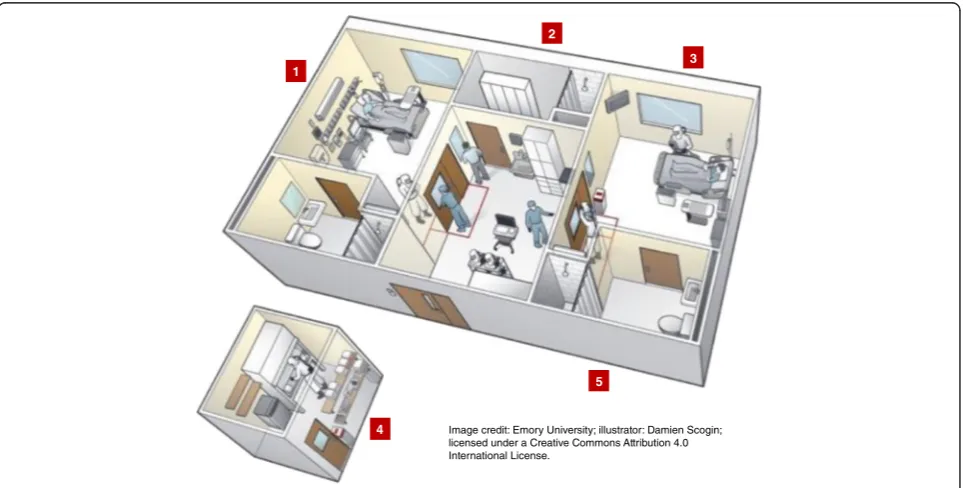
Second, the physical environment of a proposed Ebola treatment unit is a critical component of care. Ideally,
there should be a large available physical space, sufficient for multiple isolation rooms, with very generously sized antechamber areas for donning and doffing, and a shared area from where clinical observation can occur (Fig. 4). There should be sufficient adjoining space to house dedicated diagnostic (e.g., portable radiograph and ultrasound machines, potentially point-of-care labora-tory devices) and therapeutic (intravenous pumps, mech-anical ventilator and circuits, dialysis machine and supplies) equipment. There should be ample nearby space to house packaged soiled PPE and medical waste that allows pick-up and proper disposal.
Third is the necessity for sufficiently numerous and trained inter-professional team of clinical (nurses, physi-cians, respiratory therapists, others) and patient support staff (coordinators, monitors, cleaners, patient transpor-tation services, diagnostic and laboratory staff, and so forth), who are well practiced in the institutional Ebola care plan and their specific roles. Whether this team is led by infectious disease or critical care specialists, or both, is likely less important than establishing an inter-disciplinary model of continuity care throughout the hospital stay, oftentimes in a single geographic location that is institutionally appropriate.
Fourth, while EVD is accompanied by an increasingly well-characterized clinical gastrointestinal syndrome
Fig. 3Ebola treatment facility, Royal Free Hospital, London, UK—September 2014
[image:8.595.57.290.87.266.2] [image:8.595.58.540.411.655.2]leading to fluid, electrolyte, and acid-base imbalance with multisystem organ dysfunction, there are no Ebola-specific therapies yet to be proven effective. However, in-tensive care medicine comprises experiential and evidence-based organ-supportive care, which should guide the care of patients with EVD—attention to fluid, electrolyte and acid-base balance, initiation of empiric and specific anti-infective therapy, and support for renal injury and respiratory failure as occurs for other poten-tially self-limited and survivable illnesses. Among pa-tients with EVD treated in the USA and Europe, 41 % were deemed to have critical illness with 70 % receiving supplemental oxygen, 22 % with acute respiratory dis-tress syndrome, 26 % invasive mechanical ventilation, 30 % intravenous vasoactive medications, and 19 % re-quiring dialysis [40].
For the most severely ill patients, clinical judgment is always necessary to balance risks and benefits of certain resuscitation strategies, including the initiation of car-diopulmonary resuscitation (CPR) [28, 87]. While there is a lack of clinical experience with CPR in EVD pa-tients, it may be a reasonable consideration while cor-recting reversible abnormalities (i.e., hypoxia, severe electrolyte disturbance, arrhythmias) in settings where the option for advanced life-support exists. The decision to provide CPR should be guided by its medical indication and utility in that context, the ability to provide effective CPR, and the safety of those providing care including safe donning and doffing of PPE, in addition to patient preferences [88, 89].
Fifth, as with all critical illness, medical technical care is only one dimension of our support for patients and their families. Patients with EVD and their families require mechanisms to stay in audio and visual contact throughout the illness—ideally visual contact through transparent bar-riers or at safe distance, or direct contact with supervised donning and doffing of PPE—in addition to substantial psychosocial support during and after EVD.
Ebola-specific pharmacological prevention and therapeutics
Current EVD treatment focuses on supportive care [70] as there are no specific treatment options yet to be proven effective [70, 90, 91]. A number of Ebola-specific treatment strategies have undergone preliminary clinical trial investigation, including convalescent plasma, Favi-piravir, Brincidofovir and TMK-130803 [92–97]. Trans-fusion of convalescent whole blood or plasma donated by EVD survivors has been used in this and prior EVD outbreaks [98] in an uncontrolled or compassionate-use basis [25, 79, 81, 99], and in animal models [100, 101]. One of three clinical trials of convalescent plasma ther-apy [94] has been completed and reported [102]. In this nonrandomized comparison to historical controls,
transfusion of up to 500 ml convalescent plasma with unknown levels of neutralizing antibodies in 84 patients with confirmed EVD was not associated with a signifi-cant improvement in survival. While there were no ser-ious adverse reactions in this trial, transfusion-related acute lung injury was described during convalescent plasma therapy in Spain [25]. Favipiravir (Toyama, Japan) [103], a pre-existing influenza virus inhibitor, has been administered for compassionate use outside West Africa [37, 38, 42]. In a multicenter, nonrandomized clinical trial in Guinea [104], 111 patients receiving Favi-piravir had similar survival to that based upon historical control patients. The trial authors suggested that Favipir-avir should be further studied in patients with medium to high viremia, but not in those with very high viremia [105]. Brincidofovir (Chimerix, USA), a nucleotide ana-log that inhibits RNA-polymerase with in vitro activity against Ebola [106], was administered to a small number of EVD patients for uncontrolled compassionate use [42, 79, 81, 99] and was tested in a phase 2 clinical trial in Liberia [95] that was stopped after the manufacturer withdrew study support [107]. TKM-130803 is a formula-tion of lipid-nanoparticle-encapsulated small interfering ribonucleic acids (siRNA) targeting two proteins involved in Ebola virus transcription and replication (Tekmira, USA, Canada). It was used in nonhuman primate Ebola virus infection as a postexposure treatment strategy [108] and in patients medically evacuated from West Africa in uncontrolled compassionate use [79, 81]. However, a phase 2 clinical trial (RAPIDE-TKM) in Sierra Leone [96] was halted according to pre-established stopping rules [109].
ZMapp, a monoclonal antibody cocktail (Leafbio, USA) [110], has been used under the emergency investi-gational new drug approvals from the Food and Drug Administration in patients treated in the USA, West Africa, and Western Europe [40, 76, 111]. ZMapp treat-ment of rhesus macaques resulted in 100 % survival even when started 5 days after lethal EBOV infection [110]. In the only randomized controlled trial of an investigational therapeutic for EVD, ZMapp plus standard of care was compared to standard of care alone for EVD patients in four countries, including the three most impacted West African countries. Due to the decline in EVD cases, this unblinded ZMapp randomized controlled trial only enrolled 72 of the prespecified target goal of 200 EVD pa-tients; data were analyzed for 71 EVD patients, and mor-tality in the ZMapp treatment group was 22 % versus 37 % in the untreated group, but this difference was not statistically significant [112, 113].
The open-label, uncontrolled, and selected administra-tion of other agents such as amiodarone [114], HMG-CoA reductase inhibitors, and angiotensin II receptor antagonists [115], and therapies to counteract vascular
leak (FX06) [38] preclude any conclusions. In an obser-vational study examining temporal trends in mortality among patients with EVD in one ETU in Guinea, 125 of 194 (64.4 %) patients receiving artemether–lumefantrine for malaria prophylaxis died as compared with 36 of 71 patients receiving artesunate–amodiaquine (50.7 %). In adjusted analyses, the risk ratio was 0.69 (95 % confidence interval, 0.54 to 0.89), with a stronger effect observed among patients without malaria [116]. These findings have not been confirmed in a randomized clinical trial.
Two vaccine candidates demonstrated efficacy in nonhuman primates [92, 117, 118]. A recombinant, replication-competent vesicular stomatitis virus-based vaccine expressing a surface glycoprotein of Zaire
ebola-virusrVSVΔG-EBOV-GP (rVSV) [118, 119] was evaluated
in an open-label, ring vaccination trial involving 7651 people in 90 clusters, randomized to immediate or delayed (21 days) administration. The vaccine was well tolerated and in the immediate vaccination group there were no new EVD cases while in the delayed vaccination group there were 16 EVD cases [120]. Another candidate vaccine, cAd3-EBOV (cAd3) [117] remains under investi-gation [92, 121]. Other vaccine candidates are also under development and evaluation [122, 123].
Post-exposure Prophylaxis
Several healthcare personnel received post-exposure prophylaxis with different interventions, including a candidate Ebola vaccine, following potential high-risk exposures to Ebola virus; although Ebola virus disease did not occur in these individuals, no conclusions can be made about the effectiveness of these uncontrolled inter-ventions [124–126].
Ethical challenges in caring for patients with Ebola virus disease
Each of the commonly applied four principles of medical bioethics faces numerous threats in treating patients with EVD [87]. A symptomatic patient’s autonomy to not seek treatment (and not be isolated) is weighed against the threat of disease transmission by staying in the community. The injustice of treatment variability, across regions and over time, places patients at differen-tial risk of death. In acting beneficently, healthcare workers inherently place themselves at some risk. A nat-ural response is to balance that risk with the duty to help. This frequently conspires against greater numbers of health workers responding to an Ebola outbreak. The duty to nonmaleficence, doing no harm, is a daily con-undrum, through potential delays in routine diagnostic work-up for common illnesses because of a lack of diag-nostic testing, or, in resource-constrained environments,
inadequate space to separate potentially infectious suspect patients along a gradient of risk.
Post-Ebola syndrome
With over 11,000 EVD survivors, there is increased recog-nition of a post-Ebola syndrome in the convalescent period, characterized by mental health and cognitive sequelae, chronic headaches, insomnia, arthralgias, audi-tory disturbances, and ocular effects including sight-threatening uveitis [127–132]. It is uncertain whether these manifestations are due to direct viral cytopathic effect in immune-privileged compartments or postin-fectious immune-mediated inflammation [133–135].
Research directions
Although this EVD outbreak narrowed some knowledge gaps, pathophysiology and the immunological response to acute infection and convalescence is still minimally char-acterized. Access to rapid point-of-care EVD diagnostic capacity to differentiate between other common febrile illnesses [136] is critical because the early presentation of EVD has a broad differential diagnosis [5, 7, 34]. Labora-tory testing to identify prognostic indicators could help guide clinical care. Evaluation of specific antiviral therap-ies is critical as is evaluation of commonly used treatments for which there is still very limited evidence (e.g., empiric antibiotics, anti-diarrheal agents, and fluid replacement composition and volume). The safety and functionality of PPE must be improved. Standardized, easy-to-use clinical charting and human resources for data entry should be made available to support cohort studies and clinical trials. While it seems intuitive that provision of advanced supportive and critical care improves patient outcomes, operationalizing and evaluating increased levels of care to resource-challenged environments is needed. Prepared research protocols that can be rapidly adapted to country-specific settings and quickly implemented could reduce research delays in future outbreaks. Following patients who survive EVD is important to better characterize immune correlates of virus clearance and host genetic factors that contribute to survival, and to better address morbidity of the post-Ebola syndrome.
Conclusions
Additional file
Additional file 1:Weblink references for Tables 1–3. (DOCX 124 kb)
Abbreviations
CPR, cardiopulmonary resuscitation; EBOV, Ebola virus; ETU, Ebola Treatment Unit; EVD, Ebola virus disease; ICU, intensive care unit; IL, interleukin; IPC, infection prevention and control; PPE, personal protective equipment; RT-PCR, real-time reverse transcription polymerase chain reaction
Authors’contributions
RAF, AL, WAFII, TEF, and TMU conceived of the study, participated in its design and coordination, collected data, performed the statistical analysis and drafted the manuscript. NKJA, GP, FL, TMU, CC, STJ, LR, AV, JH, SM, MF, IC, EIB, MCL, JSS, DBM, DGB, NS, AC, TOD, SM, MJ, SD, and GMLIII participated in the design of the study, helped in data collection and helped to revise the manuscript. All authors read and approved the final manuscript.
Competing interests
The authors declare that they have no competing interests.
Disclaimer
The findings and conclusions in this report are those of the authors and do not necessarily represent the official position of the Centers for Disease Control and Prevention.
Author details
1Interdepartmental Division of Critical Care, University of Toronto, Toronto,
ON, Canada.2Department of Medicine, University of North Carolina, Chapel Hill, NC, USA.3Centers for Disease Control and Prevention, Atlanta, Georgia, USA.4Defence Medical Services, Whittington Barracks, Lichfield, UK. 5Liverpool School of Tropical Medicine, Liverpool, Merseyside, UK.
6Department of Critical Care Medicine, Sunnybrook Health Sciences Centre,
Toronto, ON, Canada.7Emergency NGO, Milan, Italy.8Department of Medicine, Université de Sherbrooke, Sherbrooke, Quebec, Canada. 9Polyclinique Bordeaux Nord Aquitaine, Bordeaux, France.10Department of
Medicine, University of Washington, Seattle, Washington, USA.11Department of Medicine, University of Maryland, Baltimore, MD, USA.12Centre de recherche de l’institut Universitaire de Cardiologie et de Pneumologie de Québec, Quebec City, Quebec, Canada.13Division of Infectious Diseases, University of British Columbia, Vancouver, BC, Canada.14Department of Paediatrics, University of British Columbia, Vancouver, BC, Canada. 15
Infectious Diseases Institute, College of Health Sciences, Makerere University, Kampala, Uganda.16Department of Infectious and Parasitic Diseases, Donka Hospital, Conakry, Guinea.17Department of Pediatrics, School of Medicine and School of Public Health and Tropical Medicine, Tulane University, New Orleans, LA, USA.18Department of Preventive Medicine and Biometrics, Uniformed Services University, Bethesda, MD, USA. 19Department of Pandemic and Epidemic Diseases, World Health
Organization, Geneva, Switzerland.20Division of Infectious Diseases, Sunnybrook Health Sciences Centre, Toronto, ON, Canada.21Department of Clinical Sciences, Liverpool School of Tropical Medicine, Liverpool, UK. 22Department of Medicine, University of Toronto, Toronto, ON, Canada.
23Department of Infection, Royal Free London NHS Foundation Trust,
London, UK.24Acute Medicine and Intensive Care, Derriford Hospital, Plymouth, UK.25Department of Infectious Diseases, Emory University Hospital, Atlanta, Georgia, USA.
Received: 3 March 2016 Accepted: 26 April 2016
References
1. Baize S, Pannetier D, Oestereich L, et al. Emergence of Zaire Ebola virus disease in Guinea. N Engl J Med. 2014;371(15):1418–25.
2. Bres P. The epidemic of Ebola haemorrhagic fever in Sudan and Zaire, 1976: introductory note. Bull World Health Organ. 1978;56(2):245.
3. CDC. Outbreaks Chronology: Ebola Virus Disease. http://www.cdc.gov/vhf/ ebola/outbreaks/history/chronology.html. Accessed 7 Sep 2015.
4. Feldmann H, Geisbert TW. Ebola haemorrhagic fever. Lancet. 2011;377(9768): 849–62.
5. Bah EI, Lamah MC, Fletcher T, et al. Clinical presentation of patients with Ebola virus disease in Conakry. Guinea N Engl J Med. 2015;372(1):40–7. 6. Schieffelin JS, Shaffer JG, Goba A, et al. Clinical illness and outcomes in
patients with Ebola in Sierra Leone. N Engl J Med. 2014;371(22):2092–100. 7. Fauci AS. Ebola—underscoring the global disparities in health care
resources. N Engl J Med. 2014;371(12):1084–6.
8. Kortepeter MG, Bausch DG, Bray M. Basic clinical and laboratory features of filoviral hemorrhagic fever. J Infect Dis. 2011;204 Suppl 3:S810–6. 9. Bwaka MA, Bonnet MJ, Calain P, et al. Ebola hemorrhagic fever in Kikwit,
Democratic Republic of the Congo: clinical observations in 103 patients. J Infect Dis. 1999;179 Suppl 1:S1–7.
10. Peters CJ, LeDuc JW. An introduction to Ebola: the virus and the disease. J Infect Dis. 1999;179 Suppl 1:ix–xvi.
11. WHO. Ebola haemorrhagic fever in Zaire, 1976. Bull World Health Organ. 1978;56(2):271–93.
12. WHO. Ebola haemorrhagic fever in Sudan, 1976 Report of a WHO/ International Study Team. Bull World Health Organ. 1978;56(2):247–70. 13. Gire SK, Goba A, Andersen KG, et al. Genomic surveillance elucidates Ebola virus
origin and transmission during the 2014 outbreak. Science. 2014;345(6202):1369–72. 14. CDC. 2014 Ebola Outbreak in West Africa - Reported Cases Graphs.
http://www.cdc.gov/vhf/ebola/outbreaks/2014-west-africa/cumulative-cases-graphs.html. Accessed 7 Sep 2015.
15. WHO. Ebola Situation Reports. http://apps.who.int/ebola/ebola-situation-reports, Accessed 20 May 2016.
16. Team WHOER. Ebola virus disease in West Africa—the first 9 months of the epidemic and forward projections. N Engl J Med. 2014;371(16):1481–95. 17. Victory KR, Coronado F, Ifono SO, et al. Ebola transmission linked to a single
traditional funeral ceremony—Kissidougou, Guinea, December, 2014-January 2015. MMWR Morb Mortal Wkly Rep. 2015;64(14):386–8.
18. WHO. Interim infection prevention and control guidance for care of patients with suspected or confirmed filovirus haemorrhagic fever in health-care settings, with focus on Ebola. Geneva, Switzerland: WHO; 2014. 19. Johnson E, Jaax N, White J, et al. Lethal experimental infections of rhesus
monkeys by aerosolized Ebola virus. Int J Exp Pathol. 1995;76(4):227–36. 20. Jaax N, Jahrling P, Geisbert T, et al. Transmission of Ebola virus (Zaire strain)
to uninfected control monkeys in a biocontainment laboratory. Lancet. 1995;346(8991-8992):1669–71.
21. Brearley MB, Heaney MF, Norton IN. Physiological responses of medical team members to a simulated emergency in tropical field conditions. Prehosp Disaster Med. 2013;28(2):139–44.
22. WHO. Health worker Ebola infections in Guinea, Liberia and Sierra Leone. Geneva, Switzerland: WHO; 2015.
23. Suwantarat N, Apisarnthanarak A. Risks to healthcare workers with emerging diseases: lessons from MERS-CoV, Ebola, SARS, and avian flu. Curr Opin Infect Dis. 2015;28(4):349–61.
24. Lopaz MA, Amela C, Ordobas M, et al. First secondary case of Ebola outside Africa: epidemiological characteristics and contact monitoring, Spain, September to November 2014. Euro Surveill. 2015;20(1).
25. Mora-Rillo M, Arsuaga M, Ramirez-Olivencia G, et al. Acute respiratory distress syndrome after convalescent plasma use: treatment of a patient with Ebola virus disease contracted in Madrid. Spain Lancet Respir Med. 2015;3(7):554–62. 26. Chevalier MS, Chung W, Smith J, et al. Ebola virus disease cluster in the
United States—Dallas County, Texas, 2014. MMWR Morb Mortal Wkly Rep. 2014;63(46):1087–8.
27. Tartari E, Allegranzi B, Ang B, et al. Preparedness of institutions around the world for managing patients with Ebola virus disease: an infection control readiness checklist. Antimicrob Resist Infect Control. 2015;4:22.
28. Johnson DW, Sullivan JN, Piquette CA, et al. Lessons learned: critical care management of patients with Ebola in the United States. Crit Care Med. 2015;43(6):1157–64.
29. McElroy AK, Erickson BR, Flietstra TD, et al. Ebola hemorrhagic fever: novel biomarker correlates of clinical outcome. J Infect Dis. 2014;210(4):558–66. 30. Fletcher TE, Fowler RA, Beeching NJ. Understanding organ dysfunction in
Ebola virus disease. Intensive Care Med. 2014;40(12):1936–9. 31. WHO. Case definition recommendations for Ebola or Marburg Virus
Diseases. 2014. http://www.who.int/csr/resources/publications/ebola/ebola-case-definition-contact-en.pdf, Accessed 7 Sep 2015.
32. Martin P, Laupland KB, Frost EH, et al. Laboratory diagnosis of Ebola virus disease. Intensive Care Med. 2015;41(5):895–8.
33. WHO. Clinical management of patients with viral haemorrhagic fever: a pocket guide for the front-line health worker. Geneva, Switzerland: WHO; 2014.
34. Broadhurst MJ, Kelly JD, Miller A, et al. ReEBOV Antigen Rapid Test kit for point-of-care and laboratory-based testing for Ebola virus disease: a field validation study. Lancet. 2015;386(9996):867–74.
35. Emond RT, Evans B, Bowen ET, et al. A case of Ebola virus infection. Br Med J. 1977;2(6086):541–4.
36. Nkoghe D, Leroy EM, Toung-Mve M, et al. Cutaneous manifestations of filovirus infections. Int J Dermatol. 2012;51(9):1037–43.
37. Schibler M, Vetter P, Cherpillod P, et al. Clinical features and viral kinetics in a rapidly cured patient with Ebola virus disease: a case report. Lancet Infect Dis. 2015;15(9):1034–40.
38. Wolf T, Kann G, Becker S, et al. Severe Ebola virus disease with vascular leakage and multiorgan failure: treatment of a patient in intensive care. Lancet. 2015;385(9976):1428–35.
39. Khan AS, Tshioko FK, Heymann DL, et al. The reemergence of Ebola hemorrhagic fever, Democratic Republic of the Congo, 1995. Commission de Lutte contre les Epidemies a Kikwit. J Infect Dis. 1999;179 Suppl 1:S76–86.
40. Uyeki TM, Mehta AK, Davey RT Jr, Liddell AM, Wolf T, Vetter P, Schmiedel S, Grünewald T, Jacobs M, Arribas JR, Evans L, Hewlett AL, Brantsaeter AB, Ippolito G, Rapp C, Hoepelman AI, Gutman J; Working Group of the U.S.– European Clinical Network on Clinical Management of Ebola Virus Disease Patients in the U.S. and Europe. Clinical Management of Ebola Virus Disease in the United States and Europe. N Engl J Med. 2016;18;374(7):636–46. 41. Chertow DS, Kleine C, Edwards JK, et al. Ebola virus disease in West
Africa—clinical manifestations and management. N Engl J Med. 2014; 371(22):2054–7.
42. Kreuels B, Wichmann D, Emmerich P, et al. A case of severe Ebola virus infection complicated by gram-negative septicemia. N Engl J Med. 2014; 371(25):2394–401.
43. Fitzpatrick G, Vogt F, Moi Gbabai OB, et al. The contribution of Ebola viral load at admission and other patient characteristics to mortality in a Medecins Sans Frontieres Ebola Case Management Centre, Kailahun, Sierra Leone, June–October 2014. J Infect Dis. 2015;212(11):1752–8.
44. Mupapa K, Mukundu W, Bwaka MA, et al. Ebola hemorrhagic fever and pregnancy. J Infect Dis. 1999;179 Suppl 1:S11–12.
45. Towner JS, Rollin PE, Bausch DG, et al. Rapid diagnosis of Ebola hemorrhagic fever by reverse transcription-PCR in an outbreak setting and assessment of patient viral load as a predictor of outcome. J Virol. 2004;78(8):4330–41. 46. Faye O, Andronico A, Faye O, et al. Use of viremia to evaluate the baseline
case fatality ratio of Ebola virus disease and inform treatment studies: a retrospective cohort study. PLoS Med. 2015;12(12):e1001908.
47. Lanini S, Portella G, Vairo F, et al. Blood kinetics of Ebola virus in survivors and nonsurvivors. J Clin Invest. 2015;125(12):4692–8.
48. Rollin PE, Bausch DG, Sanchez A. Blood chemistry measurements and D-dimer levels associated with fatal and nonfatal outcomes in humans infected with Sudan Ebola virus. J Infect Dis. 2007;196 Suppl 2:S364–371. 49. Fowler RA, Fletcher T. Fischer 2nd WA, et al. Caring for critically ill patients
with Ebola virus disease. Perspectives from West Africa. Am J Respir Crit Care Med. 2014;190(7):733–7.
50. West TE. von Saint Andre-von Arnim A. Clinical presentation and management of severe Ebola virus disease. Ann Am Thorac Soc. 2014;11(9):1341–50. 51. Cellarier GR, Bordes J, Karkowski L, et al. Safety, feasibility, and interest of
transthoracic echocardiography in a deployed French military Ebola virus disease treatment center in Guinea. Intensive Care Med. 2015;41(8):1491–2. 52. Auffermann WF, Kraft CS, Vanairsdale S, et al. Radiographic imaging for patients
with contagious infectious diseases: how to acquire chest radiographs of patients infected with the Ebola virus. AJR Am J Roentgenol. 2015;204(1):44–8. 53. Bausch DG, Towner JS, Dowell SF, et al. Assessment of the risk of Ebola
virus transmission from bodily fluids and fomites. J Infect Dis. 2007;196 Suppl 2:S142–7.
54. Formenty P, Leroy EM, Epelboin A, et al. Detection of Ebola virus in oral fluid specimens during outbreaks of Ebola virus hemorrhagic fever in the Republic of Congo. Clin Infect Dis. 2006;42(11):1521–6.
55. Zaki SR, Shieh WJ, Greer PW, et al. A novel immunohistochemical assay for the detection of Ebola virus in skin: implications for diagnosis, spread, and surveillance of Ebola hemorrhagic fever. Commission de Lutte contre les Epidemies a Kikwit. J Infect Dis. 1999;179 Suppl 1:S36–47.
56. Rowe AK, Bertolli J, Khan AS, et al. Clinical, virologic, and immunologic follow-up of convalescent Ebola hemorrhagic fever patients and their household contacts, Kikwit, Democratic Republic of the Congo. Commission de Lutte contre les Epidemies a Kikwit. J Infect Dis. 1999;179 Suppl 1:S28–35.
57. Rodriguez LL, De Roo A, Guimard Y, et al. Persistence and genetic stability of Ebola virus during the outbreak in Kikwit, Democratic Republic of the Congo, 1995. J Infect Dis. 1999;179 Suppl 1:S170–6.
58. Varkey JB, Shantha JG, Crozier I, et al. Persistence of Ebola virus in ocular fluid during convalescence. N Engl J Med. 2015;372(25):2423–7.
59. Deen GF, Knust B, Broutet N, et al. Ebola RNA persistence in semen of Ebola virus disease survivors—preliminary report. N Engl J Med. 2015. doi:10.1056/ NEJMoa1511410, http://www.nejm.org/doi/pdf/10.1056/NEJMoa1511410. 60. Mate SE, Kugelman JR, Nyenswah TG, et al. Molecular evidence of sexual
transmission of Ebola virus. N Engl J Med. 2015;373(25):2448–54.
61. Jacobs M, Rodger A, Bell DJ, Bhagani S, Cropley I, Filipe A, Gifford RJ, Hopkins S, Hughes J, Jabeen F, Johannessen I, Karageorgopoulos D, Lackenby A, Lester R, Liu RS, MacConnachie A, Mahungu T, Martin D, Marshall N, Mepham S, Orton R, Palmarini M, Patel M, Perry C, Peters SE, Porter D, Ritchie D, Ritchie ND, Seaton RA, Sreenu VB, Templeton K, Warren S, Wilkie GS, Zambon M, Gopal R, Thomson EC. Late Ebola virus relapse causing meningoencephalitis: a case report. Lancet. 2016 May 18. pii: S0140–6736(16)30386-5. doi:10.1016/S0140-6736(16)30386-5. [Epub ahead of print].
62. WHO. Interim advice on the sexual transmission of the Ebola virus disease. Sexual and reproductive health. http://www.who.int/reproductivehealth/ topics/rtis/ebola-virus-semen/en. Accessed 22 Sep 2015.
63. CDC. Interim Guidance for Management of Survivors of Ebola Virus Disease in U.S. Healthcare Settings. 2016. http://www.cdc.gov/vhf/ebola/healthcare-us/evaluating-patients/guidance-for-management-of-survivors-ebola.html. Accessed 6 Apr 2016. 64. Brett-Major DM, Jacob ST, Jacquerioz FA, et al. Being ready to treat Ebola
virus disease patients. Am J Trop Med Hyg. 2015;92(2):233–7. 65. Dunser MW, Festic E, Dondorp A, et al. Recommendations for sepsis
management in resource-limited settings. Intensive Care Med. 2012;38(4):557–74. 66. Maitland K, Kiguli S, Opoka RO, et al. Mortality after fluid bolus in African
children with severe infection. N Engl J Med. 2011;364(26):2483–95. 67. Andrews B, Muchemwa L, Kelly P, et al. Simplified severe sepsis protocol:
a randomized controlled trial of modified early goal-directed therapy in Zambia. Crit Care Med. 2014;42(11):2315–24.
68. Kortepeter MG, Lawler JV, Honko A, et al. Real-time monitoring of cardiovascular function in rhesus macaques infected with Zaire ebolavirus. J Infect Dis. 2011;204 Suppl 3:S1000–10.
69. Roberts I, Perner A. Ebola virus disease: clinical care and patient-centred research. Lancet. 2014;384(9959):2001–2.
70. Perner A, Fowler RA, Bellomo R, et al. Ebola care and research protocols. Intensive Care Med. 2015;41(1):111–4.
71. Chertow DS, Uyeki TM, DuPont HL. Loperamide therapy for voluminous diarrhea in Ebola virus disease. J Infect Dis. 2015;211(7):1036–7. 72. Rees PS, Lamb LE, Nicholson-Roberts TC, et al. Safety and feasibility of a
strategy of early central venous catheter insertion in a deployed UK military Ebola virus disease treatment unit. Intensive Care Med. 2015;41(5):735–43. 73. Paterson ML, Callahan CW. The use of intraosseous fluid resuscitation in a pediatric patient with Ebola virus disease. J Emerg Med. 2015;49(6):962–4. 74. Lamontagne F, Clement C, Fletcher T, et al. Doing today’s work superbly
well—treating Ebola with current tools. N Engl J Med. 2014;371(17):1565–6. 75. Murthy S. Ebola Clinical Care authors group. Ebola and provision of critical
care. Lancet. 2015;385(9976):1392–3.
76. Lyon GM, Mehta AK, Varkey JB, et al. Clinical care of two patients with Ebola virus disease in the United States. N Engl J Med. 2014;371(25):2402–9. 77. Parra JM, Salmeron OJ, Velasco M. The first case of Ebola virus disease
acquired outside Africa. N Engl J Med. 2014;371(25):2439–40.
78. Connor Jr MJ, Kraft C, Mehta AK, et al. Successful delivery of RRT in Ebola virus disease. J Am Soc Nephrol. 2015;26(1):31–7.
79. Kraft CS, Hewlett AL, Koepsell S, et al. The use of TKM-100802 and convalescent plasma in 2 patients with Ebola virus disease in the United States. Clin Infect Dis. 2015;61(4):496–502.
80. Rubin EJ, Baden LR. Out of Africa—caring for patients with Ebola. N Engl J Med. 2014;371(25):2430–2.
81. Liddell AM, Davey Jr RT, Mehta AK, et al. Characteristics and clinical management of a cluster of 3 patients with Ebola virus disease, including the first domestically acquired cases in the United States. Ann Intern Med. 2015;163(2):81–90. 82. Sueblinvong V, Johnson DW, Weinstein GL, et al. Critical care for multiple
organ failure secondary to Ebola virus disease in the United States. Crit Care Med. 2015;43(10):2066–75.
84. Boggild AK, Esposito DH, Kozarsky PE, et al. Differential diagnosis of illness in travelers arriving from Sierra Leone, Liberia, or Guinea: a cross-sectional study from the GeoSentinel Surveillance Network. Ann Intern Med. 2015;162(11):757–64. 85. Tan KR, Cullen KA, Koumans EH, et al. Inadequate diagnosis and
treatment of malaria among travelers returning from Africa during the Ebola epidemic—United States, 2014–2015. MMWR Morb Mortal Wkly Rep. 2016;65(2):27–9.
86. Decker BK, Sevransky JE, Barrett K, et al. Preparing for critical care services to patients with Ebola. Ann Intern Med. 2014;161(11):831–2.
87. Halpern SD, Emanuel EJ. Ethical guidance on the use of life-sustaining therapies for patients with Ebola in developed countries. Ann Intern Med. 2015;162(4):304–5.
88. Torabi-Parizi P, Davey Jr RT, Suffredini AF, et al. Ethical and practical considerations in providing critical care to patients with Ebola virus disease. Chest. 2015;147(6):1460–6.
89. Canadian Critical Care Society, Canadian Association of Emergency Physicians, Association of Medical Microbiology & Infectious Diseases Canada. Ebola Clinical Care Guidelines: A guide for clinicians in Canada, Report #2. 2014. Organized by the Public Health Agency of Canada. http://www.canadiancriticalcare.org/_assets/
Ebola%20Clinical%20Care%20Guidelines_ENG.pdf. Accessed 7 Sept 2015. 90. Friedrich BM, Trefry JC, Biggins JE, et al. Potential vaccines and
post-exposure treatments for filovirus infections. Viruses. 2012;4(9):1619–50. 91. Bishop BM. Potential and emerging treatment options for Ebola virus
disease. Ann Pharmacother. 2015;49(2):196–206.
92. WHO. WHO Ebola R&D Effort—vaccines, therapies, diagnostics. http://www.who. int/medicines/ebola-treatment/ebola_r_d_effort/en. Accessed 27 Sep 2015. 93. WHO. Potential new Ebola therapies and vaccines. Geneva, Switzerland: WHO; 2014. 94. ClincalTrials.gov. Studies of convalescent plasma use in Ebola Virus Disease.
https://clinicaltrials.gov/ct2/results?term=convalescent+plasma+ebola& Search=Search. Accessed 7 Sep 2015.
95. ClincalTrials.gov. An open-label, multicenter study of the safety and anti viral activity of brincidofovir (BCV, CMX001) for Ebola Virus Disease. NCT02271347. https://www.clinicaltrials.gov/ct2/show/NCT0227-1347. Accessed 27 Dec 2014. 96. PanAfricanClinicalTrialsRegistry. Rapid assessment of potential interventions
& drugs for Ebola (RAPIDE) -TKM. http://www.pactr.org/ATMWeb/ appmanager/atm/atmregistry?_nfpb=true&_windowLabel= BasicSearchUpdateController_1&BasicSearchUpdateController_1_ actionOverride=%2Fpageflows%2Ftrial%2FbasicSearchUpdate%2 FviewTrail&BasicSearchUpdateController_. Accessed 23 Sep 2015. 97. Sissoko D, Laouenan C, Folkesson E, et al. Experimental treatment with
favipiravir for Ebola virus disease (the JIKI trial): a historically controlled, single-arm proof-of-concept trial in Guinea. PLoS Med. 2016;13(3):e1001967. 98. Mupapa K, Massamba M, Kibadi K, et al. Treatment of Ebola hemorrhagic
fever with blood transfusions from convalescent patients. International Scientific and Technical Committee. J Infect Dis. 1999;179 Suppl 1:S18–23. 99. Florescu DF, Kalil AC, Hewlett AL, et al. Administration of brincidofovir and
convalescent plasma in a patient with Ebola virus disease. Clin Infect Dis. 2015;61(6):969–73.
100. Takada A, Ebihara H, Jones S, et al. Protective efficacy of neutralizing antibodies against Ebola virus infection. Vaccine. 2007;25(6):993–9. 101. Marzi A, Yoshida R, Miyamoto H, et al. Protective efficacy of neutralizing
monoclonal antibodies in a nonhuman primate model of Ebola hemorrhagic fever. PLoS One. 2012;7(4):e36192.
102. van Griensven J, Edwards T, de Lamballerie X, et al. Evaluation of convalescent plasma for Ebola virus disease in Guinea. N Engl J Med. 2016;374(1):33–42. 103. Oestereich L, Ludtke A, Wurr S, et al. Successful treatment of advanced
Ebola virus infection with T-705 (favipiravir) in a small animal model. Antiviral Res. 2014;105:17–21.
104. ClincalTrials.gov. Efficacy of favipiravir against Ebola (JIKI). NCT02329054. Available from: https://clinicaltrials.gov/ct2/show/NCT02329054. Accessed 7 Sep 2015. 105. Sissoko D, Folkesson E, Abdoul M, et al. Favipiravir in patients with Ebola virus
disease: early results of the JIKI trial in Guinea. In: Conference on Retroviruses and Opportunistic Infections. Seattle, USA: IAS–USA/CROI Foundation; 2015. http://www.croiconference.org/sessions/favipiravir-patients-ebola-virus-disease-early-results-jiki-trial-guinea. Accessed 7 Sept 2015.
106. Chimerix, Inc. Brincidofovir. http://www.chimerix.com/discovery-clinical-trials/brincidofovir/brincidofovir-for-ebola. Accessed 27 Dec 2014. 107. Trust W. Wellcome Trust-funded Ebola treatment trial stopped in Liberia.
http://www.wellcome.ac.uk/News/Media-office/Press-releases/2015/ WTP058609.htm, Accessed 7 Sep 2015.
108. Geisbert TW, Lee AC, Robbins M, et al. Postexposure protection of non-human primates against a lethal Ebola virus challenge with RNA interference: a proof-of-concept study. Lancet. 2010;375(9729):1896–905. 109. Dunning J, Sahr F, Rojek A, Gannon F, Carson G, Idriss B, Massaquoi T, Gandi
R, Joseph S, Osman HK, Brooks TJ, Simpson AJ, Goodfellow I, Thorne L, Arias A, Merson L, Castle L, Howell-Jones R, Pardinaz-Solis R, Hope-Gill B, Ferri M, Grove J, Kowalski M, Stepniewska K, Lang T, Whitehead J, Olliaro P, Samai M, Horby PW; RAPIDE-TKM trial team. Experimental Treatment of Ebola Virus Disease with TKM-130803: A Single-Arm Phase 2 Clinical Trial. PLoS Med. 2016 Apr 19;13(4):e1001997. doi:10.1371/journal.pmed.1001997. 110. Qiu X, Wong G, Audet J, et al. Reversion of advanced Ebola virus disease
in nonhuman primates with ZMapp. Nature. 2014;514(7520):47–53. 111. Goodman JL. Studying“secret serums”—toward safe, effective Ebola
treatments. N Engl J Med. 2014;371(12):1086–9.
112. Davey RTftM-NPIST, NIAID, NIH, Bethesda, MD, USA. PREVAIL II: a randomized controlled trial of ZMapp™in acute Ebola virus infection. In: Conference on Retroviruses and Opportunistic Infections (CROI). Boston, Massachusetts, USA: IAS–USA/CROI Foundation; 2016. http://www. croiconference.org/sessions/prevail-ii-randomized-controlled-trial-zmapp™ -acute-ebola-virus-infection. Accessed 23 Feb 2016.
113. Dodd LE, Proschan MA, Neuhaus J, Koopmeiners JS, Neaton J, Beigel JD, Barrett K, Lane HC, Davey RT Jr. Design of a Randomized Controlled Trial for Ebola Virus Disease Medical Countermeasures: PREVAIL II, the Ebola MCM Study. J Infect Dis. 2016 Jun 15;213(12):1906–13.
114. Gehring G, Rohrmann K, Atenchong N, et al. The clinically approved drugs amiodarone, dronedarone and verapamil inhibit filovirus cell entry. J Antimicrob Chemother. 2014;69(8):2123–31.
115. Fedson DS, Jacobson JR, Rordam OM, et al. Treating the host response to Ebola virus disease with generic statins and angiotensin receptor blockers. MBio. 2015;6(3):e00716.
116. Gignoux E, Azman AS, de Smet M, et al. Effect of artesunate-amodiaquine on mortality related to Ebola virus disease. N Engl J Med. 2016;374(1):23–32. 117. Stanley DA, Honko AN, Asiedu C, et al. Chimpanzee adenovirus vaccine
generates acute and durable protective immunity against ebolavirus challenge. Nat Med. 2014;20(10):1126–9.
118. Geisbert TW, Geisbert JB, Leung A, et al. Single-injection vaccine protects nonhuman primates against infection with marburg virus and three species of Ebola virus. J Virol. 2009;83(14):7296–304.
119. Agnandji ST, Huttner A, Zinser ME, et al. Phase 1 trials of rVSV Ebola vaccine in Africa and Europe—preliminary report. N Engl J Med. 2016;374(17):1647–1660. 120. Henao-Restrepo AM, Longini IM, Egger M, et al. Efficacy and effectiveness
of an rVSV-vectored vaccine expressing Ebola surface glycoprotein: interim results from the Guinea ring vaccination cluster-randomised trial. Lancet. 2015;386(9996):857–66.
121. De Santis O, Audran R, Pothin E, et al. Safety and immunogenicity of a chimpanzee adenovirus-vectored Ebola vaccine in healthy adults: a randomised, double-blind, placebo-controlled, dose-finding, phase 1/2a study. Lancet Infect Dis. 2016;16(3):311–320.
122. Sarwar UN, Costner P, Enama ME, Berkowitz N, Hu Z, Hendel CS, Sitar S, Plummer S, Mulangu S, Bailer RT, Koup RA, Mascola JR, Nabel GJ, Sullivan NJ, Graham BS, Ledgerwood JE; VRC 206 Study Team. Safety and immunogenicity of DNA vaccines encoding Ebolavirus and Marburgvirus wild-type glycoproteins in a phase I clinical trial. J Infect Dis. 2015;211(4): 549–57.
123. Milligan ID, Gibani MM, Sewell R, et al. Safety and Immunogenicity of Novel Adenovirus Type 26- and Modified Vaccinia Ankara-Vectored Ebola Vaccines: A Randomized Clinical Trial. JAMA. 2016;315(15):1610–1623. 124. Wong KK, Davey RT Jr, Hewlett AL, Kraft CS, Mehta AK, Mulligan MJ, Beck A,
Dorman W, Kratochvil CJ, Lai L, Palmore TN, Rogers S, Smith PW, Suffredini AF, Wolcott M, Ströher U, Uyeki TM. Use of Postexposure Prophylaxis After Occupational Exposure to Zaire ebolavirus. Clin Infect Dis. 2016 Apr 26. pii: ciw256. [Epub ahead of print].
125. Jacobs M, Aarons E, Bhagani S, Buchanan R, Cropley I, Hopkins S, Lester R, Martin D, Marshall N, Mepham S, Warren S, Rodger A. Post-exposure prophylaxis against Ebola virus disease with experimental antiviral agents: a case-series of health-care workers. Lancet Infect Dis. 2015;15(11):1300–4. 126. Lai L, Davey R, Beck A, Xu Y, Suffredini AF, Palmore T, Kabbani S, Rogers S,
Kobinger G, Alimonti J, Link CJ Jr, Rubinson L, Ströher U, Wolcott M, Dorman W, Uyeki TM, Feldmann H, Lane HC, Mulligan MJ. Emergency postexposure vaccination with vesicular stomatitis virus-vectored Ebola vaccine after needlestick. JAMA. 2015;313(12):1249–55.
127. Nanyonga M, Saidu J, Ramsay A, et al. Sequelae of Ebola virus disease, Kenema District, Sierra Leone. Clin Infect Dis. 2016;62(1):125–6. 128. Epstein L, Wong KK, Kallen AJ, et al. Post-Ebola signs and symptoms in
U.S. survivors. N Engl J Med. 2015;373(25):2484–6.
129. Qureshi AI, Chughtai M, Loua TO, et al. Study of Ebola virus disease survivors in Guinea. Clin Infect Dis. 2015;61(7):1035–42.
130. Clark DV, Kibuuka H, Millard M, et al. Long-term sequelae after Ebola virus disease in Bundibugyo, Uganda: a retrospective cohort study. Lancet Infect Dis. 2015;15(8):905–12.
131. Mattia JG, Vandy MJ, Chang JC, et al. Early clinical sequelae of Ebola virus disease in Sierra Leone: a cross-sectional study. Lancet Infect Dis. 2016;16(3):331–338.
132. Vetter P, Kaiser L, Schibler M, et al. Sequelae of Ebola virus disease: the emergency within the emergency. Lancet Infect Dis. 2016. doi:10.1016/ S1473-3099(16)00077-3, http://www.sciencedirect.com/science/article/pii/ S1473309916000773.
133. Lee-Kwan SH, DeLuca N, Adams M, et al. Support services for survivors of ebola virus disease—Sierra Leone, 2014. MMWR Morb Mortal Wkly Rep. 2014;63(50):1205–6.
134. WHO. Sierra Leone: Helping the Ebola survivors turn the page. http://www. who.int/features/2014/post-ebola-syndrome/en. Accessed 7 Jul 2015. 135. WHO. A story of Ebola survival and return. http://www.who.int/tdr/news/
2014/ebola-survival-return/en. Accessed 5 Sept 2015.
136. Schoepp RJ, Rossi CA, Khan SH, et al. Undiagnosed acute viral febrile illnesses. Sierra Leone Emerg Infect Dis. 2014;20(7):1176–82.