R E S E A R C H A R T I C L E
Open Access
Prolonged persistence of IgM against
dengue virus detected by commonly
used commercial assays
Yu-Wen Chien
1,2, Zi-Hu Liu
3, Fan-Chen Tseng
4,5, Tzu-Chuan Ho
6, How-Ran Guo
2,3, Nai-Ying Ko
7,
Wen-Chien Ko
8and Guey Chuen Perng
6,9*Abstract
Background:Initial symptoms of dengue fever are non-specific, and thus definite diagnosis requires laboratory confirmation. Detection of IgM against dengue virus (DENV) has become widely used for dengue diagnosis. Understanding the persistence of anti-DENV IgM in subjects after acute infection is essential in order to interpret test results correctly. Although the longevity of anti-DENV IgM has been vehemently investigated in symptomatic children, anti-DENV IgM persistence in adults and in asymptomatically infected people have seldom been reported. Methods:We prospectively investigated 44 adults with detectable anti-DENV IgM in a serosurvey conducted in the 2015 dengue epidemic in Tainan, Taiwan. Among subjects within the cohort, 17 were classified to be symptomatic and 27 were asymptomatic. The enzyme-linked immunosorbent assay (ELISA) from Standard Diagnostic (SD) and Focus Diagnostic were used to detect anti-DENV IgM for specimens collected initially, at 6 and 12 months. Regression analyses were used to estimate the duration of anti-DENV IgM fell below the detectable level. Rapid dengue tests from Standard Diagnostics had been widely adopted to detect anti-DENV IgM in Taiwan during the 2015 dengue outbreak. As such, collected specimens were also evaluated with the SD rapid dengue test in parallel. Results:Anti-DENV IgM was detectable in 70.5 and 46.2% of the 44 subjects at 6 months and 12 months by the SD ELISA, respectively, while 13.6 and 7.7%, respectively, by the Focus ELISA. There was no significant difference in anti-DENV IgM detection for the follow-up specimens between subjects with symptomatic and asymptomatic infections. The regression analysis estimated that anti-DENV IgM persistence fell to the undetectable level at 338.3 days (95% CI 279.7–446.9) by SD ELISA, while at 175.7 days (95% CI 121.9–221.1) by Focus ELISA. The detectable frequency of anti-DENV IgM by rapid tests was 86.4%, 68.2 and 35.9% at initial, 6 and 12 months, respectively.
Conclusion:Anti-DENV IgM was found to persist much longer than previously thought, suggesting a necessity of re-evaluation of the use of anti-DENV IgM for both the diagnosis of dengue and serological surveillance, especially when large outbreaks have occurred in the preceding year.
Keywords:Flavivirus, Immunoglobulin M, Dengue, Diagnosis
* Correspondence:gperng@mail.ncku.edu.tw
6Institute of Basic Medical Sciences, College of Medicine, National Cheng
Kung University, Tainan, Taiwan
9Department of Microbiology and Immunology, College of Medicine,
National Cheng Kung University, Tainan, Taiwan
Full list of author information is available at the end of the article
Background
Dengue, one of the most common arbovirus infections in human, is caused by the infections of four dengue virus (DENV1–4) serotypes [1]. Dengue has become a major public health threat because of greatly increased disease incidence and geographical expansion in recent decades [1]. Currently, approximately 3.9 billion people living in 128 countries are at risk of DENV infection [2]. A recent estimate suggested that DENV resulted in 58.4 million symptomatic cases annually and was responsible for 1.14 million disability-adjusted life-years in 2013 [3].
Initial symptoms of dengue are non-specific, and thus definite diagnosis requires laboratory confirmation. Ac-curate, inexpensive, and timely diagnostic tools by using a single specimen are essential for patient care, surveil-lance, outbreak investigation and control [4, 5]. Dengue viremia can be detected during the early febrile period, usually from 0 to 7 days following symptom onset by virus isolation, viral nucleic acid or antigen detection [5]. However, since many affected subjects seek for health-care quite late, the programmatic detection of viral ma-terials is a challenging task. Therefore, alternative diagnosis of dengue by detection of anti-DENV munoglobulin (Ig) M using capture enzyme-linked im-munosorbent assays (MAC-ELISA) has become widely used due to the timing of IgM appearance. In addition, this assay is relatively inexpensive and less labor inten-sive, as compared to viral isolation or nucleic acid detec-tion [4, 6]. Anti-DENV IgM can be detected as early as 3–5 days and reaches its peak around 12–14 days after symptom onset [6, 7]. Detection of IgM is a preferred diagnostic test when the specimen is collected 5 days after disease onset [4,6].
Delineating the duration for the detection of anti-DENV IgM after infection is essential for diagnosis and research. Studies in the 1980s and 1990s using MAC-ELISA showed that anti-DENV IgM persisted for about 2–3 months after symptom onset [8–10]; another study in 1997 collected blood samples from children 6 months after acute DENV infection and found anti-DENV IgM antibody fell to un-detectable level [11]. Therefore, numerous reviews and guidelines stated that anti-DENV IgM only persisted for 2– 3 months [4,5,12–14]. However, the diagnostic tools used in the last century might be less sensitive than what are used today. More recently, Prince et al. from Focus Diag-nostics (Cypress, CA, USA) used their MAC-ELISA and es-timated that anti-DENV IgM antibodies persisted longer than that which was initially reported in the 1980s and 1990s by regression analysis - approximately 179 days for patients with primary infection and 139 days for those with secondary infection [15].
Rapid diagnostic tests (RDTs) for dengue based on immu-nochromatographic methods to detect NS1 antigen with or without anti-DENV IgM and IgG antibodies have become
increasing convenient and available. They are inexpensive, easy to perform without the need for additional laboratory facilities, and can provide results within 15 min. The sensi-tivity of the NS1 RDTs ranges from 38 to 71% [16]; combin-ing the NS1 and IgM results may improve the overall sensitivity from 49 to 93% [17].
Most of DENV infection was asymptomatic or subclinical [1]. To our knowledge, however, whether the persistence of anti-DENV IgM differs between people with asymptomatic and symptomatic infection has seldom been explored. In addition, previous studies investigated anti-DENV IgM per-sistence mostly in symptomatic children [8,11] or did not specify the age distribution [9, 10]. The anti-DENV IgM persistence in adults is less well known. Furthermore, the readouts obtained from the measurement of anti-DENV IgM in hyper-endemic countries may have difficulty in dif-ferentiation between the left over from a previous infection and a recent re-infection during the follow-up period. Tai-nan is a city with a population of approximately 1.88 mil-lion located in southern Taiwan, where dengue is not considered to be endemic. A severe dengue epidemic caused by DENV2 occurred in 2015, resulting in more than 22,000 confirmed cases, mostly adults [18, 19]. A sero-prevalence survey was conducted in the general public dur-ing the declindur-ing phase of this epidemic in 2015. The aim of this study was to investigate the persistence of anti-DENV IgM antibodies among adults who were tested IgM-positive in this serosurvey and to examine whether the anti-DENV IgM persistence differed between symptomatic-ally and asymptomaticsymptomatic-ally infected adults by using two commercial ELISA tests and one RDT.
Methods
Participants
Diagnostic assays
Dengue IgM capture ELISAs manufactured by Stand-ard Diagnostics (SD; Kyonggi-do, South Korea) were used in the initial serosurvey to identify anti-DENV IgM-positive subjects; samples collected at the 6-month and 12-6-month follow-up were also tested using this commercial kit. To compare our results with the study conducted by Prince et al. [15], Dengue Virus IgM Capture DxSelect manufactured by Focus Diag-nostics was also used. Samples were tested in dupli-cate and interpreted according to the manufacturers’ instruction. Furthermore, the cut-off values in current study were implemented according to the instructions accompanied with the assays. The cut-off value for the SD ELISA was the mean optical density (OD) at 450 nm of the negative controls plus 0.30; any sample with a mean OD no less than the cut-off value was considered positive. To calculate the sample index values for the Focus ELISA, specimen mean OD values corrected for the blank readings were divided by the mean of cut-off calibrator absorbance values. An index value of greater than 1.0 was considered positive.
SD BIOLINE Dengue Duo rapid dengue tests had been widely adopted to detect anti-DENV IgM globally and more recently in Taiwan during the 2015 dengue out-break. As such, collected specimens were also evaluated with the SD rapid dengue test in parallel. In order to in-terpret the readout results, three classes were defined: definitely positive, very faint, and negative. All the tests were performed with serum samples.
Statistical analysis
The percentages of detectable anti-DENV IgM in col-lected specimens at different time points for the two ELISA kits were calculated and tabulated according to age, self-report dengue diagnosis history in 2015, and initial anti-DENV IgM level, which was defined by subtracting the mean OD values of the negative con-trols in each plate from the mean sample OD values using the SD ELISA (higher or lower than median). Categorical variables were compared using Fisher’s exact tests. For those who reported being diagnosed with DENV infection in 2015, it was assumed that the middle dates of the reported months of disease diagnosis were their symptom onset dates. Linear re-gression was used to model the log-transformed mean OD values (for the SD ELISA) or index values (for the Focus ELISA) as a function of the number of days after disease onset to estimate when anti-DENV IgM fell below the undetectable level. Analyses were per-formed using SAS 9.4 (SAS Institute, Cary NC). Results were considered statistically significant at the p< 0.05 level.
Results
A total of 1520 healthy volunteers were recruited in the serosurvey conducted in late 2015; ninety-eight subjects were defined as anti-DENV IgM-positive by the SD ELISA. Since only 44 of these subjects agreed to partici-pate at the 6-month follow-up, we therefore focused on them in this study. There was no significant difference in age (p= 0.1276) between those who agreed to partici-pate in the follow-up study and those who refused. How-ever, females appeared to be much more dominant in the follow-up study (p= 0.0043). The average age of the 44 participates was 55.0 years (range 23–74 years); 37 (84.1%) were female (Table1). Five subjects dropped out of the study at the 12-month follow-up (Table1).
Discordant results were observed between the two commercial anti-DENV IgM ELISA kits. For the SD ELISA, 70.5 and 46.2% of the 44 subjects remained anti-DENV IgM-positive at 6 and 12 months, respectively (Table1). As for the Focus ELISA, 90.9% of the 44 initial samples were anti-DENV IgM-positive; in follow-up samples, the percentages of detectable anti-DENV IgM antibodies at 6 and 12 months were 13.6 and 7.7%, re-spectively (Table 1). The differences of IgM detection between SD and Focus ELISA kits were statistically sig-nificant for samples collected at the two follow-ups (bothp< 0.0001) (Table1). Detection of anti-DENV IgM did not vary by age and sex at 6 months and 12 months no matter which ELISAs were used (Table 1). Subjects with higher initial level of anti-DENV IgM defined by the SD ELISA were significantly more likely to have de-tectable anti-DENV IgM at 6 months by the same kit (86.4 and 54.5%,p= 0.0452), but the detection difference became non-significant at 12 months (63.2 and 30.0%,p = 0.0562). In addition, the initial samples with anti-DENV IgM level higher than the median defined by the SD ELISA were all detected by the Focus ELISA; in con-trast, 4 of the specimens with IgM level lower than the median were not detected by the Focus ELISA (Table1).
Seventeen (38.6%) of the 44 subjects reported being di-agnosed with dengue in 2015. Based on the subject’s re-call, the median duration from the estimated symptom onset dates to the blood samples drawn was 51 days (range 9–98 days). There was no significant difference in the persistence of anti-DENV IgM at 6 months and 12 months between people with and without dengue diagnosis in 2015 for both ELISA tests (Table1).
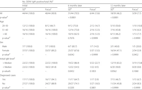
It was estimated that anti-DENV IgM became undetect-able after 338.3 days (95% CI 279.7–446.9) by the SD ELISA and 175.7 days (95% CI 121.9–221.1) by the Focus ELISA.
Results from the SD RDTs were interpreted as defin-itely positive in 63.6%, 18.2 and 5.1% initially, at 6 months and 12 months, respectively (Table2). If very faint bands on RDTs were also interpreted as positive according to manufacturer’s instruction, anti-DENV IgM were detectable in 86.4%, 68.2 and 35.9% initial, at 6 months, and 12 months (Table2).
Discussion
This study investigated the persistence of anti-DENV IgM among adults who were infected in a severe dengue epi-demic in Tainan in 2015. The main circulating serotype was DENV2 and approximately 90% of the acute patients suffered from primary DENV infection [19]. The results demonstrated that there was a discordance in the length of anti-DENV IgM detection by commonly used commer-cial ELISA tests. The estimated duration that anti-DENV IgM became undetectable by the Focus ELISA was 175.
[image:4.595.57.538.100.405.2]7 days after symptom onset in adults, which was very similar to the results reported by Prince et al., though the age distribution in their study was unknown [15]. In con-trast, regression analysis showed that the anti-DENV IgM persisted for almost 1 year by the SD ELISA, and nearly half of the participants had detectable anti-DENV IgM more than 1 year post infection. Although the discordant outcomes were a surprised finding, at this moment, we don’t know the reasons contributing to the difference in readouts. Although detection of anti-DENV IgM in one single specimen does not provide a definite diagnosis of dengue, it has been widely used, especially when blood samples are taken more than 5 days after disease onset and in non-endemic countries [4, 6]. Furthermore, it is usually difficult to obtain second convalescent specimens. However, if there is a large dengue epidemic in the previ-ous year, the recognition of anti-DENV IgM in symptom-atic patients without virology confirmation may represent anti-DENV IgM persistence from the previous year rather than an acute infection; thus alternative etiologies should be pursued and clinical management consequently be al-tered. Also, the interpretation of faint anti-DENV IgM Table 1Detection of anti-DENV IgM by two commercial ELISA tests at initial survey, 6 and 12 months
No. DENV IgM positive/total (%)a
Initial 6 months later 12 months later
SDb Focusc SDb Focusc SDb Focusc
All 44/44 (100.0) 40/44 (90.9) 31/44 (70.5) 6/44 (13.6) 18/39 (46.2) 3/39 (7.7)
p-valued < 0.0001 < 0.0001
Age
20–50 12/12 (100.0) 8/12 (66.7) 9/12 (75.0) 2/12 (16.7) 5/10 (50.0) 1/10 (10.0)
51–60 16/16 (100.0) 16/16 (100.0) 12/16 (75.0) 2/16 (12.5) 7/16 (43.8) 1/16 (6.3)
> 60 16/16 (100.0) 16/16 (100.0) 10/16 (62.5) 2/16 (12.5) 6/13 (46.2) 1/13 (7.7)
p-valued – – 0.7676 > 0.9999 > 0.9999 > 0.9999
Sex
Male 7/7 (100.0) 7/7 (100.0) 6/7 (85.7) 1/7 (14.3) 2/5 (40.0) 1/5 (20.0)
Female 37/37 (100.0) 33/37 (89.2) 25/37 (67.6) 5/37 (13.5) 16/34 (47.1) 2/34 (5.9)
p-valued – – 0.6542 > 0.9999 > 0.9999 0.3452
Initial IgM levele
> Median 22/22 (100.0) 22/22 (100.0) 19/22 (86.4) 5/22 (22.7) 12/19 (63.2) 3/19 (15.8)
< Median 22/22 (100.0) 18/22 (81.8) 12/22 (54.5) 1/22 (4.5) 6/20 (30.0) 0/20 (0.0)
p-value§ – – 0.0452 0.1853 0.0562 0.1060
Diagnosed cases
Yes 17/17 (100.0) 16/17 (94.1) 11/17 (64.7) 1/17 (5.9) 7/15 (46.7) 1/15 (6.7)
No 27/27 (100.0) 24/27 (88.9) 20/27 (74.1) 5/27 (18.5) 11/24 (45.8) 2/24 (8.3)
p-valued – – 0.5205 0.3801 > 0.9999 > 0.9999
a
Number of positive anti-DENV IgM/number of specimens tested (percentage) b
ELISA kits manufactured by Standard Diagnostics c
ELISA kits manufactured by Focus Diagnostics d
The difference in the detection rate among different groups were examined using Fisher’s exact tests e
bands on RDTs after a large dengue outbreak in the pre-ceding year should be more conservative. In addition to patient care, serological surveillance by testing of those who link to the confirmed cases epidemiologically to search for additional people with both symptomatic and asymptomatic DENV infection has also been advocated in
non-endemic areas to break the transmission cycles and contain epidemics in the early stages [20]. Nevertheless, the value of anti-DENV IgM for outbreak investigation and surveillance may also be complicated or even hin-dered if a large epidemic has occurred in the preceding year. Our study also provides important new information to the field of dengue research, especially for scholars who commonly use these tests to interpret and address their study findings.
[image:5.595.60.537.84.475.2]Previous studies barely examined anti-DENV IgM per-sistence among people asymptomatically infected with DENV because of the difficulty in knowing when the ini-tial infection occurred. In our study, we found that the anti-DENV IgM persistence did not differ between those who reported being diagnosed with DENV infection in 2015 and those who did not. In Taiwan, notification of Fig. 1Kinetic scatterplots and regression analyses of log-transformed mean optical density values at 450 nm (OD450) and mean index values (IV) for anti-DENV IgM measured by the SD ELISA and the Focus ELISA, respectively, over time in 17 people who were anti-DENV IgM positive initially defined by the SD ELISA and reported being diagnosed with DENV infection in 2015.aResults of the SD ELISA. The dashed line represents the mean cut-off OD value among different plates. (R-square: 0.5176; slope coefficient:−0.0037,p< 0.0001).bResult of the Focus ELISA. The dashed line represents the cut-off index value. (R-square: 0.5057; slope coefficient:−0.0044, p < 0.0001)
Table 2Detection of anti-DENV IgM by rapid tests from Standard Diagnostics at initial survey, 6 and 12 months
No. DENV IgM positive/total (%)a
Initial 6 months 12 months
Definitely positive 28/44 (63.6) 8/44 (18.2) 2/39 (5.1)
Very faint 10/44 (22.7) 22/44 (50.0) 12/39 (30.8)
Negative 6/44 (13.6) 14/44 (31.8) 25/39 (64.1)
a
[image:5.595.56.292.654.724.2]patients with suspected DENV infection by physicians is mandatory; one study which investigated the quality of the notification system found that the overall reporting rate was 86.6% during 2006–2007 [20, 21]. In 2015, there was an extraordinarily high awareness of dengue and febrile illness among healthcare professionals and general public due to pervasive dengue educational cam-paigns during this unprecedented dengue epidemic in Tainan; RDTs were also widely used in patients with any symptoms suspicious of DENV infection [22]. Therefore, the under-reporting of the symptomatic cases has been very low in Taiwan and probably was even lower in 2015. Thus, the majority of anti-DENV IgM positive vol-unteers without dengue diagnosis in our study could be characterized as asymptomatic infections. Our study thus provides evidence that the persistence of anti-DENV IgM does not differ between asymptomatic and asymptomatic infections.
In our study, it seems that the SD ELISA has higher anti-DENV IgM positive rate than the Focus ELISA. The performance of the two ELISAs has been evaluated pre-viously [23, 24]. One study demonstrated that the two tests were similar in sensitivity [23], while Domingo et al. showed that the Focus IgM ELISA seemed to have higher sensitivities than the SD ELISA [24]. However in that study, the tests were performed by different labora-tories, some of which might be less experienced in den-gue diagnosis and thus their results might not be directly comparable [24]. Additionally, Domingo et al. examined samples from patients infected with DENV1 and DENV3 [24] while the main circulating serotype in our study was DENV2 [19], which may also partially ex-plain the difference. We used SD ELISA in our original serosurvey to define IgM positivity and the aim of this follow-up study was to investigate IgM persistence, not to evaluate the sensitivity of the two tests. To compare their sensitivity directly, all the anti-DENV IgM-negative samples defined by the SD ELISA in the original serosur-vey should be tested by Focus ELISA simultaneously. As for the specificity, the SD ELISA has very high specificity and shows no cross-reactivity with other similar flavi-viruses including Japanese encephalitis virus and West Nile virus [23]. Although false positive results may be present in people with malaria or previous DENV infec-tion [23], Taiwan was certified by the World Health Organization (WHO) as“malaria-free” in 1965 [25] and the majority of DENV-infected people in this epidemic suffered from primary infection [19]. Furthermore, al-though the current ELISA kits may have a concern of cross-reactivity to Zika virus infection, there are no local Zika cases reported in Taiwan as of today. Consequently, the false positive rate in our study should be low.
The major strength of this study is that the samples were collected in a non-endemic area and there was no
dengue outbreak in the following year, 2016, despite ex-tensive surveillance efforts. We therefore can be sure that detection of anti-DENV IgM in the follow-up samples was a result of IgM persistence rather than re-infection of DENV during follow-up. However, there were also several limitations in our study. Firstly, we assumed that the mid-dle date of the self-reported month of diagnosis was the symptom onset for each subject. Although the assumed onset dates were based on individual’s recall and thus sub-ject to error, this should not bias our results given the time span considered in the regression analysis. Secondly, al-though there might be misclassification using self-reporting of history of dengue diagnosis as a surrogate to define symptomatic and asymptomatic infection, the mag-nitude of the bias should be low as previously mentioned. Thirdly, most of the study subjects were adults suffered from primary DENV2 infection, so our results may not be generalized to children living in endemic regions and/or other serotypes. Further studies which include a wider age span, more dengue serotypes, and secondary infection are recommended. Fourthly, the sample size in this study was small, especially for the regression analysis. Finally, al-though the current two ELISA assays are widely utilized for acute dengue diagnosis, we do not know which assay is better for persistent anti-DENV IgM measurement, since currently there is no gold standard test for persistent anti-DENV IgM. As such, despite our findings presume to be very important for dengue diagnosis, we are still unable to affirm whether a person is anti-DENV IgM positive or not if his or her anti-DENV IgM is detectable by SD ELISA kit but undetectable by Focus ELISA kit. Subse-quently, the biology of immune response after certain period of dengue virus infection could be far more com-plex and warrants further investigation.
Conclusions
It was found that anti-DENV IgM detected by some current commercial ELISA kits or RDTs persisted much longer than previously thought, which may complicate diagnosis of dengue and surveillance efforts in the follow-ing year after a large dengue epidemic. Therefore, diagno-sis of dengue using anti-DENV IgM following large dengue outbreaks should be more conservative. Our study also demonstrated that the duration of anti-DENV IgM persistence did not differ between symptomatically and asymptomatically infected individuals, which has seldom been reported in the literature.
Abbreviations
DENV:Dengue virus; ELISA: Enzyme-linked immunosorbent assay; RDT: Rapid diagnostic test
Acknowledgements
Funding
This work was supported by the Taiwan Ministry of Science and Technology (grant no. MOST 105–2634-B-006-001, MOST 103–2320 - B - 006 - 030 - MY3, NSC 105–2314-B-006 -004) and Taiwan Ministry of Health and welfare awarded to National Mosquito-Borne Diseases Control Research Center in National Health Research Institutes (MOHW105-TDU- M-212-000006).
Availability of data and materials
The datasets used and/or analyzed during the current study are available from the corresponding author on reasonable request.
Authors’contributions
YWC designed the study and wrote the first draft of the manuscript; ZHL, FCT, and TCH handled the patient specimens and performed the experiments. HRG, NKY, and WCK assisted in IRB approval and statistical analysis; GCP designed the overall study, assisted to sample analysis as well as edited the final manuscript. All authors read and approved the final manuscript.
Ethics approval and consent to participate
The study was approved by the Institutional Review Board of National Cheng Kung University Hospital (approval no. A-ER-104-386 and B-ER-104-178) and all participants were enrolled to the study after signing informed consent.
Consent for publication
Consent for the publication of identifying images or other personal or clinical details of participants that compromise anonymity is“Not applicable”. All authors have read the manuscript and approve the manuscript to be published.
Competing interests
All authors declare that they have no competing interests. However, Guey Chuen Perng is a member of editorial board of BMC Infectious Diseases.
Publisher’s Note
Springer Nature remains neutral with regard to jurisdictional claims in published maps and institutional affiliations.
Author details
1
Department of Public Health, College of Medicine, National Cheng Kung University, Tainan, Taiwan.2Department of Occupational and Environmental
Medicine, National Cheng Kung University Hospital, College of Medicine, National Cheng Kung University, Tainan, Taiwan.3Department of
Environmental and Occupational Health, College of Medicine, National Cheng Kung University, Tainan, Taiwan.4Department of Nursing, National
Taipei University of Nursing and Health Sciences, Taipei, Taiwan.5National Institute of Infectious Diseases and Vaccinology, National Health Research Institutes, Tainan, Taiwan.6Institute of Basic Medical Sciences, College of Medicine, National Cheng Kung University, Tainan, Taiwan.7Department of
Nursing, College of Medicine, National Cheng Kung University, Tainan, Taiwan.8Department of Medicine, College of Medicine, National Cheng Kung
University, Tainan, Taiwan.9Department of Microbiology and Immunology, College of Medicine, National Cheng Kung University, Tainan, Taiwan.
Received: 15 August 2017 Accepted: 22 March 2018
References
1. Guzman MG, Harris E. Dengue. Lancet. 2015;385:453–65.
2. Brady OJ, Gething PW, Bhatt S, Messina JP, Brownstein JS, Hoen AG, Moyes CL, Farlow AW, Scott TW, Hay SI. Refining the global spatial limits of dengue virus transmission by evidence-based consensus. PLoS Negl Trop Dis. 2012;6:e1760.
3. Stanaway JD, Shepard DS, Undurraga EA, Halasa YA, Coffeng LE, Brady OJ, Hay SI, Bedi N, Bensenor IM, Castaneda-Orjuela CA, et al. The global burden of dengue: an analysis from the global burden of disease study 2013. Lancet Infect Dis. 2016;16:712–23.
4. WHO: Dengue: Guidelines for Diagnosis, Treatment, Prevention, and Control. New edition. Geneva: World Health Organization; 2009.
5. Peeling RW, Artsob H, Pelegrino JL, Buchy P, Cardosa MJ, Devi S, Enria DA, Farrar J, Gubler DJ, Guzman MG, et al. Evaluation of diagnostic tests: dengue. Nat Rev Microbiol. 2010;8:S30–8.
6. Tang KF, Ooi EE. Diagnosis of dengue: an update. Expert Rev Anti-Infect Ther. 2012;10:895–907.
7. Koraka P, Suharti C, Setiati TE, Mairuhu AT, Van Gorp E, Hack CE, Juffrie M, Sutaryo J, Van Der Meer GM, Groen J, et al. Kinetics of dengue virus-specific serum immunoglobulin classes and subclasses correlate with clinical outcome of infection. J Clin Microbiol. 2001;39:4332–8.
8. Innis BL, Nisalak A, Nimmannitya S, Kusalerdchariya S, Chongswasdi V, Suntayakorn S, Puttisri P, Hoke CH. An enzyme-linked immunosorbent assay to characterize dengue infections where dengue and Japanese encephalitis co-circulate. Am J Trop Med Hyg. 1989;40:418–27.
9. Kuno G, Gomez I, Gubler DJ. An ELISA procedure for the diagnosis of dengue infections. J Virol Methods. 1991;33:101–13.
10. Nogueira RM, Miagostovich MP, Cavalcanti SM, Marzochi KB, Schatzmayr HG. Levels of IgM antibodies against dengue virus in Rio de Janeiro. Brazil Res Virol. 1992;143:423–7.
11. Vaughn DW, Green S, Kalayanarooj S, Innis BL, Nimmannitya S, Suntayakorn S, Rothman AL, Ennis FA, Nisalak A. Dengue in the early febrile phase: viremia and antibody responses. J Infect Dis. 1997;176:322–30. 12. Guzman MG, Halstead SB, Artsob H, Buchy P, Farrar J, Gubler DJ,
Hunsperger E, Kroeger A, Margolis HS, Martinez E, et al. Dengue: a continuing global threat. Nat Rev Microbiol. 2010;8:S7–16. 13. PATHO. Dengue and Dengue Hemorrhagic Fever in The Americas:
Guidelines for Prevention and Control. Wahshington, DC: Pan American Health Organization; 1994. (Scientific Publicaiton No. 548).
14. Shu PY, Huang JH. Current advances in dengue diagnosis. Clin Diagn Lab Immunol. 2004;11:642–50.
15. Prince HE, Matud JL. Estimation of dengue virus IgM persistence using regression analysis. Clin Vaccine Immunol. 2011;18:2183–5.
16. Hunsperger EA, Yoksan S, Buchy P, Nguyen VC, Sekaran SD, Enria DA, Vazquez S, Cartozian E, Pelegrino JL, Artsob H, et al. Evaluation of commercially available diagnostic tests for the detection of dengue virus NS1 antigen and anti-dengue virus IgM antibody. PLoS Negl Trop Dis. 2014;8:e3171.
17. Blacksell SD, Jarman RG, Bailey MS, Tanganuchitcharnchai A, Jenjaroen K, Gibbons RV, Paris DH, Premaratna R, de Silva HJ, Lalloo DG, et al. Evaluation of six commercial point-of-care tests for diagnosis of acute dengue infections: the need for combining NS1 antigen and IgM/IgG antibody detection to achieve acceptable levels of accuracy. Clin Vaccine Immunol. 2011;18:2095–101.
18. Chen CM, Kuo HW, Liu DP. Dengue fatal cases in Taiwan: a preliminary study for 2015 outbreak (in Chinese). Taiwan Epidemiol. Bull. 2015;31:605–7. 19. Tsai HP, Tsai YY, Lin IT, Kuo PH, Chang KC, Chen JC, Ko WC, Wang JR.
Validation and application of a commercial quantitative real-time reverse transcriptase-PCR assay in investigation of a large dengue virus outbreak in southern Taiwan. PLoS Negl Trop Dis. 2016;10:e0005036.
20. Kao JH, Chen CD, Tiger Li ZR, Chan TC, Tung TH, Chu YH, Cheng HY, Liu JW, Shih FY, Shu PY, et al. The critical role of early dengue surveillance and limitations of clinical reporting - implications for non-endemic countries. PLoS One. 2016;11:e0160230.
21. Liao SY, Yen JJ, Huang CC. Quality of the dengue surveillance system in Taiwan (in Chinese). Taiwan Epidemiol. Bull. 2010;26:324–9.
22. Shih HI, Hsu HC, Wu CJ, Lin CH, Chang CM, Tu YF, Hsieh CC, Chi CH, Sung TC. Applications of a rapid and sensitive dengue DUO rapid
Immunochromatographic test kit as a diagnostic strategy during a dengue type 2 epidemic in an Urban City. PLoS One. 2016;11:e0158437. 23. Hunsperger EA, Yoksan S, Buchy P, Nguyen VC, Sekaran SD, Enria DA,
Pelegrino JL, Vazquez S, Artsob H, Drebot M, et al. Evaluation of commercially available anti-dengue virus immunoglobulin M tests. Emerg Infect Dis. 2009;15:436–40.
24. Domingo C, Alves MJ, de Ory F, Teichmann A, Schmitz H, Muller R, Niedrig M. International external quality control assessment for the serological diagnosis of dengue infections. BMC Infect Dis. 2015;15:167. 25. Yip K. Malaria eradication: the Taiwan experience. Parassitologia.