R E S E A R C H
Open Access
MiniCD4 protein resistance mutations affect
binding to the HIV-1 gp120 CD4 binding site and
decrease entry efficiency
Katrijn Grupping
1, Philippe Selhorst
1, Johan Michiels
1, Katleen Vereecken
1, Leo Heyndrickx
1, Pascal Kessler
2,
Guido Vanham
1,3, Loïc Martin
2and Kevin K Ariën
1*Abstract
Background:Binding of the viral envelope protein (Env), and particularly of its gp120 subunit, to the cellular CD4 receptor is the first essential step of the HIV-1 entry process. The CD4 binding site (CD4bs) of gp120, and especially a recessed cavity occupied by the CD4 Phe43 residue, are known to be highly conserved among the different circulating subtypes and therefore constitute particularly interesting targets for vaccine and drug design. The miniCD4 proteins are a promising class of CD4bs inhibitors. Studying virus evolution under pressure of CD4bs inhibitors could provide insight on the gp120-CD4 interaction and viral entry.
Results:The present study reports on the resistance induction of two subtype B HIV-1 against the most active miniCD4, M48U1, and its ancestor, M48, and how these mutated positions affect CD4bs recognition, entry efficiency, and sensitivity to other CD4bs inhibitors. Resistance against M48U1 was always associated with S375R/N substitution in both BaL and SF162; M48 resistance was associated with D474N substitution in SF162 and with H105Y
substitution in BaL. In addition, some other mutations at position V255 and G471 were of importance for SF162 resistant viruses. Except for 474, all of these mutated positions are conserved, and introducing them into an SF162 Env expressing infectious molecular clone (pBRNL4.3 SF162) resulted in decreased entry efficiency. Furthermore, resistant mutants showed at least some cross-resistance towards other CD4bs inhibitors, the V3 monoclonal antibody 447-52D and some even against the monoclonal antibody 17b, of which the epitope overlaps the co-receptor binding site.
Conclusions:The mutations H105Y, V255M, S375R/N, G471R/E, and D474N are found to be involved in resistance
towards M48 and M48U1. All mutated positions are part of, or in close proximity to, the CD4bs; most are highly conserved, and all have an impact on the entry efficiency, suggesting their importance for optimal virus infectivity.
Keywords:HIV-1, Resistance, Entry inhibitors, CD4 binding site, Entry efficiency
Background
The entry process of the Human Immunodeficiency Virus type 1 (HIV-1) into host cells is an important tar-get for the development of preventive vaccines and microbicides. HIV-1 entry is a multi-step process that is mediated by the envelope surface glycoprotein gp120 and the transmembrane glycoprotein gp41 [1,2]. These two subunits constitute a functional heterotrimeric
molecule that enables the virus to interact with its pri-mary receptor, CD4 [3–10]. The gp120-CD4 interaction triggers a conformational change that allows binding of gp120 to its co-receptor, most frequently CCR5 or CXCR4, and induces refolding of gp41, finally resulting in fusion with the target cell membrane [11,12].
Three distinct gp120 core structures were revealed: (1) a heavily glycosylated outer domain that is exposed to the surface of the trimer, (2) an inner domain that inter-acts with the gp41 subunit, and (3) a four-stranded anti-parallel β-sheet (i.e. the bridging sheet) connecting the outer and inner domains. The CD4bs is formed at the
* Correspondence:[email protected] 1
Virology Unit, Department of Biomedical Sciences, Institute of Tropical Medicine of Antwerp, Antwerp, Belgium
Full list of author information is available at the end of the article
interface of these three domains and buries a large sur-face of approximately 800 Å2. However, the area of ac-tual contact between gp120 and CD4 is much smaller because of cavities formed at the interface. One of these cavities is plugged by the aromatic ring of phenylalanine 43 of the CD4 receptor and, as a consequence, named the Phe43-cavity [11]. This important region, at the interface of the outer and inner domains and the bridg-ing sheet, is well-conserved among the different HIV-1 subtypes and is crucial in the lifecycle of the virus [13].
Because of its high genetic and functional conserva-tion, the CD4bs, and in particular the Phe43-cavity, is considered an extremely interesting target for the devel-opment of HIV-1 entry inhibitors [11,13–16].
Several potent CD4bs inhibitors such as soluble CD4 (sCD4), BMS-378806, NBD-556, some llama heavy-chain antibodies (A12, D7, and C8), and various CD4bs anti-bodies have already been described in literature [17-24]. The best known broad neutralizing monoclonal antibody (mAb) is IgG1b12, which can neutralize 75% of all clade B primary viruses and 40% of all known HIV-1 isolates
in vitro. It has also been shown to protect macaques
from infection [25–29]. Furthermore, recent discoveries have led to some new potent CD4bs mAbs such as HJ16, VRC01, VRC02, VRC03, NIH45-46, 8ANC131, and 12A12 [30–32].
CD4 mimetic compounds, also called miniCD4s, con-stitute a very promising class of CD4bs inhibitors, e.g. M48 and M48U1 [23,33–38]. Upon binding with HIV-1 and similarly to the cellular CD4 receptor, M48 and M48U1 induce conformational changes in the gp120 architecture thereby exposing masked epitopes on the envelope protein. Furthermore, they were shown to have antiretroviral activities in the nanomolar range [33,35]. Besides their potent antiviral activity, these CD4 mimetic miniproteins also have very interesting physico-chemical characteristics such as their small size (27 amino acids), stable conformation in denaturing conditions such as acidic pH and high temperatures, and relative resistance towards proteolytic degradation [33]. Considering the va-ginal environment, it is clear that these characteristics are extremely relevant for microbicide candidates [39]. The most potent miniCD4, M48U1, derived from its an-cestor M48, was created by adding a flexible cyclohexyl-methoxy group in the para-position of the phenylalanine at position 23 of M48, a residue mimicking Phe43 of CD4. This results in a miniCD4 with high affinity for the conserved and vulnerable Phe43-cavity.
In this study, we investigated the evolution of HIV-1 under miniCD4 pressure to get a better understanding of the miniCD4-virus interaction. To this end, resistance induction in two subtype B viruses was performed; and the genotype, as well as the phenotype, of these viruses was characterized.
Results
In vitroresistance induction and genotyping
Resistance was induced against M48 and M48U1 by ex-posing the CCR5-tropic subtype B HIV-1 viruses BaL and SF162 to increasing concentrations of the miniCD4 mimetic proteins M48 or M48U1 in PHA/IL-2 stimu-lated donor peripheral blood mononuclear cells (PBMCs). In addition, resistance was also induced against an equipotent combination of M48 and M48U1.
In general, resistance was rapidly acquired (see Table 1), which reflects the flexible nature of the en-velope glycoprotein and confirms the low genetic barrier for development of resistance towards most entry inhibitors.
Resistance induction was repeated in two independent experiments (referred to as viruses‘a and b’). Gp120 se-quencing was done at the time when the resistance level was at least 100x above the IC50 of M48 or M48U1 and
for most virus cultures also at intermediate time points (Table 1). Sequencing indicated that the serine at pos-ition 375, part of the constant region 3 of gp120 (C3) and situated in the outer domain, was altered in all M48U1 resistant viruses. An arginine was found in both M48U1 resistant BaL viruses and in one of the M48U1 resistant SF162 viruses (rM48U1SF162_a), whereas an asparagine was observed in the second M48U1SF162 re-sistant virus (rM48U1SF162_b) (Table 1).
In addition to the S375R/N mutation, both rM48U1SF162 viruses displayed mutations at other amino acid positions. Both G335 and G471 were mutated into an arginine in rM48U1SF162_a after the S375R mutation was induced, whereas V255M and L494V mutations were observed in rM48U1SF162_b prior to the appearance of S375N. The valine at position 255 is, similar to S375, a highly conserved residue lining the Phe43-cavity. Both V255 and G471 are part of C2 and C5, respectively, and contribute to the outer domain, which makes up the largest part of the CD4bs [11,40]. Interestingly, virus rM48U1SF162_b had, besides the S375N and V255M amino acid changes, an additional mutation close to the gp120-gp41 cleavage site; i.e. L494V.
resistant to the combination of both miniproteins (rCombiSF162_b) revealed two additional mutations in the outer domain, namely G471E and L494V.
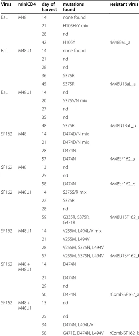
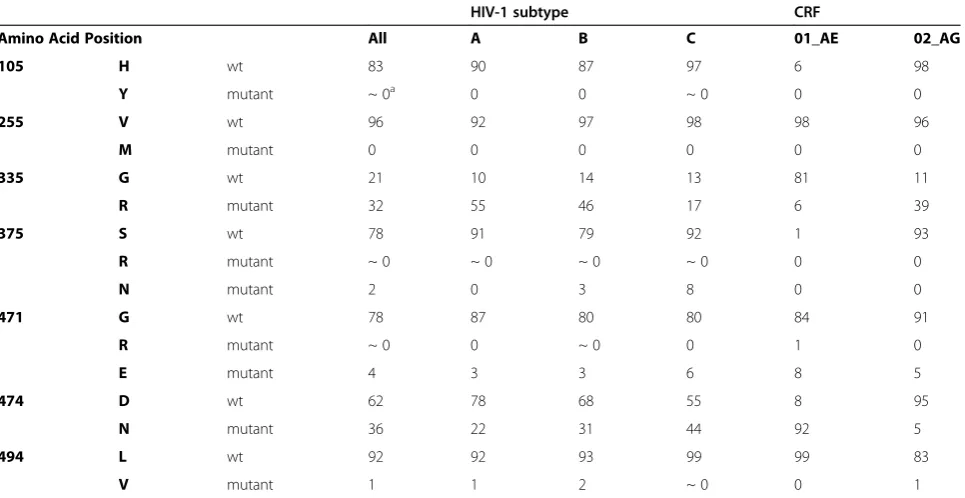
[image:3.595.55.288.111.674.2]In summary, all mutated positions except two, i.e. G335R and L494V are part of or in very close proximity to the CD4bs and particularly near to the Phe43-cavity (Figure 1). The resistance-associated mutations appear often as early as day 14 after the start of drug exposure (Table 1).
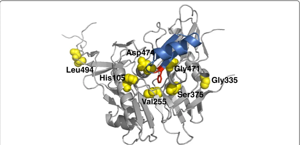
Comparative sequence analysis of 3045 HIV-1/SIV Env sequences from the Los Alamos HIV Sequence database (accessed September 2011) showed that most of the mutated positions that we identified are conserved across the most common clades (Table 2). At least 79% of the clade A, B, C and CRF02_AG sequences checked show a histidine at position 105, a valine at position 255, a serine at position 375, a glycine at position 471, and a leucine at position 494. Histidine at position 105 and serine at pos-ition 375 are less common in CRF01_AE Env sequences; however, the amino acids found in the M48 and M48U1 resistant viruses at these positions (i.e. Y and R, respect-ively) were not found in CRF01_AE isolates. In general, only 7 (out of 3045 HIV-1/SIV sequences, Table 2) natur-ally occurring sequences were found carrying the H105Y, V255M, or S375R mutations, suggesting that these muta-tions are extremely rare. Furthermore, asparagine, glu-tamic acid, and valine at positions 375, 471, and 494, respectively, were only observed in few cases (between 0% to 8% of sequences, Table 2). In contrast, a glycine to ar-ginine substitution at position 335 and an aspartic acid to asparagine substitution at position 474 appear quite com-mon acom-mong the different subtypes (Table 2).
Binding experiments with SF162 gp120 mutants
To explore the contribution of the different amino acid mutations in the interaction between gp120 and the miniCD4 proteins M48 and M48U1, site-directed mutants of SF162envwere generated carrying one of the RAMs: H105Y, V255M, G335R, S375R, S375N, G471R, D474N or L494V (see Table 3 for an overview of SDMs). These gp120 mutants were tested in binding assays to as-sess the interaction with the M48 and M48U1 miniCD4 proteins using fluorescence polarization. Figure 2 shows the fold increase in Kd for the different mutants in com-parison with the native SF162 gp120.
Mutations G335R, S375N, D474N and L494V displayed Kd values comparable to SF162 wild type (WT) gp120 and therefore did not have a significant impact on the af-finity of HIV-1 Env for both M48 and M48U1, as reflected in a fold Kd close to 1 (Figure 2A). Residues G335 and L494 are located away from the CD4bs (Figure 1) and are accompanied by either S375R/N and/ or V255M. Therefore, it is not surprising that they do not influence the affinity for miniCD4 protein binding. We
Table 1 Resistance development in virus isolates exposed to increasing amounts of miniCD4
Virus miniCD4 day of harvest
mutations found
resistant virus
BaL M48 14 none found
21 H105H/Y mix
28 nd
42 H105Y rM48BaL_a
BaL M48U1 14 none found
21 nd
28 nd
36 S375R
45 S375R rM48U1BaL_a
BaL M48U1 14 nd
20 S375S/N mix
27 nd
35 nd
48 S375R rM48U1BaL_b
SF162 M48 14 D474D/N mix
21 D474D/N mix
28 D474N
57 D474N rM48SF162_a
SF162 M48 13 nd
25 nd
58 D474N rM48SF162_b
SF162 M48U1 14 S375S/R mix
22 S375R
28 nd
59 G335R, S375R,
G471R
rM48U1SF162_a
SF162 M48U1 14 V255M, L494L/V mix
21 V255M, L494V
28 V255M, S375N, L494V
57 V255M, S375N, L494V rM48U1SF162_b
SF162 M48 + M48U1
14 D474N
21 D474N
29 nd
50 D474N rCombiSF162_a
SF162 M48 + M48U1
13 nd
25 nd
34 D474N, L494L/V
58 G471E, D474N, L494V rCombiSF162_b
identified D474N as single resistance-associated mutation in the viruses rM48SF162_a, rM48SF162_b, and rCom-biSF162_a. D474N is situated at the edge of the inner do-main, close to the CD4bs and known to be a direct CD4 and miniCD4 contact residue [36,41]. Nevertheless, loss of the aspartic negative charge does not affect the binding affinity of M48, M48U1 or sCD4 to gp120. This is expected because Asp474 faces the hydrophobic part of miniCD4D-Pro21 and the side chain of CD4 Gln25, and is located too far away for a hydrogen bond.
On the contrary, the mutations H105Y, V255M, S375R, and G471R, all located close to or in the CD4bs (Figure 1), have a clear impact on the Kd values. Envelopes carrying these mutations lost affinity for the M48 miniCD4 protein, whereas only the arginine on position 375 resulted in an extreme reduction in affinity for M48U1, the most potent miniCD4 protein specifically targeting the Phe43-cavity (Figure2A). Of note, the S375N mutation did not signifi-cantly impact the binding affinity.
To further validate these observations, the H105Y, V255M, S375R/N, G471R and D474N mutant SF162 envelopes were also evaluated in the context of replica-tion competent pBRNL4.3 molecular clones (Figure 2B). With the exception of H105Y, results correlated with binding affinity studies and confirmed the importance of V255M and S375R/N in resistance towards M48 and M48U1. Whereas binding of M48 to H105Y mutant
monomer gp120 was affected (approx. 40-fold increase in Kd), the impact of this mutation was less apparent in the context of the pBRNL4.3 molecular clone, where gp120 is in its natural trimeric conformation. Similarly, the G471R mutation resulted in small increases in Kd, but had no apparent effect in the context of replication competent HIV. Because the infectivity of the V255M substitution was dramatically reduced, no IC50 could be
calculated.
Furthermore, S375R and D474N mutations were intro-duced in different gp120 backgrounds (a primary subtype C strain VI829 and a primary CRF02_AG strain VI1090; Figure 2C), again confirming the pivotal role of the 375 residue in the interaction of M48U1 with envelope. All mutant S375R pseudoviruses had a dramatic increase in IC50 for the miniCD4 M48U1 compared to wild type
pseudovirus IC50values (Figure 2C).
Of note, although D474N was found as resistance-associated mutation in 4 out of 5 viruses made resistant against M48, this residue did not significantly affect the binding (Figure 2A), nor the sensitivity to inhibition by M48 or M48U1 (Figure 2B and 2C).
Impact of M48 and M48U1 resistance associated mutants on entry efficiency
The observed impact of some mutations on the binding affinities, together with the conserved nature of most
Gly335
Leu494
Ser375
His105
Asp474
Gly471
[image:4.595.57.541.89.322.2]Val255
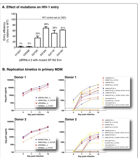
mutated residues, raises the question whether these mutations influence the entry efficiency. To answer this question, mutations H105Y, V255M, S375R, S375N, G471R, and D474N were introduced in the SF162 WT
envgene using site-directed mutagenesis (Table 3). About 30% of all sequences have an Arg residue at pos-ition G335; and given that the G355R mutant does not im-pact on the miniCD4’s affinity, this mutant was excluded from further analysis. Also the L494V mutant, although more conserved, but located away from the miniCD4 inter-action site and not affecting CD4 binding, was excluded. Al-though a third of the known gp120 sequences share an Asn474 residue, it was the only mutation found to be
associated with M48 resistance in SF162, and therefore it was tested alongside the H105Y, V255M, S375R/N and G471R mutants.
Briefly, mutant envelopes were cloned in a delta-Env pBRNL4.3 molecular clone. Reverse transcriptase (RT) activity of each mutant virus was determined, and equal amounts (40 pg of RT) were used to infect TZM-bl cells. The entry efficiency of each mutant was measured rela-tive to the pBRNL4.3 clone bearing the WT SF162 envelope.
[image:5.595.58.541.101.350.2]Two mutations, H105Y and V255M, had a dramatic negative effect on entry efficiency, resulting in respectively 1% and 2% of infection relative to WT virus (Figure 3A). As mentioned before, these two residues are highly conserved among the most prevalent subtypes (Tyr105 in only 3 out of 3045 sequences and no Met255), are in close proximity to the CD4bs, and substantially affect the binding affinities for M48 (Figure 2A). Nevertheless, the H105Y mutation was only observed in the BaL virus and not in SF162, sug-gesting that this mutation may be BaL-specific. The mo-lecular clone with an arginine instead of a serine at position 375 had an entry efficiency of only 33% relative to WT virus. The other mutated positions, S375N, G471R, and D474N affected entry efficiency to a lesser extent, with mean per-centages of infection relative to the WT clone ranging from 50 to 68%. However, an alanine at position 474 was described previously to make a BaL pseudovirus completely non-infectious, where the same mutation introduced in a
Table 2 Prevalence of observed mutations in HIV-1 sequences
HIV-1 subtype CRF
Amino Acid Position All A B C 01_AE 02_AG
105 H wt 83 90 87 97 6 98
Y mutant ~ 0a 0 0 ~ 0 0 0
255 V wt 96 92 97 98 98 96
M mutant 0 0 0 0 0 0
335 G wt 21 10 14 13 81 11
R mutant 32 55 46 17 6 39
375 S wt 78 91 79 92 1 93
R mutant ~ 0 ~ 0 ~ 0 ~ 0 0 0
N mutant 2 0 3 8 0 0
471 G wt 78 87 80 80 84 91
R mutant ~ 0 0 ~ 0 0 1 0
E mutant 4 3 3 6 8 5
474 D wt 62 78 68 55 8 95
N mutant 36 22 31 44 92 5
494 L wt 92 92 93 99 99 83
V mutant 1 1 2 ~ 0 0 1
Prevalence (in percentage) of certain amino acids for the most prevalent HIV-1 subtypes at positions mutated in the resistant viruses is given. All: 3045 HIV-1/SIV Env sequences (Los Alamos National Lab data bank 2011); A, B and C correspond to HIV-1 subtypes. Number of Env sequences: subtype A = 186; subtype B = 1001;
subtype C = 756; CRF01_AE = 186; CRF02_AG = 100.a
~ 0 means less than 0.3%.
Table 3 Summary of site-directed mutants
SF162 gp120 Pseudovirus clones
Monomer gp120 for binding studies
pBRNL4.3 clones
SF162 BaL VI829 VI1090
H105Y H105Y
V255M V255M
G335R
S375R S375R S375R S375R S375R
S375N S375N
G471R G471R
[image:5.595.56.291.579.731.2]YU-2 pseudovirus did not have any effect on the infectivity [42].
Together, these observations suggest that (1) position 474 could be of importance for optimal virus infectivity, and (2) the introduced mutations are context-dependent, i.e. the envelop environment is of importance for the phenotype.
Viral growth kinetics on CD4 negative HOS cells and primary monocyte-derived macrophages (MDM)
Viruses adapted to replicate in CD4 negative cells and in cells expressing low levels of CD4 have been described in literature [43]. Blockage of the CD4 receptor using e.g. CD4 mimetics could skew viruses towards a phenotype of less CD4-dependency, which would theoretically offer the virus the opportunity to replicate in a very diverse subset of cells. To address this, we investigated the viral growth kinetics on a CD4 negative HOS cell line and on primary monocyte-derived macrophages (MDM). MDM express low levels of surface CD4 and represent anin vivotarget cell population for HIV replication.
All WT and resistant viruses were able to grow in HOS CD4+ CCR5+ cells, whereas none of the viruses replicated in HOS CD4- CCR5+ cells (data not shown). Because MDM are a more representative model, we determined the viral growth in this cell type. MDM were infected with WT and resistant viruses at a multiplicity of infection (MOI) of 10-3and cultures were maintained for 15 days. Viral replica-tion was monitored at different time points using Gag p24 production as a measure of viral growth. Figure 3B shows the results from two independent blood donors. Overall, H105YV255MG335
R
S375RS375NG47 1R
D47 4N
L494V 0
10 20 30 40 50 1000 2000
M48 M48U1
Mutant monomer gp120
F
o
ld
in
c
re
a
s
e
in
K
d
A.
Binding affinities of miniCD4 proteinsBaL S37
5R
VI8 29
S37 5R
VI1 090
S375 R
SF162 D474
N
BaL D47
4N 0
10 20 30 40 50 1000
7000 M48
M48U1
C.
Sensitivity of mutant pseudovirusesfor miniCD4 proteins
Pseudoviruses with mutant env
Fol
d
in
c
rea
s
e
in
IC
5
0
>
> >
H10 5Y
S37 5R
S37 5N
G471 R
D47 4N
0 20 40 60 80 1000 8000 15000
M48 M48U1
B.
Sensitivity of Env-mutant pBRNL4.3 molecularclones for miniCD4 proteins
F
o
ld
in
c
re
a
s
e
in
IC
5
0
> > >
Mutant SF162 Env
BaL & SF162: subtype B VI829: subtype C VI1090: CRF02_AG
Figure 2
A. Effect of mutations on HIV-1 entry
H105Y V255M S37
5R S375
N
G471R D47
4N
0 20 40 60 80 100 120
pBRNL4.3 with mutant SF162 Env
Ent
ry
e
ffic
ien
cy
(%
re
la
ti
v
e
to
W
T
) WT control set at 100%
1% 2%
33% 68%
50% 61%
B. Replication kinetics in primary MDM
Donor 1
4 7 11 15
100 1000 10000 100000 1000000
wtM48U1BaL_b rM48U1BaL_b_S375R wtM48BaL_a rM48BaL_a_H105Y
Gag p2
4
(pg/
m
l)
4 7 11 15
100 1000 10000 100000
wtM48SF162_b rM48SF162_b_D474N
wtCombiSF162_b
rCombiSF162_b_G471E, D474N, L494V wtM48U1SF162_b
rM48U1SF162_b_V255M, S375N, L494V wtM48SF162_a
rM48SF162_a_D474N
wtCombiSF162_a rCombiSF162_a_D474N wtM48U1SF162_a
rM48U1SF162_a_G335R, S375R, G471R
4 7 11 15
100 1000 10000 100000
4 7 11 15
100 1000 10000 100000 1000000
Day post infection Day post infection
Day post infection
Gag p2
4
(pg/
m
l)
wtM48U1BaL_b rM48U1BaL_b_S375R wtM48BaL_a rM48BaL_a_H105Y
Donor 2
Donor 1
Day post infection
Donor 2
wtM48SF162_b rM48SF162_b_D474N
wtCombiSF162_b
rCombiSF162_b_G471E, D474N, L494V wtM48U1SF162_b
rM48U1SF162_b_V255M, S375N, L494V wtM48SF162_a
rM48SF162_a_D474N
wtCombiSF162_a rCombiSF162_a_D474N wtM48U1SF162_a
[image:7.595.57.540.78.629.2]rM48U1SF162_a_G335R, S375R, G471R
the WT viruses replicated more efficiently in MDM than the resistant viruses (Figure 3B).
The mutant virus carrying H105Y showed a clear reduc-tion in infectivity (approximately 1 log) in MDM, confirm-ing the low entry efficiency observed in TZM-bl cells. More dramatic effects were seen with virus carrying V255M, S375N and L494V and the virus carrying D474N, with ex-tremely poor replication in MDM.
Together with the observations done in the HOS cells, this finding suggests that resistance induction towards the miniCD4 M48 and M48U1 is not substantially driv-ing the viruses to a CD4 independent phenotype.
Mapping the sensitivity towards HIV-1 inhibitors and antibodies
To identify the phenotype of the different resistant viruses, we examined potential cross-resistance against various entry inhibitors, including the CD4bs inhibitors s(oluble)CD4, the mAb IgG1b12 and the llama nanobody A12 (Figure 4A), the CD4-induced (CD4i) mAb 17b, and the V3-directed mAb 447-52D (Figure 4B), and the non-gp120 binders 4E10, 2F5 (directed against gp41 MPER) and dapivirine (TMC120; a non-nucleoside reverse transcriptase inhibitor) (Figure 4C) in a TZM-bl assay. The non-gp120 binding agents were included as controls, since they are not expected to be influenced by miniCD4 protein resistance. We used“fold change in resistance”as a measure of cross-resistance, defined as the IC50 (50% inhibitory
concentra-tion) of the resistant viruses divided by the IC50of the
con-trol viruses BaL and SF162, which were cultured in parallel with each resistance induction. As expected, there was no substantial difference in sensitivity of M48 and M48U1 re-sistant versus wild type viruses (fold change between 0.36 and 4.6) towards the control compounds 4E10, 2F5 and TMC120, confirming that the M48 and M48U1 RAMs in gp120 have no effect on the accessibility of the MPER in gp41, nor on RT function (Figure 4C).
However, cross-resistance towards several CD4bs inhibi-tors was demonstrated. The virus rM48BaL (H105Y) was not only resistant to M48, but also to M48U1 and sCD4. The other M48 resistant viruses, rM48SF162_a and rM48SF162_b (D474N), showed a different cross-resistance profile. Besides resistance against the two miniproteins and sCD4, a high level of resistance towards the nanobody A12 was seen. Not surprisingly, the two viruses rCombiSF162, possessing the D474N mutation show a similar resistance profile as both rM48SF162 viruses, i.e. resistant towards M48, M48U1, sCD4, and A12.
Both rM48U1BaL viruses (S375R) showed extensive cross-resistance to A12, no cross-resistance to M48; and further they were the only mutant viruses which remained completely sensitive to sCD4. The two M48U1 resistant SF162 viruses were cross-resistant to A12, but in contrast to the BaL mutants, displayed also resistance against M48
and sCD4. Finally, SF162 viruses with an asparagine on position 474 presented the highest loss in sensitivity to the mAb b12. Since side chain interactions on this position are involved in b12 binding, this result was not surprising [44].
We also tested whether miniCD4 protein induced re-sistance affected the sensitivity towards the CD4-induced (CD4i) monoclonal antibody 17b and confirmed that mutant viruses carrying D474N or S375R/N or V255M were resistant to inhibition by 17b (Figure 4B). This anti-body recognizes an epitope overlapping the conserved co-receptor binding site, indicating that the changes in the gp120 region documented here for M48 and M48U1 resistance have an impact on the accessibility of the co-receptor binding surface. Furthermore, SF162 viruses re-sistant towards M48 and the combination of M48 and M48U1, all carrying the D474N mutation, showed sub-stantial cross-resistance to the V3 mAb 447-52D, whereas the viruses resistant towards M48U1 only showed marginal cross-resistance to this mAb (Figure 4B).
Overall, we can conclude that all our mutant viruses showed some cross-resistance to different CD4bs inhibi-tors, to the CD4i mAb 17b, and to some extent towards the V3-directed mAb 447-52D.
Discussion
M48 1 10 100 1000 10000 100000 > > TCO Fol d c h a n ge re la ti v e to IC 5 0 w t v ir u s M48U1 1 10 100 1000 10000 100000 > > > TCO b12 1 10 100 1000 10000 100000 TCO
M48 M48U18U1 combicombi
A. CD4 binding site inhibitors
C. non-gp120 inhibitors
TCO
H105Y
D474N D474N
S375R S375R G335R S375R G471R
V255M S375N L494V
D474N
G471E D474N L494V
rBaL rSF 162_ a rSF1 62_ b rBa L_a rBa L_b rSF 162_a rSF16 2_b rSF1 62_ a rSF1 62_b H105Y D474N D474N S375R S375R
G335R S375R G471R
V255M S375N L494V
D474N
G471E D474N L494V
H105Y
D474N
D474N
S375R
S375R
G335R S375R G471R V255M S375N L494V
D474N
G471E D474N L494V
17b rSF16 2_a rSF16 2_a rSF1 62_b rSF 162_ a 1 10 100 1000 10000 100000 > > D474N
G335R S375R G471R V255M S375N L494V
D474N rBaL rSF1 62_ a rSF16 2_b rBaL _a rBaL_ b rSF 162_ a rSF 162_ b rSF1 62_a rSF 162_b sCD4 1 10 100 1000 10000 100000 TCO F o ld ch an g e re la ti ve to IC 5 0 w t v iru s H105Y D474N D474N S375R S375R G335R S375R G471R
V255M S375N L494V
D474N
G471E D474N L494V
rBaL rSF1 62_a rSF 162_ b rBaL _a rBaL _b rSF1 62_a rSF 162_b rSF 162_ a rSF16
2_b rBa
L rSF 162_ a rSF 162_ b rBaL _a rBaL _b rSF16 2_a rSF 162 _b rSF 162_ a rSF 162_b A12 1 10 100 1000 10000 100000
> > >
> > > TCO H105Y D474N D474N S375R S375R G335R S375R G471R
V255M S375N L494V
D474N
G471E D474N L494V
rBa L
rSF1 62_a
rSF 162_brBaL_arBaL_
b rSF16 2_a rSF1 62_b rSF1 62_ a rSF 162 _b 447-52D 1 10 100 1000 10000 100000 > TCO Fo ld c h a nge re la ti v e toI C 5 0 w t v ir u s H105Y D474N D474N
S375R S375R G335R S375R G471R V255M S375N L494V
D474N G471E D474N L494V
rBaL rSF 162_ a rSF1 62_ b rBaL _a rBaL _b rSF 162_a rSF1 62_b rSF1 62_a rSF1 62_b TMC120 0.1 1 10 100 1000 10000 100000 TCO H105Y D474N D474N S375R S375R
G335R S375R G471R V255M S375N L494V
D474N
G471E D474N L494V
rBaL rSF1 62_a rSF1 62_ b rBaL_ a rBa L_b rSF 162_a rSF 162_b rSF1 62_a rSF 162_ b 4E10 0.1 1 10 100 1000 10000 100000 TCO Fo ld c h a nge re la ti v e to IC 5 0 w t v ir us
H105Y D474N D474N S375R S375R G335R S375R G471R
V255M S375N L494V
D474N
G471E D474N L494V
rBaL rSF1 62_ a rSF1 62_ b rBa L_a rBaL _b rSF 162_ a rSF 162 _b rSF1 62_ a rSF16 2_b 2F5 0.1 1 10 100 1000 10000 100000 TCO H105Y D474N D474N S375R S375R
G335R S375R G471R V255M S375N L494V
D474N
G471E D474N L494V
[image:9.595.56.539.89.623.2]rBaL rSF1 62_a rSF1 62_b rBaL _a rBa L_b rSF1 62_ a rSF 162_ b rSF1 62_ a rSF1 62_b
B. V3 and CD4i inhibitors
the binding of CD4 in CD4bs mAbs and are therefore critical for the CD4bs epitope [11,45–51]
Here, we describe mutations found in viruses resistant to the miniCD4 proteins M48 and M48U1, two highly ac-tive CD4bs inhibitors. Resistance was induced to evaluate the evolution of the virus under miniCD4 pressure. Over-all, most mutations found are situated in the outer do-main, which makes up the major part of the CD4bs. Two mutated residues, V255 and S375, both highly conserved, contact the Phe43-cavity and are known to influence the interaction of CD4 or CD4bs antibodies with the envelope protein [11,54–57]. A methionine at position 255, found in one of the SF162 M48U1 resistant viruses, together with a S375N and L494V substitution presum-ably destabilizes and/or occluded the Phe43-cavity (Figure 5A). A decrease in affinity of M48 towards the SF162 gp120 V255M mutant suggests that the CD4bs region changed to some extent. Previously, a V255E sub-stitution was shown to be responsible forin vitroselected sCD4 resistant viruses [54]. The polar amino acid serine at position 375 is mutated in all M48U1 resistant viruses. The arginine side chain at this position is predicted to fill the gp120 Phe43-cavity, the main target of M48U1,
implying a steric hindrance to the approach of the cyclo-hexylmethoxy moiety harbored by the modified Phe23 of M48U1 (Figure 5B). As the ancestor miniCD4 protein M48 is lacking this extra moiety, it does not penetrate as deep into the Phe43-cavity, and therefore attachment of this miniCD4 protein is still possible in the presence of Arg375 (Figure 5B). In concordance, our binding studies revealed a significant decrease in affinity of M48U1, com-pared to the wild type gp120 affinities, towards the SF162 gp120 S375R mutant, but the same was not observed for M48. In addition, we showed that an asparagine instead of a serine on 375, which does not obstruct the Phe43-cavity, had no dramatic effect on the interaction between both M48U1 and M48 and the mutant gp120. Previously, other groups have reported on the importance of position 375 in interactions of gp120 with CD4 and with CD4bs inhibitors. McKeatinget al.described a virus where a sin-gle S375N substitution conferred the virus resistant to a neutralizing human serum containing CD4bs antibodies and another group reported on this substitution in viruses resistant towards sCD4 and NBD-556, a small molecule that mimics CD4 [54,55]. Next, a tryptophan substitution on 375 fills the Phe43-cavity and forces
Ser375
Val255
Met255
A.
B.
WT
Mutant
[image:10.595.58.539.374.663.2]Arg375
Figure 5Close-up views of both V255M and S375R mutants in interaction with M48U1.(A) Close-up of the Val255 residue in space fill (left) and of the mutant Met255 residue in space fill (right) showing a steric clash with the cyclohexylmethoxy moiety at the para position of
gp120 into a CD4-bound conformation, which seems to contradict with the observed cross-resistance against 17b in this study [57]. This mutation was also involved in re-sistance towards some CD4bs compounds from the BMS family of entry inhibitors (806, #155, and BMS-488043) [54,56,58].
The histidine at position 105, highly conserved and part of the inner domain, was only found mutated in the M48 BaL resistant virus; but nevertheless H105Y con-ferred resistance, not only to M48, but also to M48U1 and sCD4. However, since this mutation was only found in BaL, it may be strain-specific.
Somewhat controversial data were collected for the D474N substitution. The SF162 viruses resistant towards M48 and one of the SF162 viruses resistant against the combination of M48 and M48U1 (rCombiSF162_a) all selected for this D474N substitution as a single mutation. This mutation was shown to decrease the entry efficiency into TZM-bl cells by 39%, but no significant differences were found in binding affinities for M48 and M48U1 to-wards the D474N mutant SF162 gp120 protein. Further-more, the pBRNL4.3 replication competent clone and the mutant pseudoviruses carrying D474N failed to re-produce the resistance observed with the SF162 resistant strains. Moreover, this mutation appears quite common in naturally occurring viruses (Table 2). A previous study reported a D474A mutant with nearly wild type affinity for CD4-Ig, but with a marked decrease in neutralization sensitivity [59]. This mutant was also shown to impair viral fusion and fitness, especially for the BaL strain [42]. However, as observed in gp120 three-dimensional struc-tures, Asp474 makes a strong hydrogen bond with Arg476, which is impossible with an alanine residue at this position. Notably, the D474A mutation was also not detected in naturally occurring viruses (Los Alamos HIV-sequences database). Taken together, we do not have a valuable mechanistic explanation for this D474N resistant mutant to date.
It is important to take into consideration the drawbacks of the different techniques used. First, in silicomodeling and fluorescence polarization binding studies using mono-meric gp120 proteins are not fully representative for the native gp120 and gp41 structure; nor do they model the gp120-gp41 interaction, the interaction of the variable loops, and the interaction between the three units that make up a functional spike completely correct. Secondly, there is considerable evidence suggesting that the genetic environment is of importance for optimal envelope inter-actions and functioning [60–62]. Expressing Env in a non-isogenic backbone could affect the quaternary structure of the envelope protein and hence its function.
By analyzing 3045 sequences of the Los Alamos Data-base, we found that most mutated amino acid residues are strongly conserved across HIV-1 clades. This
conserved nature of the mutated positions strongly sug-gests that they are critical for the survival of the virus. So, the next question we wanted to address was if the different mutations had an impact on entry efficiency. Therefore, we infected TZM-bl cells with different pBRNL4.3 viruses containing the envelope from wild type or mutant SF162. We showed that all gp120 mutants tested entered target cells less efficiently com-pared to WT virus. Two mutations resulted in a severe reduction in entry efficiency, with almost no infection observed for H105Y and V255M. The S375R substitution was responsible for a 67% reduction in entry efficiency. Finally, the effect on entry was less pronounced for the viruses bearing the S375N, G471R, and D474N substitu-tions. Surprisingly, a D474A substitution was previously shown to have a severe effect on viral infectivity in a BaL pseudovirus, but the same was not true for a YU-2 pseu-dovirus [42]. Again, these results show the importance of the envelope environment for the phenotype.
Skewing viruses towards a CD4 independent pheno-type can be a concern when using CD4 mimics. There-fore, we evaluated the viral growth on CD4 negative HOS cells and on CD4low MDM. There was no viral growth observed on CD4 negative HOS cells, whereas all viruses were able to grow on HOS CD4+ CCR5+ cells. Evaluation of the growth kinetics in MDM revealed that WT viruses were more efficiently replicating in MDMs than the resistant ones. Taken together, there is no evi-dence that the mutations we have identified as key to the development of resistance against the miniCD4 proteins M48 and M48U1 are rendering the virus less dependent on CD4 for entry.
of some viruses was not surprising. Some low level cross-resistance against the mAb b12 was observed for the M48 SF162 resistant viruses and for the viruses re-sistant towards the combination. All these viruses had the exact same position (Asp474) mutated which could account for the observed resistance.
All SF162 resistant viruses were cross-resistant towards the mAb 17b, which targets a CD4-induced region, over-lapping the conserved co-receptor binding site. Also some cross-resistance towards the V3 mAb 447-52D was observed for all SF162 resistant viruses carrying the D474N mutation. These results, together with the decreased affinity for 17b, suggest a more occluded co-receptor region in the resistant viruses.
CD4 mimetics are interesting antiretrovirals, mainly because they target a highly conserved site of the HIV envelope protein and confer broad and extremely potent neutralizing capacity. A point of concern, as with many antivirals targeting the envelope, is the relative ease of resistance acquisition against these molecules. However, changes in the highly conserved CD4bs often come at a cost for the virus. Combining miniCD4s with other entry inhibitors or physically linking CD4 miniproteins with molecules targeting e.g. the CD4i site, may increase the barrier for resistance. These and other strategies are cur-rently under investigation.
Conclusions
The mutations H105Y, V255M, S375R/N, G471R/E, and D474N are found to be of importance for resistance to-wards the miniCD4 proteins, M48 and M48U1, in sub-type B viruses. All mutated positions are part of or in close proximity to the CD4bs and most are highly con-served. Introduction of these mutations into a pBRNL4.3 chimeric virus carrying the SF162 Env had an effect on the entry efficiency, suggesting that these positions are of importance for optimal virus infectivity. Mutant viruses were not able to replicate in CD4-negative HOS cells but did replicate in MDM, a cellular model of low surface CD4 expression. Finally, cross-resistance towards other CD4bs inhibitors, the mAb 17b, and the mAb 447-52D was observed to varying extents.
Methods
Antiretroviral compounds and antibodies
Both CD4 mimetic miniproteins, M48 and M48U1, were designed, synthesized and purified at the Com-missariat à l’ Energie Atomique (CEA), Institute of Biology and Technologies of Saclay, Gif sur Yvette, France. The non-nucleoside reverse transcriptase in-hibitor (NNRTI) dapivirine (TMC120) was kindly donated by Tibotec BVBA, Mechelen, Belgium; the mAb 17b by Dr James Robinson, Tulane University Medical Center and the llama nanobody A12 by Dr.
Theo Verrips, the University of Utrecht, Utrecht, the Netherlands. The mAbs b12, 4E10, and 2F5 were purchased from Polymun Scientific, Vienna, Austria while soluble CD4 (sCD4) was purchased from Pro-genics Pharmaceuticals, New York, USA. The mAb 447-52D was provided by the NIBSC, Hertfordshire, UK.
Primary cells and cell lines
The Antwerp Blood Transfusion Centre kindly provided buffy coats from healthy donors. Human peripheral blood mononuclear cells (PBMCs) were isolated using Ficoll density gradient centrifugation. PBMCs were cul-tured and stimulated for 48 hours in RPMI-1640 medium enriched with 1% L-glutamine, 50μg/mL genta-micin, 10% heat-inactivated fetal bovine serum (FBS) (Lonza, Verviers, Belgium), and 2 μg/mL phytohem-agglutinin (PHA) (Remel, Kent, United Kingdom). After 48 hours, cells were centrifuged and subsequently main-tained in RPMI-1640 medium containing 15% FBS, 1% L-glutamine, 50 μg/mL gentamicin, 1 ng/mL IL-2 (Gen-taur, Brussel, Belgium), 2 μg/mL polybrene (Sigma-Aldrich, Bornem, Belgium) and 5μg/mL hydrocortisone (Calbiochem, Leuven, Belgium).
To identify the phenotype of the resistant viruses and their respective control viruses, the adherent CD4 and CCR5 expressing TZM-bl cell line, with a firefly reporter gene under HIV LTR control, was used (NIH AIDS Research and Reference Reagent Program, Germantown, USA). Cells were cultured in Dulbecco’s Minimum Essential Medium (DMEM) (Lonza) containing 1% L-glutamine, 10% heat-inacti-vated FBS, and 50 μg/mL gentamicin and were incu-bated by 37°C and 5% CO2.
293 T cells, used to produce replicate competent mutants, were cultured in DMEM medium (Sigma-Aldrich) containing 1% L-glutamine, 10% heat-inactivated FBS, and 50 μg/mL gentamicin and were incubated by 37°C and 5% CO2.
HOS CD4 R5 and HOS R5 cells (NIBSC, Hertford-shire, UK), used to evaluate CD4-independency, were cultured in DMEM (Lonza) containing 1% L-glutamine, 10% heat-inactivated FBS, 0.1% gentamycine, and 1 μg/ ml puromycine dihydrochloride (Sigma-Aldrich).
In vitroresistance induction
Resistance was induced by the serial passage of two CCR5-tropic reference subtype B HIV-1 viruses, BaL and SF162. Four million PHA/IL-2 stimulated PBMCs were infected with the viruses at a multiplicity of infec-tion of 10-3 in the presence of 50% inhibitory concen-tration (IC50) of M48 and/or M48U1. As a negative
control, similarly infected PBMCs from the same donor were cultured in parallel without compound. Once a week, viral replication was evaluated using an in-house p24 antigen capture ELISA [63] and cultures were refreshed as described by Oliviera et al. [64]. Once the preferred resistance level was reached, titration of the resistant viruses and their control viruses was performed on TZM-bl cells. Briefly, 100μl of TZM-bl cells (1x10⁵ cells/mL) supplemented with 30 μg/mL DEAE dextran and 100 μl of a serial dilution of virus were incubated together for 48 hours in a 96-well tissue culture plate at 37°C and 5% CO2.Subsequently, 120μL of supernatants
were removed and 75 μl of Steadylite HTS (Perkin Elmer, Life Sciences, Zaventem, Belgium) were added. Next, the luciferase activity (a measure for the amount of infectious virus particles) was measured using a TriS-tar LB941 luminometer (Berthold Technologies GmbH & Co. KG., Bad Wildbad, Germany) and expressed as relative light units (RLU). Finally, the method of Reed and Muench was used to calculate the tissue culture dose for 50% infectivity (TCID50) [65].
Genotyping of resistant and control viruses
Mutations in the envelope gene associated with re-sistance to miniCD4 were determined by population sequencing. Viral RNA was extracted from culture supernatant using the QIAamp viral RNA kit (QIAGEN, Venlo, the Netherlands). The one step Expand High Fidelity PCR system (Roche Applied Science, Lewes, UK) was used to reverse transcribe and amplify the HIV-1envsequence. To transcribe viral RNA into cDNA primer Wou29 (5′ -TGTAAGTCATTGGTCTTAAAGG-TACCTG-3′) was used. First round PCR was performed using Wou 26 (5′ -GCATCTCCTATGGCAGGAA-GAAG-3′) and Wou29. Nested PCR was performed with primers Wou28_NotI (5′ -CCGGCGGCCGCTTTGAC-CACTTGCCACCCAT-3′) and JFES (5′ -CGCTGAATT-CAGAGCAGAAGACAGTGGCAATG-3′) as described [66]. Purification of the PCR products was done using the WizardW SV Gel and PCR Clean-Up Kit (Promega, Leiden, the Netherlands). Next, samples were sent for nu-cleotide sequencing to the VIB Genetic Service Facility (Wilrijk, Belgium). Primers JFES, ED5 (5′ -ATGGGATCA-AAGCCTAAAGCCATGTG-3′), MSD_S (5′ -AATTGG-CTGTGGTATATAAAATTATTCATAAT-3′), Henv6154 (5′-AGAGTGGGGTTAATTTTACACATGG-3′), H1E100 (5′-CGGAATTCAGIACAGTACAATGTACACATGG-3′)
and gp41R1 (5′ -AACGACAAAGGTGAGTATCCCTGCC-TAA-3′) were used. Finally, DNAsis software (Hitachi Software Engineering, Molecular Biology Insights, Colorado, USA) and BioEdit Sequence Alignment Editor (Ibis Therapeutics, CA, USA) were used to analyze the sequences. The residue numbering is based upon that of the prototypic HxBc2 HIV-1 en-velope glycoproteins, according to current convention (Korber, B. Numbering positions in HIV relative to HxBc2, Los Alamos National Laboratory, 1998).
Fluorescence polarization binding experiments
The resistance associated mutations were introduced into recombinant gp120 by site-directed mutagenesis. The mutant proteins were expressed transiently using the Freestyle 293 expression system (Invitrogen, Paisley, UK) and purified by affinity column as previously described [67].
The concentrations of the purified gp120 proteins were then standardized by ELISA using the antibody D7324 (Aalto Bio Reagents). The affinities of all the mutants to both fluorescent labeled M48 and M48U1 were deter-mined by fluorescence polarization as previously described [68] on a LJL Analyst reader (LJL Biosystems, Sunnyvale, CA) and by fitting data with a non-linear re-gression program (Prism, GraphPad software Inc., San Diego, USA). Then, the fold increases in Kds for the dif-ferent mutants in comparison with the native SF162 gp120 were calculated.
Site-directed mutagenesis
backbone using the calcium phosphate transfection method (Promega, Madison, WI) to produce pseudovirions.
Evaluation of entry efficiency of env-mutant viruses Mutants were quantified on RT activity using the HS-Lenti RT Activity Kit (Cavidi AB, Uppsala, Sweden) as described in the instructions from the manufacturer. Next, 100 μl of TZM-bl cells (1x10⁵ cells/mL) supple-mented with 30 μg/mL DEAE dextran were seeded in a 96-well tissue culture plate and 100μl of virus dilution, containing a fixed amount of RT activity, were added to the cells. Plates were incubated for 48 hours at 37°C and 5% CO2, subsequently luciferase activity was measured
as described above.
Representation of the mutated residues and sequence analysis
PyMOL 1.0 (DeLano Scientific, San Carlos, CA, USA) was used to visualize the location of the resistance asso-ciated mutations. In order to show these residues, the coordinates of the pdb accession code [PDB: 3JWD] were used to report the structure of the gp120 core and the CD4 was replaced by the M48 structure found in [PDB: 2I60] based on the gp120 from the subtype B virus YU2 in complex with sCD4 and the mAb 17b. The CD4 mini-proteins M48 and M48U1 were manually docked into the structure. To determine the degree of amino acid conservation on the altered positions in the gp120 enve-lope protein, the Los Alamos HIV Database was used (http://www.hiv.lanl.gov/content/sequence/HIV/main-page.html).
Drug sensitivity
The inhibitory activity of the different compounds and antibodies was measured in a TZM-bl assay. Fifty μl of (pseudo)virus solution and 50 μl of a serial dilution of compound or 50μl medium (negative control) have been pre-incubated for 30 minutes at 37°C, 5% CO2. Next,
100μL of TZM-bl cells (at 1x10⁵/mL) supplemented with 30μg/mL DEAE dextran were added to each well and the 96-well plates have been incubated for 48 hours at 37°C, 5% CO2. After incubation, luciferase activity was
mea-sured. Finally, the inhibitory activity was calculated in GraphPad Prism 5.03 using non-linear regression (GraphPad Software, San Diego, CA, USA).
Technical cut-off (TCO) values were used to define the susceptibility of each virus to a given inhibitor. TCOs were defined as the means and standard deviations (SD) of the IC50 values obtained for the control wild-type
viruses BaL and SF162 according to the following for-mula: TCO = 1 + 2 SD/mean.
Viral growth on MDM and HOS cells
Stimulated PMBCs were used to titer control wild-type and resistant viruses. Briefly, 100μl of a serial dilution of virus were added to 100μl of PBMCs (0.75 x 10⁶/ml) in a 96-well tissue culture plate, which were incubated for 2 h at 37°C, 7% CO2. After incubation the inoculum was
washed away, and cultures were incubated for 7 days at 37°C, 7% CO2. At day 7, p24 was measured using an
in-house p24 antigen capture ELISA. The method of Reed and Muench was used to calculate the tissue culture dose for 50% infectivity (TCID50) [65]. The monocyte aliquots,
coming from the same buffy coat, were thawed and incu-bated for seven days in 10% FCS medium containing 50 ng/mL human macrophage colony-stimulating factor (MCSF) (PeproTech, London, UK), medium was refreshed at day 4. At the end of the incubation period, macrophages were gently scraped (Greiner Bio-One) from the plate and 150 μl of cells (75 x 103 cells) were seeded in a 96-well tissue culture plate in 10% FCS medium. Subsequently, 50 μl of virus were added at a multiplicity of infection of 10-3and plates were incubated for 24 h. After incubation, plates were washed thoroughly to wash away the inoculum. Next, we harvested superna-tants at different time points and measured p24, using our in-house p24 antigen capture ELISA, to evaluate viral growth in the macrophage cultures.
Viral growth on HOS R5 CD4 and HOS R5 cells was determined by titrating viral stocks on these cell lines. Therefore, 100μl of a serial dilution of the viruses were incubated with 100μl of cells (0.10 x 10⁶/ml) for 24 h by 37°C, 7% CO2. Inoculum was washed away after
incuba-tion and cells were incubated for 7 days. Half of the medium was refreshed on day 3 of incubation. At day 7, medium was harvested and viral growth was measured by Gag p24 quantification in the culture supernatant.
Competing interests
The authors declare that they have no competing interests.
Acknowledgements
We thank Drs. Frank Kirchhoff (University of Ulm) and Bruno Verhasselt (Ghent University) for the pBRNL4.3 molecular clone, and Dr. James Robinson for kindly providing the 17b antibody. This work was supported by the European Community’s Seventh Framework programme (FP7/2007-2013) under grant agreement no. 242135 (CHAARM) and the Fund for Scientific Research-Flanders (FWO-Vlaanderen). The research leading to these results has received funding from the European Community’s Seventh Framework Program NGIN (FP7/2008-2012) under grant agreement no. 201433.
Author details
1Virology Unit, Department of Biomedical Sciences, Institute of Tropical
Authors’contributions
KG performed the majority of the study design, the experimental work, and the data analysis. PS, LM, LH, KKA contributed to the study design, the experimental work and the data analysis. JM and KV contributed to some experimental work. GV and PK contributed to the study design. KG and KKA drafted the manuscript. All authors were involved in critically revising the manuscript. All authors read and approved the final manuscript.
Received: 13 October 2011 Accepted: 2 May 2012 Published: 2 May 2012
References
1. Weissenhorn W, Dessen A, Harrison SC, Skehel JJ, Wiley DC:Atomic structure of the ectodomain from HIV-1 gp41.Nature1997,387:426–430. 2. Center RJ, Leapman RD, Lebowitz J, Arthur LO, Earl PL, Moss B:Oligomeric structure of the human immunodeficiency virus type 1 envelope protein on the virion surface.J Virol2002,76:7863–7867.
3. Dalgleish AG, Beverley PC, Clapham PR, Crawford DH, Greaves MF, Weiss RA:
The CD4 (T4) antigen is an essential component of the receptor for the AIDS retrovirus.Nature1984,312:763–767.
4. Klatzmann D, Champagne E, Chamaret S, Gruest J, Guetard D, Hercend T, Gluckman JC, Montagnier L:T-lymphocyte T4 molecule behaves as the receptor for human retrovirus LAV.Nature1984,312:767–768. 5. Maddon PJ, Dalgleish AG, McDougal JS, Clapham PR, Weiss RA, Axel R:The
T4 gene encodes the AIDS virus receptor and is expressed in the immune system and the brain.Cell1986,47:333–348.
6. McDougal JS, Kennedy MS, Sligh JM, Cort SP, Mawle A, Nicholson JK:
Binding of HTLV-III/LAV to T4+ T cells by a complex of the 110 K viral protein and the T4 molecule.Science1986,231:382–385.
7. Lasky LA, Nakamura G, Smith DH, Fennie C, Shimasaki C, Patzer E, Berman P, Gregory T, Capon DJ:Delineation of a region of the human
immunodeficiency virus type 1 gp120 glycoprotein critical for interaction with the CD4 receptor.Cell1987,50:975–985.
8. Sattentau QJ, Clapham PR, Weiss RA, Beverley PC, Montagnier L, Alhalabi MF, Gluckmann JC, Klatzmann D:The human and simian immunodeficiency viruses HIV-1, HIV-2 and SIV interact with similar epitopes on their cellular receptor, the CD4 molecule.AIDS1988,2:101–105.
9. Moore JP:Simple methods for monitoring HIV-1 and HIV-2 gp120 binding to soluble CD4 by enzyme-linked immunosorbent assay: HIV-2 has a 25-fold lower affinity than HIV-1 for soluble CD4.AIDS1990,4:297–305. 10. Sattentau QJ, Moore JP:Conformational changes induced in the human immunodeficiency virus envelope glycoprotein by soluble CD4 binding.J
Exp Med1991,174:407–415.
11. Kwong PD, Wyatt R, Robinson J, Sweet RW, Sodroski J, Hendrickson WA:
Structure of an HIV gp120 envelope glycoprotein in complex with the CD4 receptor and a neutralizing human antibody.Nature1998,393:648– 659.
12. Myszka DG, Sweet RW, Hensley P, Brigham-Burke M, Kwong PD, Hendrickson WA, Wyatt R, Sodroski J, Doyle ML:Energetics of the HIV gp120-CD4 binding reaction.Proc Natl Acad Sci USA2000,97:9026–9031. 13. Moore JP, McCutchan FE, Poon SW, Mascola J, Liu J, Cao Y, Ho DD:
Exploration of antigenic variation in gp120 from clades A through F of human immunodeficiency virus type 1 by using monoclonal antibodies.
J Virol1994,68:8350–8364.
14. Moore JP, Binley J:HIV Envelope’s letters boxed into shape.Nature1998,
393:630–631.
15. Zhou T, Xu L, Dey B, Hessell AJ, Van Ryk D, Xiang SH, Yang X, Zhang MY, Zwick MB, Arthos J,et al:Structural definition of a conserved neutralization epitope on HIV-1 gp120.Nature2007,445:732–737. 16. Li Y, Migueles SA, Welcher B, Svehla K, Phogat A, Louder MK, Wu X,
Shaw GM, Connors M, Wyatt RT, Mascola JR: Broad HIV-1 neutralization mediated by CD4-binding site antibodies.Nat Med2007,13:1032– 1034.
17. Smith DH, Byrn RA, Marsters SA, Gregory T, Groopman JE, Capon DJ:
Blocking of HIV-1 infectivity by a soluble, secreted form of the CD4 antigen.Science1987,238:1704–1707.
18. Fisher RA, Bertonis JM, Meier W, Johnson VA, Costopoulos DS, Liu T, Tizard R, Walker BD, Hirsch MS, Schooley RT,et al:HIV infection is blocked in vitro by recombinant soluble CD4.Nature1988,331:76–78.
19. Lin PF, Blair W, Wang T, Spicer T, Guo Q, Zhou N, Gong YF, Wang HG, Rose R, Yamanaka G,et al:A small molecule HIV-1 inhibitor that targets the HIV-1 envelope and inhibits CD4 receptor binding.Proc Natl Acad Sci USA 2003,100:11013–11018.
20. Wang T, Zhang Z, Wallace OB, Deshpande M, Fang H, Yang Z, Zadjura LM, Tweedie DL, Huang S, Zhao F,et al:Discovery of 4-benzoyl-1-[(4-methoxy-1 H- pyrrolo[2,3-b]pyridin-3-yl)oxoacetyl]-2- (R)-methylpiperazine (BMS-378806): a novel HIV-1 attachment inhibitor that interferes with CD4-gp120 interactions.J Med Chem2003,46:4236–4239.
21. Si Z, Madani N, Cox JM, Chruma JJ, Klein JC, Schon A, Phan N, Wang L, Biorn AC, Cocklin S,et al:Small-molecule inhibitors of HIV-1 entry block receptor-induced conformational changes in the viral envelope glycoproteins.Proc Natl Acad Sci USA2004,101:5036–5041.
22. Forsman A, Beirnaert E, Aasa-Chapman MM, Hoorelbeke B, Hijazi K, Koh W, Tack V, Szynol A, Kelly C, McKnight A,et al:Llama antibody fragments with cross-subtype human immunodeficiency virus type 1 (HIV-1)-neutralizing properties and high affinity for HIV-1 gp120.J Virol2008,82:12069–12081. 23. Madani N, Schon A, Princiotto AM, Lalonde JM, Courter JR, Soeta T, Ng D,
Wang L, Brower ET, Xiang SH,et al:Small-molecule CD4 mimics interact with a highly conserved pocket on HIV-1 gp120.Structure2008,16:1689– 1701.
24. Hinz A, Lutje Hulsik D, Forsman A, Koh WW, Belrhali H, Gorlani A, de Haard H, Weiss RA, Verrips T, Weissenhorn W:Crystal structure of the neutralizing Llama V(HH) D7 and its mode of HIV-1 gp120 interaction.PLoS One2010,
5:e10482.
25. Barbas CF, Bjorling E, Chiodi F, Dunlop N, Cababa D, Jones TM, Zebedee SL, Persson MA, Nara PL, Norrby E:Recombinant human Fab fragments neutralize human type 1 immunodeficiency virus in vitro.Proc Natl Acad
Sci USA1992,89:9339–9343.
26. Burton DR, Pyati J, Koduri R, Sharp SJ, Thornton GB, Parren PW, Sawyer LS, Hendry RM, Dunlop N, Nara PL,et al:Efficient neutralization of primary isolates of HIV-1 by a recombinant human monoclonal antibody.Science 1994,266:1024–1027.
27. Parren PW, Marx PA, Hessell AJ, Luckay A, Harouse J, Cheng-Mayer C, Moore JP, Burton DR:Antibody protects macaques against vaginal challenge with a pathogenic R5 simian/human immunodeficiency virus at serum levels giving complete neutralization in vitro.J Virol2001,
75:8340–8347.
28. Binley JM, Wrin T, Korber B, Zwick MB, Wang M, Chappey C, Stiegler G, Kunert R, Zolla-Pazner S, Katinger H,et al:Comprehensive cross-clade neutralization analysis of a panel of anti-human immunodeficiency virus type 1 monoclonal antibodies.J Virol2004,78:13232–13252.
29. Walker LM, Phogat SK, Chan-Hui PY, Wagner D, Phung P, Goss JL, Wrin T, Simek MD, Fling S, Mitcham JL,et al:Broad and potent neutralizing antibodies from an African donor reveal a new HIV-1 vaccine target.
Science2009,326:285–289.
30. Corti D, Langedijk JP, Hinz A, Seaman MS, Vanzetta F, Fernandez-Rodriguez BM, Silacci C, Pinna D, Jarrossay D, Balla-Jhagjhoorsingh S,et al:Analysis of memory B cell responses and isolation of novel monoclonal antibodies with neutralizing breadth from HIV-1-infected individuals.PLoS One2010,
5:e8805.
31. Wu X, Yang ZY, Li Y, Hogerkorp CM, Schief WR, Seaman MS, Zhou T, Schmidt SD, Wu L, Xu L,et al:Rational design of envelope identifies broadly neutralizing human monoclonal antibodies to HIV-1.Science 2010,329:856–861.
32. Scheid JF, Mouquet H, Ueberheide B, Diskin R, Klein F, Oliveira TYK, Pietzsch J, Fenyo D, Abadir A, Velinzon K,et al:Sequence and structural convergence of broad and potent HIV antibodies that mimic CD4 binding.Science2011,333:1633–1637.
33. Martin L, Stricher F, Misse D, Sironi F, Pugniere M, Barthe P, Prado-Gotor R, Freulon I, Magne X, Roumestand C,et al:Rational design of a CD4 mimic that inhibits HIV-1 entry and exposes cryptic neutralization epitopes.Nat
Biotechnol2003,21:71–76.
34. Huang C-c, Stricher F, Martin L, Decker JM, Majeed S, Barthe P, Hendrickson WA, Robinson J, Roumestand C, Sodroski J,et al:Scorpion-toxin mimics of CD4 in complex with human immunodeficiency virus gp120 crystal structures, molecular mimicry, and neutralization breadth.Structure2005,
13:755–768.
compounds with promising activity as microbicides.J Antimicrob
Chemother2008,61:818–826.
36. Stricher F, Huang CC, Descours A, Duquesnoy S, Combes O, Decker JM, Kwon YD, Lusso P, Shaw GM, Vita C,et al:Combinatorial optimization of a CD4-mimetic miniprotein and cocrystal structures with HIV-1 gp120 envelope glycoprotein.J Mol Biol2008,382:510–524.
37. Haim H, Si Z, Madani N, Wang L, Courter JR, Princiotto A, Kassa A, DeGrace M, McGee-Estrada K, Mefford M,et al:Soluble CD4 and CD4-mimetic compounds inhibit HIV-1 infection by induction of a short-lived activated state.PLoS Pathog2009,5:e1000360.
38. Narumi T, Ochiai C, Yoshimura K, Harada S, Tanaka T, Nomura W, Arai H, Ozaki T, Ohashi N, Matsushita S, Tamamura H:CD4 mimics targeting the HIV entry mechanism and their hybrid molecules with a CXCR4 antagonist.Bioorg Med Chem Lett2010,20:5853–5858.
39. Arien KK, Jespers V, Vanham G:HIV sexual transmission and microbicides.
Rev Med Virol2011,21:110-133.
40. Chen B, Vogan EM, Gong H, Skehel JJ, Wiley DC, Harrison SC:Structure of an unliganded simian immunodeficiency virus gp120 core.Nature2005,
433:834–841.
41. Huang C-c, Venturi M, Majeed S, Moore MJ, Phogat S, Zhang M-Y, Dimitrov DS, Hendrickson WA, Robinson J, Sodroski J,et al:Structural basis of tyrosine sulfation and VH-gene usage in antibodies that recognize the HIV type 1 coreceptor-binding site on gp120.Proc Natl Acad Sci U S A 2004,101:2706–2711.
42. Pietzsch J, Scheid JF, Mouquet H, Klein F, Seaman MS, Jankovic M, Corti D, Lanzavecchia A, Nussenzweig MC:Human anti-HIV-neutralizing antibodies frequently target a conserved epitope essential for viral fitness.J Exp Med 2010,207:1995–2002.
43. Bhattacharya J, Peters PJ, Clapham P:CD4-independent infection of HIV and SIV: implications for envelope conformation and cell tropism in vivo.
AIDS2003,173(Suppl 4):35–43.
44. Wu X, Zhou T, O’Dell S, Wyatt RT, Kwong PD, Mascola JR:Mechanism of human immunodeficiency virus type 1 resistance to monoclonal antibody B12 that effectively targets the site of CD4 attachment.J Virol 2009,83:10892–10907.
45. Olshevsky U, Helseth E, Furman C, Li J, Haseltine W, Sodroski J:Identification of individual human immunodeficiency virus type 1 gp120 amino acids important for CD4 receptor binding.J Virol1990,64:5701–5707. 46. Thali M, Olshevsky U, Furman C, Gabuzda D, Li J, Sodroski J:Effects of
changes in gp120-CD4 binding affinity on human immunodeficiency virus type 1 envelope glycoprotein function and soluble CD4 sensitivity.
J Virol1991,65:5007–5012.
47. Thali M, Olshevsky U, Furman C, Gabuzda D, Posner M, Sodroski J:
Characterization of a discontinuous human immunodeficiency virus type 1 gp120 epitope recognized by a broadly reactive neutralizing human monoclonal antibody.J Virol1991,65:6188–6193.
48. McKeating JA, Thali M, Furman C, Karwowska S, Gorny MK, Cordell J, Zolla-Pazner S, Sodroski J, Weiss RA:Amino acid residues of the human immunodeficiency virus type I gp120 critical for the binding of rat and human neutralizing antibodies that block the gp120-sCD4 interaction.Virology1992,190:134–142.
49. Thali M, Furman C, Ho DD, Robinson J, Tilley S, Pinter A, Sodroski J:
Discontinuous, conserved neutralization epitopes overlapping the CD4-binding region of human immunodeficiency virus type 1 gp120 envelope glycoprotein.J Virol1992,66:5635–5641.
50. Wyatt R, Kwong PD, Desjardins E, Sweet RW, Robinson J, Hendrickson WA, Sodroski JG:The antigenic structure of the HIV gp120 envelope glycoprotein.Nature1998,393:705–711.
51. Pantophlet R, Ollmann Saphire E, Poignard P, Parren PW, Wilson IA, Burton DR:Fine mapping of the interaction of neutralizing and nonneutralizing monoclonal antibodies with the CD4 binding site of human
immunodeficiency virus type 1 gp120.J Virol2003,77:642–658. 52. Ho DD, McKeating JA, Li XL, Moudgil T, Daar ES, Sun NC, Robinson JE:
Conformational epitope on gp120 important in CD4 binding and human immunodeficiency virus type 1 neutralization identified by a human monoclonal antibody.J Virol1991,65:489–493.
53. Posner MR, Cavacini LA, Emes CL, Power J, Byrn R:Neutralization of HIV-1 by F105, a human monoclonal antibody to the CD4 binding site of gp120.J Acquir Immune Defic Syndr1993,6:7–14.
54. Yoshimura K, Harada S, Shibata J, Hatada M, Yamada Y, Ochiai C, Tamamura H, Matsushita S:Enhanced exposure of human immunodeficiency virus
type 1 primary isolate neutralization epitopes through binding of CD4 mimetic compounds.J Virol2010,84:7558–7568.
55. McKeating JA, Bennett J, Zolla-Pazner S, Schutten M, Ashelford S, Brown AL, Balfe P:Resistance of a human serum-selected human immunodeficiency virus type 1 escape mutant to neutralization by CD4 binding site monoclonal antibodies is conferred by a single amino acid change in gp120.J Virol1993,67:5216–5225.
56. Madani N, Perdigoto AL, Srinivasan K, Cox JM, Chruma JJ, LaLonde J, Head M, Smith AB 3rd, Sodroski JG:Localized changes in the gp120 envelope glycoprotein confer resistance to human immunodeficiency virus entry inhibitors BMS-806 and #155.J Virol2004,78:3742–3752.
57. Xiang S-H, Kwong PD, Gupta R, Rizzuto CD, Casper DJ, Wyatt R, Wang L, Hendrickson WA, Doyle ML, Sodroski J:Mutagenic stabilization and/or disruption of a CD4-bound state reveals distinct conformations of the human immunodeficiency virus type 1 gp120 envelope glycoprotein.
J Virol2002,76:9888–9899.
58. Zhou N, Nowicka-Sans B, Zhang S, Fan L, Fang J, Fang H, Gong YF, Eggers B, Langley DR, Wang T,et al:In vivo patterns of resistance to the HIV attachment inhibitor BMS-488043.Antimicrob Agents Chemother2011,
55:729–737.
59. Li Y, O’Dell S, Walker LM, Wu X, Guenaga J, Feng Y, Schmidt SD, McKee K, Louder MK, Ledgerwood JE,et al:Mechanism of neutralization by the broadly neutralizing HIV-1 monoclonal antibody VRC01.J Virol2011,
85:8954–8967.
60. Edwards TG, Hoffman TL, Baribaud F, Wyss S, LaBranche CC, Romano J, Adkinson J, Sharron M, Hoxie JA, Doms RW:Relationships between CD4 independence, neutralization sensitivity, and exposure of a CD4-induced epitope in a human immunodeficiency virus type 1 envelope protein.J Virol2001,75:5230–5239.
61. Zhang PF, Bouma P, Park EJ, Margolick JB, Robinson JE, Zolla-Pazner S, Flora MN, Quinnan GV:A variable region 3 (V3) mutation determines a global neutralization phenotype and CD4-independent infectivity of a human immunodeficiency virus type 1 envelope associated with a broadly cross-reactive, primary virus-neutralizing antibody response.J Virol2002,
76:644–655.
62. Taylor BM, Foulke JS, Flinko R, Heredia A, DeVico A, Reitz M:An alteration of human immunodeficiency virus gp41 leads to reduced CCR5
dependence and CD4 independence.J Virol2008,82:5460–5471. 63. Beirnaert E, Willems B, Peeters M, Bouckaert A, Heyndrickx L, Zhong P,
Vereecken K, Coppens S, Davis D, Ndumbe P,et al:Design and evaluation of an in-house HIV-1 (group M and O), SIVmnd and SIVcpz antigen capture assay.J Virol Meth1998,73:65–70.
64. Oliveira M, Brenner BG, Wainberg MA: InIsolation of Drug-Resistant Mutant HIV Variants Using Tissue Culture Drug Selection, HIV Protocols, Volume Volume 485. second editionth. Edited by Prasad VR, Kalpana GV. New York: Humana Press; 2009:427–433 [Walker JM (Series Editor):Methods in molecular biology]. 65. Reed LJ, Muench H:A simple method of estimating fifty percent
endpoints.Am J Hyg1938,27:493–497.
66. Beels D, Heyndrickx L, Vereecken K, Vermoesen T, Michiels L, Vanham G, Kestens L:Production of human immunodeficiency virus type 1 (HIV-1) pseudoviruses using linear HIV-1 envelope expression cassettes.J Virol Meth2008,147:99–9107.
67. Martin G, Sun Y, Heyd B, Combes O, Ulmer JB, Descours A, Barnett SW, Srivastava IK, Martin L:A simple one-step method for the preparation of HIV-1 envelope glycoprotein immunogens based on a CD4 mimic peptide.Virology2008,381:241–250.
68. Stricher F, Martin L, Barthe P, Pogenberg V, Mechulam A, Menez A, Roumestand C, Veas F, Royer C, Vita C:A high-throughput fluorescence polarization assay specific to the CD4 binding site of HIV-1 glycoproteins based on a fluorescein-labelled CD4 mimic.Biochem J2005,390:29–39.
doi:10.1186/1742-4690-9-36
Cite this article as:Gruppinget al.:MiniCD4 protein resistance mutations