A STUDY OF CK 19 EXPRESSION IN PAPILLARY
LESIONS OF THYROID
Dissertation submitted in partial fulfilment of the
Requirement for the award of the Degree of
M.D. DEGREE
–
BRANCH III
PATHOLOGY
MAY 2018
TIRUNELVELI MEDICAL COLLEGE HOSPITAL
THE TAMIL NADU DR.M.G.R. MEDICAL UNIVERSITY,
CHENNAI,
CERTIFICATE
This is to certify that the dissertation titled
“
A STUDY OF CK 19
EXPRESSION IN PAPILLARY LESIONS OF THYROID
”,
is a bonafide work
done by Dr.T.GEETHA Post Graduate Student, Department of Pathology,
Tirunelveli Medical College, Tirunelveli
–
627011, in partial fulfilment of the
university rules and regulations for the award of MD DEGREE in PATHOLOGY
BRANCH-III, under my guidance and supervision, during the academic period from
2015 to 2018.
Prof.Dr. SITHY ATHIYA MUNAVARAH, MD,
Dean,
CERTIFICATE
I hereby certify that this dissertation entitled “A STUDY OF CK 19 EXPRESSION
IN PAPILLARY LESIONS OF THYROID”
is a record of work done byDr.T. GEETHA
, in the Department of Pathology, Tirunelveli Medical College,Tirunelveli, during his postgraduate degree course period from 2015- 2018. This work has
not formed the basis for previous award of any degree.
Prof. Dr.K. SHANTARAMAN. MD,
Department of pathology,
Tirunelveli Medical College,
Tirunelveli- 627011.
Prof. Dr.K. SHANTARAMAN.MD,
Professor and Head, Department of Pathology, Tirunelveli Medical College
DECLARATION
I solemnly declare that the dissertation titled“
A STUDY OF CK 19 EXPRESSION
IN PAPILLARY LESIONS OF THYROID”
was done by me at Tirunelveli MedicalCollege, Tirunelveli– 627011, during the period 2015 to 2018 under the guidance and supervision of Prof.K.SHANTARAMAN, MD, to be submitted to The Tamil Nadu Dr.
M.G.R. Medical University towards the partial fulfilment of requirements for the award of
MD DEGREE in PATHOLOGY BRANCH-III.
Place : Tirunelveli
Date :
Dr.T. GEETHA,
Post Graduate Student,
Department of Pathology
Tirunelveli Medical College,
ACKNOWLEDGEMENT
I thank Professor Dr. SITHY ATHIA MUNAVARAH, M.D, Dean, Tirunelveli
Medical College, for having permitted me to conduct the study and use the hospital
resources in the study.
I express my heartfelt gratitude to Professor Dr. SHANTARAMAN. K. MD,
Professor and Head, Department of Pathology, for his inspiration, advice and guidance in
making this work complete.
I am extremely thankful to Professors , DR. SWAMINATHAN .K. M.D.,
DR.SURESH DURAI. J. M.D., DR.ARASI RAJESH, M.D, DR.VASUKI, M.D,
Additional Professors, Associate professor, DR. BAKKIYALAKSHMI,M.D.,
Department of Pathology, for guiding me during the period of study.
I am extremely thankful to Assistant Professors, Dr.Johnsy Merla M.D,
Dr.HidhayaFathima M.D, Dr.Sindhuja M.D, Dr.Mahalakshmi M.D, Department of
Pathology, for guiding me academically and professionally during the period of study.
I also thank all the lab technicians and my fellow postgraduates for their cooperation
which enormously helped me in the study. Without their humble cooperation, this study
would not have been possible.
I thank GOD AND MY PARENTS, for blessing me not only in this study,
CONTENTS
1. INTRODUCTION 1
2. AIMS OF THE STUDY 3
3. REVIEW OF LITERATURE 4
4. MATERIALS AND METHODS 52
5. RESULTS AND STASTICAL ANALYSIS 60
6. DISCUSSION 73
7. SUMMARY 78
8. CONCLUSION 79
9. BIBILIOGRAPHY
10. ANNEXURES
CERTIFICATE - II
This is certify that this dissertation work title
“
A STUDY OF CK 19
EXPRESSION IN PAPILLARY LESIONS OF THYROID
”
of the candidate
Dr.T. GEETHA with registration Number 201513303 for the award of
M.D. in the branch of PATHOLOGY . I personally verified the urkund.com website
for the purpose of plagiarism check. I found that the uploaded thesis file contains
from introduction to conclusion page and result shows 11 percentage of plagiarism
in the dissertation.
INTRODUCTION:
Thyroid malignancies are the most common endocrine malignancy. The
most common malignancy in thyroid are Papillary thyroid carcinoma. About
80% of all thyroid cancers are papillary carcinoma thyroid1. Papillary carcinoma
typically arises as an irregular, solid or cystic mass that comes from otherwise
normal thyroid tissue. Papillary carinoma has a high cure rate with 10-year
survival rates for all patients with papillary carcinoma thyroid estimated at 80%
to 90%.
The presence of lymph node metastasis in these cervical areas causes a
higher recurrence rate but not associated with a higher mortality rate. Distant
metastasis is rare, but lung and bone are the most common sites if the papillary
carcinoma does spread. Tumors that invade or extend beyond the thyroid
capsule have a much worse prognosis because of a high local recurrence rate.
Peak onset ages are 30 to 50 years old.Papillary thyroid carcinoma is most
common in females than in males in a 3:1 ratio.The prognosis directly related to
tumor size. That is tumor size is Less than 1.5 cm has a good prognosis.2
The identification of papillary thyroid carcinoma mainly on the presence
of papillary architecture. The current accepted diagnosis of this entity is based on
nuclear features that include optical clearing, elongation, overlapping and
irregular contours with grooves and pseudoinclusions . However, identification
of these features remains at times controversial and the distinction of papillary
difficult. One of these benign lesions is the autoimmune hyperthyroidism
(Grave's disease) that is predominantly seen in females. The thyroid in Graves’
disease may contain foci showing papillary formation microfollicles, vesicular
nuclei, and nuclear grooves, and it may be hard to distinguish these foci from
papillary carcinoma depending only on microscopic features . other condition
with difficulty may also occur in papillary formations of multinodular goiter .
Cytokeratin polypeptide 19 (CK19) is a type I intermediate filament
protein and is the smallest known keratin and is remarkable in that, contrary to
all other keratins, it does not have a designated partner for the formation of
filaments implying that regulation of its expression, so it is different from other
keratin encoding genes . Cytokeratin 19 concentrates at sarcomeres of striated
muscle and copurify with the dystrophin glycoprotein complex, perhaps through
the interaction of the cytokeratin with the actin-binding domain of dystrophin. In
vitro studies showed that dystrophin binds directly and specifically to CK19 .
CK19 is synthesized in simple and stratified epithelia. This study was designed
to determine the effectiveness of CK19 to differentiate in distinguishing papillary
AIMS AND OBJECTIVES
1. To study the expression of ck 19 in benign and malignant papillary lesions
of thyroid
2. To assess the significance of expression of this markers in the papillary
neoplasms of thyroid.
3. To ascertain the usefulness of these markers for diagnostic purpose. This
study is to determine the effectiveness of CK19 in distinguishing papillary
thyroid carcinoma and papillary carcinoma-like changes other benign
conditions like multi nodular goiter, graves disease and other lesions with
REVIEW OF LITERATURE
I. EMBRYOLOGY
Thyroid begins to develop between 2 to 3 weeks of gestation and
completed by 11 week.4
-The gland develop from 3 structures
One median analage- develop from base of tongue to its final position in
the anterior neck along the thyroglossal duct. Following the median analage
descent and expansion to its final position, the thyroglossal duct then atrophies
although the duct may persist, becomecystic in nature,&possibily even develop
papillary thyroid carcinoma in thyroid tissue present in its wall. The two lateral
analage develop from fourth to fifth branchial pouch, which contain
utimobranchial body. Ultimobranchial body is associated with calcitonin
secreting cells. Fusion of median and both lateral analage occurs in upper lateral
aspect of gland.
II. ANATOMY :
The normal adult thyroid consists of two lobes connected by an isthumus.
The thyroid gland usually located below and anterior to the larynx. The normal
gland ranges from 14-18 g, depends on sex, size, and nutritional status of
individuals. 5,6The superior and inferor arteries supply the gland. Intraglandular
III. Physiology
In response to hypothalamic factors , TSH is released by thyrotrophs in the
anterior pitutitary into the circulation.The binding of thyrotropin to its receptor
on the thyroid follicular epithelium results in activation of the receptor, allowing
it to associate with Gs protein.Activation of G protein stimulates downstream
events that result in an increase in intrcellular c AMP levels, which stimulates
thyroid growth and thyroid hormone synthesis and release via c AMP- dependent
protein kinases.
Thyroid follicular epithelial cells convert thyroglobulin into thyroxine and
lesser amounts of triiodothyronin. 8T4 and T3 are released into the systemic
circulation, where most of these peptides are reversibly bound to circulating
plasma proteins, such as thyroxin binding globulin and transthyretin.
The binding proteins act as a buffer that maintains the serum unbound T3
and T4 concentrations within narrow limits, while ensuring that the hormones are
readily available to the tissues. The function of the thyroid gland can be inhibited
by a variety of chemical agents, collectively reffered to as goitrgens. Because
they suppress T3 and T4 synthesis, the level of TSH increase, and subsequent
IV. HISTOLOGY
The thyroid tissue composed of follicles lined by epithelial cell that
surround central colloid. 20 -40 follicles make up a lobule. Ultrstructral
studies of normal thyroid shows that the follicular cells arranged in single layer
around the colloid. 9The cells contain liposomes and acomplement of
endoplasmic reticulum,and small mitochondria.
The nuclei are round with homogenous chromatin. In the intersutium
numerous fenestrated capillaries are noted. The c cells are found with in the
confines of the basement membranes of follicles.
V. WHO (2017)
Tumours of the thyroid gland
o Follicular adenoma
o Hyalinizing trabecular tumour
o Other encapsulated follicular patterned thyroid tumours
o Tumours of uncertain malignant potential
o Noninvasive follicular thyroid neoplasm with papillary-like nuclear features
o Papillary thyroid carcinoma
o Follicular thyroid carcinoma
o Hürthle (oncocytic) cell tumours
o Poorly differentiated thyroid carcinoma
o Medullary thyroid carcinoma
o Mixed medullary and follicular thyroid carcinoma
o Mucoepidermoid carcinoma
o Sclerosing mucoepidermoid carcinoma with eosinophilia
o Mucinous carcinoma
o Ectopic thymoma
o Spindle epithelial tumour with thymus-like differentiation
o Intrathyroid thymic carcinoma
o Paraganglioma and mesenchymal / stromal tumours
Paraganglioma
Peripheral nerve sheath tumours
Benign vascular tumours
Angiosarcoma
Smooth muscle tumours
Solitary fibrous tumour
o Hematolymphoid tumours
Langerhans cell histiocytosis
Rosai-Dorfman disease
Follicular dendritic cell sarcoma
Primary thyroid lymphoma
VI. PAPILLARY LESIONS OF THYROID:
Papillary thyroid lesions include papillary carcinoma, papillary
hyperplasia, & papillary change in adenoma or adenomatous follicular nodule,
medullary carcinoma with papillary areas.
a. PAPILLARY HYPERPLASIA:
It has been classified by their mechanism of production, morphologic
feature and clinical manifestations.
a (1). Dyshormogenic goiter:
Goiter resulting from defects in hormone synthesis due to lack of
responsiveness to TSH, defects in iodide transport ,ossification ,coupling ,
abnormalities in thyroglobulin synthesis.10,11
Grossly the gland is enlarged and multinodular.
Microscopical findings shows ,hypercellular nodules a variety of architectural
apperances with a predominance of solid and microfollicular patterns. In some
cases shows papillary and insular formation. Fibrosis with nuclear atypia and
minimal amounts of colloid.10
Cases of thyroid carcinoma have beeen reported in patients with dyshormogenic
goitre, but the number of well documented cases is very low. Most have been
follicular type, others micropapillary carcinoma.10
Differential diagnosis:
a. (ii) .Graves disease:
It is also known as basedow disease,thyrotoxicosis, diffuse toxic goiter.
Commonly presents in young adult females, with muscle weakness , weight loss,
irritability, tachycardia. Increased t4 and free t4 and bound t3 level increased.12
Grossly the gland shows a mild to moderate symmetric diffuse enlargement ,
reddish ,has a consistency of pancreatic tissue.
Microscopical finding shows the follicles are hyperplastic with prominent
papillary infolding , it may confuse with papillary carcinoma. The lesion lined by
columnar with basally located hypercromatic nuclei , and a clear cytoplasm, that
may contain fat and glycogen. The colloid is pale, finely vacuolated with
prominent scalloping. The stroma contains lymphoid aggregate.13Incidental
carcinomas have been found in the glands removed for hyperthyroidism ,
incidence is 1 to 9 %.14
Graves disease is autoimmune disease, thought to be intiated by IgG antibodies
against domain of TSH receptor.
Electron microscopy shows deposits of immune complexes in the follicular
basement membrane.
a. (iii). Nodular hyperplasia:
It is an endemic goiter, due to low iodine content of the water and soil.
Iodine deficiency leads to increased TSH secretion , which results in a
hyperactive thyroid with tall follicular epithelium, and small amounts of colloid.
Grossly , capsule may be stretched , multiple nodules with secondary change
haemarrhage, calcification and cystic degeneration are common.
Microscopical finding shows varying appearances, some with large follicles
lined by flattened epithelium, others have papillary projection, it may confuse
with papillary carcinoma. Rupture of follicle may lead to a granulomatous
reaction, prominent vascularisation. Thyroid nodules are clinically important for
several reasons. They may cause thyroid dysfunction and, rarely, compressive
symptoms, but they are primarily important because of the need to exclude
thyroid cancer. The reported prevalence of malignancy in thyroid nodules
evaluated by biopsy ranges from 4.0% to 6.5% and is largely independent of the
nodule size. Despite this, papillary microcarcinomas (smaller than 1 cm)
incidentally found at the time of surgery are much more common (up to 36%)16,17
Differential diagnosis:
Papillary thyroid carcinoma - esp. papillary thyroid carcinoma follicular
variant.
Follicular thyroid adenoma - contained in a fibrous capsule.
Follicular thyroid carcinoma - has fibrous capsule and invasion through it.17
b. PAPILLARY CARCINOMA THYROID
Thyroid carcinoma is the most common endocrine malignancy. It
comprises 1% of all cancers.18
cancer include the surgical removal of the entire thyroid gland (total
thyroidectomy), radioactive iodine therapy, and molecular-targeted therapies
with tyrosine kinase inhibitors.19
This is the most common malignant tumor of the gland having iodine
sufficient or iodine excess diet and comprises approximately 80% thyroid
malignancies.(7) The tumors invade lymphatics leading to multifocal lesion.
Most tumors are diagnosed in patients in the third and fifth decades. 20Women
are affected more than men in ratios of 2:1 to 4:1.21
b.(i).Etiologic factors: Etiologic factors for papillary carcinoma not well
established. Various cellular and genetic mechanism and targets have been
studied in the development of papillary carcinoma.
Iodine: The addition of iodine to the diet in endemic goiter areas in Europe and
south America has been associated with a decreased incidence of follicular
cancer and an increased in papillary carcinoma. Thyroid follicular cells
proliferate only slowly under normal conditions, but in iodine deficient animals,
serum TSH increases and the proliferation rate of thyroid cells increases by 5 to
30-fold, leading to marked thyroid hyperplasia and hypertrophy. Rapidly
proliferating thyrocytes are likely more vulnerable to mutagens such as radiation,
chemical carcinogens and oxidative stress, and may accumulate a higher number
of genetic alterations. Thyroid hyperplasia induced by iodine deficiency results
in chromosomal changes in the rat thyroid, with an increased number of
iodine deficiency are due to chronic TSH overstimulation, possibly working
together with epidermal growth factor and insulin-like growth factor I22
External radiation: External radiation probably plays an role in the
development of papillary carcinoma. Papillary thyroid cancer with a relative risk
incidence of approximately 80 % per se is typical for thyroid cancer in childhood
and adolescence however, after exposure to radioiodine this relative frequency is
increased close to 100%. Latency times between radiation exposure and
development of thyroid cancer, range between a minimum of 3-7 years and a
maximum of 40-50 years. Risk decreases significantly with increasing age of
exposure with little risk apparent after the age of 20 years.23
The great increase in the incidence of papillary carcinoma in Belarus and
Ukraine has been apparent since the Chernobyl nuclear accident.23 Most
reported tumors following this accident have been papillary carcinoma. Many of
which show aggressive histologic features including extracapsular invasion and
vascular invasion.
Auto immune disease : Patients with graves disease have a higher than expected
incidence of papillary carcinoma. 13Many studies indicate that upto one third of
papillary carcinoma arise in the setting of chronic thyroiditis. 24However recent
molecular data have shown that foci of atypical follicular epithelium in chronic
thyroidits do have loss of heterozygosity for various tumor suppressor gene and
Hormone and reproductive factors: Papillary carcinoma have been described
in patients with familial adenomatous polyposis coli, cowden syndrome ,
hereditary nonpolyposis colon cancer syndrome, peutz- jeghers syndrome and
ataxia tenangietasia. Familial adenomatosus polyposis coli is caused by germline
mutation of the adenomatosus polyposis coli gene. Thyroid carcinoma mostly
papillary carcinoma occurs in 1-2 % of patients with familial adenomatosus
polyposis coli, all these show germ line mutation of adenomatosus polyposis coli
gene.26
However somatic mutation or loss of heterozygosity for adenomatosus
polyposis coli gene are not found in thyroid tumors. A majority of these tumors
do show activation of RET/PTC1 in thyroid tumors, suggesting a possible
association between APC and RET/PTC in the development of this particular
subset of familial papillary carcinoma.26
Cowden syndrome is characterized by formation of hamartomas in several
organ and a high risk of developing breast and thyroid cancer. Genetic loss for
cowden syndrome has been mapped to chromosome 10q23.3 and is also known
as PTEN .27,28 PTEN mutation or gene deletion is noted in 26% of benign
tumors , but only in 6% in malignant tumors of thyroid28. 28
Thyroid and parathyroid adenomas
Occasionally papillary carcinoma arise in benign nodules or adenomas .
Papillary carcinoma and parathyroid adenoma or hyperplasia, both types of
Pathology of papillary carcinoma thyroid:
Gross appearance of papillary carcinoma is variable.
The lesion may appear anywhere in the gland. By definition, typical
papillary carcinoma are greater than 1 to 1.5 cm often averaging to 2 to 3 cm,
although lesions may be quite large.2 The lesions are firm and usually white in
colour with an invasive appearance. Lesional calcification is a common feature.
Necrosis is a not a feature of typical papillary carcinoma and suggests a higher
grade lesion. HT is the most prevalent autoimmune disease and one of the most
common endocrine diseases. This condition is the most common cause of
hypothyroidism, excluding cases secondary to thyroidectomy, that are
predominant among females. The association between PTC and HT was first
described in 1955 by Dailey et al., and became evident because of an increase in
new cases of thyroiditis diagnosed by anatomopathological exams over the past
decades. The concept of chronic inflammation as a risk factor for the
development of malignancies has been well established for other tumors.
However, with respect to these two entities, the association of cause and effect
between them remains uncertain.29
Microscopical finding of shorts certains features of papillary carcinoma . The
neoplastic papillae contain a central core of fibrovascular tissue lined by one or
occasionally several layers of cells with crowded oval nuclei. In papillary
change,there is infolding of the lining epithelium composed of columnar cells
with basal round and uniform nuclei.
There is either no central core or a core of edematous or myxomatous
paucicellular stroma often including small follicles. Papillary carcinoma,
psammoma bodies that represent the ghosts of dead papillae are differentiated
from dystrophic calcifications by lamellations. True psammoma bodies are
formed by focal areas of infarction of the tips of papillae, attracting calcium that
is deposited on the dying cells.2 Psammoma bodies are usually present with in
the cores of papillae or in the cores of papillaeor in the tumor stroma but not in
the neoplastic follicles. The finding of psammoma bodies in a cervical
lymphnode is astrong evidence of papillary carcinoma in the thyroid.
The nuclei of papillary carcinoma have been described as clear, ground
glass,empty or orphan annie eyed nuclei. These nuclei are larger and moreoval
than normal follicular nuclei, and contain hypodense chromatin.(21)
In papillary carcinoma these nuclei are often overlap one another.
Although cleared nuclei are characteristic of papillary carcinoma, autoimmune
thyroiditis, particularly often shows similar nuclear changes. Intranuclear
inclusions of cytoplasm are often found. Another characteristic of the papillary
carcinoma nucleus is nuclear groove. Nuclear grooves may be seen in other
thyroid lesions including hashimotos disease, adenomatous hyperplasia and
grooves more commonly seen in papillary carcinoma ,mere presence of nuclear
grooves is not a diagnostic of papillary carcinoma.
Many of papillary carcinoma contain foci of squamous differentiation
approximately 15 t0 45%.30. Almost all papillary carcinoma shows areas of
desmoplasia either in the central portion of the tumor or at the peripheral zones
of the lesion. Cyst formation may occur in fact may be so striking that the
diagnosis of papillary carcinoma is difficult to make, particularly if the lesion has
metastasized to neck lymph nodes making the distinction from a branchial cleft
cyst difficult. Papillary carcinoma invades the glandular lymphatics, which
accounts for high incidence of regional node metastases. 31,32Regional lymph
node metastases are extremely common at initial presentation of usual papillary
cancer. This feature does not affect long term prognosis. Papillary carcinoma can
also present as multifocal tumors with in the same gland . it has been shown that
papillary carcinoma are clonal proliferation.32
Venous invasion can be identified in up to 7% of papillary cancer. It has
been suggested that cases with histologic vascular invasion may be considered as
a sign of an increased tendency toward haematogenic invasion and consequent
increase in the relative percentage of metastases. 32Distant metastases of
papillary carcinoma to lungs and bones occur in less than 10 %. despite the
presence of multiple metastases ,however survival may still be prolonged.
Especially if the metastases can be treated with radio iodine. In ordinary
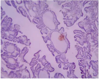
34figure 1. Papillary carcinoma thyroid histopathology
The electron microscopic appearance of papillary carcinoma includes a nucleus
with dispersed chromatin and highly infolded nuclear membrane,cytoplasmic
intrnuclear inclusions , and a cytoplasm that contain many mitochontria and
numerous cytoplasmic filaments. 2,35Keratohyaline granules have been found in
tumors with squamous foci.
b.iii.VARIANTS OF PAPILLARY CARCINOMA
1.Follicular variant: follicular variant of PTC (FVPTC) is the most common
subset of papillary carcinoma and is found in 9% to 22.5% of patients with PTC
growth pattern, or an encapsulated pattern. The follicles vary in size and shape
are often elongated or irregular shaped with abortive papillary formation, and the
colloid is usually deep stainedand scalloped.
Psammoma bodies and sclerosis may be present. The diagnosis made by
identification of the typical nuclear features. Follicular variant of papillary
carcinoma has two entities ;
1. Infiltrative type
2. Encapsulated type.
The infitrative non encapsulated type shows obvious infiltration of thyroid
parenchyma often accompanied by sclerosis. The growth is similar to that of
conventional papillary csarcinoma, except the papillae is absent. The
encapsulated type is surrounded by a fibrous capsule ,and it may or may not
exhibit invasion of the capsule or blood vessels.
Clinically low frequency of lymphnode metastasis; encapsulated solitary
tumor , infrequent intratumoral scelerosis. Presence of RAS mutation or
PAX8-PPARγ37translocation
Kakudo and colleagues , recently proposed the name well differentiated
thyroid tumor of uncertain behaviour to encompass this variant and encapsulated
Solid variant
This variant shows more than 50% solid, or trabecular growth pattern.
The tumor is traversed by delicat capillaries and the nuclear features of
conventional papillary carcinoma.38,39This variant occurs with a disproportionate
frequency among nuclear accident associated papillary thyroid carcinoma. It is
associated with high frequency of distant metastasis and less favorable
prognosis.40
Encapsulated variant
This variant constitutes 4% to 14% of all papillary carcinoma.41,42 The
fibrous capsule may or may not show invasion by tumor, but lymph node
metastasis can occur even in the absence of capsular or vascular invasion. The
patients tend to be younger,and the frequency of lymphnode metastasis is lower
.42The prognosis is excellent.43 The findings that the encapsulated follicular
variant of papillary carcinoma behaves more like follicular adenoma and
carcinoma.
2.Diffuse sclerosing variant
This rare variant mostly affects children44 and young adults. Patients
present with unilateral or bilateral symetrical thyroid swelling. Serum anti
thyroglobulin or antimicrosomal antibodies may be positive . 45This variant
shows more aggressive than conventional type, as manifested by higher
incidence of extra thyroidal extension ,lymphnode metastasis and distant
The thyroid shows diffuse replacement of the parenchyma by white firm
tisue,which isoften gritty on cutting.
The typical histologic features include
1. Diffuse involvement of one or both obes.
2. Scelerosis
3. Heavy lymphoplasmacytic infiltrate.
4. Abundant psammoma bodies.
5. Scatterd small islands of papllary carcinoma with prominent
squamous or squamoid differentiation.
6. Extensive lymphatic permeation.44
3.Diffuse follicular variant
This is a rare aggressive form of papillary carcinoma, it occurs in young
patints.15,46 This variant is characterised by diffuse enlargement of the entire
thyroid without formation of distinct nodules, predominent follicular pattern,
and absence of fibrosis. Histology showed a well-differentiated thyroid cancer
with a diffuse follicular growth pattern, mainly constituted by micro-follicles
containing a small amount of colloid, organized in confluent nodules of small to
medium size with dense collagenous septa without a detectable tumor capsule.46
The diffuse follicular variant shows a high frequency of lymph node ,pulmanory
and bone metastasis.47 it is a clinically aggressive tumor and had a poor
4.Tall cell variant
This variant composed predominantly of cells with height at least three
times their width compared with conventional papillary carcinoma proposed by
WHO.48This type shows following features,
1. Slightly older age group (50-57 years)48
2. Bulkier tumors
3. Higher frequency of BRAF mutation.(80%)48
4. More likely to show extrathyroidal extention(42-82%)49
5.More aggressive49
One third of cases ehibit RET/PTC rearrangements, selectively
RET/PTC350 exhibits more potent than RET/PTC 1. The tall cell variant is
highly papilliferous and invasive. The nuclei are typical those of papillary
carcinoma conventional type,are mostly basally located. The cytoplasm is
plentyful and oxyphilic because of accumulation of mitochondria.Focal clearing
of the cytoplasm is some times present.
5.Columnar cell variant
The columnar cell variant was first reported by evans as a rare thyroid
neoplasm.51More aggressive than differentiated thyroid carcinoma.52The tumors
are invasive. The Mean age is 57 years. 53 A more frequently metastases to
lung vertebra, and regional lymph node metastases exists.51The mortality rate is
1. Mixed papillary ,glandular,cribriform and solid patterns.
2. The papillae and glands are lined by tall columnar cells with
pseudostratified hyperchromatic oval or elongated nuclei.
3. Sub nuclear vacuoles and cytoplasmic clearing may be present.
4. The cells in the solid areas are often smaller and polyglonal.
5. The tumor is thyroglobulin positive.54
6. BRAF Mutation is demonstrable in one third of cases.53
6.Oxyphilic variant:
The latest World Health Organization International Classification
defines papillary thyroid carcinoma by its “follicular cell differentiation, as well as characteristic nuclear changes”. However the oxyphilic (Hürthle cell)
papillary carcinoma have nuclei which generally resemble the nuclei seen in
oxyphilic follicular carcinomas, and such oxyphilic papillary tumors may
behave more aggressively than typical papillary cancers55. The tumor is formed
predominantly by cells with abundant eosinophilic granular cytoplasm by
accumulation of mitochondria. It is important to differentiate this neoplasm
from Hürthle cell follicular neoplasms of thyroid .56
7.Warthin tumor like variant
Rare variant of papillary carcinoma, features similar to warthin tumor of
salivary gland.The tumor composed of a papillary pattern and a rich
8.Clear cell variant :
Rare variant of of papillary carcinoma.58 These tumors comprised of
mainly clear cells have a papillary architecture and cytological features of PTC.
Some tumors may have oncocytic and clear cell features . Clear cell appearance
believed to be due to mitochondrial expansion or accumulation of glycogen or
mucin Electron microscopy reveals dilated empty mitochondria.
Also believed to be related to TSH overstimulation58. Immunostaining for
TTF-1 and thyroglobulin may be necessary to distinguish these tumors from a
metastatic clear cell carcinoma.57
9.Macrofollicular variant
This type has the more than 50% of their area composed of large follicles.
59The cells that line the macrofollicles are attenuated and may not show the
characteristic nuclear features of papillary carcinoma. Assesement is made on the
smaller follicles.Uncommonly, dedifferentiation to undifferentiated carcinoma
can occur.60
10.Trabecular variant
This variant shows the trabecular growth pattern ,more than 50% of cells.
The cells are cuboidal or columnar, with cells perpenticular in long and straight
trabeculae. The tumors are large and invasive. This variant has been associated
with poor prognosis. This variant has also been considered by some authors as a
poorly differentiated variant of papillary carcinoma.differential diagnosis
typified by a trabecular or alveolar architecture with extensive hyaline and
colloid deposits. Their cellular features of cytoplasmic inclusions, nuclear
pseudoinclusions, nuclear grooves, and psammoma bodies often cause them to
be mistaken for papillary thyroid carcinomas on FNA. They represent benign or
low-malignant-potential tumors that are adequately treated by thyroid
lobectomy.61
11.Cribriform morular variant
This is a uncommon variant. It is microscopically shows prominent
cribriform pattern,with interspersed squamoid islands. The nuclei filled with
lightly eosinophilic ,homogenous ,biotin containing inclusions. Cells arranged in
closely packed follicles ,papillae, trabeculae. The characterstic feature ,the
luminal spaces shows absence of colloid. The tumor cells are cells are columnar
or cuboidal. The nuclei are chromatin rich, typical of nuclear features of
papillary carcinoma can often seen focally.
Some of cells showing spindle shaped nuclei,forms the fascicles and
whorls.
The tumor shows circumscribed, encapsulated,with or with out capsular
or vascular invasion. This variant of papillary carcinoma can occur as a sporadic
tumor , or associated with familial adenomatosus polyposis coli syndrome.
Female predominance ( 1:17), the mean age of diagnosis is 27.7 years. The
outcome of this tumor is favorable. This type commonly show RET/PTC
The APC gene shows either germline or somatic,germline mutation in
FAP associate cases, somatic mutation in some sporadic cases. Somatic mutation
in exon 3 of the beta catenin gene, which results in nuclear translocation of beta
catenin. BRAF mutation is not found in this type. Cytokeratin 19 (CK19) is
commonly strongly expressed in papillary thyroid carcinomas. It is also positive
in the squamous metaplasia in diffuse sclerosing variant of papillary thyroid
carcinoma . In contrast, CK19 may express only weakly in the morules in
CMV-PTC. Cytoplasmic staining of CD10 and nuclear staining of bcl-2 are strongly
positive in the morular cells but weak or not positive in the non-morular area .63
12.Papillary carcinoma with lipomatous stroma
In rare circumstances , adipose cells are interspersed with in the papillary
carcinoma.64
13.Variant with exuberant nodular fascitis like stroma
It is a rarely ,papillary carcinoma associated, with an abundant nodular
fascitis or fibromatosis like stroma. Microscopically shows the stroma
composed of spindle cells lying in a vascularised fibromyxoid matrix with
extravasated red cells. The spindle cells are myofibroblasticThese cells had oval
to elongated nuclei with fine chromatin and small distinct nucleoli . There was
no pleomorphism, and mitotic figures were not identified. Some extravasated red
blood cells were present in addition to interspersed lymphocytes and mast cells.
The interaction between the stroma and tumor results in unusal histologic pattern
like fibroadenoma and phylloides tumor of breast. The carcinomatous component
calcitonin. Cytokeratin staining highlighted occasional isolated tumor cells in the
stroma. Some tumor cells also showed vimentin and S-100 protein positivity.
The spindle cells were positive for vimentin and muscle-specific actin; a small
proportion also stained for desmin. They were negative for S-100 protein.65
14.Variant with spindle cell metaplasia
Rare variant of papillary carcinoma shows a component of spindle tumor
cells, constitute a major or minor proportion. The bland looking spindle cells
form short fascicles, along with the papillary carcinoma component.
This case reported as spindle cell transformation of papillary carcinoma
is a dedifferentiated papillary carcinoma rather than an example of this variant.66
15.Hobnail variant
It is a aggressive and rare variant of papillary carcinoma. Commonly
patient present with Cervical lymphadenopathy . Distant metastasis are common.
The tumor is multifocal, with microscopic finding shows variably sized papillae
covered by cells with atypically placed nucleus ,forming a surface bulge. BRAF
mutation commonly present in about half of the cases.67
16.Micropapillary variant
This is a rare type and having a prognosis. It is most commonly present
with lympho vascular invasion, lymph node metastasis, and distant metastasis are
seen. This variant is microscopically shows the , micropapillary growth pattern
17.Adenoid cystic carcinoma like variant
Papillary carcinoma rarely present with abundant deposits of globular
hyaline material in focal areas of tumor, like a adenoid cystic carcinoma of
salivary gland. Fine-needle aspiration smears from the thyroid nodule were
highly cellular and showed follicular cells arranged inpapillary clusters and in
monolayered sheets with d cytoplasmic vacuolization, including marginal
vacuoles. In some areas, structures resembling follicles with central hyaline
globules, reminiscent of adenoid cystic carcinoma increased frequency of nuclear
grooves. In May-Grun wald-Giemsa-stained smears, the neoplastic cells were
cuboidal to low columnar with clear to light-red cytoplasm an. These globules
were pink to magenta in colour.(68)
18.Dedfferentiated papillary carcinoma
This type coexistenceof papillary carcinoma with an undifferentiated or
poorly differentiated thyroid carcinoma. This transformation can occur either in
primary tumor or metastatic deposits. The prognosis is bad.
19. Microcarcinoma
It is also called as papillary microtumor.
It is defined as a tumor less than 1 cm.It has a excellent prognosis even in
lymphnode metastasis or distant metastasis cases. Rare cases with
microcarcinoma who have an unfavorable outcome are those with
lymphadenopathy greater than 3 cm , and a nonencapsulated type of papillary
In the 2004 WHO classification ,the definition of micropapillary
carcinoma, include only a incidental finding with less than 1 cm but not
clinically evident small papillary carcinoma.
b.iii.MOLECULAR PATHOLOGY OF PAPILLARY CARCINOMA: There
are three molecular alterations are recognised in papillary carcinomas, and result
in activation of the mitogen activated protein kinase path way.
1. RET mutation
2. BRAF Mutation
3. RAS Mutation
Activation of the proto oncogene ret or trk by intrachromosomal inversion
or chromosomal translocation
This occurs in 10 to 30 % of cases. Transfection of primary cultures of
human thyroid epithelial cells with a RET/PTC retroviarl construct results in
nuclear changes including irregular nuclear contour and euchromatic appearance,
suggesting that the genetic alteration may cause the characterstic nuclear feature
of papillary carcinoma. Oligoglonal RET/PTC rearrangement can occur in non
neoplastic thyroid tissues such as hashimotos thyroditis and benign nodules.
The tyrosine kinase of RET gene can be fused with a number of genes
constitutively expressed in thyroid epithelial cells, such as PTC -1, THROUGH
INV (10) (q11.2q21), PTC 2 through t(10;17),q(11.2q23),PTC 3through
cytogenetically undetectable paracentric inversion within 10q11.2,PTC4,PTC 5.
activation of the tyrosine kinase.RET/PTC 1 is most common,followed by
RET/PTC 3.70
The frequency of RET/PTC gene fusion is higher among children and
young patients,(50- 60%), chernobyl accident associated papillary carcinoma( 60
-80%) , mostly RET/ PTC 3, and who had received external radiation therapy
(60- 80%),70 most commonly RET/PTC 1. RET/PTC 1 is correlated with
papillary carcinoma with predominate papilary structure and papillary
microcarcinoma.RET/ PTC 3 is correlated with the tall cell and solid variants.
BRAF mutation
BRAF belongs to RAF family of protein kinase that play a role in
transduction of signals along the RAS/RAF/MEK/MAPK pathway,mediating
cell growth, differentiation and survival. Some study from, Animal experiments
discovered that normal rat thyroid cells with BRAF V600E mutation were likely
to promote tumor invasion by expressing certain gene products and degradating
the extracellular matrix of PTC.71,72
In addition, researchers also found that the BRAFV600E mutation in PTC
was correlated with transportation and metabolism of iodide, and resulted in
radioiodine treatment failure . Furthermore, the BRAF V600E mutation induced
silence of several tumor-suppressor genes and further promoted the invasiveness
of PTC. Xing et al discovered that the BRAF V600E mutation in PTC was
correlated with methylation of following tumor suppressor genes including tissue
inhibitor of metalloproteinase-3 (TIMP3), SLC5A8, death-associated protein
The most common mutation is a missense mutation at nucleotide 1799
with T-A transversion that results in substitution of valine by glutamic acid. The
frequency of BRAF V600E mutation in papillary carcinoma has risen gradually
over past two decades.
BRAF V600E mutation is less in less frequent in papillary carcinoma of
children and young patients.73
The prognontic significance of BRAF mutation is controversial, but most
studies report this features to be correlated with aggressive features such as old
age ,extrathyroidal extension , more advansed stage,, lymph node metastasis, and
tumor recurrence.
RAS mutation
Mutation of the RAS gene is reported approximately 15% of papillary
carcinoma , and the all positive cases were follicular variant. Mutations in RAS is
the the second most commonly identified genetic alteration in thyroid
malignancies. RAS mutations are primarily found in follicular-patterned tumors,
including Follicular adenoma, follicular cancer, and the follicular variant of
papillary cancer. There is increasing evidence that RAS mutation status has
significant diagnostic utility when used concurrently with FNAB.74
A small proportion of cases of the follicular variant of papillary
carcinoma ( 7%) show a mutation in the BRAF gene different from V600E , with
b.iv. PROGNOSTIC FACTORS
1. Age : Papillary carcinoma mostly developing in children and adolescents
having good prognosis. Nearly all deaths from papillary carcinoma commonly
occur when it is occur after the age of 45 years75.
2. Sex: Better prognosis in females than males.
3.Extra thyroidal extension : papillary carcinoma with extrathyroidal
extension having the bad prognosis.
4.Previous irradiation : it does not seem to significantly differ from others.
5.Tumor size: inverse correlation is present between size and prognosis.
6.capsule and margins : Better prognosis in encapsulated tumor.
7.Multicentricity and distant metastasis : patients with metastasis having the
bad prognosis;
8.Poorly differentiated ,squamous or anaplastic foci: bad prognosis. It has
only 5% of presentation.
9.DNA ploidy : it has a good correlation between a aneupliody and having an
aggressive behaviour in papillary carcinoma.
10.BRAF: BRAF mutation shows aggressive tumor. Unresponsive to
radioactive iodine.76
c.MEDULLARY CARCINOMA :
Medullary carcinoma is a rare , 10 % all thyroid malignancies. The tumor
is a aggressive in nature. It most commonly associated with MEN 2A, MEN
2B, familial non- MEN MTC. C cell hyperplasia is a precursor lesion. Most of
associated with mutations in RET oncogen, identified on chromosome 10. It
occur at any age group ,mostly adults in the age group of 50 years. children
may be affected in familial medullary carcinoma. It commonly Present with
painless firm nodule. Nodal metastasis are common. Distant metasasis to lung ,
bone are liver.
Pathology: Medullary carcinoma arises in the area of more on c- cell
concentration, lateral upper 2/3 of gland. In familial cases present with multiple
small nodules. Grossly it is a circumscribed, focal areas shows necrosis and
haemorrhage. Medullary thyroid carcinoma, tumor of the para follicular
cells (C cells) of the thyroid gland. It occurs both sporadically and genetically,
affecting multiple members of families who carry gene mutations associated with
the disease. In some families medullary thyroid carcinomas are the only tumor
that appear, whereas in other families medullary thyroid carcinomas are
associated with multiple endocrine neoplasia type 2 (MEN2). Medullary thyroid
carcinomas are moderately malignant tumors that invade nearby tissues in the
neck and spread to distant organs, such as the lungs and liver. A characteristic
feature of these tumors is hypercalcitoninemia, an abnormally high serum
concentration of a protein hormone called calcitonin, which is secreted by C
cells. Calcitonin normally lower the concentration of calcium in the blood when
it rises above the normal value. However, despite marked increases in serum
calcitonin concentrations, patients with medullary thyroid carcinoma do not have
carcinoma or MEN2 have hereditary mutations in the RET (rearranged
during transfection) proto-oncogene (a gene that can become a cancer-causing
gene, or oncogene). Patients with medullary thyroid carcinoma should be
analysed for mutations in RET; if a mutation is detected, other family members
should also be tested. Some people with hereditary mutations in RET will
develop medullary thyroid carcinoma at a young age. Therefore, any individual
carrying a RET mutation usually undergoes thyroidectomy (removal of the
thyroid gland) at an early age, before a tumour appears.77
Microscopically , circumscribed, cells arranged in nest pattern, separated by
stroma. Cells being round ,oval or spindle shaped cells. Nuclei are uniform.
Nuclear cytoplasmic ratio is low. Intranuclear inclusion, mitotic figures will be
present.The stroma contains amyloid. Variants have been papillary variant,
papillary or pseudopapillary growth is paresent. It should be differentiated from
tyoical papillary thyroid carcinoma by typical nuclear features.
VII. DIFFERENTIAL DIAGNOSIS OF PAPILLARY LESIONS OF
THYROID.
Fine needle aspiration cytology:
Fine needle aspiration cytology of the thyroid has been increasingly
utilized for the investigations of thyroid lesions. Since cancer is more common
in solitary cold nodules, prevalence of malignancy in solitary cold nodules
ranges from 10% to 44.5 %78
The main indications of fine needle aspiration in thyroid lesions are the
1. Evaluation of solitary thyroid nodules with a view to distinguish benign
from malignant.
2. Evaluation of diffuse thyroid lesions with a view to distinguish
inflammatory and autoimmune lesions from nodular goiter
3. Confirmation and categorization of clinically obvious thyroid malignancy.
especially anaplastic carcinoma that may require preoperative palliative
treatment, and lymphoma and metastatic malignancy where surgery is
usually not indicated.
4. To obtain material for ancillary test and prognostic parameters.
5. Evaluation of lesions detected initially by imaging, measuring 1-1.5 cm
diameter with features suspicious of malignancy.
Fine needle aspiration has been shown to the safest and most accurate of
diagnostic tools in thyroid lesion. Reporting of thyroid fine needle aspiration
specimen should follow a standard format that is clinically relevant in order to
direct management. At the national cancer center institute sponsored thyroid
state of the science conference in Bethesda in October 2007. Consensus was
reached regarding indications, pre-fine needle aspiration, techniques, diagnostic
terminology. The Bethesda system reporting terminology includes six categories
1. Non diagnostic
2. Benign
3. Atypia of undetermined origin
6. Malignant.
Cytologic diagnosis is generally accurate in thyroiditis, usual type of
papillary carcinoma, medullary carcinoma, anaplastic carcinoma and high grade
lymphoma. False negatives have been minimized by using ultrasound guided
fine needle aspiration.
Normal structure: Follicular epithelial cells and colloid are regular features in
normal thyroid and in colloid goiter. thyroid and in colloid goiter. Follicular
cells show fragile blue cytoplasm or pale blue cytoplasm with indistinct or fuzzy
cell border. Coarse blue cytoplasmic granules may be seen. Thick colloid
appears as round, dense clumps, of deep blue, violet, or magenta coloured
acellular materal.
FINE NEEDLE ASPIRATION CYTOLOGY
LESIONS FNAC FINDINGS DIFFFERENTIAL
DIAGNOSIS Nodular goiter Flat monolayered sheets
of epithelial cells has frayed edges. Abundant thick and thin collid. Cytoplasm is indistinct.
80
Cystic papillary
carcinoma-May contain abundant colloid. can cause diagnostic difficulties. Hyperplastic papillae containing follicles and intact dilated follicles in cell block preparation shows a beningnity of nodule.
Graves disease Colloid free bloody
background.monolayered sheets with moderate amount of pale cob web
like vacuolated
cytoplasm.81
with well defined margins .
Papillary carcinoma Flat sheets ,three dimentional structures, and papillary fragments ,shows anatomical bordering. Enlarged ovoid nuclei ,fine
granular powdery
chromatin in PAP stain. Chewing gum colloid. Intranuclear cytoplasmic inclusions and nuclear grooves . psammoma bodies variable.82
Positive immunostaining for CK 19, CD 44, HBME 1.
Papillry foci are present in hyperplastic nodular goiter and graves didease.
Cysti papillary
carcinoma.
Large cell size, pseudo inclusions, nuclear grooves, well defined vaculoes in atypical histiocytoid cells. 79
Histiocytic cells in cystic nodular goiter mimic papillary carcinoma. Immunostainig for CD 68 may be useful.
Follicular variant of papillary carcinoma
Follicular architexure, nuclear features shows powdery pale chromatin, and nuclear grooves.
Follicular neoplasm-combined use of hbme 1 and ck 19 may be useful to distinguish between follicular neoplasm and follicular variant of papillary carcinoma. Macrofollicular
encapsulated variant
Predominant
macrofollicles, large, cuboidal cells with fine nuclear chromatin,
grooves,
pseudo-inclusions and dense eosinophilic colloid.
Macrofollicular adenoma and nodular goiter
Oncocytic variant Papillary and follicular architexture, abundant coarsely granular cytoplasm with nuclear features of classical papillary carcinoma.
Papillary hurthle cell tumor – macronucleoli absent in oncocytic variant.
Hashimotos thyroiditis.
infiltrate in papillary stalks83.
lymphocytes and plasma cells intermingle with the cell clusters, in hashimoto s thyroiditis. Cribriform morular
variant
Closely packed
follicles,papillae, solid areas with islands of squamoid morules. Nuclear chromatin fine powdery with occasional grooves and inclusions. Adenoid cystic variant Papilliform clusters,
monolayered sheets, many nuclear grooves and inclusions. Light pink to purple hyaline globules surrounded by neoplastic cells.84
Nodular facitis like stroma
Predominate stromal component, with bland spindle cells,irreguar in shape and size. Sparse epithelial groups shows features of papillary carcinoma.
Tall cell variant Solid areas,follicles and papillae lined by oxyphilic cells twice as tall as other than variants. Oxyphilic cells shows reddish or cyanophilic granular eosinophilic vacuolated cytoplasm with nuclear grooves and inclusions.49,50
usually not seen.52
Diffuse scerlosing variant
Prominent fibrosis with papillary pattern many psammoma bodies with lymphoplasmacytic infiltrate. Squamous metaplastic cells will be present.85,86
Hashimoto s thyroiditis.
Solid /trabecular variant Solid and trabecular pattern with irregular nuclear contours with few cells showing nucear groooves and inclusions. Medullary carcinoma Plasmacytoid, spindle
cell , small cell pattern with nuclei showing moderate anisonucleosis,
uniform stippled
chromatin , few scattered cells coarse red cytoplasmic granularity ( MGG) seen.
Papillary carcinoma –
SURGICAL PATHOLOGY
LESIONS GROSS FINDINGS MICROSCOPY
FINDINGS DIFFERENTI AL DIAGNOSIS Graves disaese
the gland shows a mild to moderate symmetric diffuse enlargement , reddish ,has a consistency of pancreatic tissue.
Microscopically the
follicles are
hyperplastic with prominent papillary infolding , it may
confuse with
papillary carcinoma. The lesion lined by
columnar with
basally located hypercromatic
nuclei , and a clear cytoplasm, that may contain fat and glycogen.
The collid is pale, finely vacuolated with prominent scalloping.
The stroma contains lymphoid aggregate
Papillary carcinoma
Nodular hyperplasia
Grossly , capsule may be stretched , multiple nodules with secondary change haemarrhage,
calcification and cystic degeneration are common.risation
Microscopically varying appearances, some with large follicles lined by flattened epithelium, others have papillary projection, it may
confuse with
papillary carcinoma. Rupture of follicle may lead to a granulomatous reaction, prominent vascularisation Papillary carcinoma Follicular carcinoma Classical papillary carcinoma thyroid.
Average 1.5 to 3 cm in size. Lesions are firm ,white in colour with an invasive tumor. Lesional
cells arranged in complex papillae
,with central
fibrovascular core. Cells lining the
Hyperplasia of thyroid follicle –
calcification is seen. Necrosis may be
seen. Cystic
formation may be associated with it.
papillae shows nuclear stratification ,irregular nuclear membrane with clear nuclear chromatin ,eccentric placed nucleoli.
Intranuclear grooves and inclusions paresent.
Psammoma bodies may be present.
change.
Follicular variant of papillary carcinoma
Well circumscribed encapsulated nodule.
Encapsulated
microfollicles and macrofollicles,capsu lar invasion or vascular invasion may be present. Cells lining the follicles shows scant to eosinophilic
cytoplasm with
nuclear features of classical papillary carcinoma.
Follicular
adenoma /
carcinoma.-Papillary microcarcino ma
Tumor less than 1 cm.
Nuclear features shows classical papillary carcinoma. Tumor is less than 1 to 1.5 cm.
Tall cell variant
Large , infiltrative , white tan lesions. The tumor more than 6 cm .
Papillary growth pattern, cells showing cells height 2 to 3 times width with oncocytic cytoplasm. Focal to diffuse prominent nucleoli. Multiple intrnucear inclusions present. Vascular
invasion and
extrathyroidal
extension more common.
Hurthle cell tumor- oncocytic
cells may
circumscribed tumor.(brucen, thompson.)
columnar cells ,
nuclei are
hyperchromatic ,
with punctate
chromatin. Extrathyroidal
extension most common.
Warthin like variant.
Solid infiltrative ,ill defined tumor.
Paillary structure with cells showing abundant
eosinophilic
cytoplasm ,papillary
core contains
lymphoplasmacytic infiltrate.
Hashimotos thyroiditis and hurthle cell carcinoma. Papillary thyroid carcinoma with nodular fascitis like stroma
Cells arranged in cords tubules and papillae with nuclear features of classical papillary carcinoma. Also shows varying degree of squamous metaplasia.
Anaplastic differentiation of papillary thyroid carcinoma.- in this spindle cell positive for cytokeratin. In nodular facitis spindle cell negative for cytokeratin. Cribriform morular
variant of papillary carcinoma.
well-circumscribed, somewhat lobulated tan masses ranging from 1.5-2 cm.
Cribriform ,solid and spindle cell growth pattern with nuclear features of papillary carcinoma. Diffuse
scelerosing variant
Diffuse growth with fibrosis
Tumor cells lined by papillae, with psammoma bodies. Lymphocytic
infiltrates around tumor foci.
Hobnail variant
Tumor cell nuclei located in the middle
or apex of
cytoplasm. Encapsulated
variant
Encapsulated tumor. Look like a adenoma groosly.
Microscopically
shows total
psammoma bodies. Hurthle cell
variant Encapsulated,mahoga ny brown in colour.
Papillary
structures at least focally, may be
mixed with
follicular growth pattern.
Predominantly oncocytic
cytoplasm but papillary nuclear features.
Hurthle cell tumor –benign or malignant.- it shows papillary areas.
Medullary carcinoma thyroid
Circumscribed,
infiltrative tumor. Also admixed with
necrosis and
haemmorrhage.
Typical medullary carcinoma shows nests of tumor cells shows, composed of round to oval nuclei or spindle shaped cells.papillary
variant of medullary carcinoma shows papillary
architexture ,it may be pseudopapillary or true papillary .
Papillary carcinoma thyroid.
IMMUNOHISTOCHEMISTRY APPLICATION IN PAPILLARY
LESIONS:
Immunohistochemistry is the application of immunologic principles and
techniques to demonstrate specific antigens in cells and tissues based on antigen
antiody interaction and it explore the specificity at light microscopic level.
Various methods of immunohistochemistry include eroxidase –antiperoxidase method, alkaline phosphatase labelling method, avidin biotin method, and two
STEPS OF IMMUNOHISTOCHEMISTRY:
Antigen retrieval:
Antigen retrival is done to unmask the antigen determinants of fixed tissue
sections. This can be done by
1. Proteolytic enzyme digestion
2. Microwave antigen retieval
3. Microwave and trypsin antigen retrieval
4. Pressure cooker antigen retrieval87
Proteolytic enzyme digestion
Enzymes like trypsin and proteinases are used to breakdown the formalin
cross linkages and unmask the antigen determinants. But there is a disadvantage
of antigen destruction and inadequate digestion.
MICROWAVE ANTIGEN RETRIEVAL
In this formalin fixed paraffin sections are boiled in varoius buffers for
rapid and uniform heating. This is the most common method used now.
PRESSURE COOKER ANTIGEN RETRIEVAL:
In this method also the tissue sections are boiled in buffers to unmask
the antigens . this method is used to retrieve large number of slides.
DETECTIONS SYSTEMS:
After adding specific antibodies ,the antigens, the antigen antibody
DIRECT METHOD:
The primary antibody is directly conjucated with flurochrome. Commonly
used flurochromes are horse radish peroxidase and alkaline phosphatase.
INDIRECT METHOD:
It is a two step method. First the labelled secondary antibody reacts with
primary antibody which is bound to specific antigen. The use of peroxidase
enzyme complex or avidin biotin complex further increases yhe sensitivity of
immunohistoc
Immunostaining shows that most papillary cancer contains thyroglobulin
and thyroid transcription factor -1 .
A number of markers used for the differential diagnosis of thyroid lesions ,such
are CD 56, HBME 1, CK 19, Galectin 3.
RET
The Ret gene is located on chromosome 10 q and encodes a tyrosine
kinase transmembrane receptor. It is typically absent in the normal thyroid
follicular cells; however, gene rearrangement occurs in most PTCs. This
oncogene is believed to be specific to PTC and encodes an oncoprotein product
that contains the cytoplasmic portion of Ret gene. Therefore, some investigators
believe that IHC expression of Ret oncoprotein is a reliable marker for PTC .
Cheung et al showed immune expression of Ret in 78% of PTC, 63% of FVPC
and 57% of Hurthle cell carcinoma, while all benign nodules were non-immuno
found that Ret had focal or moderate immune reactivity in benign lesions while it
showed prevalent cytoplasmic expression in classic papillary carcinoma and its
variants. The general conclusion among researchers is that diffuse immune
expression represents a good supportive evidence for the diagnosis of papillary
carcinomas; however, focal staining is often found in other lesions including
benign nodules. Perhaps, a more important finding is that immune reactivity of a
panel that includes Ret, HBME-1 and CK19 is very specific for papillary
carcinoma. Cheung et al concluded that HBME-1 positivity indicates
malignancy, whereas diffuse CK19 and/or Ret positivity confirm papillary
differentiation .88
THYROID TRANSCRIPTION FACTOR 1
Thyroid Transcription Factor 1, also named NKX2 homeobox 1
(NKX2.1), is a nuclear protein, approximately 38 kDa, composed of a single
polypeptide of 371 amino acids belonging to the family of homeodomain
transcription factors. Thyroid transcription factor 1 plays a crucial role in the
organogenesis and differentiation of thyroid and lung.
Thyroid transcription factor 1 expression by immunohistochemical analysis was
initially exclusively identified in thyroid and lung epithelial tissues, including
normal, benign, and malignant tissues. In routine practice, TTF1 became one of
the most commonly used immunomarkers to identify thyroid or lung primary
tumor in the setting of metastasis and to differentiate adenocarcinoma from
and parafollicular cells show diffuse expression of TTF1. In thyroid neoplasm,
TTF1 expression was reported in nearly 100% of PTCs, FTCs, and follicular
adenomas (FAs); in approximately 90% of papillary carcinomas and Medullary
carcinomas; and in none to fewer than 25% of undifferentiated carcinoma.89
CD 56
CD 56 is a neural cell adhesion molecule . its expression may affect the
migratory capability of tumor cells. Loss of CD 56 correlates with metastatic
potentials and poor prognostic outcome in some malignancies. The marker
pattern and intensity of staining were scored. Positive expression of the markers
in 10% or more of follicular epithelium within the tumor or lesional cells was
considered positive. An expression of <10% was considered to be negative.
Diffuse CD56 expression was consistently present in normal, lesional, and
neoplastic follicular epithelium, except for PTC, including the follicular variant.
We concluded that CD56 is of value to distinguish PTC from other thyroid
follicular pathology/histology with a sensitivity of 100% and a specificity of
100%. We suggest that CD56 is extremely useful in the diagnosis of PTC,
including the follicular variant, and to distinguish it from other follicular
cell-derived thyroid tumors/lesions. Application of CD56 by a group of expert
pathologists on a larger series of follicular thyroid neoplasms of uncertain
HBME 1
HBME-1 is a monoclonal antibody that was initially promoted as a marker
of mesothelial cells;91 it is directed against an unknown epitope. In the thyroid,
HBME-1 is almost exclusively expressed in malignant neoplasms, including
papillary carcinoma, whereas benign lesions are negative. HBME-1 is the most
specific marker of thyroid malignancy, but it may not be very sensitive because
oncocytic lesions are generally negative; also, many malignancies are not stained
by this antibody. HBME-1 positivity is characterized by predominantly
membranous staining with variable cytoplasmic staining