0022-538X/10/$12.00 doi:10.1128/JVI.01902-09
Copyright © 2010, American Society for Microbiology. All Rights Reserved.
Fine Mapping of Pre-S Sequence Requirements for Hepatitis B Virus
Large Envelope Protein-Mediated Receptor Interaction
䌤
Andreas Schulze,
1Alexa Schieck,
2Yi Ni,
1Walter Mier,
2and Stephan Urban
1*
Department of Infectious Diseases, Molecular Virology, Otto-Meyerhof-Zentrum, University Hospital Heidelberg, Im Neuenheimer Feld 350, 69120 Heidelberg, Germany,1and Department of Nuclear Medicine, University Hospital Heidelberg,
Im Neuenheimer Feld 400, 69120 Heidelberg, Germany2
Received 8 September 2009/Accepted 30 November 2009
Previous studies showed that the N-terminal 75 amino acids of the pre-S1 domain of the hepatitis B virus (HBV) L protein are essential for HBV and hepatitis delta virus (HDV) infectivity. Consistently, synthetic lipopeptides encompassing this sequence or only parts of it efficiently block HBV and HDV infection, presum-ably through specific interference with a cellular receptor. Crucial for both virus infectivity and the inhibitory activity of the peptides are N-terminal myristoylation and a highly conserved motif within the N-terminal 48 amino acids. To refine the sequence requirements, we synthesized a series of HBV pre-S1 peptides containing
deletions, point mutations,D-amino acid exchanges, or genotype-specific sequence permutations. Using the
HepaRG cell line and a genotype D-derived virus, we determined the specific inhibitory activities of the peptides and found that (i) lipopeptides with an artificial consensus sequence inhibit HBV genotype D infection
more potently than the corresponding genotype D peptides; (ii) point mutations,D-amino acid exchanges, or
deletions introduced into the highly conserved part of the pre-S1 domain result in an almost complete loss of activity; and (iii) the flanking sequences comprising amino acids 2 to 8, 16 to 20, and, to a less pronounced extent, 34 to 48 gradually increase the inhibitory activity, while amino acids 21 to 33 behave indifferently. Taken together, our data suggest that HBV pre-S1-mediated receptor interference and, thus, HBV receptor recogni-tion form a highly specific process. It requires an N-terminal acyl moiety and a highly conserved sequence that is present in primate but not rodent or avian hepadnaviruses, indicating different entry pathways for the different family members.
Hepatitis B viruses (HBVs) are small, enveloped DNA vi-ruses that replicate their genome via reverse transcription of a pregenomic RNA transcript in the cytoplasm of infected hepa-tocytes (31). They are classified into the familyHepadnaviridae
and are adapted to mammals (primates and rodents) and birds, where they cause acute and persistent infections. At present about 360 million people are chronically infected with the human HBV. Due to HBV-related progressive liver failure (cirrhosis or hepatocellular carcinoma) ⬇650,000 people die each year (10). HBV, like duck hepatitis B virus (DHBV) or woodchuck hepatitis B virus (WHV), shows a pronounced species specificity and a remarkable liver tropism. These fea-tures have been assigned to specific early infection events, most likely receptor recognition. However, it cannot be excluded that other steps during or postentry contribute to the host restrictions of infection (13).
The HBV replication cycle has been deciphered in some detail. However, studies on the early infection events were hampered by the lack of appropriatein vitroinfection systems. The establishment of the HBV-susceptible cell line HepaRG and systems based on primary human hepatocytes (PHH) and primaryTupaia belangeri hepatocytes resolved this issue and facilitated investigations on cellular and viral determinants in-volved in HBV entry. It became clear that infectivity can be
assigned to different subdomains in two of the three viral envelope proteins (34). These three envelope proteins are termed large (L), middle (M), and small (S) proteins. They form a proteinaceous outer virus shell, which is embedded in an endoplasmic reticulum (ER)-derived lipid bilayer. Their coding mRNAs originate from one single open reading frame (4). Since their three start codons are in phase, L, M, and S proteins share the C-terminal 226 amino acids (termed the S domain in the context of the L and M proteins), which anchor the proteins via four putative transmembrane helices (TM domains) in the lipid bilayer. The S protein drives particle formation and accordingly serves an important function in virus assembly (4, 5). The S domain has further been shown to participate in virus entry since antibodies against the antigenic loop of S, located between TM domains 2 and 3, are neutral-izing (11), and mutations introduced in this loop render as-sembled hepatitis delta virus (HDV) particles noninfectious (1, 20, 29). Linked to the N terminus of the S domain, the M and L proteins bear two hydrophilic extensions, one of 55 amino acids and, depending on genotype, one of 108 (genotype D), 118 (genotypes E and G), or 119 (genotypes A, B, C, F, H) amino acids called pre-S2 (M) and pre-S1 (L), respectively. The L proteins of all hepadnaviruses contain recognition mo-tifs for N-myristoyltransferase (36, 37) and are accordingly subjected to this modification. The lack of a classical secretion signal in L results in an initial cytoplasmic orientation of its pre-S domains. After synthesis, myristoylation, and incorpora-tion of the TM helices into the ER membrane, the pre-S domain of L traverses the lipid bilayer, allowing parts of it to become accessible at the particle exterior (7).
* Corresponding author. Mailing address: Molecular Virology, Otto Meyerhof Zentrum, University of Heidelberg, Im Neuenheimer Feld 350, 69120 Heidelberg, Germany. Phone: 49 6221 562910. Fax: 49 6221 561946. E-mail: [email protected].
䌤Published ahead of print on 9 December 2009.
1989
on November 8, 2019 by guest
http://jvi.asm.org/
Crucial elements for virus attachment, specific receptor binding, or fusion have been identified within the L, M, and S proteins by two complementary approaches. One approach using HBV particles that were enveloped with mutated L pro-teins showed that myristoylation of L is mandatory for virus infectivity (6, 16). Successive deletions of 5 amino acids within the N-terminal pre-S1 sequence of residues 1 to 78 abrogated HBV infectivity (24). In contrast, most parts of the pre-S2 domain were dispensable for infection (23). Using HDV, these results were confirmed, indicating that both viruses share very similar entry modes (3). Moreover it became obvious that the S domain, either as part of the HBV L protein or as the S protein, being the major constituent of the viral envelope, is required (1, 20, 29). The second approach followed the obser-vation that a synthetic myristoylated peptide consisting of the N-terminal amino acids 2 to 78 of the pre-S1 domain (HBVpreS/2-78myr) prevents HBV infection of PHH and
Hep-aRG cells even at nanomolar concentrations (17). Surprisingly, C-terminal shortening of this sequence up to amino acid 48 increases the inhibitory activity of the peptide (15). This indi-cates two separable and distinct functions within the pre-S1 infectivity determinant consisting of residues 1 to 78: one is related to the inhibitory activity of the peptide through inter-ference with a hepatocyte-specific receptor, and the other, involving amino acids 49 to 78, is still unknown. The require-ment of the N-terminal myristic acid moiety in the inhibitory activity is to some extent variable. Replacement of myristic acid by other hydrophobic saturated and unsaturated fatty acid moieties or even by cholesterol allows the modulation of spe-cific activities according to the relative hydrophobicity (14, 15, 27). Thus, lipophilicityper seand not a specific lipid structure seems to play the key role for this part of the molecule.
Previous studies using synthetic peptides or recombinantly produced fusion proteins carrying internal deletions and point mutations identified a highly conserved pre-S1 sequence ele-ment (amino acids 9 to 18 in genotype D) that is very impor-tant for infection competition (2, 9, 14). Single amino acid exchanges in this region abrogate the antiviral activity of the peptides and render the respective recombinant hepatitis B virions noninfectious (9).
Our recent observation that HBV pre-S1-derived lipopep-tides are also potent inhibitors of HBV infectionin vivo(27) suggests that the lead substance of this group of entry inhibi-tors may be suitable for clinical applications in cases of acute and possibly chronic HBV and HDV infection in the future.
In this report, we examined the capacity of synthetic HBV pre-S1-derived peptides to interfere with the HBV infection of HepaRG cells. By using this approach we (i) further narrowed down the minimal amino acid sequence requirements and the sensitivity to amino acid exchanges for interference with the HBV infection, (ii) characterized in more detail the role of N-and C-terminal deletions, N-and (iii) defined the consensus se-quence derived from an alignment of the different HBV geno-types as the most active peptide and the lead substance for clinical development.
MATERIALS AND METHODS
Cell lines.HepaRG cells were grown in Williams’ medium E supplemented
with 10% heat-inactivated fetal calf serum, 50 U of penicillin/ml, 50g of
streptomycin/ml, 5g of insulin/ml, and 50M hydrocortisone hemisuccinate.
The cells were passaged every 2 weeks at a ratio of 1:5. Fourteen days before infection, cell differentiation was induced by adding 2% dimethyl sulfoxide to the maintenance medium (17). The medium was exchanged every 3 days.
Infection competition assays.As an infectious inoculum, HepAd38 (22) or HepG2.2.15 (32) cell culture supernatant, concentrated approximately 100-fold, was used. The inoculum was prepared from cell culture supernatants by precip-itating viral particles with 6% polyethylene glycol 8000 (Sigma-Aldrich) for 12 to
16 h at 4°C. The precipitates were recovered by centrifugation (at 7,000⫻gfor
1 h at 4°C) and resuspended in phosphate-buffered saline (PBS) supplemented
with 25% fetal calf serum. Aliquots were stored at⫺80°C. For infection,
differ-entiated HepaRG cells (1⫻106cells/well of a 12-well plate) were incubated with
a 20-fold dilution of the concentrated virus stock in medium (corresponding to
⬇4⫻1010genome equivalents) supplemented with 4% polyethylene glycol 8000
for 16 h at 37°C. At the end of the incubation, the cells were washed three times and further cultivated. Medium was changed every 3 days. Competition experi-ments were performed in 12-well plates. To ensure comparability within one concentration series, all assays with one peptide were done in a single 12-well plate. To guarantee the best comparability between different peptides, the re-spective tests of different concentration series were performed in plates of the same batch of cells. HepaRG cells were preincubated for 30 min at 37°C with the chemically synthesized acylated HBV pre-S1 peptides, followed by coincubation of cells with the peptides and virus for 16 h at 37°C. To quantify the infection, amounts of hepatitis B surface antigen (HBsAg) and hepatitis B e antigen (HBeAg) secreted into the culture supernatant from day 7 to 11 postinfection (p.i.) were determined by enzyme-linked immunosorbent assay ([ELISA] AxSYM; Abbott). All competition experiments were performed in duplicate and repeated at least two times independently.
Quantification of cellular HBV binding.Attachment of HBV to HepaRG cells was determined by quantitative PCR to detect the viral DNA. Binding/uptake
was performed for 4 h at 37°C in the absence or presence of 200g/ml heparin
or 1M HBVpreS/2-48myr(a synthetic myristoylated peptide consisting of
N-terminal amino acids 2 to 48 of the pre-S1 domain). Unbound virus was removed by washing. Viral DNA was prepared from the cellular lysates with a NucleoSpin Blood Kit (Macherey-Nagel) according to the manufacturer’s protocol. For the SYBR green-based real-time PCR (Invitrogen), the primer set Taq-HBV-F
(5⬘-TCCCAGAGTGAGAGGCCTGTA-3⬘) and Taq-HBV-R (5⬘-ATCCTCGA
GAAGATTGACGATAAGG-3⬘) was used. Quantitative PCR was performed in
an ABI Prism 7000 Sequence Detection System (Applied Biosystems). The reaction process started at 50°C for 2 min and 95°C for 15 min, followed by 45 cycles at 95°C for 15 s and 60°C for 1 min. All experiments were performed at least twice.
Peptide synthesis, purification, and analysis.All peptides were synthesized by standard solid-phase peptide synthesis employing the fluorenylmethoxycarbonyl/
t-butyl (Fmoc/tBu) strategy and purified by semipreparative reversed-phase
high-performance liquid chromatography (HPLC) on a Cromolith SemiPrep column (Merck). The purity of all peptides was verified by analytical HPLC on a Per-formance Cromolith column (Merck). As solvents for analytical HPLC, 0.1% aqueous trifluoroacetic acid (solvent A) and acetonitrile containing 0.1% triflu-oroacetic acid (solvent B) were used. The analysis was performed by applying a linear gradient (flow rate of 4 ml/min) from 0 to 100% solvent B within 5 min. The identity of the peptides was verified by mass spectrometry. The peptide
WHVpreS/2-81myr (2-GNNIKVTFNPDKIAAWWPAVGTYYTTTYPQNQSVF
QPGIYQTTSLINPKNQQELDSVLINRYKQIDWNTWQGFPVDQKLP-81) was purchased from Peptide Specialty Laboratories GmbH (Heidelberg, Germany). For infection competition experiments, stock solutions of the peptides in di-methyl sulfoxide were prepared, prediluted in medium, and added to the cells at the concentrations indicated in the figures.
Immunofluorescence (IF) analyses.At 11 days p.i. the HepaRG cells were washed with PBS, fixed with 4% paraformaldehyde in PBS for 15 min at room temperature, and permeabilized with 0.25% (vol/vol) Triton X-100 in PBS (30 min at room temperature). After a washing step, a hepatitis B core antigen (HBcAg)-specific polyclonal antibody (H363; provided by Heinz Schaller, ZMBH, University of Heidelberg, Germany) at a dilution of 1:1,000 in PBS containing 5% skim milk powder was added. Following overnight incubation at 4°C, cells were washed and incubated in the dark for 1 h at room temperature with a 1:500 dilution of an Alexa Fluor 488-conjugated secondary antibody (Invitrogen/Molecular Probes) in PBS containing 5% skim milk powder. After a
washing step, cells were incubated for 30 min with a 1g/ml solution of 4⬘,
6⬘-diamidino-2⬘-phenylindole dihydrochloride (DAPI; Roche Applied Science).
Cells were washed three times with PBS. Images were acquired with an inverted fluorescence microscope (Leica, Germany). All images were made under iden-tical settings and afterwards handled ideniden-tically. The images were analyzed using ImageJ software. The nucleus counter function of the Wright Cell Imaging
1990 SCHULZE ET AL. J. VIROL.
on November 8, 2019 by guest
http://jvi.asm.org/
Facility (WCIF) plug-in was used to count the number of labeled nuclei (DAPI). The cell counter plug-in was used to count the number of HBcAg-positive cells. For each peptide and concentration, four images were analyzed.
RESULTS
Sequence-specific and acylation-dependent inhibition of HBV infection by synthetic lipopeptides derived from the
N-terminal pre-S1 domain of the HBV L protein.To substantiate
the specificity of infection inhibition by HBV pre-S1-derived peptides, we infected differentiated HepaRG cells with HBV (genotype D) in the presence of increasing concentrations (1, 5, 25, 100, and 1,000 nM) of HBVpreS/2-48stearoyl, the
N-terminally stearoylated pre-S1-derived peptide of genotype D; HBVpreS/2-48scrambledstearoyl, an N-terminally stearoylated
peptide of the same amino acid composition but with random-ized sequence; and HBVpreS/1-48, the nonstearoylated pre-S1 peptide carrying instead of a lipid moiety the initially biosyn-thesized methionine in position 1 (Table 1). Prior to the infec-tion inhibiinfec-tion experiments, the purity of all synthesized pep-tides was controlled by HPLC, as shown for the three peppep-tides in Fig. 1A. The peptides eluted in a single peak with no de-tectable side products or contaminations. A clear increase in the retention time (2.3 to 3.7 min) was observed for the
stearoylated peptides compared to the nonacylated HBVpreS/ 2-48, indicating that nonacylated side products were com-pletely removed.
Consistent with earlier results (15), HBVpreS/2-48stearoyl
in-hibited HBV infection by more than 80% at a concentration of 1 nM (as measured by secreted HBsAg between days 7 to 11 p.i.) (Fig. 1B). Concentrations of⬎1 nM reduced HBsAg in the supernatant to almost the detection limit of the ELISA. In contrast, neither HBVpreS/2-48scrambledstearoylnor the
non-stearoylated pre-S1 peptide was able to interfere with infection even at 1M. This confirms the specificity of the assay within this concentration range and excludes nonspecific effects caused by, for example, the acyl moiety. In a separate experi-ment performed under the same conditions but applying lower peptide concentrations, the half-maximal inhibitory concentra-tion (IC50) of HBVpreS/2-48stearoylwas determined to be⬇400
pM (data not shown). Using a WHV-derived peptide (WH-VpreS/2-81myr) no interference with thein vitroHBV infection
was observed while a parallel control experiment using 1M HBVpreS/2-48myrcompletely abolished infection.
[image:3.585.73.516.81.420.2]It has been shown that inoculation of HepaRG cells with acylated pre-S1 peptides prior to infection results in inhibition, indicating that the peptides address a cellular factor (15). In
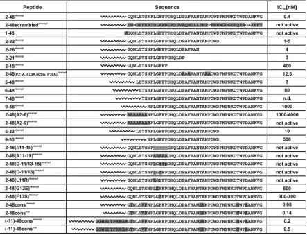
TABLE 1. Name, primary sequence, andin vitroinhibitory activity of the tested HBV peptidesa
a
To conserve space, all peptide names were shortened by deletion of the prefix “HBVpreS/.” Sequence variations in comparison to HBVpreS/ 2-48stearoyl
(genotype D) are marked by gray boxes. IC50s are given according to the corresponding HBsAg measurements. Not active, peptide is not
active in the concentration range tested; ND, not determined in the concentration range tested.
on November 8, 2019 by guest
http://jvi.asm.org/
FIG. 1. HBVpreS/2-48stearoylefficiently inhibits HBV infection of HepaRG cells, requires N-terminal acylation, and does not obstruct HBV
attachment at concentrations up to 1M. (A) Elution profiles of analytical HPLC runs of HBVpreS/2-48stearoyl, HBVpreS/2-48scrambledstearoyl,
and HBVpreS/2-48. Note that, due to size limitations, all peptide names have been shortened and are represented by their amino acid sequences (e.g., 2-48 for HBVpreS/2-48). Peptide bonds were detected by UV absorbance at 214 nm and are given as arbitrary units. (B) At left, data are shown for HBV infection inhibition using increasing concentrations (0 to 1,000 nM) of HBVpreS/2-48stearoyl, HBVpreS/2-48scrambledstearoyl, and
HBVpreS/1-48. At right, data are shown for HBV infection inhibition using increasing concentrations (0 to 10,000 nM) of WHVpreS/2-81myr. As
control, 1M HBVpreS/2-48myrwas used in parallel. The amount of HBsAg secreted from days 7 to 11 p.i. was determined by ELISA and is given
1992 SCHULZE ET AL. J. VIROL.
on November 8, 2019 by guest
http://jvi.asm.org/
addition, we along with others identified heparan sulfate pro-teoglycans (HSPG) as the primary cellular attachment sites of HBV (25, 30). To investigate if pre-S1-derived lipopeptides interfere with binding of the viral particles to cellular HSPG, we compared the HBV cell association in the presence of heparin or HBVpreS/2-48myr(Fig. 1C). Addition of 200g/ml
heparin led to a 62% reduction of cell-associated viral genome equivalents in comparison to the untreated control. In con-trast, the presence of 1M HBVpreS/2-48myr, a concentration
that blocks infection, had no influence on HBV cell associa-tion, indicating that the HBV pre-S1 peptides do not affect primary attachment. To analyze whether HBV pre-S1 lipopep-tides act at a postfusion step, we added HBVpreS/2-48stearoyl
16 h after the beginning of virus inoculation (Fig. 1D). While coincubation with the virus led to a more than 80% reduction of infection, addition of the peptide at 16 h postinoculation was without effect. To verify the results of the infection inhibition experiments on a single-cell level, we performed HBcAg-spe-cific IF analyses. While a subset of differentiated HepaRG cells stained positively for HBcAg in the untreated control infec-tion, only sporadic infection events could be observed in the presence of 250 nM or 2.5M HBVpreS/2-48stearoyl(Fig. 1E).
As determined by cell counting, this corresponds to 0.06% (at 250 nM) and 0.02% (at 2.5M) of HBcAg-positive cells of the control infection (Fig. 1F). Under the same conditions HBVpreS/2-48scrambledstearoyl did not lead to a significant
reduction of HBcAg-expressing cells (98% at 250 nM and 77% at 2.5 M). Parallel quantification of HBsAg secreted from days 7 to 11 p.i. showed a reduction to 18% of the control infection for 250 nM and 2.5M HBVpreS/2-48stearoyl (Fig.
1G), indicating that some part of the observed HBsAg in the measurement is most likely due to remaining input virus. In accordance with the IF results, the addition of 250 nM or 2.5
M HBVpreS/2-48scrambledstearoylhad no effect on the HBV
infection. HBcAg-specific IF was also performed for the sub-sequent experiments. These analyses confirmed the results ob-tained by the HBsAg/HBeAg measurements (data not shown).
Influence of C-terminal truncations of HBVpreS/2-48stearoyl .
It has been previously shown that successive C-terminal short-ening of HBVpreS/2-48stearoylup to amino acid 18 results in a
significant but not complete loss of the inhibitory activity of the peptide (2, 14). To refine the contribution of the sequence elements located within this segment and to specifically ad-dress the role of the pre-S1 amino acids 15 to 18, we chemically synthesized the peptides HBVpreS/2-33stearoyl,
HBVpreS/2-26stearoyl, HBVpreS/2-21stearoyl, and HBVpreS/2-15stearoyland
quantified their HBV-inhibitory activity in concentrations ranging from 1 nM to 1M. As shown in Fig. 2,
HBVpreS/2-33stearoylexhibited about a 10-fold reduced activity compared
to HBVpreS/2-48stearoyl. Further truncations up to amino acid
21 had only a marginal influence on the respective IC50s, in-dicating that this part of the pre-S1 sequence, although con-taining the two major neutralizing B-cell epitopes 20-DPAF-23 (19) and 26-NTANPDW-32 (28), does not contribute substan-tially to the inhibitory activity of the peptide. In contrast, re-moval of the next six adjacent amino acids increased the IC50
by an extra factor of⬇100 to⬇400 nM. This demonstrates an important contribution of amino acids 16-DHQLD-20 and, to a lesser extent, of amino acids 34 to 48 to infection inhibition. To analyze the influence of the two aforementioned neutral-izing B-cell epitopes in more detail, HBVpreS/2-48(P21A F23A N29A P30A)stearoyl, a peptide comprising the four point
mutations P21A, F23A, N29A, and P30A in this region, was tested. It exhibited about a 25-fold reduction in the IC50
com-pared to HBVpreS/2-48stearoyl (Fig. 2, right), supporting the
notion that changes in these immunogenic epitopes have only moderate effects although the corresponding monoclonal an-tibodies block infection very efficiently (12, 26; also data not shown).
Effects of incremental N-terminal single amino acid dele-tions on infection inhibition.Using HDV as a surrogate system to study HBV-receptor interactions, Barrera et al. previously demonstrated that neither insertion of a methionine between residue G-2 and the myristic acid nor deletion of amino acids 2 to 4 and fusion of the hydrophobic moiety to residue L-5 results in abrogation of the inhibitory activity (2). This indicates that the N-terminal positioning of the lipid as well as the length of the lipid-bound amino acid sequence can be varied to some ex-tent. To quantify the contribution of single amino acids within this very N-terminal part of the peptide, we performed comparative competition experiments using peptides with consecutive N-ter-minal truncations (HBVpreS/5-48stearoyl, HBVpreS/6-48stearoyl,
HBVpreS/7-48stearoyl, and HBVpreS/9-48stearoyl) and peptides
that carry polyalanine stretches of the respective length instead of the authentic sequence [HBVpreS/2-48stearoyl(A2-8) and
HBVpreS/2-48stearoyl(A2-9), with alanines at residues 2 to 8
and 2 to 9, respectively].
As shown in Fig. 3A, removal of the three N-terminal amino acids 2-GQN-4 (corresponding to the peptide HBVpreS/5-48stearoyl) reduced the IC
50 of the full-length peptide
HBVpreS/2-48stearoyl by a factor of ⬇7.5 (IC
50 of ⬇3 nM).
Deletion of the next four amino acids (matching the peptide HBVpreS/9-48stearoyl) resulted in a ⬇2,500-fold reduced but
still significant specific activity (IC50of⬇1,000 nM). This
em-phasizes an important contribution of the motif 5-LSTS-8 for the peptide activity. Incremental shortening from amino acid 5
as a percentage of the control infection. The sensitivity of the ELISA (cutoff) is indicated by the dotted line. (C) Quantification of HBV binding/uptake (4 h at 37°C) to HepaRG cells in the absence (left) and presence of 200g/ml heparin (middle) or 1M HBVpreS/2-48myr(right).
Cell-associated HBV genome equivalents were quantified by real-time PCR. (D) Effect of 10M HBVpreS/2-48stearoylon HBV infection when
added together with virus (middle) or 16 h after the start of virus inoculation (right). HBsAg amounts from days 7 to 11 p.i. were determined by ELISA. (E) Analysis of the inhibitory activities of HBVpreS/2-48stearoyl(upper row) and HBVpreS/2-48scrambledstearoyl(lower row) by
HBcAg-specific IF. At 30 min prior to infection HepaRG cells were incubated with buffer (left) or 250 nM (middle) or 2.5M (right) of the respective peptides and subsequently infected with HBV. At 11 days p.i. cells were analyzed for HBcAg (red) and nuclei (DAPI; blue). (F) Quantification of the IF analyses shown in panel E. In total 61,304 DAPI-positive cells were counted for HBVpreS/2-48scrambledstearoyl; for HBVpreS/2-48stearoyl
all HBcAg-positive cells in the corresponding well were counted. (G) Measurement of the secreted HBsAg (days 7 to 11 p.i.) of the infection shown in panel E. The results are depicted as a percentage of the control infection. The sensitivity of the assay (cutoff) is indicated by the dotted line.
on November 8, 2019 by guest
http://jvi.asm.org/
to 9 (corresponding to the peptides HBVpreS/5-48stearoyl,
HBVpreS/6-48stearoyl, HBVpreS/7-48stearoyl, and
HBVpreS/9-48stearoyl) led to steadily decreased IC
50s (Fig. 3B). HBVpreS/
7-48stearoyl, which includes residues S-8 and T-7, exhibited an
ability to interfere with HBV infection that is comparable to that of HBVpreS/9-48stearoyl while the presence of S-6
en-hanced the activity by a factor of about 14.
To test whether the nine N-terminal amino acids serve a specific function or simply provide a spacer and/or myristoyl-ation recognition sequence, we replaced amino acids 2 to 8 [HBVpreS/2-48(A2-8)stearoyl] or 2 to 9
[HBVpreS/2-48(A2-9)stearoyl] by alanine stretches and compared the activities of
these peptides with activity of the deletion mutant HBVpreS/9-48stearoyl(Fig. 3C). To better evaluate the specific activities, the
maximal peptide concentration in these assays was raised to 10
M. HBVpreS/9-48stearoyl and HBVpreS/2-48(A2-8)stearoyl
dis-played comparable activities (IC50of 1 to 4M; 80% inhibitory concentration [IC80] of⬇10M), indicating that the N-terminal
seven amino acids of the pre-S1 sequence contribute to the in-hibitory activity in a sequence-specific manner. Notably, replace-ment of N-9 by alanine [HBVpreS/2-48(A2-9)stearoyl] led to a
complete abrogation of infection inhibition at concentrations up to 10M. We thus consider residue N-9 essential.
Combined N- and C-terminal truncations of HBVpreS/2-48stearoyl do not cooperatively influence the infection
inhibi-tion. To determine whether the previously defined N- and
C-terminal sequence elements cooperatively influence each other, we tested peptides with combined deletions of the N and C termini (HBVpreS/5-33stearoyl and HBVpreS/9-33stearoyl)
and compared their infection inhibition activity with the pre-viously determined IC50s. As shown in Fig. 4, combination of the truncations did not further reduce the peptide activity.
HBVpreS/5-33stearoyl displayed an IC
50 of approximately 10
nM while HBVpreS/9-33stearoylhad an IC
50of⬇500 nM.
The integrity of a highly conserved pre-S sequence of pri-mate hepadnaviruses encompassing amino acids 11 to 15 is
crucial for infection inhibition. Our previous studies using
Escherichia coli-derived myristoylated HBV pre-S–glutathione
S-transferase (GST) fusion proteins revealed that amino acids 11 to 15 play a profound role in both the inhibitory activity of the peptides (shown by deletions and point mutations in the pre-S1 part of the fusion proteins) and the infectivity of the corresponding HBV mutants (shown by infection assays using genetically altered viruses produced in hepatoma cells) (9). To more precisely analyze the role of this highly conserved se-quence, we chemically synthesized peptides that either com-prise a complete deletion of this sequence [HBVpreS/2-48(⌬11-15)stearoyl], carry alanine substitutions for these five
residues [HBVpreS/2-48(A11-15)stearoyl], or have incorporated
the respectiveD-enantiomeric amino acids at position 11, 13, 14, and 15 [HBVpreS/2-48(D-11/13-15)stearoyl]. Consistent with
our previous results, deletion of amino acids 11 to 15 resulted in a complete loss of the inhibitory activity even at concentra-tions of 1M (Fig. 5A) and 2.5M (data not shown). Re-placement of 11-LGFFP-15 by alanines could not rescue the activity, confirming the sequence requirement. Interestingly, replacement of L-11, F-13, F-14, and P-15 with the respective
D-amino acids also led to the complete inactivation of the peptide. The same held true when only two of these four
D-amino acids, at positions 11 and 13, were exchanged (Fig. 5B). This not only points at the importance of these residues but also indicates a stereo-selective interaction of the peptide with its putative cellular receptor. To investigate the contribution of single amino acids within this region, we compared the
inhib-FIG. 2. Infection inhibition activities of C-terminally shortened and antigenically altered HBV pre-S1 lipopeptides. HBV infection competition experiments on HepaRG cells using increasing concentrations (0 to 1,000 nM) of the indicated peptides. All peptide names have been shortened due to size limitation. The amount of secreted HBsAg (days 7 to 11 p.i.) was determined by ELISA. Values are presented as a percentage of the control infection. The detection limit of the assay is indicated by the dotted line. Uncomp, uncompeted.
1994 SCHULZE ET AL. J. VIROL.
on November 8, 2019 by guest
http://jvi.asm.org/
[image:6.585.136.452.68.308.2]itory activity of peptides carrying the exchanges L11R, G12E, or F13S. They have been reported to render the respective virus noninfectious (9). While HBVpreS/2-48(G12E)stearoyl and
HBVpreS/2-48(F13S)stearoyl showed a residual activity at 1,000
nM, HBVpreS/2-48(L11R)stearoylhad no effect on infection at this
concentration (Fig. 5C). These data indicate that L-11 (similar to N-9) contributes with a high impact to the peptide activity. These results were supported by the IF analyses using 250 nM or 2.5M peptides (Fig. 5D and E).
Genotypic cross-inhibition with HBVpre-S1-derived li-popeptides encompassing the consensus sequences of
geno-types A to H. We have previously shown that the IC50s of
[image:7.585.348.493.71.270.2]pre-S1-derived peptides can be modulated by changing the length and, as a result, the hydrophobicity of the N-terminal acyl moiety (15). Through modification with palmitic or stearic acid, it was possible to generate inhibitors with about 10-fold increased specific activities compared to the authentically myr-istoylated peptides. Furthermore, preincubation experiments led us to the hypothesis that HBV pre-S1-derived lipopeptides address a cellular receptor and render it nonfunctional for supporting the subsequent virus entry (15). Assuming that the
FIG. 3. Infection inhibition activity of N-terminally shortened or mod-ified HBVpre-S1 lipopeptides. (A to C) HBV infection inhibition using increasing concentrations (0 to 1 M or 0 to 10 M) of HBVpreS/5-48stearoyland HBVpreS/9-48stearoyl(A); HBVpreS/5-48stearoyl,
HBVpreS/6-48stearoyl, HBVpreS/7-48stearoyl, and HBVpreS/9-48stearoyl
(B); or HBVpreS/2-48(A2-8)stearoyl, HBVpreS/2-48(A2-9)stearoyl, and
HBVpreS/9-48stearoyl(C). All peptide designations have been shortened
due to size limitations. The amount of HBsAg or HBeAg secreted from days 7 to 11 p.i. was determined by ELISA. The values are given as a percentage of the untreated control infection. The sensitivity of the ELISA (cutoff) is indicated by a dotted line.
FIG. 4. Infection inhibition activity of combined N- and C-termi-nally deleted HBV pre-S1 lipopeptides. HBV infection competition using 0, 1, 5, 25, 100, and 1,000 nM HBVpreS/5-33stearoylor HBVpreS/
9-33stearoyl. All peptide names have been shortened due to size
limita-tions. The amount of HBsAg secreted between days 7 to 11 p.i. was determined by ELISA. The sensitivity of the ELISA is indicated by the dotted line.
on November 8, 2019 by guest
http://jvi.asm.org/
[image:7.585.55.268.75.707.2]1996 SCHULZE ET AL. J. VIROL.
on November 8, 2019 by guest
http://jvi.asm.org/
different HBV genotypes (as well as other primate hepadna-viruses containing the aforementioned conserved pre-S se-quence) address the same receptor, we would expect a peptide consisting of the consensus sequence of genotypes A to H to be inhibitory for a genotype D-derived virus.
The alignment of the HBV genotypes A to H (Fig. 6A) showed that, in comparison to genotype D, all other genotypes contain an additional 10 (E and G) or 11 (A, B, C, F, and H) N-terminal pre-S1 amino acids. Furthermore, all genotypes share the conserved region comprising amino acids 9 to 21.
FIG. 5. Infection inhibition activity of HBV pre-S1 lipopeptides carrying mutations in the genotypically highly conserved region 11-LGFFP-15. (A to C) HBV infection competition experiments using 0 to 1,000 nM HBVpreS/2-48(⌬11-15)stearoyl, HBVpreS/2-48(A11-15)stearoyl, HBVpreS/
2-48(D-11/13-15)stearoyl(A), HBVpreS/2-48(
D-11/13-15)stearoyland HBVpreS/2-48(
D-11/13)stearoyl(B), or HBVpreS/2-48(L11R)stearoyl, HBVpreS/
2-48(G12E)stearoyl, and HBVpreS/2-48(F13S)stearoyl(C). All peptide names have been shortened due to size limitation. The amount of secreted
[image:9.585.132.454.74.466.2]HBsAg of days 7 to 11 p.i. was quantified by ELISA and is shown as a percentage of the control infection. (D) HBcAg-specific IF analyses (red) and nuclei staining (DAPI; blue) of infection competition experiments on HepaRG cells using a 250 nM or 2.5M concentration of the above-mentioned peptides. (E) Quantification of the IF analyses shown in panel D. In total, 143,678 DAPI-positive cells were counted. The number of HBcAg-positive cells is presented as a percentage of the untreated control.
FIG. 6. Infection inhibition activity of myristoylated and stearoylated HBVpre-S1-derived peptides comprising the complete or a shortened consensus sequence. (A) Sequence alignment of the human HBV genotypes A to H and the chimpanzee, gorilla, and woolly monkey hepatitis B viruses. The short (residues 2 to 48) and long (residues⫺11 to 48) consensus sequences derived from HBV genotypes A to H are depicted in the middle. Identical amino acids are marked by dark-gray boxes. Nonidentical amino acids used for the consensus sequences are marked by light-gray boxes. (B) HBV infection competition using increasing concentrations (0 to 1,000 nM) of the aforementioned consensus peptides. All peptide names have been shortened due to size limitations. The amount of secreted HBsAg between days 7 to 11 p.i. was measured by ELISA and is presented as a percentage of the control infection.
on November 8, 2019 by guest
http://jvi.asm.org/
This sequence is also present in the hepatitis B viruses isolated from chimpanzee, gorilla, and woolly monkey. Based on this, we synthesized four peptides which differ in length (58 versus 47 amino acids) and in their N-terminal modifications (stearoy-lation versus myristoy(stearoy-lation). Since genotypes A, B, C, F, and H include an additional 11 amino acids and genotypes E and G have an additional 10 N-terminal pre-S1 amino acids in com-parison to genotype D, the consensus peptide consists of 58 amino acids (starting at position⫺11 relative to genotype D sequence). This peptide, termed HBVpreS/(⫺11)-48cons, and a corresponding peptide which was truncated by this 11 addi-tional amino acids (HBVpreS/2-48cons) were modified with stearic or myristic acid and tested for their inhibitory potential. HBVpreS/(⫺11)-48consstearoyl had an IC
50 (IC50 ⬇200 pM)
similar to that of the stearoylated genotype-D derived peptide lacking the 11 amino acids (IC50of⬇400 pM) (Fig. 6B and
data not shown). This indicates a genotype-spanning inhibitory activity and shows that insertion of additional amino acids at the amino terminus of pre-S1—in contrast to the deletions described above—is tolerated. Consistent with earlier findings (15), replacement of the authentic myristic acid by stearic acid resulted in about a 2.5-fold reduction of the IC50(200 to 500
pM). Interestingly, removal of the 11 N-terminal amino acids from the consensus peptides resulted in a significant (2.5- to 5-fold) increase of their activities, with IC50s for HBVpreS/2-48consmyrand HBVpreS/2-48consstearoylof 140 pM and 80 pM,
respectively. Thus, the artificially shortened consensus se-quence-derived lipopeptides are more potent in inhibiting a genotype D HBV infection than the peptides with the respec-tive authentic sequence. This again indicates that the peptides act via targeting a cellular factor. Moreover, our results define the consensus sequence-derived peptide as the most promising inhibitor for future preclinical and clinical development.
DISCUSSION
In this study we extended and refined previous investigations that characterized synthetic lipopeptides or recombinant fu-sion proteins derived from the N-terminal half of the pre-S1 domain of the L protein with respect to their inhibitory effect on HBV entry into hepatocytes (2, 9, 14, 15, 17, 27). Taking advantage of solid-phase peptide synthesis in contrast to pro-duction of recombinant fusion proteins as described previously (9), we were able to apply higher peptide concentrations and thus determine IC50s of only weak inhibitors. Moreover, the opportunity to acylate at any position in the primary sequence allowed us to discriminate sequence requirements for recogni-tion ofN-myristoyltransferase from those that are important in receptor binding. Regarding the results of our mutational ana-lyses, the N-terminal pre-S1 part could be dissected into dif-ferent sequence elements. (i) Amino acids N-9, L-11, G-12, and F-13, located in a highly conserved part of the pre-S1 domain (amino acids 9 to 21), are essential for the peptide activity since deletions or amino acid exchanges lead to an abrogation of infection. Since the exchange of amino acids 11 and 13 or 11 and 13 to 15 by their respectiveD-enantiomers also resulted in a complete loss of activity, we have to assume a stereo-selective interaction of this “active site” of the peptide with a receptor on the hepatocyte surface. In addition to the previous data of Engelke et al., who demonstrated the
impor-tance of these amino acids using recombinant pre-S–GST fu-sion proteins, we could demonstrate by using higher peptide concentrations that L-11 has the highest impact on peptide activity. Following these observations, we narrowed down the active site of the peptide to the amino acid motif 9-NPLGFF P-15. (ii) The 8 amino acids following the myristoylation site in genotype D serve a specific role that exceeds the function of a simple spacer or recognition sequence for the N -myristoyl-transferase. (iii) Two sequence elements (amino acids 16 to 20 and 34 to 48) fulfill accessory functions in inhibition activity while amino acids 21 to 33, containing the two major B-cell epitopes of the pre-S1 domain, serve a different role. The extent to which the two sequences with accessory function contribute to the HBV infection in the context of the pre-S1 domain of the HBV L protein in the viral particle remains to be determined. (iv) An artificial peptide consisting of the con-sensus sequence of all known primate genotypes is able to inhibit infection of genotype D virus with about a 5-fold in-crease in inhibitory activity. Besides a deeper understanding of the determinants for HBV receptor recognition, our results provide important clinical implications regarding the choice of the lead substance of an HBV entry inhibitor and the cross-genotype applicability of the substance.
Evidence for a highly specific interaction of the pre-S1 do-main of the L protein with a hepatocyte receptor at some step during HBV entry comes from the observation that an HBV pre-S1 peptide with a scrambled amino acid sequence is unable to significantly interfere with infection at concentrations up to 2.5M (Fig. 1B and G). This excludes unspecific effects that could have been mediated by the interaction of the myristic acid with membranes. The observation that addition of the wild-type lipopeptide 16 h after the initiation of infection has no effect further confirmed that we specifically interfered with an early entry event, such as receptor recognition or membrane fusion.
The pre-S1 sequence identified here as the essential part of a more extended HBV receptor binding site is common in the different human HBV genotypes and the primate hepadnavi-ruses isolated from chimpanzee, woolly monkey, gorilla, and orangutan. It shows homology neither to the pre-S domain of other mammalian hepadnaviruses, e.g., from woodchucks and ground squirrels, nor to the pre-S domain of the avian hepad-naviruses. The absence of this motif in rodent and avian L proteins together with its requirement for infection (in the context of the viral L protein) and infection inhibition (as part of an inhibitory lipopeptide) leads to the hypothesis that avian, rodent, and mammalian hepadnaviruses address three differ-ent differ-entry receptors and follow differdiffer-ent differ-entry pathways. For the avian viruses, it has been shown that carboxypeptidase D plays a crucial role in virus entry (21, 33). For HBV all attempts to assign a role of human carboxypeptidase D in entry were un-successful (S. Urban, unpublished results). The fact that myr-istoylated pre-S-derived peptides of duck and heron hepatitis B virus block DHBV infection of primary duck hepatocytes (35) but, on the other hand, do not substantially interfere with the human HBV infection (9, 14, 15) supports the aforementioned hypothesis. For WHV the situation has not been fully investi-gated. The lack of sequence homology to DHBV and HBV pre-S as well as the absence of inhibitory activity of WHVpreS/ 2-81myr in HBV (Fig. 1B) and HDV (18) infections further
1998 SCHULZE ET AL. J. VIROL.
on November 8, 2019 by guest
http://jvi.asm.org/
strengthens the hypothesis of different entry receptors for ro-dent and primate hepadnaviruses. However, taking into ac-count that a WHV surface protein-enveloped human HDV can infect PHH (18) and HepaRG cells (unpublished results), we have to assume that these cells of human origin possess a pre-S receptor for both viruses. Since there is no report for the productive infection of PHH by WHV, we have to assume that additional restrictions besides entry are involved in the species-specific replication of hepadnaviruses. Support for this as-sumption comes from the additional observation that a woolly monkey hepatitis B virus (WMHBV)-derived pre-S peptide is able to inhibit HBV infection (15); also WMHBV cannot in-fect PHH (8). Thus, the pronounced host specificity of hepad-naviruses can only partially be explained by the presence or absence of a specific receptor. Other postentry steps may play important roles as well.
Although the precise mechanism of the inhibitory effect of the HBV pre-S1-derived peptides is not understood, there is accumulating evidence that the lipopeptides primarily address a specific target on differentiated hepatic cells. This is based on the observation that preincubation of PHH or HepaRG cells with HBVpreS/2-48myrresulted in the abrogation of infection
for several hours even when the peptide was absent during virus inoculation (15). Our observation that myristoylated and stearoylated peptides made up of an artificial consensus se-quence show increased inhibitory activities compared to the respective peptides composed of the identical sequence present in the L protein of the infectious inoculum further supports this conclusion indirectly. Direct evidence for a se-quence-specific and acyl moiety-dependent interaction with hepatocytes came from binding studies using fluorescently la-beled lipopeptide conjugates (15). Using peptides with the point mutations defined in the infection inhibition experiments above, we could demonstrate specific binding of only the pep-tides with inhibitory activity to differentiated HepaRG cells and PHH and, surprisingly, also to primary hepatocytes of other non-HBV-susceptible species (unpublished results). It has been hypothesized that pre-S1-derived peptides block the primary attachment sites of HBV on the surface of hepatocytes since binding of L protein-enriched subviral particle prepara-tions to primary Tupaia hepatocytes can be inhibited by HBVpreS/2-48myr at micromolar concentrations (14).
How-ever, the lack of binding inhibition at the concentrations which are sufficient to block infection (Fig. 1C) and the observation that the IC50s for heparin-mediated binding and infection com-petition are comparable (30) suggest a multistep entry process. This process may start with a low-affinity binding to cellular but not only hepatocyte-specific HSPG. It is followed by a high-affinity transfer to a hepatocyte-specific receptor that is blocked by acylated peptides in a complex manner (e.g., a dominant negative effect in the course of cooperative receptor binding). With regard to the viral particle, it is reasonable that the N-terminal pre-S sequence as part of the L protein and represented by the peptide is not freely accessible on the virus surface but may be released only after primary binding in the presence of the receptor.
Our previous observation that pre-S1-derived lipopeptides inhibit HBV infection of PHH-transplanted uPA/RAG-2 mice after subcutaneous application at very low dosing (27) opens a new therapeutic opportunity for the treatment of HBV- and
HDV-infected patients. The demonstration that a peptide en-compassing the consensus sequence of all known primate ge-notypes is able to inhibit infection of genotype D virus more potently than the respective autologous peptide provides im-portant clinical implications regarding the choice of the lead substance of an HBV entry inhibitor. The observation that recombinant pseudotyped HBV with L proteins of genotypes B, C, E, and G are equally sensitive to inhibition with a con-sensus peptide (data not shown) indicates the cross-genotype applicability of the substance and strengthens the hypothesis that the peptide addresses a cellular receptor. Moreover, since the shortened versions of this sequence, HBVpreS/2-48consmyr
and HBVpreS/2-48consstearoyl, are the most active peptides
characterized so far, they represent promising lead substances for preclinical studies and possible future clinical trials.
ACKNOWLEDGMENTS
We thank Martina Spille for technical assistance and Thomas Mu ¨l-ler for the synthesis of some peptides. We are grateful to Heinz Schaller for providing the H363 antibody. We are indebted to Ralf Bartenschlager, who constantly supports our work.
This work was funded by the Bundesministerium fu¨r Bildung und Forschung (BMBF), Innovative Therapieverfahren, grant number 01GU0702.
REFERENCES
1.Abou-Jaoude, G., and C. Sureau.2007. Entry of hepatitis delta virus requires the conserved cysteine residues of the hepatitis B virus envelope protein antigenic loop and is blocked by inhibitors of thiol-disulfide exchange. J.
Vi-rol.81:13057–13066.
2.Barrera, A., B. Guerra, L. Notvall, and R. E. Lanford.2005. Mapping of the hepatitis B virus pre-S1 domain involved in receptor recognition. J. Virol.
79:9786–9798.
3.Blanchet, M., and C. Sureau.2007. Infectivity determinants of the hepatitis B virus pre-S domain are confined to the N-terminal 75 amino acid residues.
J. Virol.81:5841–5849.
4.Bruss, V.2007. Hepatitis B virus morphogenesis. World J. Gastroenterol.
13:65–73.
5.Bruss, V., and D. Ganem.1991. The role of envelope proteins in hepatitis B
virus assembly. Proc. Natl. Acad. Sci. U. S. A.88:1059–1063.
6.Bruss, V., J. Hagelstein, E. Gerhardt, and P. R. Galle.1996. Myristylation of the large surface protein is required for hepatitis B virus in vitro infectivity.
Virology218:396–399.
7.Bruss, V., X. Lu, R. Thomssen, and W. H. Gerlich.1994. Post-translational alterations in transmembrane topology of the hepatitis B virus large
enve-lope protein. EMBO J.13:2273–2279.
8.Chouteau, P., J. Le Seyec, I. Cannie, M. Nassal, C. Guguen-Guillouzo, and P. Gripon.2001. A short N-proximal region in the large envelope protein harbors a determinant that contributes to the species specificity of human
hepatitis B virus. J. Virol.75:11565–11572.
9.Engelke, M., K. Mills, S. Seitz, P. Simon, P. Gripon, M. Schnolzer, and S. Urban.2006. Characterization of a hepatitis B and hepatitis delta virus
receptor binding site. Hepatology43:750–760.
10.Ganem, D., and A. M. Prince.2004. Hepatitis B virus infection—natural
history and clinical consequences. N. Engl. J. Med.350:1118–1129.
11.Glebe, D.2006. Attachment sites and neutralising epitopes of hepatitis B
virus. Minerva Gastroenterol. Dietol.52:3–21.
12.Glebe, D., M. Aliakbari, P. Krass, E. V. Knoop, K. P. Valerius, and W. H. Gerlich.2003. Pre-s1 antigen-dependent infection ofTupaiahepatocyte
cul-tures with human hepatitis B virus. J. Virol.77:9511–9521.
13.Glebe, D., and S. Urban.2007. Viral and cellular determinants involved in
hepadnaviral entry. World J. Gastroenterol.13:22–38.
14.Glebe, D., S. Urban, E. V. Knoop, N. Cag, P. Krass, S. Grun, A. Bulavaite, K. Sasnauskas, and W. H. Gerlich.2005. Mapping of the hepatitis B virus
attachment site by use of infection-inhibiting preS1 lipopeptides andTupaia
hepatocytes. Gastroenterology129:234–245.
15.Gripon, P., I. Cannie, and S. Urban.2005. Efficient inhibition of hepatitis B virus infection by acylated peptides derived from the large viral surface
protein. J. Virol.79:1613–1622.
16.Gripon, P., J. Le Seyec, S. Rumin, and C. Guguen-Guillouzo.1995. Myristy-lation of the hepatitis B virus large surface protein is essential for viral
infectivity. Virology213:292–299.
17.Gripon, P., S. Rumin, S. Urban, J. Le Seyec, D. Glaise, I. Cannie, C. Guyomard, J. Lucas, C. Trepo, and C. Guguen-Guillouzo.2002. Infection of
on November 8, 2019 by guest
http://jvi.asm.org/
a human hepatoma cell line by hepatitis B virus. Proc. Natl. Acad. Sci.
U. S. A.99:15655–15660.
18.Gudima, S., Y. He, N. Chai, V. Bruss, S. Urban, W. Mason, and J. Taylor.
2008. Primary human hepatocytes are susceptible to infection by hepatitis delta virus assembled with envelope proteins of woodchuck hepatitis virus.
J. Virol.82:7276–7283.
19.Heermann, K. H., U. Goldmann, W. Schwartz, T. Seyffarth, H. Baumgarten, and W. H. Gerlich.1984. Large surface proteins of hepatitis B virus
con-taining the pre-S sequence. J. Virol.52:396–402.
20.Jaoude, G. A., and C. Sureau.2005. Role of the antigenic loop of the hepatitis B virus envelope proteins in infectivity of hepatitis delta virus.
J. Virol.79:10460–10466.
21.Kuroki, K., F. Eng. T. Ishikawa, C. Turck, F. Harada, and D. Ganem.1995. gp180, a host cell glycoprotein that binds duck hepatitis B virus particles, is encoded by a member of the carboxypeptidase gene family. J. Biol. Chem.
270:15022–15028.
22.Ladner, S. K., M. J. Otto, C. S. Barker, K. Zaifert, G. H. Wang, J. T. Guo, C. Seeger, and R. W. King.1997. Inducible expression of human hepatitis B virus (HBV) in stably transfected hepatoblastoma cells: a novel system for screening potential inhibitors of HBV replication. Antimicrob. Agents
Che-mother.41:1715–1720.
23.Le Seyec, J., P. Chouteau, I. Cannie, C. Guguen-Guillouzo, and P. Gripon.
1998. Role of the pre-S2 domain of the large envelope protein in hepatitis B
virus assembly and infectivity. J. Virol.72:5573–5578.
24.Le Seyec, J., P. Chouteau, I. Cannie, C. Guguen-Guillouzo, and P. Gripon.
1999. Infection process of the hepatitis B virus depends on the presence of
a defined sequence in the pre-S1 domain. J. Virol.73:2052–2057.
25.Leistner, C. M., S. Gruen-Bernhard, and D. Glebe.2008. Role of glycosami-noglycans for binding and infection of hepatitis B virus. Cell Microbiol.
10:122–133.
26.Maeng, C. Y., C. J. Ryu, P. Gripon, C. Guguen-Guillouzo, and H. J. Hong.
2000. Fine mapping of virus-neutralizing epitopes on hepatitis B virus PreS1.
Virology270:9–16.
27.Petersen, J., M. Dandri, W. Mier, M. Lutgehetmann, T. Volz, W. F. von, U. Haberkorn, L. Fischer, J. M. Pollok, B. Erbes, S. Seitz, and S. Urban.2008.
Prevention of hepatitis B virus infection in vivo by entry inhibitors derived
from the large envelope protein. Nat. Biotechnol.26:335–341.
28.Pizarro, J. C., B. Vulliez-le Normand, M. M. Riottot, A. Budkowska, and G. A. Bentley.2001. Structural and functional characterization of a mono-clonal antibody specific for the preS1 region of hepatitis B virus. FEBS Lett.
509:463–468.
29.Salisse, J., and C. Sureau.2009. A function essential to viral entry underlies
the hepatitis B virus “a” determinant. J. Virol.83:9321–9328.
30.Schulze, A., P. Gripon, and S. Urban.2007. Hepatitis B virus infection initiates with a large surface protein-dependent binding to heparan sulfate
proteoglycans. Hepatology46:1759–1768.
31.Seeger, C., and W. S. Mason.2000. Hepatitis B virus biology. Microbiol. Mol.
Biol. Rev.64:51–68.
32.Sells, M. A., M. L. Chen, and G. Acs.1987. Production of hepatitis B virus particles in Hep G2 cells transfected with cloned hepatitis B virus DNA.
Proc. Natl. Acad. Sci. U. S. A.84:1005–1009.
33.Tong, S., J. Li, and J. R. Wands.1995. Interaction between duck hepatitis B virus and a 170-kilodalton cellular protein is mediated through a neutralizing epitope of the pre-S region and occurs during viral infection. J. Virol.
69:7106–7112.
34.Urban, S.2008. New insights into hepatitis B and hepatitis delta virus entry.
Future Med.3:253–264.
35.Urban, S., and P. Gripon.2002. Inhibition of duck hepatitis B virus infection by a myristoylated pre-S peptide of the large viral surface protein. J. Virol.
76:1986–1990.
36.Utsumi, T., K. Nakano, T. Funakoshi, Y. Kayano, S. Nakao, N. Sakurai, H. Iwata, and R. Ishisaka.2004. Vertical-scanning mutagenesis of amino acids in a model N-myristoylation motif reveals the major amino-terminal
se-quence requirements for protein N-myristoylation. Eur. J. Biochem.271:
863–874.
37.Utsumi, T., M. Sato, K. Nakano, D. Takemura, H. Iwata, and R. Ishisaka.
2001. Amino acid residue penultimate to the amino-terminal Gly residue strongly affects two cotranslational protein modifications, N-myristoylation
and N-acetylation. J. Biol. Chem.276:10505–10513.
2000 SCHULZE ET AL. J. VIROL.