0022-538X/09/$12.00 doi:10.1128/JVI.00931-09
Copyright © 2009, American Society for Microbiology. All Rights Reserved.
Nucleosome Assembly Proteins Bind to Epstein-Barr Virus Nuclear
Antigen 1 and Affect Its Functions in DNA Replication and
Transcriptional Activation
䌤
Shan Wang and Lori Frappier*
Department of Molecular Genetics, University of Toronto, 1 Kings College Circle, Toronto, Canada M5S 1A8
Received 11 May 2009/Accepted 27 August 2009
The EBNA1 protein of Epstein-Barr virus (EBV) plays several important roles in EBV latent infection,
including activating DNA replication from the latent origin of replication (oriP) and activating the
transcrip-tion of other latency genes within the EBV chromatin. These functranscrip-tions require EBNA1 binding to the DS and
FR elements withinoriP, respectively, although how these interactions activate these processes is not clear. We
previously identified interactions of EBNA1 with the related nucleosome assembly proteins NAP1 and TAF-I, known to affect the replication and transcription of other chromatinized templates. We have further
investi-gated these interactions, showing that EBNA1 binds directly to NAP1 and to theisoform of TAF-I (also called
SET) and that these interactions greatly increase the solubility of EBNA1 in vitro. These interactions were confirmed in EBV-infected cells, and chromatin immunoprecipitation with these cells showed that NAP1 and TAF-I both localized with EBNA1 to the FR element, while only TAF-I was detected with EBNA1 at the DS
element. In keeping with these observations, alteration of the NAP1 or TAF-Ilevel by RNA interference and
overexpression inhibited transcriptional activation by EBNA1 in FR reporter assays. In addition, EBNA1-mediated DNA replication was stimulated when TAF-I (but not NAP1) was downregulated and was inhibited
by TAF-I overexpression. The results indicate that the interaction of EBNA1 with NAP1 and TAF-I is
important for transcriptional activation and that EBNA1 recruits TAF-I to the DS element, where it negatively regulates DNA replication.
Epstein-Barr virus (EBV) is a ubiquitous human gammaher-pesvirus which establishes latent infections in B lymphocytes and, to a lesser degree, in epithelial cells (66, 78). Since latent infection can involve immortalization of the infected cell, EBV latent infection is causatively associated with several types of B-cell lymphomas and carcinomas. During latent infection, the circularized viral chromosomes are maintained as mul-ticopy episomes that are assembled into nucleosomes with spacing typical of cellular chromatin (15, 59). Two viral com-ponents, the latent origin of DNA replication (oriP) and the Epstein-Barr nuclear antigen 1 protein (EBNA1), are required for stable maintenance of the viral genome (75), which repli-cates once per cell cycle and segregates equally to the daughter cells in mitosis (2, 16).
oriP is comprised of two functional elements separated by approximately 1 kb, namely, the dyad symmetry (DS) element and the family of repeats (FR) (52). The DS contains four EBNA1 recognition sites and functions as an initiation site for DNA replication withinoriPwhen bound by EBNA1 (21, 24, 76). The FR is a cluster of 20 EBNA1 binding sites and governs the mitotic segregation of the EBV genomes ororiP-containing plasmids (39, 52). In addition, FR acts as a transcriptional enhancer when bound by EBNA1, activating the expression of other EBV latency genes and of reporter genes placed under FR control (22, 52, 74).
The mechanisms by which EBNA1 binding to the DS and FR elements activates DNA replication and transcription, re-spectively, are currently unclear, although considerable infor-mation is accumulating on the mechanism of initiation of DNA replication from the DS element. For example, it has been shown that EBNA1 dimers must assemble on at least two of the adjacent binding sites in the DS element to activate repli-cation, that the spacing between these sites must be maintained, and that EBNA1 dimers assemble cooperatively on these sites (24, 64, 76). The assembly of EBNA1 on the DS element does not melt the DNA but causes sharp bending of the DS region and localized distortion within the EBNA1 binding sites due to insertion of an EBNA1 peptide along the minor groove of the DNA recognition site (6, 17, 18, 29). In addition, several stud-ies have shown that the host cell origin recognition complex (ORC) and minichromosome maintenance complex are recruited tooriP, most likely through interactions between EBNA1 and ORC (8, 14, 33, 47, 56). A third cellular factor, the telomere repeat-associated factor TRF2, has also been shown to bind the DS element in an EBNA1-dependent manner, where it appears to facilitate ORC recruitment (4, 12). Alterations to the chromatin structure at the DS element are also likely to be important for origin activation. For example, it has been shown that the DS element is flanked by positioned nucleosomes that are destabilized and undergo transient deacetylation at G1/S
(80) and that EBNA1 can destabilize nucleosomes formed at the DS element (5).
The transcriptional activation function of EBNA1 is known to require two distinct EBNA1 regions, an N-terminal se-quence from amino acids 61 to 83 and the central Gly-Arg repeat from amino acids 325 to 376 (7, 36, 73). The 61-83 * Corresponding author. Mailing address: Department of Molecular
Genetics, University of Toronto, 1 Kings College Circle, Toronto, Canada M5S 1A8. Phone: (416) 946-3501. Fax: (416) 978-6885. E-mail: [email protected].
䌤Published ahead of print on 2 September 2009.
11704
on November 8, 2019 by guest
http://jvi.asm.org/
region mediates an interaction with the host bromodomain protein Brd4 (38). This appears to contribute to transcriptional activation, since Brd4 silencing decreases EBNA1-mediated transcription and Brd4 can be detected with EBNA1 at the FR (38). The 325-376 region has been found to interact with P32/ TAP, a promiscuous binder of basic proteins, and EBP2, a nucleolar protein whose interaction with EBNA1 in mitosis is important for EBNA1-mediated segregation (27, 61, 71). However, neither of these interactions seems likely to mediate EBNA1-specific transcriptional activation.
To gain a more comprehensive understanding of EBNA1-host protein interactions important for EBNA1 functions in EBV latency, we used two proteomic approaches, EBNA1 affinity column profiling and in vivo tandem affinity purification tag-ging, to screen for human proteins that are specifically recog-nized by EBNA1 (28, 62). Both approaches identified ubiq-uitin-specific protease 7 (USP7), casein kinase 2, and protein arginine methyltransferase 5 (PRMT5) as specific binding partners of EBNA1. In addition, the affinity column approach identified interactions with three related histone chaperone pro-teins, namely, nucleosome assembly protein 1 (NAP1), tem-plate-activating factor TAF-I␣, and TAF-I(also called SET). NAP1 and TAF-I were shown to interact specifically with EBNA1 and not with a charge-matched control protein, and the recovery of NAP1 (but not TAF-I␣ and TAF-I) was diminished when affinity column profiling was performed with an EBNA1 mutant lacking the 325-376 region (28).
NAP1, TAF-I␣, and TAF-Ibelong to the nucleosome as-sembly family of proteins that have a highly conserved central domain (the NAP domain) and acidic C-terminal sequences (50). All of these proteins bind histones and have roles in nucleosome assembly and disassembly and chromatin remod-eling. TAF-I␣ and TAF-I are two alternatively spliced iso-forms that differ only at the extreme N terminus. They were first identified as factors that stimulate the transcription and replication of adenovirus core particles in vitro (40, 46) and were shown to form homo- and heterodimers (42, 45). TAF-I␣ and TAF-Iare also components of the inhibitor of histone acetylation (INHAT) complex and therefore can inhibit acet-ylation of some proteins (43, 57). Conversely, TAF-Ican bind p300 and CBP and stimulate acetylation by CBP (34, 60).
NAP1 is well known for its ability to assemble nucleosomes in vitro but also has been implicated in several other processes (82). NAP1 can function similarly to TAF-I in the activation of replication and transcription of adenovirus core particles (35) and in the recruitment of p300 and CBP acetyltransferases to chromatin and stimulation of their acetylation activity (60). NAP1 is also utilized by a number of viruses, binding some viral proteins to promote viral transcription (51, 58, 60, 69). NAP1 is highly homologous (70%) to NAP2, which is found only in higher eukaryotes. Not surprisingly, these proteins ap-pear to share several functions. Both NAP1 and NAP2 bind histones and can transfer them onto DNA, both are involved in nucleocytoplasmic shuttling, and both can bind and stimulate p300 (44, 48, 54, 60).
The association of nucleosome assembly proteins with his-tone modifications and nucleosome remodeling, known to in-fluence transcription and DNA replication, suggests that their interactions with EBNA1 may be important for the replication and transcriptional activation functions of EBNA1. In this
study, we further defined the nature of the interactions of EBNA1 with NAP1, TAF-I␣, and TAF-Iand examined EBNA1 interactions with NAP2. We also investigated the functional significance of these interactions through RNA interference, overexpression, and chromatin immunoprecipitation (ChIP) approaches. The results point to the importance of the nucleo-some assembly proteins in transcriptional activation by EBNA1 and to a specific role for TAF-I in regulating EBNA1-mediated DNA replication.
MATERIALS AND METHODS
Cell lines.CNE2Z is an EBV-negative nasopharyngeal carcinoma cell line (31). Raji is an EBV-positive Burkitt’s lymphoma cell line. D98/Raji cells were generated by fusion of D98 and Raji cells and retain the Raji cell EBV (23). AGS-rEBV cells are AGS gastric carcinoma cells that have been infected with recombinant EBV in culture (63) (kindly provided by Lawrence Young).
Plasmids and siRNAs.pET15b-NAP1, pET15b-NAP2, and pET14b-TAF-I␣ were gifts from J. Pelletier (McGill University), P. Rodriguez (University of Pittsburgh) (54), and K. Nagata (University of Tsukuba) (42), respectively. A TAF-Ihuman clone was purchased from ATCC and inserted between the BamHI and NdeI sites of pET15b (Novagen). cDNAs for NAP1, NAP2, TAF-I␣, and TAF-Iwere amplified using the appropriate primers and inserted between the BamHI and EcoRI sites of the mammalian vector pCMVmyc, downstream of a myc tag. Small interfering RNA (siRNA) for NAP1 (GCCGAUAUUUUCC AGUUCUUACAACA) was purchased from Integrated DNA Technologies Inc. siRNAs for NAP2 (UCAGGUGAUGCAGAAUCCUCGAGUU), TAF-I (oligo 1, UCUCUCCAAAGAAUUUCAUCUGAAU; and oligo 2, UUUACUGACU UUGGCCGCCUGCGCC), and EBNA1 (GGAGGUUCCAACCCGAAAUTT) were synthesized by Invitrogen. siRNA against green fluorescent protein (GFP) (GAACUUCAGGGUCAGCUUGCCG) was used as a negative control.
Expression and purification of recombinant proteins.His-tagged NAP1, NAP2, TAF-I␣, and TAF-Iwere expressed inEscherichia coliBL21(DE3)pLysS from pET15b or pET14b plasmids. Freshly transformed colonies were grown in 2 liters of Luria broth at 37°C to anA600 of⬃0.7, and expression of recombinant proteins was induced overnight at 15°C by the addition of IPTG (isopropyl- -D-thiogalactopyranoside) to a final concentration of 0.5 mM. Sonicated cell lysates were subjected to a Qiagen Ni-nitrilotriacetic acid column for purification according to the manufacturer’s protocol. The recombinant proteins were dia-lyzed against buffer A (50 mM HEPES [pH 7.5], 200 mM NaCl, 10% glycerol, 0.5 mM EDTA, 0.1 mM dithiothreitol). EBNA1 (lacking most of the Gly-Ala repeat region) was expressed as a hexahistidine fusion from a baculovirus and purified from insect cell nuclei as previously described (18). The recombinant EBNA1 protein was dialyzed in buffer A with 1 M NaCl.
Western blotting and antibodies. Rabbit polyclonal antibodies to TAF-I (Abcam) and actin (Santa Cruz Biotechnology) were purchased and used ac-cording to the manufacturers’ protocols. Anti-EBNA1 monoclonal antibody OT1x (kindly provided by Jaap Middeldorp) was used for Western blotting at a 1:5,000 dilution, while anti-EBNA1 rabbit serum R4 (28) was used in ChIP assays. Polyclonal rabbit anti-hNAP-2 antibody was a gift from Pedro Rogriguez (55) and was used at a 1:1,000 dilution for Western blotting. A monoclonal antibody against NAP1 was kindly provided by Yoshiko Ishimi and used for Western blotting at a 1:500 dilution. Rabbit antibodies were generated against full-length NAP1 and TAF-I␣purified from bacteria and were used for coim-munoprecipitation and ChIP assays. For Western blotting, antibodies were di-luted in blocking buffer containing 5% milk in TBS-T buffer (50 mM Tris [pH 7.5], 150 mM NaCl, 0.1% Tween 20). Blots were washed three times with TBS-T buffer and then incubated with horseradish peroxidase-conjugated secondary antibodies (Santa Cruz Biotechnology) for 1 h. Signals were detected by en-hanced chemiluminescence (ECL) assay (Perkin Elmer Life and Analytical Sci-ences).
Coimmunoprecipitation. Coimmunoprecipitation was performed on EBV-positive Raji Burkitt’s lymphoma cells. Raji cells were lysed in hypotonic buffer, and the nuclei were harvested and extracted in RIPA buffer (50 mM Tris-Cl [pH 8.0], 150 mM NaCl, 1% NP-40, 0.5% sodium deoxycholate, 0.1% sodium dodecyl sulfate [SDS], 1 mM phenylmethylsulfonyl fluoride, and protease inhibitors). The cell extract was precleared by incubation with protein A-agarose (Santa Cruz) for 30 min at 4°C, followed by 1 hour of incubation at 4°C with ExactaCruz F IP matrix (Santa Cruz). Antibodies against NAP1, NAP2, and TAF-I␣and -were conjugated to ExactaCruz F IP matrix as described by the manufacturer and then incubated with 1 mg of nuclear lysate at 4°C overnight. The immunocomplexes
on November 8, 2019 by guest
http://jvi.asm.org/
were collected by centrifugation, washed in cold phosphate-buffered saline, and analyzed by SDS-polyacrylamide gel electrophoresis (SDS-PAGE) followed by Western blotting.
Glycerol gradient sedimentation assays.Purified NAP1 (110g), TAF-I␣(73 g), or TAF-I(73g) was incubated with or without EBNA1 (100g) for 60 min at room temperature in buffer A in a final volume of 500l. A 12-ml glycerol gradient containing 10 to 30% buffer A was generated using a Gradient master apparatus (Biocomp). Protein samples applied to the gradient were subjected to centrifugation at 34,000 rpm in an SW41 rotor (Beckman) for 20 h at 4°C. Twenty-three fractions of 500l each were collected from the top of the gradient and analyzed by SDS-PAGE and silver staining. An identical gradient was also performed with aldolase (158 kDa) and catalase (232 kDa) (Amersham) as molecular mass standards.
EBNA1 solubility assays.Purified NAP1 (11g), TAF-I␣(7.3g), TAF-I (7.3g), and EBNA1 (10g) were incubated separately in 50l of buffer A in which the salt concentration was varied from 50 to 250 mM. The same amount of NAP1, TAF-I␣, or TAF-Iwas also coincubated with 10g of EBNA1 in 50 l of buffer A in which the salt concentration was varied from 50 to 250 mM. After 1 h at room temperature, samples were spun at 13,000 rpm in a micro-centrifuge to separate the pellet and supernatant fractions. Pellets were then resuspended in 50l of buffer A, and the whole supernatant and pellet fractions were analyzed by SDS-PAGE and Coomassie blue staining.
CAT reporter transcriptional activation assays.Transcriptional activation as-says were performed using the pFRTKCAT plasmid (kindly provided by Bill Sugden) under conditions either silencing or overexpressing nucleosome assem-bly proteins. For assays involving silencing, 1⫻105CNE2Z cells were plated in one well of a six-well plate at⬃30% confluence and then transfected with 40 pmol siRNA against NAP1, NAP2, TAF-I, or siGFP (negative control), using Lipofectamine 2000 (Invitrogen). This transfection was repeated 24 h later. Forty-eight hours after the first siRNA treatment, cells were transfected with 1.0 g of pFRTKCAT reporter plasmid (52), 5 ng of pc3OriPEBNA1 or pc3OriP (61), and 0.5g of plasmid CMVPLAP expressing secreted alkaline phosphatase (SEAP) (72), using Fugene HD (Roche). At 48 h posttransfection, cells were harvested to measure chloramphenicol acetyltransferase (CAT) activity as pre-viously described (73). The amount of acetylated product produced at each time point was used to determine the acetylation rate for each lysate. SEAP levels were measured at 405 nm as described previously (72). Changes in CAT activity were standardized to changes in SEAP to correct for any general effects of the treatments on gene expression. Standard two-tailed Studentttests were per-formed to determine statistically significant differences between treated cells and the siGFP negative control.
For the transactivation assays involving protein overexpression, 4 ⫻ 105 CNE2Z cells were plated in a 6-cm dish. The next day, cells were cotransfected with 1.0 g of pFRTKCAT reporter, 0.5 g of SEAP plasmid, 5 ng of pc3OriPEBNA1 or pc3OriP, and 3g of pMyc-CMV plasmid expressing myc-tagged NAP1, NAP2, TAF-I␣, TAF-I, or nothing (empty vector). At 48 h posttransfection, cells were harvested and CAT and SEAP levels were deter-mined as described above.
Transient DNA replication assays.For replication experiments involving pro-tein silencing, 4⫻105CNE2Z cells in one 6-cm dish were transfected twice on subsequent days with 80 pmol siRNA against NAP1, NAP2, TAF-I, or GFP (negative control), using Lipofectamine 2000. Forty-eight hours after the first siRNA treatment, cells were transfected with 4g of pc3OriPEBNA1 or pc3OriP, using Fugene HD. Seventy-two hours later, cells were harvested and plasmids were isolated by Hirt’s method as described previously (7, 26). The extracted plasmids were linearized with XhoI, and 9/10 of the linearized DNA was further digested with DpnI. The remained 1/10 of the linearized samples was used as an input control for the recovery efficiency of the plasmids. Finally, the plasmid samples were separated in 1% agarose gels, transferred to Hybond-XL membranes (Am-ersham), and probed with32
P-labeled pc3OriPEBNA1. Bands were visualized by autoradiography and quantified by PhosphorImager analysis using ImageQuant software (Molecular Dynamics).
For replication assays involving overexpression, 1⫻106CNE2Z cells in one 10-cm dish were cotransfected with 4g of pc3OriPEBNA1 or pc3OriP plasmid and 4g of pMycCMV expressing myc-tagged NAP1, NAP2, TAF-I␣, or TAF-I. At 72 h posttransfection, plasmid DNA was isolated by Hirt’s extraction and processed as described above.
ChIP assays.Raji cells were subjected to 1% paraformaldehyde cross-linking for 15 min and then incubated with hypotonic buffer (10 mM HEPES [pH 7.9], 10 mM KCl, 1 mM EDTA, 10% glycerol, 1 mM phenylmethylsulfonyl fluoride, and protease inhibitor cocktail) on ice for 30 min. After Dounce homogeniza-tion, nuclei were collected by centrifugation and lysed in RIPA buffer. Chromatin was sheared by sonication to an average DNA length of 500 to 1,000 bp, using a
Branson 450 sonifier, and precleared by incubation with 50% (vol/vol) salmon sperm DNA–protein A-agarose (Upstate Biochemicals). Fifty micrograms of sheared chromatin was then incubated with 2.0g of rabbit immunoglobulin G (IgG) (Santa Cruz), anti-EBNA1 R4 rabbit antibody, or a rabbit antibody against either NAP1 or TAF-I␣overnight at 4°C with rotation. Immune complexes were recovered by incubation with 50l of salmon sperm DNA–protein A-agarose with rotation for 1 h at 4°C. After reversal of the cross-links, phenol-chloroform extraction, and ethanol precipitation, immunoprecipitated DNA was resus-pended in 50l of 10 mM Tris-Cl (pH 8.0). Quantitative real-time PCR was performed using 1/50 of the ChIP DNA and Platinum SYBR green qPCR SuperMix-UDG (Invitrogen) in a Rotorgene qPCR system (Corbett Research). Real-time PCR was also performed on samples directly after the shearing step (input samples), using 1/2,500 of each sample, and values obtained for ChIP samples were normalized to those for input samples with the same primer sets. The primers for amplification of the DS element and the BZLF1 promoter region are as described by Deng et al. (13), while primers for the FR region correspond to oligonucleotides SC3F and SC3B of Schepers et al. (56). In experiments involving silencing of EBNA1, D98/Raji and AGS-rEBV cells (in 6-cm dishes) were subjected to three rounds of transfection with 100 pmol of siRNA against EBNA1, and then ChIP assays were performed as described above.
RESULTS
EBNA1 interacts with NAP1, NAP2, and TAF-I in
EBV-infected cells.We have previously shown that related
[image:3.594.349.481.67.154.2]nucleo-some assembly proteins, NAP1 and TAF-I (both ␣ and  subunits), can interact with EBNA1, as they were isolated from HeLa cell lysates on EBNA1 affinity columns. To determine if these interactions occur in EBV-infected cells, coimmunopre-cipitation experiments were performed on endogenous pro-teins in EBV-positive Raji Burkitt’s lymphoma cells. Immuno-precipitation was performed with equal amounts of Raji nuclear lysates with antibodies against NAP1, TAF-I (both␣ and  subunits are recognized), or TAF-I or with a nonspecific rabbit IgG antibody. Western blots for EBNA1 showed that it coimmunoprecipitated with all three specific antibodies but not with the nonspecific antibody, indicating that EBNA1 was able to interact specifically with these nucleosome assembly proteins under physiological conditions (Fig. 1). TAF-I␣ anti-body recovered both TAF-I␣ and TAF-I in addition to EBNA1. However, despite the fact that TAF-I␣ and - are known to heterodimerize, TAF-Iimmunoprecipitation recov-ered EBNA1 and TAF-Ibut not TAF-I␣. Failure to recover TAF-I␣ is likely a property of the TAF-I antibody, which reacts with the N-terminal dimerization region. The results FIG. 1. Coimmunoprecipitation of EBNA1 with nucleosome as-sembly proteins. Nucleosome asas-sembly proteins were immunoprecipi-tated from Raji nuclear lysates by use of antibodies against NAP1, NAP2, TAF-I (␣ andforms), or TAF-I, and recovered proteins were Western blotted (WB) for EBNA1, NAP1, NAP2, or TAF-I. Immunoprecipitation was also performed with a nonspecific antibody (IgG) as a negative control. A 1/20 sample of the nuclear lysate used for immunoprecipitation is also shown (input).
on November 8, 2019 by guest
http://jvi.asm.org/
indicate that EBNA1 can interact with thesubunit of TAF-I in the absence of the␣subunit.
NAP2 is 70% homologous to NAP1 and is ubiquitously expressed (30), prompting us to ask whether EBNA1 also interacts with NAP2. Note that the major peptides identified as NAP1 in matrix-assisted laser desorption ionization–time-of-flight analyses from the EBNA1 affinity column experiments (28) are also present in NAP2, and additional peptides were recovered that match only NAP1 or NAP2 (but not both), suggesting that both NAP1 and NAP2 were present in the same band (data not shown). Immunoprecipitation with NAP2-specific antibody recovered EBNA1, although it is possible that this interaction was mediated by NAP1, since NAP1 was also re-covered (Fig. 1).
EBNA1 directly interacts with NAP1 and TAF-I.Next, we
wanted to determine if the interactions that we observed be-tween EBNA1 and NAP1, NAP2, TAF-I␣, and TAF-Iwere direct. To this end, we purified each of the proteins and incu-bated EBNA1 with each nucleosome assembly protein at a 1:1 molar ratio. The complexes were then analyzed by glycerol gradient sedimentation followed by SDS-PAGE of gradient fractions. The positions of the proteins from these samples were compared to identical analyses performed on each pro-tein alone, to identify any shifts in the gradient positions of the proteins that resulted in cosedimentation, as would occur if the proteins directly interacted. Comparison of the sedimentation of NAP1 and EBNA1, alone and together (Fig. 2A), showed little change in the position of NAP1 (the larger of the two proteins), which migrated as a 150-kDa species. Since a NAP1
monomer is 58 kDa, its migration suggests that NAP1 interacts with itself, and this is consistent with previous reports of NAP-1 dimerization (41). However, in the presence of NAP1, EBNA1 shifted from a peak at gradient fractions 3 and 4 (a position consistent with its dimeric nature) (19) to a peak at fraction 5. Importantly, this EBNA1 shift resulted in cosedi-mentation with NAP1, indicating a direct interaction between the two proteins.
A similar analysis of EBNA1 with TAF-I(Fig. 2B) showed a shift in the sedimentation position of TAF-Ito that of the larger EBNA1 protein upon incubation with EBNA1. Thus, like NAP1, TAF-I was able to bind directly to EBNA1. In contrast, the sedimentation profiles of TAF-I␣ and EBNA1 did not change when they were coincubated, suggesting that TAF-I␣does not stably bind to EBNA1 and that any interaction of EBNA1 with TAF-I heterodimers occurs through TAF-I.
We also performed glycerol gradient assays with NAP2. NAP2 on its own was found to migrate in the glycerol gradient at the same position as EBNA1 alone, and the migration of these two proteins did not change when they were combined. Since the two proteins comigrate even when not combined, we could not make conclusions about whether or not they were interacting by using this method (data not shown).
NAP-1 and TAF-I affect EBNA1 solubility. EBNA1 is a
highly basic protein that is known to require relatively high salt concentrations to remain soluble in vitro (19). During the course of our in vitro studies, we noticed an effect of the nucleosome assembly proteins on the solubility of EBNA1, which was then examined in more detail. To this end, purified EBNA1 was FIG. 2. Glycerol gradient sedimentation analysis of EBNA1-nucleosome assembly protein complexes. Equal molar ratios of EBNA1 and NAP1 (A) or EBNA1 and TAF-Ior TAF-I␣(B) were preincubated, analyzed on a glycerol gradient, and compared to the same proteins analyzed individually. Equal-volume fractions were collected from the top of each gradient and analyzed by SDS-PAGE and silver staining, and the pellet at the bottom of the tube (P) is also shown. Sedimentation positions are shown (at the top) for the molecular mass markers aldolase (158 kDa) and catalase (232 kDa), which were analyzed on an identical gradient. A sample of the protein(s) loaded on the gradients is also shown (input).
on November 8, 2019 by guest
http://jvi.asm.org/
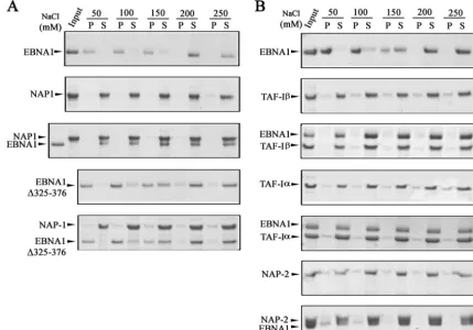
incubated on its own or at a 1:1 molar ratio with purified NAP1, NAP2, TAF-I␣, or TAF-I, with various salt concen-trations. Soluble and insoluble proteins were then separated by centrifugation, and the entire soluble and pellet fractions were analyzed by SDS-PAGE and Coomassie staining (Fig. 3). EBNA1 alone was insoluble at NaCl concentrations of 150 mM or less and became soluble at 200 mM NaCl, resulting in a shift from the pellet to the supernatant fraction (Fig. 3A and B, top panels). This is in contrast to the solubility profiles of NAP1, TAF-I␣, and TAF-I, each of which was largely soluble at all salt concentrations tested (50 to 250 mM). The solubility pro-file of EBNA1 changed significantly when it was incubated with NAP1 (Fig. 3A, third panel), TAF-I, or TAF-I␣ (Fig. 3B, third and fifth panels), in that EBNA1 became soluble at salt concentrations as low as 50 mM and fractionated with NAP1, TAF-I␣, and TAF-Iin the supernatant. Therefore, these re-sults provide additional evidence that EBNA1 interacts di-rectly with these nucleosome assembly proteins and show that these interactions can alter the properties of EBNA1.
In comparison to the other nucleosome assembly proteins, NAP2 had only a partial effect on EBNA1 solubility (Fig. 3B, bottom panel). NAP2 itself was soluble at all the salt concen-trations tested and caused⬃50% of the EBNA1 to shift to the soluble fraction in 50 mM salt and ⬃75% of EBNA1 to be
soluble in 100 mM salt. This suggests a weaker interaction of EBNA1 with NAP2 than with NAP1.
We previously observed that less NAP1 was recovered from an EBNA1 affinity column containing the⌬325-376 EBNA1 mutant, which lacks the large Gly-Arg-rich region, than from a wild-type EBNA1 column, suggesting that this region is important for NAP1 binding (28). We further examined the importance of the 325-376 region for the NAP1 interaction by repeating the solu-bility assays with EBNA1⌬325-376. EBNA1⌬325-376 alone had a solubility profile similar to that of wild-type EBNA1 (Fig. 3A, fourth panel). However, unlike the case for wild-type EBNA1, the solubility profile of EBNA1⌬325-376 did not change when it was incubated with NAP1, with the protein remaining insoluble at 50 and 100 mM NaCl (Fig. 3A, bottom panel). These results confirm that the 325-376 region is important for EBNA1 binding to NAP1.
Nucleosome assembly proteins contribute to
EBNA1-medi-ated transcriptional activation. Interactions of EBNA1 with
[image:5.594.77.506.72.371.2]the EBV oriP DS and FR sequences are known to activate DNA replication and to enhance transcription of other EBV latency genes, respectively (52, 77). Since both of these pro-cesses are likely to be regulated by nucleosome positioning and histone modifications, we examined whether NAP1, NAP2, and TAF-I affected these EBNA1 functions. We first tested the FIG. 3. Effects of nucleosome assembly proteins on EBNA1 solubility. (A) EBNA1 and NAP1 were incubated alone or together (at an equal molar ratio) in buffer containing 50 to 250 mM NaCl. Soluble (S) and precipitated (P) proteins were then separated by centrifugation and analyzed by SDS-PAGE and Coomassie blue staining. A sample of the protein(s) prior to incubation is shown as “input.” The same experiment was also performed with an EBNA1 mutant lacking amino acids 325 to 376 (EBNA1⌬325-376). (B) Same experiment as in panel A, except that TAF-I, TAF-I␣, or NAP2 was used in place of NAP1.
on November 8, 2019 by guest
http://jvi.asm.org/
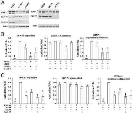
effects of silencing these proteins on transcriptional activation by EBNA1, using a standard reporter assay in which expression of a CAT gene is under the control of theoriPFR transcrip-tional element (52). Assays were performed in an EBV-nega-tive nasopharyngeal carcinoma cell line, CNE2Z, which is a physiologically relevant background for EBNA1, since devel-opment of this carcinoma is closely tied to EBV latent infec-tion. siRNAs specific to NAP1, NAP2, and a region of TAF-I conserved in both the␣and  subunits were used to down-regulate these proteins individually prior to cotransfection of the cells with the FR-CAT reporter plasmid, a SEAP reporter plasmid that is not regulated by EBNA1, and an EBNA1 ex-pression plasmid. As shown in Fig. 4A, introduction of the siRNAs decreased the endogenous expression of the target protein(s) compared to that with the control siRNA against GFP. As expected, the TAF-I siRNA decreased the expression of both TAF-I␣ and TAF-I without affecting NAP1, and
NAP1 siRNA decreased NAP1 without affecting TAF-I or NAP2. However, NAP2 siRNA partially decreased NAP1 in addition to dramatically decreasing NAP2 levels.
[image:6.594.80.503.71.433.2]Transcriptional assays were performed on the cells treated with each of the specific siRNAs and the results compared to those for cells treated with siGFP. EBNA1-mediated transcrip-tion was measured by assaying for CAT, while transcriptranscrip-tional effects that are independent of EBNA1 were determined by SEAP assays. We consistently found that EBNA1-mediated transcriptional activity was significantly reduced when NAP1 expression was decreased (P ⬍ 0.01) and more dramatically reduced upon downregulation of NAP-2 or TAF-I expression (Fig. 4B, left panel) (P ⬍ 0.01), perhaps due to the better silencing of these proteins than that of NAP1. NAP1 and TAF-I downregulation did not significantly affect the expres-sion of SEAP, showing that the effects were specific for EBNA1-mediated transcriptional activation (Fig. 4B, middle FIG. 4. Effects of nucleosome assembly proteins on transcriptional activation by EBNA1. (A) Western blots of extracts of CNE2Z cells after transfection with siRNA against GFP (negative control), NAP1, NAP2, or TAF-I. Duplicate samples are shown. (B) After transfection with the indicated siRNA (or with siGFP in the second columns), cells were transfected with an EBNA1 expression plasmid or an empty plasmid (first column in each histogram), an FR-CAT reporter plasmid that is EBNA1 dependent, and a SEAP reporter plasmid that is independent of EBNA1. Effects on CAT expression (left) and SEAP expression (middle) were determined separately. CAT levels were also normalized to SEAP levels to account for any nonspecific transcriptional effects (right).ⴱⴱ,P⬍0.001 relative to the EBNA1 positive control. (C) CNE2Z cells were transfected with a plasmid overexpressing NAP1, NAP2, TAF-I␣, or TAF-Ior with empty plasmid (second columns) prior to transfection with EBNA1 expression, CAT reporter, and SEAP reporter plasmids and calculation of transcriptional activities as in panel B.
on November 8, 2019 by guest
http://jvi.asm.org/
panel). In contrast, NAP2 silencing did significantly decrease EBNA1-independent SEAP expression, although not as dra-matically as dependent transcription. The EBNA1-dependent results were then normalized to the EBNA1-inde-pendent results to account for any general effects of silencing the nucleosome assembly proteins (Fig. 4B, right panel). We concluded that NAP1, TAF-I, and possibly NAP2 can posi-tively contribute to transcriptional activation by EBNA1.
We also examined the effect of overexpressing NAP1, NAP2, TAF-I␣, or TAF-I on EBNA1-mediated transcrip-tional activation by including a plasmid expressing each myc-tagged nucleosome assembly protein (or myc tag alone) along with the reporter and EBNA1 expression plasmids. The results from three independent experiments with duplicate samples showed that EBNA1-dependent transcriptional activation was two- to threefold lower when cells overexpressed NAP1, NAP2, TAF-I␣, or TAF-Ithan when they expressed the myc tag alone (Fig. 4C, left panel) (P ⬍0.01). However, overex-pression of these proteins did not affect exoverex-pression of SEAP from the EBNA1-independent SEAP reporter (Fig. 4C, mid-dle panel), showing that the effect has specificity for EBNA1. These results further support a role for the nucleosome assem-bly proteins in contributing to EBNA1-mediated transcrip-tional activation and indicate that the level of these proteins is important for regulation of this EBNA1 function.
TAF-I contributes to EBNA1-mediated replication. To
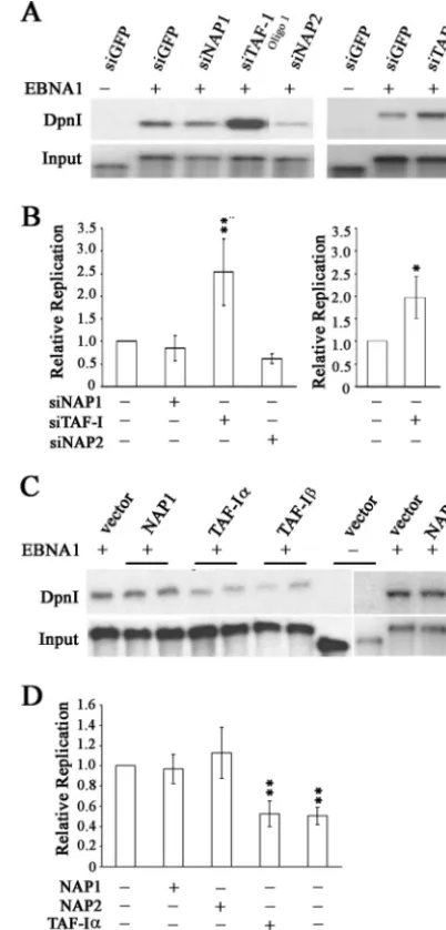
in-vestigate the roles of NAP-1, NAP-2, TAF-I␣, and TAF-Iin EBNA1 replication activity, we performed transient replica-tion assays in CNE2Z cells after downregulareplica-tion of the nucleo-some assembly proteins with siRNA treatment or with the control GFP siRNA. The replication of an oriP plasmid ex-pressing EBNA1 (or without EBNA1 as a negative control) was then assayed at 3 days posttransfection by DpnI resistance assays, in which the level of the DpnI-resistant plasmid (re-flecting replication in the mammalian cells) was compared to the total amount of plasmid recovered from the cells. As shown in Fig. 5A, silencing of TAF-I caused a pronounced increase in EBNA1-mediated plasmid replication, while silencing of NAP1 or NAP2 had less obvious effects. Composite results from multiple experiments confirmed that TAF-I silencing sig-nificantly stimulated EBNA1 replication activity (P⬍0.01) but that NAP1 and NAP2 silencing had no significant effect (Fig. 5B). To verify that the stimulation of DNA replication seen after TAF-I silencing was not due to off-target effects of the siRNA, we repeated the experiments using a second siRNA sequence against TAF-I (oligo 2). As shown in the right panels of Fig. 5A and B, this siRNA had similar effects on the replication of oriP plasmids to those with the initial TAF-I siRNA, confirming that the effects were due to the loss of TAF-I.
We further examined the contributions of NAP1, NAP2, TAF-I␣, and TAF-Ito EBNA1-mediated replication by con-ducting transient replication assays in the presence of plasmids that overexpress the myc-tagged nucleosome assembly proteins or the myc tag alone (Fig. 5C and D). Overexpression of either TAF-I␣or TAF-Iconsistently caused a twofold repression of EBNA1-mediated replication (P⬍0.01), while overexpression of NAP1 or NAP2 did not significantly change EBNA1 repli-cation activity. These results support the effects of RNA inter-ference, indicating that TAF-I negatively regulates
EBNA1-FIG. 5. Effects of nucleosome assembly proteins on DNA replica-tion activity of EBNA1. (A) CNE2Z cells were treated with siRNA against NAP1, NAP2, TAF-I, or GFP (negative control) and then transfected with anoriPplasmid expressing EBNA1 or lacking EBNA1 (first lane only). For TAF-I, two different siRNA sequences were used (oligo 1 and oligo 2). Three days later,oriPplasmids were harvested from the cells and linearized. One-tenth of the sample was saved as a recovery control (input), and then the remaining sample was digested with DpnI to digest any transfected plasmid that had not replicated in the human cells. Samples were analyzed by Southern blotting and probing for the oriP plasmid. The DpnI-resistant plasmid band is shown in the top panel. (B) Quantification of multiple experiments performed as in panel A. For TAF-I, the left panel is for oligo 1 and the right panel is for oligo 2. Values are shown relative to that for the sample with EBNA1 and siGFP within each experiment, which was set to 1.0.Pvalues of⬍0.01 (ⴱⴱ) and⬍0.05 (ⴱ) are indicated. (C) CNE2Z cells were transfected with plasmids overexpressing NAP1, NAP2, TAF-I␣, or TAF-Ior with empty plasmid (vector) along with theoriP
plasmid expressing EBNA1 or lacking EBNA1. Replication of theoriP
plasmids was then determined as in panel A. (D) Quantification of multiple experiments performed as in panel C. Values are shown relative to that for EBNA1 with an empty overexpression plasmid within each experiment, which was set to 1.0, andPvalues of⬍0.01 are indicated.
on November 8, 2019 by guest
http://jvi.asm.org/
[image:7.594.315.516.79.498.2]mediated replication but that NAP1 and NAP2 do not contribute to this process.
Association of nucleosome assembly proteins withoriP
func-tional elements.Transcriptional activation by EBNA1 requires
EBNA1 binding to the FR element oforiP, while DNA repli-cation fromoriPrequires EBNA1 binding to the DS element, and both of these elements are constitutively bound by EBNA1 in EBV-infected cells (10, 53). Therefore, proteins that are functioning directly with EBNA1 to modulate transcription and replication should localize with EBNA1 on the FR and DS sequences, respectively. To better understand the mechanisms by which NAP1 contributes to EBNA1-mediated tional activation and TAF-I contributes to both the transcrip-tion and replicatranscrip-tion functranscrip-tions of EBNA1, we performed ChIP assays with Raji cells to determine whether NAP1 or TAF-I localized with EBNA1 to the FR and DS elements within the EBV episome. NAP1 and TAF-I are both expected to have general chromatin interactions in addition to being targeted to specific sites. Therefore, ChIP assays were also performed to examine association with the BZLF1 promoter region, which is not regulated by EBNA1. In all cases, the levels of the DNA fragments recovered from sheared Raji cell DNA were deter-mined by quantitative real-time PCR.
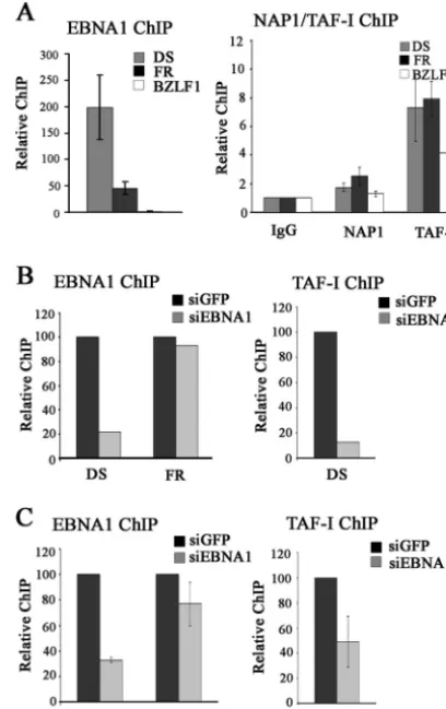
ChIP assays conducted with EBNA1 antibody confirmed that EBNA1 was bound to the DS and FR elements but not to the BZLF1 promoter (Fig. 6A, left panel), with better recovery of the DS element, as previously reported (13, 38, 56). In contrast, NAP1 antibody preferentially recovered the FR DNA fragment over either the DS (P⫽0.018) or BZLF1 (P⫽0.001) fragment. TAF-I antibody (which binds both TAF-I␣ and TAF-I) recovered all three DNA fragments to some degree but resulted in significantly more immunoprecipitation of both the DS and FR fragments than of the BZLF1 fragment (P⫽
0.009 for the DS element andP⫽00005 for the FR element). Therefore, TAF-I is preferentially localized with EBNA1 on both the DS and FR elements, while NAP1 is preferentially detected at the FR element. These results support the tional assays indicating that TAF-I modulates EBNA1 func-tions at both the FR and DS elements and that NAP1 contrib-utes to EBNA1 function only at the FR element.
We also investigated the requirement of EBNA1 for the recruitment of NAP1 and TAF-I tooriP elements by down-regulating EBNA1 in EBV-positive cells, using siRNA treat-ment. These experiments could not be performed with Raji cells due to their low transfection efficiency and instead were performed with D98/Raji fusion cells and AGS-rEBV gastric carcinoma cells, both of which contain EBV. In both cases, downregulation of the cellular levels of EBNA1 resulted in decreased EBNA1 binding to the DS element, as determined by ChIP (Fig. 6B and C, left panels). However, EBNA1 silenc-ing did not significantly decrease the level of EBNA1 bound to the FR element (Fig. 6B and C, left panels), which is known to contain higher-affinity EBNA1 binding sites that are more sta-bly bound than those in the DS element (3, 19, 32). Therefore, our analysis of the EBNA1 requirement for recruitment of nucleosome assembly proteins tooriPelements was limited to TAF-I recruitment to the DS element. ChIPs performed using antibody against TAF-I showed that EBNA1 silencing resulted in a decreased association of TAF-I with the DS element in
both D98/Raji and AGS-rEBV cells, consistent with EBNA1-mediated recruitment of TAF-I (Fig. 6B and C, right panels).
DISCUSSION
[image:8.594.317.521.71.395.2]We have previously shown that three nucleosome assembly proteins, NAP1, TAF-I␣, and TAF-I, are recovered from cell extracts on EBNA1 affinity columns (28). We have now con-firmed the interactions of EBNA1 with NAP1 and TAF-Iby glycerol gradient sedimentation and, in the process, have shown that these interactions are direct. NAP1 and TAF-I also altered the biochemical properties of EBNA1 in vitro, such that its solubility under low-salt conditions was dramati-cally increased, a property that might reflect the ability of these proteins to act as chaperones for EBNA1 in vivo. An interac-tion between EBNA1 and TAF-I␣was not detected by glycerol FIG. 6. Localization of NAP1 and TAF-I tooriPelements by ChIP. (A) ChIP experiments were performed with Raji cells, using antibodies against EBNA1 (left), NAP1, TAF-I, or nonspecific rabbit IgG (right). Recovered DNA fragments were quantified by real-time PCR, using primer sets for theoriPDS and FR elements and the BZLF1 promoter region. The amplification signals were normalized to those from the same cell lysates prior to IP, using the same primer pairs. Signals from NAP1 and TAF-I antibody samples were expressed relative to that for the control IgG samples, which was set to 1. The results shown are from three independent experiments, with PCR quantification per-formed in triplicate for each experiment. (B and C) D98/Raji (B) and AGS-rEBV (C) cells were treated with siRNA against EBNA1 or GFP, and ChIP assays were performed for EBNA1 (left) and TAF-I (right) as in panel A.
on November 8, 2019 by guest
http://jvi.asm.org/
gradient sedimentation, but like NAP1 and TAF-I, TAF-I␣ did increase the solubility of EBNA1 in vitro, indicating an interaction with EBNA1. Since gradient sedimentation analy-ses would require a more stable interaction than the solubility assays, the results indicate that EBNA1 can interact to some degree with either TAF-I␣or TAF-Ibut interacts more stably with TAF-I. This is consistent with our previous affinity col-umn experiments that identified TAF-Ias interacting more specifically with EBNA1 than TAF-I␣does. Since it is known that TAF-I␣ and TAF-I can heterodimerize (40, 42), the retention of TAF-I␣on EBNA1 affinity columns could have been indirect, mediated by TAF-I. In support of this model, coimmunoprecipitation assays showed that EBNA1 could be isolated in a complex with TAF-Ithat did not contain TAF-I␣, further pointing to TAF-Ias the more important TAF-I interaction.
Since NAP2 is a close homologue of NAP1 that is found in the same cells, we examined possible interactions of EBNA1 with NAP2. NAP2 antibody immunoprecipitated EBNA1 from Raji cells, but NAP1 was also recovered. Since NAP1 and NAP2 are known to interact (60), it is not clear whether the recovered EBNA1 was bound to NAP2 or to NAP1. On the other hand, NAP1 antibody recovered EBNA1 in the absence of NAP2. The results of glycerol gradient sedimentation with EBNA1 and NAP2 were ambiguous, since both proteins comi-grated even when analyzed individually. However, NAP2 did increase the solubility of EBNA1 in vitro, although to a lesser degree than that with NAP1. Taken together, the results sug-gest that the more prominent interaction of EBNA1 with the NAPs is through NAP1.
The fact that EBNA1 interacts with both NAP1 and TAF-I suggests that the interaction occurs through a region conserved in the two proteins. These proteins share a NAP domain and C-terminal acidic sequences, either of which might mediate the EBNA1 interaction. NAP1 also contains a long N-terminal sequence, but this does not appear to be important for the EBNA1 interaction, since an N-terminally truncated form of NAP1 that is present in human cell extracts is retained on EBNA1 affinity columns along with full-length NAP1 (28). In addition, TAF-Ilacks the N-terminal region present in NAP1 and TAF-I␣ contains only a very short N-terminal sequence. The observation that TAF-I␣interacts less stably with EBNA1 than TAF-I does suggests that this N-terminal sequence is inhibitory to binding, perhaps due to steric hindrance of the adjacent NAP domain.
Downregulation of NAP1, NAP2, and TAF-I (both␣and subunits) resulted in decreased transcriptional activation by EBNA1. Silencing of NAP1 and TAF-I occurred specifically without affecting the levels of the other nucleosome assembly proteins and did not affect the transcription of a reporter gene that was not under EBNA1 control. Therefore, NAP1 and TAF-I appear to contribute directly to EBNA1-mediated tran-scriptional activation, and in keeping with these results, both proteins were preferentially detected at the EBNA1-controlled FR transcriptional element. Due to technical limitations, we have not been able to determine whether the recruitment of NAP1 and TAF-Irequires EBNA1, but this is the most likely explanation. The results for NAP2 are more complicated to interpret because (i) NAP2 silencing also decreased NAP1 levels and (ii) NAP2 silencing also inhibited transcription of
the EBNA1-independent reporter, although not as dramati-cally as that of the EBNA1-dependent reporter. Therefore, although NAP2 could contribute directly to the transcriptional activity of EBNA1, it could also act indirectly through NAP1 and through general effects on chromatin structure. Note that while the degree to which downregulation of NAP1, NAP2, and TAF-I inhibited EBNA1-mediated transcription was only 1.5- to 3-fold, this is what is typically seen for effects of NAP1 and TAF-I on specific reporter genes under the control of their binding partners (65, 69).
Overexpression of any of the nucleosome assembly proteins was found to specifically inhibit EBNA1-dependent transcrip-tion. The fact that both silencing and overexpression of these proteins inhibited EBNA1-dependent transcription suggests that a squelching mechanism is at play, in which the formation of a particular complex containing the nucleosome assembly proteins is disrupted by either treatment. Similar observations were made withXenopusNAP1, as overexpressing and silenc-ing this protein both resulted in similar embryonic defects (1, 20). NAP1 is known to multimerize, forming complexes rang-ing from dimers to hexadecamers, in a concentration-depen-dent manner, where the largest complexes are impaired for histone binding (67). Therefore, altering the level of NAP1 in the cell may affect the nature of the NAP1 complexes formed, and hence their ability to interact with histones. In addition, nucleosome assembly proteins are known to mediate interac-tions between the p300 coactivator and specific transcription factors, which are important for their stimulatory effects on these transcription factors (51, 60, 69). Both overexpression and silencing of the nucleosome assembly proteins would be expected to inhibit such ternary interactions and therefore could account for the effects we observed on EBNA1-mediated transcriptional activation.
While NAP1 and TAF-I had similar effects on the transcrip-tional activity of EBNA1, only TAF-I had an obvious effect on EBNA1-mediated DNA replication, and in keeping with this observation, only TAF-I was specifically detected at the DS replication element. TAF-I downregulation stimulated DNA replication while overexpression inhibited it, indicating a reg-ulatory role for TAF-I in EBNA1-mediated replication. In addition to its interactions with p300 and related histone acety-lases, TAF-I forms part of the INHAT complex and inhibits acetylation of histones and, in some cases, other DNA-binding transcriptional activators (37, 43, 57). Histone acetylation has long been linked to transcriptional activation and is also known to influence the initiation of DNA replication from specific origins (11, 49, 70, 79). In addition, the cell cycle-dependent acetylation of histone H3 atoriPhas been reported (80) and has been shown to control the time in S phase at which repli-cation initiates fromoriP(81). Therefore, the inhibitory effect of TAF-I on replication fromoriPis likely to involve effects on histone acetylation.
An increasing number of examples suggest that viral pro-teins often usurp cellular nucleosome assembly propro-teins for their own purposes. For example, TAF-I, which was originally identified as a factor stimulating the replication and transcrip-tion of adenovirus core particles in vitro (40, 46), was recently shown to interact with the adenovirus core protein VII and to affect adenovirus early gene transcription in vivo (25). In ad-dition, the herpes simplex virus type 1 tegument protein VP22
on November 8, 2019 by guest
http://jvi.asm.org/
was found to bind TAF-I and to inhibit its chromatin assembly activity in vitro (68). NAP1 interacts with the E2 protein of papillomavirus (51) and the Tat protein of human immunode-ficiency virus type 1 (HIV-1) (69), in both cases activating transcription from viral elements bound by these proteins. In addition, NAP1 was found to be utilized by human T-cell leukemia virus type 1 in promoting transcription-independent nucleosome disruption at the viral promoter (58). Finally, NAP1 can form a complex with HIV-1 Rev, which stimulates Rev’s ability to export HIV RNA (9). We have now shown that the EBNA1 protein of EBV uses NAP1 and TAF-I in the activation of viral gene expression and uses TAF-I to regulate replication fromoriP, indicating that the nucleosome assembly proteins also play important roles in EBV latent infection.
ACKNOWLEDGMENTS
We gratefully acknowledge Pedro Rodriguez for NAP2 antibodies and the pET15b-NAP2 expression construct, Yukio Ishimi for NAP1 monoclonal antibody, and Lawrence S. Young for AGS-rEBV cells. We also thank Jerry Pelletier for pET15b-NAP1, K. Nagata for pET14b-TAF-I␣, and Alan Cochrane for the SEAP reporter plasmid. Finally, we thank Nirojini Sivachandran for assistance with the revi-sions.
This work was funded by the Canadian Cancer Society. L.F. is a tier 1 Canada Research Chair in Molecular Virology.
REFERENCES
1.Abu-Daya, A., W. M. Steer, A. F. Trollope, C. E. Friedeberg, R. K. Patient, A. W. Thorne, and M. J. Guille.2005. Zygotic nucleosome assembly protein-like 1 has a specific, non-cell autonomous role in hematopoiesis. Blood 106:514–520.
2.Adams, A.1987. Replication of latent Epstein-Barr virus genomes. J. Virol. 61:1743–1746.
3.Ambinder, R. F., W. A. Shah, D. R. Rawlins, G. S. Hayward, and S. D. Hayward.1990. Definition of the sequence requirements for binding of the EBNA-1 protein to its palindromic target sites in Epstein-Barr virus DNA. J. Virol.64:2369–2379.
4.Atanasiu, C., Z. Deng, A. Wiedmer, J. Norseen, and P. M. Lieberman.2006. ORC binding to TRF2 stimulates OriP replication. EMBO Rep.7:716–721. 5.Avolio-Hunter, T. M., P. N. Lewis, and L. Frappier.2001. Epstein-Barr nuclear antigen 1 binds and destabilizes nucleosomes at the viral origin of latent DNA replication. Nucleic Acids Res.29:3520–3528.
6.Bochkareva, E., L. Frappier, A. M. Edwards, and A. Bochkarev.1998. The RPA32 subunit of human replication protein A contains a single-stranded DNA binding domain. J. Biol. Chem.273:3932–3936.
7.Ceccarelli, D. F., and L. Frappier.2000. Functional analyses of the EBNA1 origin DNA binding protein of Epstein-Barr virus. J. Virol.74:4939–4948. 8.Chaudhuri, B., H. Xu, I. Todorov, A. Dutta, and J. L. Yates.2001. Human
DNA replication initiation factors, ORC and MCM, associate with oriP of Epstein-Barr virus. Proc. Natl. Acad. Sci. USA98:10085–10089.
9.Cochrane, A., L. L. Murley, M. Gao, R. Wong, K. Clayton, N. Brufatto, V. Canadien, D. Mamelak, T. Chen, D. Richards, M. Zeghouf, J. Greenblatt, C. Burks, and L. Frappier.2009. Stable complex formation between HIV Rev and the nucleosome assembly protein, NAP1, affects Rev function. Virology 388:103–111.
10.Daikoku, T., A. Kudoh, M. Fujita, Y. Sugaya, H. Isomura, and T. Tsurumi. 2004. In vivo dynamics of EBNA1-oriP interaction during latent and lytic replication of Epstein-Barr virus. J. Biol. Chem.279:54817–54825. 11.Danis, E., K. Brodolin, S. Menut, D. Maiorano, C. Girard-Reydet, and M.
Mechali.2004. Specification of a DNA replication origin by a transcription complex. Nat. Cell Biol.6:721–730.
12.Deng, Z., C. Atanasiu, J. S. Burg, D. Broccoli, and P. M. Lieberman.2003. Telomere repeat binding factors TRF1, TRF2, and hRAP1 modulate repli-cation of Epstein-Barr virus OriP. J. Virol.77:11992–12001.
13.Deng, Z., C. Atanasiu, K. Zhao, R. Marmorstein, J. I. Sbodio, N. W. Chi, and P. M. Lieberman.2005. Inhibition of Epstein-Barr virus OriP function by tankyrase, a telomere-associated poly-ADP ribose polymerase that binds and modifies EBNA1. J. Virol.79:4640–4650.
14.Dhar, S. K., K. Yoshida, Y. Machida, P. Khaira, B. Chaudhuri, J. A. Wohlschlegel, M. Leffak, J. Yates, and A. Dutta.2001. Replication from oriP of Epstein-Barr virus requires human ORC and is inhibited by geminin. Cell 106:287–296.
15.Dyson, P. J., and P. J. Farrell.1985. Chromatin structure of Epstein-Barr virus. J. Gen. Virol.66:1931–1940.
16.Frappier, L.2004. Viral plasmids in mammalian cells, p. 325–339.InB. E. Funnell and G. J. Phillips (ed.), Plasmid biology. ASM Press, Washington, DC. 17.Frappier, L., and M. O’Donnell.1992. EBNA1 distortsoriP, the
Epstein-Barr virus latent replication origin. J. Virol.66:1786–1790.
18.Frappier, L., and M. O’Donnell.1991. Epstein-Barr nuclear antigen 1 me-diates a DNA loop within the latent replication origin of Epstein-Barr virus. Proc. Natl. Acad. Sci. USA88:10875–10879.
19.Frappier, L., and M. O’Donnell.1991. Overproduction, purification, and characterization of EBNA1, the origin binding protein of Epstein-Barr virus. J. Biol. Chem.266:7819–7826.
20.Friedeberg, C., G. Scarlett, J. McGeehan, A. Abu-Daya, M. Guille, and G. Kneale.2006. Identification of a structural and functional domain in xNAP1 involved in protein-protein interactions. Nucleic Acids Res.34:4893–4899. 21.Gahn, T. A., and C. L. Schildkraut.1989. The Epstein-Barr virus origin of
plasmid replication, oriP, contains both the initiation and termination sites of DNA replication. Cell58:527–535.
22.Gahn, T. A., and B. Sugden.1995. An EBNA-1-dependent enhancer acts from a distance of 10 kilobase pairs to increase expression of the Epstein-Barr virus LMP gene. J. Virol.69:2633–2636.
23.Glaser, R., and M. Nonoyama.1974. Host cell regulation of induction of Epstein-Barr virus. J. Virol.14:174–176.
24.Harrison, S., K. Fisenne, and J. Hearing.1994. Sequence requirements of the Epstein-Barr virus latent origin of DNA replication. J. Virol.68:1913– 1925.
25.Haruki, H., M. Okuwaki, M. Miyagishi, K. Taira, and K. Nagata.2006. Involvement of template-activating factor I/SET in transcription of adeno-virus early genes as a positive-acting factor. J. Virol.80:794–801. 26.Hirt, B.1967. Selective extraction of polyoma DNA from infected mouse cell
cultures. J. Mol. Biol.26:365–369.
27.Holowaty, M. N., Y. Sheng, T. Nguyen, C. Arrowsmith, and L. Frappier. 2003. Protein interaction domains of the ubiquitin-specific protease, USP7/ HAUSP. J. Biol. Chem.278:47753–47761.
28.Holowaty, M. N., M. Zeghouf, H. Wu, J. Tellam, V. Athanasopoulos, J. Greenblatt, and L. Frappier.2003. Protein profiling with Epstein-Barr nu-clear antigen-1 reveals an interaction with the herpesvirus-associated ubiq-uitin-specific protease HAUSP/USP7. J. Biol. Chem.278:29987–29994. 29.Hsieh, D.-J., S. M. Camiolo, and J. L. Yates.1993. Constitutive binding of
EBNA1 protein to the Epstein-Barr virus replication origin, oriP, with dis-tortion of DNA structure during latent infection. EMBO J.12:4933–4944. 30.Hu, R. J., M. P. Lee, L. A. Johnson, and A. P. Feinberg.1996. A novel human
homologue of yeast nucleosome assembly protein, 65 kb centromeric to the p57KIP2 gene, is biallelically expressed in fetal and adult tissues. Hum. Mol. Genet.5:1743–1748.
31.Huang, D. P., J. H. Ho, Y. F. Poon, E. C. Chew, D. Saw, M. Lui, C. L. Li, L. S. Mak, S. H. Lai, and W. H. Lau.1980. Establishment of a cell line (NPC/ HK1) from a differentiated squamous carcinoma of the nasopharynx. Int. J. Cancer26:127–132.
32.Jones, C. H., S. D. Hayward, and D. R. Rawlins.1989. Interaction of the lymphocyte-derived Epstein-Barr virus nuclear antigen EBNA-1 with its DNA-binding sites. J. Virol.63:101–110.
33.Julien, M. D., Z. Polonskaya, and J. Hearing.2004. Protein and sequence requirements for the recruitment of the human origin recognition complex to the latent cycle origin of DNA replication of Epstein-Barr virus oriP. Virol-ogy326:317–328.
34.Karetsou, Z., G. Martic, G. Sflomos, and T. Papamarcaki.2005. The histone chaperone SET/TAF-Ibeta interacts functionally with the CREB-binding protein. Biochem. Biophys. Res. Commun.335:322–327.
35.Kawase, H., M. Okuwaki, M. Miyaji, R. Ohba, H. Handa, Y. Ishimi, T. Fujii-Nakata, A. Kikuchi, and K. Nagata.1996. NAP-I is a functional ho-mologue of TAF-I that is required for replication and transcription of the adenovirus genome in a chromatin-like structure. Genes Cells1:1045–1056. 36.Kennedy, G., and B. Sugden.2003. EBNA-1, a bifunctional transcriptional
activator. Mol. Cell. Biol.23:6901–6908.
37.Kutney, S. N., R. Hong, T. Macfarlan, and D. Chakravarti.2004. A signaling role of histone-binding proteins and INHAT subunits pp32 and Set/TAF-Ibeta in integrating chromatin hypoacetylation and transcriptional repres-sion. J. Biol. Chem.279:30850–30855.
38.Lin, A., S. Wang, T. Nguyen, K. Shire, and L. Frappier.2008. The EBNA1 protein of Epstein-Barr virus functionally interacts with Brd4. J. Virol.82: 12009–12019.
39.Lupton, S., and A. J. Levine.1985. Mapping of genetic elements of Epstein-Barr virus that facilitate extrachromosomal persistence of Epstein-Epstein-Barr vi-rus-derived plasmids in human cells. Mol. Cell. Biol.5:2533–2542. 40.Matsumoto, K., K. Nagata, M. Ui, and F. Hanaoka.1993. Template
activat-ing factor I, a novel host factor required to stimulate the adenovirus core DNA replication. J. Biol. Chem.268:10582–10587.
41.McBryant, S. J., and O. B. Peersen. 2004. Self-association of the yeast nucleosome assembly protein 1. Biochemistry43:10592–10599.
42.Miyaji-Yamaguchi, M., M. Okuwaki, and K. Nagata.1999. Coiled-coil struc-ture-mediated dimerization of template activating factor-I is critical for its chromatin remodeling activity. J. Mol. Biol.290:547–557.
43.Miyamoto, S., T. Suzuki, S. Muto, K. Aizawa, A. Kimura, Y. Mizuno, T.
on November 8, 2019 by guest
http://jvi.asm.org/
Nagino, Y. Imai, N. Adachi, M. Horikoshi, and R. Nagai.2003. Positive and negative regulation of the cardiovascular transcription factor KLF5 by p300 and the oncogenic regulator SET through interaction and acetylation on the DNA-binding domain. Mol. Cell. Biol.23:8528–8541.
44.Mosammaparast, N., C. S. Ewart, and L. F. Pemberton.2002. A role for nucleosome assembly protein 1 in the nuclear transport of histones H2A and H2B. EMBO J.21:6527–6538.
45.Muto, S., M. Senda, Y. Akai, L. Sato, T. Suzuki, R. Nagai, T. Senda, and M. Horikoshi. 2007. Relationship between the structure of SET/TAF-Ibeta/ INHAT and its histone chaperone activity. Proc. Natl. Acad. Sci. USA 104:4285–4290.
46.Nagata, K., H. Kawase, H. Handa, K. Yano, M. Yamasaki, Y. Ishimi, A. Okuda, A. Kikuchi, and K. Matsumoto.1995. Replication factor encoded by a putative oncogene, set, associated with myeloid leukemogenesis. Proc. Natl. Acad. Sci. USA92:4279–4283.
47.Norseen, J., A. Thomae, V. Sridharan, A. Aiyar, A. Schepers, and P. M. Lieberman.2008. RNA-dependent recruitment of the origin recognition complex. EMBO J.27:3024–3035.
48.Okuwaki, M., K. Kato, H. Shimahara, S. Tate, and K. Nagata.2005. As-sembly and disasAs-sembly of nucleosome core particles containing histone variants by human nucleosome assembly protein I. Mol. Cell. Biol.25:10639– 10651.
49.Pappas, D. L., Jr., R. Frisch, and M. Weinreich.2004. The NAD(⫹ )-depen-dent Sir2p histone deacetylase is a negative regulator of chromosomal DNA replication. Genes Dev.18:769–781.
50.Park, Y. J., and K. Luger.2006. Structure and function of nucleosome assembly proteins. Biochem. Cell Biol.84:549–558.
51.Rehtanz, M., H.-M. Schmidt, U. Warthorst, and G. Steger.2004. Direct interaction between nucleosome assembly protein 1 and the papillomavirus E2 proteins involved in activation of transcription. Mol. Cell. Biol.24:2153– 2168.
52.Reisman, D., and B. Sugden.1986.trans-Activation of an Epstein-Barr viral transcriptional enhancer by the Epstein-Barr viral nuclear antigen 1. Mol. Cell. Biol.6:3838–3846.
53.Ritzi, M., K. Tillack, J. Gerhardt, E. Ott, S. Humme, E. Kremmer, W. Hammerschmidt, and A. Schepers.2003. Complex protein-DNA dynamics at the latent origin of DNA replication of Epstein-Barr virus. J. Cell Sci. 116:3971–3984.
54.Rodriguez, P., D. Munroe, D. Prawitt, L. L. Chu, E. Bric, J. Kim, L. H. Reid, C. Davies, H. Nakagama, R. Loebbert, A. Winterpacht, M. J. Petruzzi, M. J. Higgins, N. Nowak, G. Evans, T. Shows, B. E. Weissman, B. Zabel, D. E. Housman, and J. Pelletier.1997. Functional characterization of human nu-cleosome assembly protein-2 (NAP1L4) suggests a role as a histone chap-erone. Genomics44:253–265.
55.Rodriguez, P., J. Pelletier, G. B. Price, and M. Zannis-Hadjopoulos.2000. NAP-2: histone chaperone function and phosphorylation state through the cell cycle. J. Mol. Biol.298:225–238.
56.Schepers, A., M. Ritzi, K. Bousset, E. Kremmer, J. L. Yates, J. Harwood, J. F. Diffley, and W. Hammerschmidt.2001. Human origin recognition com-plex binds to the region of the latent origin of DNA replication of Epstein-Barr virus. EMBO J.20:4588–4602.
57.Seo, S. B., P. McNamara, S. Heo, A. Turner, W. S. Lane, and D. Chakravarti. 2001. Regulation of histone acetylation and transcription by INHAT, a human cellular complex containing the set oncoprotein. Cell104:119–130. 58.Sharma, N., and J. K. Nyborg.2008. The coactivators CBP/p300 and the
histone chaperone NAP1 promote transcription-independent nucleosome eviction at the HTLV-1 promoter. Proc. Natl. Acad. Sci. USA105:7959– 7963.
59.Shaw, J., L. Levinger, and C. Carter.1979. Nucleosomal structure of Ep-stein-Barr virus DNA in transformed cell lines. J. Virol.29:657–665. 60.Shikama, N., H. M. Chan, M. Krstic-Demonacos, L. Smith, C. W. Lee, W.
Cairns, and N. B. La Thangue.2000. Functional interaction between nu-cleosome assembly proteins and p300/CREB-binding protein family coacti-vators. Mol. Cell. Biol.20:8933–8943.
61.Shire, K., D. F. Ceccarelli, T. M. Avolio-Hunter, and L. Frappier.1999.
EBP2, a human protein that interacts with sequences of the Epstein-Barr virus nuclear antigen 1 important for plasmid maintenance. J. Virol.73: 2587–2595.
62.Shire, K., P. Kapoor, K. Jiang, M. N. Hing, N. Sivachandran, T. Nguyen, and L. Frappier.2006. Regulation of the EBNA1 Epstein-Barr virus protein by serine phosphorylation and arginine methylation. J. Virol.80:5261–5272. 63.Stewart, S., C. W. Dawson, K. Takada, J. Curnow, C. A. Moody, J. W. Sixbey,
and L. S. Young.2004. Epstein-Barr virus-encoded LMP2A regulates viral and cellular gene expression by modulation of the NF-kappaB transcription factor pathway. Proc. Natl. Acad. Sci. USA101:15730–15735.
64.Summers, H., J. A. Barwell, R. A. Pfuetzner, A. M. Edwards, and L. Frap-pier.1996. Cooperative assembly of EBNA1 on the Epstein-Barr virus latent origin of replication. J. Virol.70:1228–1231.
65.Telese, F., P. Bruni, A. Donizetti, D. Gianni, C. D’Ambrosio, A. Scaloni, N. Zambrano, M. G. Rosenfeld, and T. Russo.2005. Transcription regulation by the adaptor protein Fe65 and the nucleosome assembly factor SET. EMBO Rep.6:77–82.
66.Thorley-Lawson, D. A., and A. Gross.2004. Persistence of the Epstein-Barr virus and the origins of associated lymphomas. N. Engl. J. Med.350:1328– 1337.
67.Toth, K. F., J. Mazurkiewicz, and K. Rippe. 2005. Association states of nucleosome assembly protein 1 and its complexes with histones. J. Biol. Chem.280:15690–15699.
68.van Leeuwen, H., M. Okuwaki, R. Hong, D. Chakravarti, K. Nagata, and P. O’Hare.2003. Herpes simplex virus type 1 tegument protein VP22 interacts with TAF-I proteins and inhibits nucleosome assembly but not regulation of histone acetylation by INHAT. J. Gen. Virol.84:2501–2510.
69.Vardabasso, C., L. Manganaro, M. Lusic, A. Marcello, and M. Giacca.2008. The histone chaperone protein nucleosome assembly protein-1 (hNAP-1) binds HIV-1 Tat and promotes viral transcription. Retrovirology5:8. 70.Vogelauer, M., L. Rubbi, I. Lucas, B. J. Brewer, and M. Grunstein.2002.
Histone acetylation regulates the time of replication origin firing. Mol. Cell 10:1223–1233.
71.Wang, Y., J. E. Finan, J. M. Middeldorp, and S. D. Hayward.1997. P32/TAP, a cellular protein that interacts with EBNA-1 of Epstein-Barr virus. Virology 236:18–29.
72.Woolaway, K., K. Asai, A. Emili, and A. Cochrane.2007. hnRNP E1 and E2 have distinct roles in modulating HIV-1 gene expression. Retrovirology4:28. 73.Wu, H., P. Kapoor, and L. Frappier.2002. Separation of the DNA replica-tion, segregareplica-tion, and transcriptional activation functions of Epstein-Barr virus nuclear antigen 1. J. Virol.76:2480–2490.
74.Wysokenski, D. A., and J. L. Yates.1989. Multiple EBNA1-binding sites are required to form an EBNA1-dependent enhancer and to activate a minimal replicative origin within oriP of Epstein-Barr virus. J. Virol.63:2657–2666. 75.Yates, J., N. Warren, D. Reisman, and B. Sugden.1984. A cis-acting element from the Epstein-Barr viral genome that permits stable replication of re-combinant plasmids in latently infected cells. Proc. Natl. Acad. Sci. USA 81:3806–3810.
76.Yates, J. L., S. M. Camiolo, and J. M. Bashaw.2000. The minimal replicator of Epstein-Barr virus oriP. J. Virol.74:4512–4522.
77.Yates, J. L., N. Warren, and B. Sugden.1985. Stable replication of plasmids derived from Epstein-Barr virus in various mammalian cells. Nature313: 812–815.
78.Young, L. S., and A. B. Rickinson.2004. Epstein-Barr virus: 40 years on. Nat. Rev. Cancer4:757–768.
79.Zhou, J., C. Chau, Z. Deng, W. Stedman, and P. M. Lieberman.2005. Epigenetic control of replication origins. Cell Cycle4:889–892.
80.Zhou, J., C. M. Chau, Z. Deng, R. Shiekhattar, M. P. Spindler, A. Schepers, and P. M. Lieberman.2005. Cell cycle regulation of chromatin at an origin of DNA replication. EMBO J.24:1406–1417.
81.Zhou, J., A. R. Snyder, and P. M. Lieberman.2009. Epstein-Barr virus episome stability is coupled to a delay in replication timing. J. Virol.83: 2154–2162.
82.Zlatanova, J., C. Seebart, and M. Tomschik.2007. Nap1: taking a closer look at a juggler protein of extraordinary skills. FASEB J.21:1294–1310.