JOURNAL OFVIROLOGY, Apr. 2010, p. 3147–3161 Vol. 84, No. 7 0022-538X/10/$12.00 doi:10.1128/JVI.02587-09
Copyright © 2010, American Society for Microbiology. All Rights Reserved.
A V3 Loop-Dependent gp120 Element Disrupted by CD4 Binding
Stabilizes the Human Immunodeficiency Virus
Envelope Glycoprotein Trimer
䌤
Shi-Hua Xiang,
1Andre
´s Finzi,
1Beatriz Pacheco,
1Kevin Alexander,
1Wen Yuan,
1Carlo Rizzuto,
1Chih-Chin Huang,
2Peter D. Kwong,
2and Joseph Sodroski
1,3*
Department of Cancer Immunology and AIDS, Dana-Farber Cancer Institute, Department of Pathology, Division of AIDS, Harvard Medical School, Boston, Massachusetts 021151; Vaccine Research Center, National Institute of Allergy and
Infectious Diseases, National Institutes of Health, Bethesda, Maryland 208922; and Department of
Immunology and Infectious Diseases, Harvard School of Public Health, Boston, Massachusetts 021153
Received 10 December 2009/Accepted 5 January 2010
Human immunodeficiency virus (HIV-1) entry into cells is mediated by a trimeric complex consisting of noncovalently associated gp120 (exterior) and gp41 (transmembrane) envelope glycoproteins. The binding of gp120 to receptors on the target cell alters the gp120-gp41 relationship and activates the membrane-fusing capacity of gp41. Interaction of gp120 with the primary receptor, CD4, results in the exposure of the gp120 third variable (V3) loop, which contributes to binding the CCR5 or CXCR4 chemokine receptors. We show here that insertions in the V3 stem or polar substitutions in a conserved hydrophobic patch near the V3 tip result in decreased gp120-gp41 association (in the unliganded state) and decreased chemokine receptor binding (in the CD4-bound state). Subunit association and syncytium-forming ability of the envelope glycoproteins from primary HIV-1 isolates were disrupted more by V3 changes than those of laboratory-adapted HIV-1 envelope glycoproteins. Changes in the gp1202,19,20, and21 strands, which evidence suggests are proximal to the V3 loop in unliganded gp120, also resulted in decreased gp120-gp41 association. Thus, a gp120 element composed of the V3 loop and adjacent beta strands contributes to quaternary interactions that stabilize the unliganded trimer. CD4 binding dismantles this element, altering the gp120-gp41 relationship and rendering the hydrophobic patch in the V3 tip available for chemokine receptor binding.
The entry of human immunodeficiency virus type 1 (HIV-1) is mediated by the viral envelope glycoproteins (9, 79). The HIV-1 envelope glycoproteins are synthesized as an ⬃ 850-amino acid precursor, which trimerizes and is posttranslation-ally modified by carbohydrates to create a 160-kDa glycopro-tein (gp160). The gp160 envelope glycoproglycopro-tein precursor is proteolytically processed in the Golgi apparatus, resulting in a gp120 exterior envelope glycoprotein and a gp41 transmem-brane envelope glycoprotein (16, 17, 66, 76). In the mature HIV-1 envelope glycoprotein trimer, the three gp120 subunits are noncovalently bound to three membrane-anchored gp41 subunits (32).
HIV-1 entry involves the binding of gp120 in a sequential fashion to CD4 and one of the chemokine receptors, CCR5 or CXCR4 (1, 8, 15, 18, 25, 36). CD4 binding triggers the forma-tion of an activated intermediate that is competent for binding to CCR5 or CXCR4 (29, 69, 73, 78). These chemokine tors are G protein-coupled, 7-transmembrane segment recep-tors with relatively short N termini. The choice of chemokine receptors is dictated primarily by the sequence of a gp120 region, the third variable (V3) loop, that exhibits variability among HIV-1 strains and becomes exposed upon CD4 binding
(4, 8, 10, 33, 37, 38, 49, 59, 75). X-ray crystal structures of CD4-bound HIV-1 gp120 have revealed that the gp120 “core” consists of a gp41-interactive inner domain, a surface-exposed and heavily glycosylated outer domain, and a conformationally flexible bridging sheet (38, 43, 79). In the CD4-bound state, the V3 loop projects 30 Å from the gp120 core, toward the che-mokine receptor (38). The V3 loop in these structures consists of three elements: (i) conserved antiparallel  strands that contain a disulfide bond at the base of the loop; (ii) a confor-mationally flexible stem; and (iii) a conserved tip (37, 38). During the virus entry process, the base of the gp120 V3 loop and elements of the bridging sheet interact with the CCR5 N terminus, which is acidic and contains sulfotyrosine residues (12–14, 23, 24). Sulfotyrosine 14 of CCR5 is thought to insert into a highly conserved pocket near the V3 base, driving fur-ther conformational rearrangements that result in the rigidifi-cation of the V3 stem (37). The conserved-turn at the tip of the V3 loop, along with some residues in the V3 stem, is believed to bind the “body” of CCR5, i.e., the extracellular loops and membrane-spanning helices. CCR5 binding is thought to induce further conformational changes in the HIV-1 envelope glycoproteins, leading to the fusion of the viral and target cell membranes by the gp41 transmembrane enve-lope glycoproteins.
CCR5 binding involves two points of contact with the gp120 V3 loop: (i) the CCR5 N terminus with the V3 base and (ii) the CCR5 body with the V3 tip and distal stem (12–14, 23, 24, 37, 38). The intervening V3 stem can tolerate greater
conforma-* Corresponding author. Mailing address: Dana-Farber Cancer In-stitute, 44 Binney Street, CLS 1010, Boston, MA 02115. Phone: (617) 632-3371. Fax: (671) 632-4338. E-mail: [email protected] .edu.
䌤Published ahead of print on 20 January 2010.
3147
on November 8, 2019 by guest
http://jvi.asm.org/
tional and sequence variation, features that might decrease HIV-1 susceptibility to host antibodies (30). Despite amino acid variation, the length of the V3 loop is well conserved among naturally occurring group M (major group) HIV-1 strains (30, 42). This conserved length may be important for aligning the two CCR5-binding elements of the V3 loop. In addition to allowing optimal CCR5 binding, the conserved V3 length and orientation may be important for CCR5 binding to exert effects on the conformation of the HIV-1 envelope gly-coproteins. We examine here the consequences of introducing extra amino acid residues into the V3 stem. The residues were introduced either into both strands of the V3 loop, attempting to preserve the symmetry of the structure, or into one of the strands, thereby kinking the loop. The effects of these changes on assembly, stability, receptor binding, and the membrane-fusing capacity of the HIV-1 envelope glycoproteins were as-sessed. In addition to effects on chemokine receptor binding, unexpected disruption of gp120-gp41 association was ob-served. Further investigation revealed a conserved patch in the tip of the V3 loop that is important for the association of gp120 with the trimeric envelope glycoprotein complex, as well as for chemokine receptor binding. Apparently, the V3 loop and adjacent gp120 structures contribute to the stability of the trimer in the unliganded HIV-1 envelope glycoproteins. These structures are known to undergo rearrangement upon CD4 binding, suggesting their involvement in receptor-induced changes in the virus entry process.
MATERIALS AND METHODS
HIV-1 envelope glycoprotein and CCR5 mutants.Mutations were introduced into the pSVIIIenv plasmid expressing the full-length envelope glycoproteins from the ADA, YU2, 89.6, HXBc2, and MN27 HIV-1 strains (68), using a QuikChange II XL site-directed mutagenesis protocol (Stratagene). The pres-ence of the desired mutations was confirmed by DNA sequencing. The V3 insertion mutants are designated m1, m2, m3, etc. (Fig. 1). The gp120 substitu-tion mutants are designated with the amino acid residue to the right of the number substituted for the amino acid residue to the left of the number. All residues are numbered according to those of the prototypic HXBc2 sequence, as per current convention (41).
The predicted amino acid sequence of the HIV-1 ADA⌬V3 mutant is:
296CTgagHC332, where the lower case gag represents a glycine-alanine-glycine
linker. The predicted amino acid sequence of the HIV-1 HXBc2⌬V3 and
⌬V3-20 mutants, respectively, are: 296CTgagHC332 and 296CTRPNNNTRK
RgagKIGNMRQAHC332
(82).
The CCR5-GG mutant contains a two-residue insertion in the N terminus of human CCR5. The sequence of the CCR5-GG protein near the insertion is: . . .S PIYDINYYTSEGGPCQKINV. . ., with the inserted residues underlined.
Syncytium formation assay.The ability of the HIV-1 envelope glycoprotein
variants to mediate cell-cell fusion was determined by the␣-complementation
assay (35). In this assay, the N-terminal (␣) fragment of -galactosidase is
coexpressed with the HIV-1 envelope glycoproteins; the C-terminal () fragment
of-galactosidase is expressed in the receptor-bearing target cells. Upon fusion
of the envelope glycoprotein-expressing cells and the target cells, the
-galacto-sidase is reconstituted, and its activity can be measured. Briefly, 293T cells in
six-well plates were transfected with plasmids expressing the-galactosidase␣
fragment, the HIV-1 envelope glycoproteins and the HIV-1 Tat protein in a 4:4:1 weight ratio. Cf2Th-CD4/CCR5 cells (for the HIV-1 ADA, YU2, and 89.6 envelope glycoproteins) or Cf2Th-CD4/CXCR4 cells (for the HIV-1 89.6, HXBc2, and MN27 envelope glycoproteins) in 100-mm dishes were transfected
with 3g of a plasmid expressing the-galactosidasefragment. On the next
day, the Cf2Th target cells were lifted from the plates with 5 mM EDTA–
phosphate-buffered saline (EDTA-PBS) and reseeded at 105
cells/well in 96-well plates. One day later, the transfected 293T cells were lifted from the plates with
5 mM EDTA-PBS and diluted to 2⫻105
cells/ml with Dulbecco modified Eagle
medium (DMEM). Approximately 2⫻104293T cells (in 100l) expressing the
HIV-1 envelope glycoproteins were added in triplicate to each well of a 96-well
plate containing the target cells. The cocultivated cells were cultured at 37°C in
a CO2incubator for 4 h. The medium was then removed, and the cells were
washed once with PBS and lysed in 20l of lysis buffer. The plate was covered
with Parafilm, wrapped in plastic film and place at⫺70°C for 30 min. The
-galactosidase activity was measured with a Galacto-Star-galactosidase
re-porter gene assay system for mammalian cells (Tropix, Bedford, MA), using a Berthold Microplate Luminometer LB 96V (Promega).
The ability of the wild-type and mutant HIV-1 ADA envelope glycoproteins expressed transiently in 293T cells to form syncytia with target Cf2Th-CD4 cells expressing CD4 and either the wild-type CCR5 protein or the CCR5-GG mutant
was assessed by using the␣-complementation assay described above. The 293T
cells were transfected with 0.8g of the envelope glycoprotein expressor
plas-mid, 0.8g of the plasmid expressing the␣-fragment of-galactosidase, and 0.2
g of the Tat-expressing plasmid. The Cf2Th-CD4 cells in 100-mm dishes were
transfected with 2.5g of the plasmid expressing either wild-type CCR5 or
CCR5-GG and 2.5g of the plasmid expressing the-fragment of
-galactosi-dase. On the next day, the transfected Cf2Th-CD4 cells were harvested with 5
mM EDTA-PBS and reseeded in a 96-well plate (2⫻104
cells/well). The similar surface expression level of the wild-type CCR5 and the CCR5-GG proteins was verified by fluorescence-activated cell sorting using phycoerythrin-conjugated 2D7 antibody (BD Pharmingen). The medium of the 293T cells was changed on the day after transfection. The following day, the 293T cells were harvested with
5 mM EDTA-PBS, diluted to 2⫻105cells/ml with DMEM, and then
coculti-vated with the Cf2Th-CD4 cells as described above. After 4 h, the cells were lysed
and-galactosidase activity measured, as described above.
In some cases, an additional assay was used to measure the syncytium-forming ability of the HIV-1 envelope glycoprotein variants (Table 1). In this assay, cells coexpressing the HIV-1 envelope glycoproteins and Tat were cocultivated with TZM-bl cells. Cell-cell fusion was quantitated by measuring luciferase activity (A. Finzi et al., unpublished data).
Infection by single-round luciferase-expressing HIV-1. Recombinant lucif-erase-expressing HIV-1 viruses were produced by transfection of 293T cells with the pCMV Gag-Pol packaging plasmid, the pHIV-luc vector and the pSVIIIenv plasmids (50). At 3 days after transfection, the cell supernatants were harvested. The amount of virus in the supernatants was assessed by measurement of reverse transcriptase (RT) (57).
For infection, Cf2Th-CD4/CCR5 or Cf2Th-CD4/CXCR4 cells were plated at
a density of 6⫻103cells/well in a 96-well plate. The following day, the cells were
incubated with 2,500 RT units of recombinant virus per well. Two days later, the cells were lysed, and the luciferase activity measured in the Berthold microplate luminometer LB 96V (Promega).
Immunoprecipitation of HIV-1 envelope glycoproteins.293T cells were trans-fected with pSVIIIenv plasmids expressing the HIV-1 envelope glycoproteins.
One day later, the cells were metabolically labeled for 16 h with35
S-Protein Labeling Mix (Perkin-Elmer). The cell lysates (containing gp160 and gp120) and
media (containing gp120) were used for immunoprecipitation. Briefly, 400l of
clarified cell lysate or medium was incubated with 100l of 10% protein
A-Sepharose beads (Amersham Biosciences) and 4l of a mixture of a sera from
HIV-1-infected individuals. For some experiments, 1g of a monoclonal
anti-body was used instead of the serum mixture. The mixtures were brought to a volume of 1 ml with PBS and incubated on a shaking platform at room temper-ature for 1 h. The immunoprecipitation of HIV-1 gp120-containing cell super-natants by the G3-299 monoclonal antibody was carried out at 4°C for 2 h in the presence of protease inhibitors (one tablet Complete protease inhibitor cocktail [mini, EDTA-free] per 10-ml binding reaction; Roche Applied Science, Ger-many). The pelleted beads were washed once with 0.5 M NaCl-PBS and twice
with PBS. The beads were then suspended in a 2⫻loading buffer, boiled, and
applied to a 10% sodium dodecyl sulfate-polyacrylamide gel.
CCR5 binding assay.To assess CCR5-binding ability, normalized amounts of radiolabeled gp120 envelope glycoproteins from transfected 293T cell superna-tants were incubated with sCD4 and Cf2Th-CCR5 cells. Briefly, Cf2Th-CCR5 cells were lifted from the plate by using 5 mM EDTA-PBS (pH 7.5). After a wash with DMEM, the cells were resuspended in DMEM and added to 1.5-ml
micro-centrifuge tubes (3⫻106cells/tube). The radiolabeled gp120-containing cell
supernatants (500l) and 10g of sCD4 were added to the tube, and the volume
was adjusted to 1 ml with DMEM. The tubes were rocked at room temperature
for 1 h. The cells were then washed once with PBS and lysed in 1 ml of 1⫻NP-40
buffer. The cell lysates were precipitated with a mixture of sera from HIV-1-infected individuals and protein A-Sepharose beads (Amersham Biosciences) at 4°C for 2 h. The precipitated gp120 was analyzed by SDS-PAGE and autora-diography.
on November 8, 2019 by guest
http://jvi.asm.org/
RESULTS
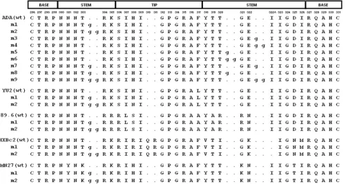
HIV-1 V3 loop insertion mutants.To examine the effects of changes in the length or orientation of the HIV-1 gp120 V3 loop, a panel of insertion mutants derived from the ADA HIV-1 isolate was created (Fig. 1). The ADA virus is a pri-mary, CCR5-using (R5) HIV-1 isolate (27). In one set of mu-tants, one or two glycine residues were introduced into one strand of the conformationally flexible stem of the V3 loop; these asymmetric insertions would presumably kink the loop. In a second set of mutants, identical insertions were placed symmetrically into the N- and C-terminal strands of the V3 stem; the intent of these changes was to extend the length of the V3 loop while minimizing any alterations in the orientation of the tip.
The ability of the V3 insertion mutants to mediate the fusion of envelope glycoprotein-expressing cells and cells bearing CD4 and CCR5 was assessed. Plasmid DNAs expressing the mutant envelope glycoproteins and the␣fragment of -galac-tosidase were transfected into 293T cells. The 293T cells were cocultivated with canine Cf2Th cells expressing human CD4, human CCR5 and thefragment of-galactosidase. Success-ful fusion of the Env-expressing cells with the Cf2Th-CD4/ CCR5 cells results in reconstitution of enzymatically active
-galactosidase (35). In this cell-cell fusion assay, most of the HIV-1 ADA V3 insertion mutants exhibited⬍5% of the ac-tivity observed for the wild-type ADA envelope glycoproteins (Fig. 2A). Mutants 3, 4, 5, and 6 exhibited syncytium-forming abilities between 5 and 60% of that seen for the wild-type
ADA envelope glycoproteins. These four mutants contain gly-cine insertions in the C-terminal strand of the V3 stem.
[image:3.585.45.546.70.337.2]To examine the generality of these results, V3 mutants 1 and 2 were created in the context of envelope glycoproteins derived from two additional primary viruses, the R5 YU2 strain and the dualtropic (R5X4) 89.6 strain (11, 47), and from two CXCR4-using (X4) laboratory-adapted viruses, HXBc2 and MN27 (26, 64) (Fig. 1). The syncytium-forming abilities of these envelope glycoproteins were assessed as described above. For the envelope glycoproteins from the HXBc2 and MN27 HIV-1 strains, the mutant 1 glycoproteins, which contain a single glycine inserted into the N-terminal strand of the V3 stem, exhibited 80 to 85% of the syncytium-forming abilities of the respective wild-type envelope glycoproteins (Fig. 2B). In the context of the YU2 and 89.6 envelope glycoproteins, the mutant 1 insertion more significantly reduced syncytium-form-ing ability. Likewise, the HXBc2 and MN27 mutant 2 envelope glycoproteins, which contain two glycine residues inserted into the N-terminal strand of the V3 stem, exhibited ca. 50 and 70% of the syncytium-forming ability of the respective wild-type envelope glycoproteins. In contrast, the mutant 2 variants of the YU2 and 89.6 envelope glycoproteins induced only very low numbers of syncytia. The dualtropic 89.6 envelope glyco-protein variants induced fusion with target cells expressing CD4 and CCR5 or CD4 and CXCR4 equivalently. Thus, in-sertions in the V3 stem generally reduced the ability of HIV-1 envelope glycoproteins to induce cell-cell fusion. The extent of this reduction depended upon the number of residues inserted
FIG. 1. V3 loop insertion mutants. The V3 loop sequences of the wild-type (wt) and insertion mutant envelope glycoproteins from the indicated HIV-1 strains are aligned. The numbering of the gp120 residues corresponds to that of the HXBc2 prototype, according to current convention (41). The corresponding segments of the V3 loop structure in the CD4-bound state (38) are shown above the alignment. The inserted glycine residues are shown in lower case.
VOL. 84, 2010 HIV-1 gp120 V3 LOOP REGULATES TRIMER STABILITY 3149
on November 8, 2019 by guest
http://jvi.asm.org/
and upon the particular HIV-1 envelope glycoproteins altered, with the laboratory-adapted HIV-1 envelope glycoproteins be-ing functionally more tolerant of these changes than the pri-mary HIV-1 envelope glycoproteins.
Infectivity of HIV-1 with V3 loop insertions.The ability of the V3 insertion mutants to support HIV-1 infection was as-sessed in a single-round Env complementation assay (31). Re-gardless of the HIV-1 strain from which the envelope glyco-proteins were derived, all of the V3 mutants were markedly defective in mediating HIV-1 entry (Fig. 2C and D). Complete defectiveness was also observed for an ADA envelope glyco-protein mutant with an alanine substitution in place of the glycine insert in mutant 1 (data not shown).
Expression, processing, and subunit association of the HIV-1 Env mutants.To investigate the basis for the reduced activities of the V3 mutants in syncytium-forming ability and virus replication, cells transiently expressing the wild-type and mutant envelope glycoproteins were radiolabeled. Cell lysates and supernatants were precipitated by a polyclonal mixture of sera from HIV-1-infected individuals. In the cell lysates, the wild-type gp160 envelope glycoprotein precursor and the mature gp120 envelope glycoprotein were evident
(Fig. 3A and B). For the primary HIV-1 ADA, YU2, and 89.6 envelope glycoproteins, although the levels of the V3 mutant gp160 envelope glycoproteins were generally similar to those of the wild-type counterparts, the levels of cell-associated gp120 glycoproteins were relatively reduced (Fig. 3A and B). For these HIV-1 envelope glycoproteins, the amounts of gp120 shed into the medium were increased for the V3 mutants compared to the wild-type glycoproteins. These results suggest that the V3 loop insertions decrease the association of the gp120 and gp41 subunits in the unli-ganded envelope glycoprotein complex. The amount of cell-associated gp120 glycoprotein was lower for the wild-type HXBc2 and MN27 envelope glycoproteins than for the pri-mary HIV-1 envelope glycoproteins, under these labeling conditions (Fig. 3B). Slight decreases in the amount of cell-associated gp120 were observed for the HXBc2 m1 and m2 mutants, relative to the wild-type HXBc2 envelope glyco-proteins; however, no differences between the phenotypes of the MN27 wild-type and mutant envelope glycoproteins with respect to gp160 precursor processing or gp120-gp41 asso-ciation were evident. Thus, the V3 loop insertions decrease gp120-gp41 association of primary HIV-1 envelope
glyco-FIG. 2. Syncytium-forming activity and ability of V3 insertion mutants to support HIV-1 infection. (A) 293T cells expressing the wild-type (wt) or the indicated HIV-1 ADA envelope glycoprotein mutants were cocultivated with Cf2Th-CD4/CCR5 cells and syncytium formation measured as described in Materials and Methods. The negative control cells were transfected with a plasmid expressing the HIV-1 HXBc2 envelope glycoproteins. The syncytium-forming activity of the mutants is reported relative to that seen for the wild-type HIV-1 ADA envelope glycoproteins. Means and standard deviations derived from four replicate assays are shown. (B) The relative syncytium-forming activities of the mutant 1 and 2 envelope glycoproteins are shown for each of the indicated HIV-1 strains. Cf2Th-CD4/CXCR4 target cells were used for the HXBc2, MN27 and 89.6 envelope glycoproteins, and Cf2Th-CD4/CCR5 target cells were used for the YU2 and 89.6 envelope glycoproteins. The experiments were conducted as described in panel A. The means and standard deviations derived from four replicate assays are shown. (C and D) The wild-type (wt) or mutant (m1, m2, etc.) envelope glycoproteins from the indicated HIV-1 strain were assessed for the ability to support the infection of recombinant HIV-1 expressing luciferase, using Cf2Th-CD4/CCR5 target cells (for the ADA, YU2, and 89.6 HIV-1 envelope glycoproteins) or Cf2Th-CD4/CXCR4 target cells (for the HXBc2 and 89.6 HIV-1 envelope glycoproteins). The infectivity of the mutants relative to that seen for the respective wild-type envelope glycoproteins is shown. The experiments were repeated with comparable results.
on November 8, 2019 by guest
http://jvi.asm.org/
[image:4.585.111.474.70.350.2]proteins; this phenotype is less evident for envelope glyco-proteins derived from laboratory-adapted HIV-1 isolates.
Decreased stability of the envelope glycoprotein complex explains the lower function of the V3 loop mutants in mediat-ing virus entry compared to cell-cell fusion. Because a greater time interval elapses between envelope glycoprotein synthesis and engagement of the target cell in the virus entry assay, decreases in the functional stability of the HIV-1 envelope glycoproteins are more apparent than in the syncytium forma-tion assay (84; Finzi et al., unpublished).
Interaction of mutant envelope glycoproteins with ligands.
To assess the effect of the V3 loop insertions on gp120 formation, the recognition of the gp120 glycoprotein by con-formation-dependent ligands was assessed. Radiolabeled gp120 glycoproteins from transfected cell supernatants were precipitated by a mixture of sera from HIV-1-infected individ-uals, which recognizes gp120 independently of its conforma-tional integrity, or by specific gp120-directed ligands whose recognition depends upon gp120 conformation. The ligands include: CD4-Ig, in which the amino-terminal two domains of CD4 are fused to an immunoglobulin Fc; 17b and 412d, two antibodies that recognize CD4-induced epitopes near the che-mokine receptor-binding surface of gp120 (58, 72); and 39F, which recognizes a conformation-dependent V3 epitope (45). All of the HIV-1 ADA gp120 mutants tested bound CD4 efficiently, as evidenced by the precipitation of these glycopro-teins by CD4-Ig (Fig. 4A). The 17b and 412d antibodies pre-cipitated all of the gp120 mutants, although the recognition of some of the mutants by the 17b antibody was less efficient than that of the wild-type gp120 glycoprotein. This result is consis-tent with the proximity of the 17b epitope to the base of the V3 loop (43, 58). The 39F anti-V3 antibody precipitated most of the mutants equivalently to the wild-type gp120, with three exceptions. Mutants 4, 6, and 9 were precipitated less
effi-ciently than the wild-type gp120 glycoprotein by the 39F anti-body. All three mutants have a two-residue insertion in the carboxy-terminal half of the V3 stem, suggesting that changes in this V3 region can disrupt the 39F epitope. These results suggest that the gp120 V3 insertion mutants are not globally misfolded but exhibit specific alterations near or within the V3 loop.
The ability of the gp120 mutants to bind CCR5 was exam-ined (78). Radiolabeled gp120 glycoproteins from transfected cell supernatants were incubated in the presence of soluble CD4 with Cf2Th cells expressing CCR5. After a washing step, the amount of gp120 bound to the cells was determined. Figure 4B shows that all of the V3 insertions decreased the efficiency of CCR5 binding. This decrease was readily apparent for the gp120 glycoproteins from the R5 HIV-1 strains, ADA and YU2. The wild-type gp120 glycoprotein from the R5X4 (dual-tropic) 89.6 HIV-1 strain exhibits a lower affinity for CCR5 than those of R5 gp120 glycoproteins (2, 3). Therefore, higher concentrations of the 89.6 gp120 are required to demonstrate CCR5 binding. At these higher concentrations, mutants 1 and 2 from the 89.6 strain bound CCR5 less efficiently than the wild-type 89.6 gp120 glycoprotein (Fig. 4B). Thus, insertions into the V3 stem of the HIV-1 gp120 glycoprotein result in significant decreases in the efficiency of CCR5 binding.
[image:5.585.45.542.70.290.2]Partial compensation of the syncytium-forming ability of V3 insertion mutants by a CCR5 protein with an extended amino terminus.Current models of HIV-1 gp120-CCR5 interaction suggest that the CCR5 N terminus binds near the V3 base and the body of CCR5 binds the V3 tip (12–14, 23, 24, 37, 38). Thus, the gp120 mutants with insertions in the V3 stem might utilize CCR5 less efficiently because of poor alignment of these two binding contacts. In this case, the mutants might be more effective if the CCR5 N terminus were extended further from the body of the chemokine receptor. To test this, two glycine
FIG. 3. Processing and subunit association of HIV-1 envelope glycoprotein variants. (A to D) 293T cells expressing the wild-type (wt) or mutant envelope glycoproteins from the indicated HIV-1 strains were radiolabeled. The cells were pelleted and lysed. The radiolabeled cell lysates and media were precipitated by a mixture of sera from HIV-1-infected individuals. The envelope glycoproteins precipitated from the cell lysates and media were analyzed by SDS-PAGE. The gp160 and gp120 envelope glycoproteins are indicated.
VOL. 84, 2010 HIV-1 gp120 V3 LOOP REGULATES TRIMER STABILITY 3151
on November 8, 2019 by guest
http://jvi.asm.org/
residues were inserted into the CCR5 sequence, between the N terminus and the initial cysteine residue. This mutant, CCR5-GG, and wild-type CCR5 were expressed in Cf2Th-CD4 cells, which were assessed for the ability to form syncytia with cells expressing the wild-type or mutant ADA envelope glycopro-teins. The syncytium-forming ability of some of the V3 mutant envelope glycoproteins, relative to that of the wild-type ADA envelope glycoproteins, was greater with target cells expressing the CCR5-GG mutant than with target cells expressing wild-type CCR5 (Fig. 5). This phenowild-type was particularly evident for envelope glycoprotein mutants (mutants 7, 8, and 9) with symmetrical substitutions in the V3 loop and was also seen for mutant 1 with a single glycine insertion. The defective entry of viruses with the mutant envelope glycoproteins was not com-pensated by the expression of the CCR5-GG mutant on the target cell (data not shown). These results are consistent with a model suggesting that one of the consequences of the V3
insertions is a disruption of the optimal spacing between gp120 regions important for binding the CCR5 N terminus and body.
A conserved V3 element that is important for gp120-gp41 association. The effect of V3 loop insertions on gp120-gp41 association could result from an incompatibility of a kinked or extended loop with the proper packing of the gp120 subunits in the trimeric spike. Alternatively, specific structures in the V3 loop could positively contribute to subunit association. Al-though the V3 region is not absolutely essential for gp120-gp41 association in the laboratory-adapted HXBc2 envelope glyco-proteins (see Fig. 3C), mild increases in the amount of gp120 spontaneously shed into the medium have been observed for V3 loop-deleted envelope glycoproteins compared to the wild-type HXBc2 envelope glycoproteins (81). To examine the role of the V3 loop in maintaining the integrity of the trimeric envelope glycoprotein complex in a primary HIV-1 isolate, the V3 loop of the ADA gp120 glycoprotein was deleted in pre-cisely the same manner as that used previously for the HXBc2 envelope glycoproteins from a laboratory-adapted HIV-1. The V3 deletion resulted in a decrease in gp120-gp41 association comparable to that which resulted from an insertion into the V3 region (see Fig. 3D). Thus, the absence of the V3 loop can weaken gp120-gp41 interactions in the envelope glycoprotein trimer of a primary HIV-1 isolate.
The result described above raised the possibility that a V3 loop element contributes in a positive way to subunit associa-tion. Our observation that V3 insertions decreased the stability of the envelope glycoprotein trimers from multiple primary HIV-1 strains suggested that such a V3 element might be conserved in different HIV-1 isolates. Thus, we shifted our attention away from the more variable V3 stem to the rela-tively conserved V3 tip. Although the structure of the V3 loop in the unliganded HIV-1 envelope glycoproteins is unknown, in the CD4-bound gp120 (38), a-hairpin at the V3 tip juxta-poses residues 307, 309, and 317 (Fig. 6, column at far right). Some variability is tolerated in these residues in HIV-1 strains; however, the hydrophobic character of these residues is almost always maintained (Fig. 7A). We hypothesized that this hydro-phobic patch in the V3 tip serves to strengthen gp120-gp41 association in the unliganded HIV-1 envelope glycoprotein trimer. Amino acid residues of different degrees of hydropho-bicity were introduced into residues 307, 309, and 317 of the HIV-1 YU2 envelope glycoproteins. Although hydrophobic substitutions did not significantly disrupt gp120-gp41 associa-tion, substitutions of alanine or hydrophilic residues resulted in severe decreases in subunit association (Fig. 7B to D). Thus, the hydrophobic patch in the V3 tip contributes to subunit association in the unliganded HIV-1 envelope glycoprotein trimer.
[image:6.585.43.281.69.395.2]Contribution of the V3 hydrophobic patch to HIV-1 enve-lope glycoprotein function.The V3 tip is known to contribute to CCR5 binding (8, 9, 12, 14, 34, 58). The effect of changes in the hydrophobic V3 patch on CCR5 binding and syncytium formation were examined. Decreases in CCR5-binding affinity resulted from hydrophilic substitutions in residues 307, 309, and 317 (Fig. 4C and Table 1). These decreases were accom-panied by defects in the ability to mediate the formation of syncytia (Table 1). The effects of changes in the hydrophobic V3 patch were in some cases more disruptive of gp120-gp41 association than of CCR5 binding and syncytium formation; in
FIG. 4. Binding of ligands to HIV-1 envelope glycoprotein variants. (A) 293T cells expressing the wild-type (wt) or mutant (m1 to m9) HIV-1 ADA envelope glycoproteins were radiolabeled. The cell su-pernatants were precipitated by a mixture of sera from HIV-1-infected individuals (PS), by CD4-Ig, or by the indicated monoclonal antibody. The gp120 glycoproteins precipitated from the media were analyzed by SDS-PAGE and autoradiography. (B and C) Radiolabeled wild-type (wt) or mutant gp120 envelope glycoproteins from the indicated HIV-1 strains were incubated at room temperature for 1 h with Cf2Th-CCR5 cells in the presence of sCD4. The cells were washed and lysed, and the bound gp120 was precipitated by a mixture of sera from HIV-1-in-fected individuals. The bound gp120 glycoprotein was analyzed by SDS-PAGE and autoradiography.
on November 8, 2019 by guest
http://jvi.asm.org/
general, however, the phenotypic effects of the V3 changes on all of these properties correlated.
Involvement of V3-proximal gp120 regions in association with gp41. Previous studies have suggested that, in the unli-ganded gp120 glycoprotein, the V3 loop may interact with other gp120 regions, particularly the19,20, and21 strands (the fourth conserved [C4] region) and the V1/V2 stem-loop structure (51–53, 67, 74, 80). Several residues conserved among the gp120 glycoproteins of different HIV-1 strains were iden-tified in these regions. The effect of alteration of these residues in the HIV-1YU2envelope glycoproteins on the association of gp120 with gp41 was examined (Table 1). Some changes in the
19,20, and21 strands resulted in decreased subunit asso-ciation (Table 1 and Fig. 7D). In addition, changes in leucine 120 and valine 122 in the2 strand of the conserved V1/V2 stem also reduced gp120-gp41 association. Thus, changes in several HIV-1 gp120 regions that may be proximal to the V3 loop in the unliganded state resulted in decreases in the asso-ciation of gp120 with gp41.
Previous studies have examined the effect of the HIV-1 gp120 changes on gp120-gp41 association (32, 63, 71, 84; Finzi
et al., unpublished). The results of these studies, as well as the results generated in the present study, are summarized in Fig. 6. The N and C termini and inner domain of gp120, particularly the inner domain-sandwich, are the major contributors to the noncovalent interaction with gp41 (43, 55, 63, 84; Finzi et al., unpublished). Our results indicate that other gp120 regions (the V3 loop,2,17,19,20, and21) can also influence the stability of the association of gp120 and gp41. A compar-ison of the different crystallized gp120 structures indicates that these regions can change conformation in response to ligand binding or alterations introduced into gp120 to promote crys-tallization. Although the structure of the unliganded HIV-1 gp120 glycoprotein is unknown, in light of this conformational flexibility, these gp120 elements are potentially in proximity in the unliganded envelope glycoprotein trimer.
Effects of gp120 changes on a discontinuous V3-C4 epitope.
[image:7.585.135.451.69.379.2]The G3-299 monoclonal antibody has been previously shown to recognize a discontinuous HIV-1 gp120 epitope that is apparently composed of elements from the V3 loop and the fourth conserved (C4) region (51, 52). The G3-299 antibody neutralizes laboratory-adapted HIV-1 strains (60,
FIG. 5. Syncytium-forming ability of V3 insertion mutants with target cells expressing a CCR5 mutant. (A to C) 293T cells expressing the wild-type (wt) or V3 mutant envelope glycoproteins from the indicated HIV-1 strain were cocultivated with Cf2Th cells expressing CD4 and either wild-type (wt) CCR5 (white) or the CCR5-GG mutant (gray). CCR5-GG has two glycines inserted after residue 18, thus extending the N terminus. Syncytium-forming ability was measured as described in Materials and Methods. In panels A and B, the syncytium-forming abilities of the envelope glycoproteins are all normalized to that of the wild-type HIV-1 envelope glycoproteins on target cells expressing CD4 and wild-type CCR5. In panel C, the syncytium-forming abilities of the mutant envelope glycoproteins are normalized to that of the wild-type envelope glycoproteins on target cells expressing CD4 and the corresponding CCR5 variant. The means and standard deviations derived from three experiments are shown. The delta V3 envelope glycoprotein is missing the gp120 V3 loop and is a fusion-defective negative control (81). 293T cells transfected with the empty pcDNA vector serve as an additional negative control.
VOL. 84, 2010 HIV-1 gp120 V3 LOOP REGULATES TRIMER STABILITY 3153
on November 8, 2019 by guest
http://jvi.asm.org/
70), indicating that this epitope is both intact and exposed on the unliganded envelope glycoprotein trimers of at least some HIV-1 variants. The effect of gp120 changes on the integrity of this epitope was examined by precipitation of a large panel of wild-type and mutant gp120 envelope glyco-proteins by the G3-299 antibody. Of note, changes in the V3 loop (residues 307, 309, and 317) or in the 21 strand (residues 434 and 435) significantly decreased HIV-1 gp120 recognition by the G3-299 antibody (Tables 1 and 2). In the CD4-bound state, the only gp120 conformation for which a V3 loop structure is available (37, 38), these two elements are separated (Fig. 8). This is consistent with the observa-tion that CD4 binding decreases the recogniobserva-tion of HIV-1 gp120 by the G3-299 antibody (52).
The contribution of V3 loop and21 residues to the G3-299 epitope suggests that, in the unliganded HIV-1 gp120
glyco-protein, these gp120 elements are proximate and form a dis-continuous structural element. To gain additional insight into this possibility, we examined the structure of HIV-1 gp120 bound to F105, a CD4-binding site (CD4BS) antibody that recognizes a gp120 conformation distinct from that seen by CD4 (6, 83). F105 and G3-299 can simultaneously bind gp120 (6), indicating that G3-299 can recognize the F105-bound con-formation. With one exception, serine 375 (discussed below), the gp120 residues implicated in G3-299 recognition are dis-tinct from those that contact F105 (Fig. 8). In the F105-gp120 crystal, the V3 loop is intact but disordered (6). Nonetheless, in this conformation, the V3 loop projects toward the21 strand, potentially interacting with the21 residues implicated in G3-299 binding (Fig. 8).
Substitution of a tryptophan residue for serine 375 resulted in a decrease in the recognition of gp120 by the G3-299
anti-FIG. 6. Changes in HIV-1 gp120 resulting in decreased association with gp41. A ribbon diagram of the unliganded simian immunodeficiency virus (SIV) gp120 core structure (5) is shown in the left column. In the three columns on the right, the HIV-1 gp120 glycoprotein is shown in the different conformations observed in available crystal structures, complexed with the Fab fragments of the b12 or F105 neutralizing antibodies (6, 87), or with two-domain CD4 (38). The gp120 structures are viewed from the perspective seen by the Fab or CD4 proteins; the outer domains of the gp120 cores are aligned. The trimeric axis of the envelope glycoprotein complex is located on the left side of each structure, in approximately the vertical orientation (44, 48). The gp120 core in the b12-bound structure was modified to predispose the protein to assume the CD4-bound conformation (87). In the top row, the HIV-1 domains are colored as follows: outer domain (yellow), inner domain (red) and bridging sheet components (blue for the20-21 loop and green for the2-3 V1/V2 stem). In the cases where the V3 loop structure was not determined, the position of the V3 base is indicated. In the CD4-bound structure, the elements of the V3 loop are labeled. The gp120 beta strands (defined in the CD4-bound structure [43]) relevant to the present study are also labeled. In the bottom row, the gp120 residues are colored according to the gp120-gp41 association index (red, association index⬍0.5; green, association indexⱖ0.5) observed upon mutagenesis of the HIV-1YU2 and HXBc2 gp120 glycoproteins (32, 63, 71, 84; the present study; Finzi et al., unpublished). In the CD4-bound gp120 structure, the three hydrophobic residues in the tip of the V3 loop that were implicated in gp120-gp41 association are labeled.
on November 8, 2019 by guest
http://jvi.asm.org/
[image:8.585.78.502.68.402.2]body (see Table 1 footnotes). Because serine 375 contacts the F105 antibody, which does not compete with G3-299 for gp120 binding (6), the effects of the S375W change on G3-299 bind-ing are likely indirect. Indeed, it has been shown that the
S375W mutant favors the CD4-bound conformation (83), which is recognized less efficiently by the G3-299 antibody (52).
[image:9.585.43.540.80.594.2]The V3 loop and adjacent gp120 elements in the unliganded HIV-1 envelope glycoprotein trimer.The X-ray crystal structure
FIG. 7. The hydrophobic V3 patch. (A) The V3 variable loops of the primate immunodeficiency viruses are aligned. The degree of variation in residues 307, 309, and 317 in the HIV-1 gp120 V3 tip is shown (42). The percentage of sequences in each virus group that contain the indicated residue is shown as a superscript to the right of the single-letter amino acid designation. Amino acid variants that are found in less than 1% of the sequences surveyed are not listed. (B to D) 293T cells expressing the wild-type (wt) or mutant HIV-1 YU2 envelope glycoproteins were radiolabeled. The cells were pelleted and lysed. The radiolabeled cell lysates and media were precipitated by a mixture of sera from HIV-1-infected individuals. The envelope glycoproteins precipitated from the cell lysates and medium were analyzed by SDS-PAGE. The gp160 and gp120 envelope glycoproteins are indicated.
VOL. 84, 2010 HIV-1 gp120 V3 LOOP REGULATES TRIMER STABILITY 3155
on November 8, 2019 by guest
http://jvi.asm.org/
of the CD4-bound HIV-1 gp120 envelope glycoprotein with an intact V3 loop (38) has been fitted to tomograms derived from cryo-electron microscopy studies of HIV-1 virion spikes (48). In the CD4-bound state, the exposed V3 loop projects obliquely from the outer domain toward both the trimer axis and the target cell (Fig. 6). Although the detailed structure of unliganded HIV-1 gp120 with an intact V3 loop is unknown, low-resolution electron cryotomograms of the unliganded HIV-1 virion spike are avail-able (48, 88). The unliganded SIV gp120 core structure (5) cannot be accommodated within the electron density of the unliganded HIV-1 envelope glycoprotein spike (48). Thus, the structure ob-served in the crystallized SIV gp120 core is distinct from that in the unliganded HIV-1 envelope glycoprotein complex, due to real or artifactual differences between the unliganded SIV and HIV-1 gp120 glycoproteins. Both the CD4-bound and the b12
antibody-bound HIV-1 gp120 core crystal structures can be readily fitted to the electron cryotomograms of the unliganded HIV-1 envelope glycoprotein spike (6, 48). The position and orientation of the V3 base in these models and the deduced proximity of the V3 loop to the21 strand in the unliganded HIV-1 envelope glycoproteins (see above) suggests that the V3 loops likely project toward the trimer axis (Fig. 9 and data not shown). Thus, in the unliganded HIV-1 envelope glycoproteins, the three V3 loops are potentially poised for interactions with gp41 and/or the other gp120 elements implicated in stabilizing the association with gp41.
DISCUSSION
Here we show that the insertion of amino acid residues into the stem of the V3 variable loop of the gp120 envelope
glyco-TABLE 1. Phenotypes of HIV-1YU2mutants
Envelope
glycoprotein Residue location
Indexa Binding
Cell-to-cell fusiond
G3-299 recognitione
Association Processing CD4b
CCR5c
Wild-type YU2 1.00 1.00 1.00 ⫹⫹⫹⫹ 1.00 1.00
V120S 2 0.29 1.28 1.20 ND ND 1.82
L122R 2 0.44 1.15 0.77 ND ND 1.62
V200A 3 ND ND 1.10 ND ND 0.93
V200S 3 0.7 1.69 0.60 ND ND 1.21
I307A V3 loop 0.25 1.10 ND ⫹⫹ 0.05 ND
I307E V3 loop 0.30 1.03 0.86 ⫹ 0.02 0.20
I307L V3 loop 0.63 1.34 0.79 ⫹⫹⫹⫹ 1.00 0.26
I307S V3 loop ND ND 0.69 ND ND 0.10
I309A V3 loop 0.34 1.45 0.71 ⫹⫹⫹ 0.81 0.32
I309S V3 loop 0.28 1.25 0.81 ⫹⫹⫹ 0.48 0.10
I309L V3 loop 1.07 1.31 0.98 ⫹⫹⫹⫹ 1.01 0.81
L317A V3 loop 0.32 1.52 ND ⫹⫹⫹ 0.84 ND
L317E V3 loop 0.29 1.45 0.88 ⫹⫹ 0.02 0.56
L317S V3 loop 0.21 1.83 0.78 ⫺ 0.03 0.60
L317F V3 loop 0.82 1.05 0.72 ND ND 0.61
L317I V3 loop 1.29 1.07 0.66 ⫹⫹⫹⫹ 1.05 0.35
I420S 19 0.28 1.65 0.30 ⫹ 0.79 1.83
I423S 20 0.19 2.16 0.51 ⫹⫹⫹⫹ 0.03 1.75
I423R 20 0.14 2.54 0.60 ⫹⫹⫹⫹ 0.01 0.83
I423L 20 0.15 2.59 0.82 ⫹⫹⫹⫹ 0.91 0.91
I424S 20 0.21 1.91 0.45 ⫹⫹⫹⫹ 0.44 1.22
M434S 21 0.27 1.28 0.47 ND ND 0.15
M434R 21 0.68 0.50 0.45 ND ND 0.16
a
The processing and association indices were determined by precipitation of radiolabeled cell lysates and supernatants with mixtures of sera from HIV-1-infected individuals. The association index is a measure of the ability of the mutant gp120 molecule to remain associated with the envelope glycoprotein complex on the
expressing cell, relative to that of the wild-type envelope glycoproteins. The association index is calculated as follows: association index⫽(关mutant gp120兴cell⫻
关wild-type gp120兴supernatant)/(关mutant gp120兴supernatant⫻ 关wild-type gp120兴cell). The processing index is a measure of the conversion of the mutant gp160 envelope
glycoprotein precursor to mature gp120, relative to that of the wild-type envelope glycoproteins. The processing index was calculated by the formula: processing index⫽
(关total gp120兴mutant⫻ 关gp160兴wild type)/(关gp160兴mutant⫻ 关total gp120兴wild type). Additional HIV-1 YU2 gp120 mutants that exhibited association indices of⬍0.20 were
K121D, R298G, E381R, K421A, and P437A (data not shown). ND, not determined.
b
Radiolabeled wild-type and mutant gp120 glycoproteins in the supernatants of envelope-expressing 293T cells were incubated with various amounts of sCD4-Ig for
2 h at 37°C in the presence of 70l of 10% protein A-Sepharose (American Biosciences). At a near-saturating concentration of sCD4-Ig for the wild-type gp120
glycoprotein, the relative ratio of the mutant gp120 glycoprotein precipitated is reported. ND, not determined.
c
The CCR5-binding ability of the HIV-1 gp120 envelope glycoprotein variants was determined as described in Materials and Methods. The amount of bound mutant
gp120 glycoprotein was compared to the amount of bound wild-type gp120 glycoprotein. The relative CCR5-binding ability is reported as follows:⫹⫹⫹⫹, 75 to 100%
of the wild-type gp120 level;⫹⫹⫹, 50 to 74% of the wild-type level;⫹⫹, 25 to 49% of the wild-type level;⫹, 5 to 24% of the wild-type level; and –,⬍5% of the wild-type
level. ND, not determined.
d
To assess cell-to-cell fusion, 3⫻105
293T cells were cotransfected by the calcium phosphate method with an HIV-1 Tat-expressing plasmid, pLTR-Tat, and the
pSVIIIenv plasmid expressing the HIV-1YU2envelope glycoproteins. At 2 days after transfection, 3⫻10
4
293T cells were added to TZM-bl target cells that were seeded
at a density of 3⫻104
cells/well in 96-well luminometer-compatible tissue culture plates (Dynex) 24 h before the assay. Cells were coincubated for 6 h at 37°C, after
which they were lysed by the addition of 30l of passive lysis buffer (Promega) and three freeze-thaw cycles. The luciferase activity in each well was measured as
described above. The reported value represents the ratio of the luciferase activity observed for the mutant envelope glycoproteins relative to that of the wild-type envelope glycoproteins. ND, not determined.
e
Radiolabeled wild-type and mutant gp120 glycoproteins in the supernatants of 293T cells expressing the HIV-1 YU2 envelope glycoproteins were precipitated by the G3-299 antibody for 2 h at 4°C in the presence of Complete protease inhibitor cocktail (Roche Applied Science). Precipitates were analyzed as described in Materials and Methods. At a near-saturating concentration of G3-299 antibody for the wild-type gp120 glycoprotein, the relative ratio of the mutant gp120 glycoprotein precipitated is reported. Relative values for G3-299 recognition for mutant HIV-1 YU2 gp120 glycoproteins not shown in the table were as follows: m1, 1.07; m2, 0.77; H66A, 1.91; W69L, 2.17; L111A, 0.59; S375W, 0.26; H66A/S375W, 0.55; W69L/S375W, 0.73; and L111A/S375W, 0.60. ND, not determined.
on November 8, 2019 by guest
http://jvi.asm.org/
[image:10.585.43.545.80.333.2]protein from primary HIV-1 isolates resulted in decreased sub-unit association in the envelope glycoprotein trimer and de-creased chemokine receptor binding. Similar phenotypes were observed for alteration of a conserved hydrophobic patch in the V3 tip. The displacement of the V3 hydrophobic patch by the V3 stem insertions represents a unifying explanation of the pheno-types, but more direct contributions of the V3 stem to subunit association and chemokine receptor binding are formally possi-ble. The results support functional roles for the V3 loop in both the unliganded state (i.e., maintaining subunit association) and the CD4-bound state (i.e., chemokine receptor binding).
Detailed structures of the assembled HIV-1 envelope glyco-proteins are not yet available to explain precisely how V3 loop alterations lead to disruption of the gp120-gp41 interaction in the unliganded envelope glycoprotein trimer. Not even the structure of an unliganded gp120 monomer with an intact gp41-interactive inner domain or V3 loop is available for guid-ance. Nonetheless, X-ray crystal structures of gp120 in various conformations, in some cases induced by the binding of neu-tralizing antibodies, have been fitted into low-resolution
cryo-electron tomograms of the unliganded HIV-1 virion spike (6, 48, 55). These models are consistent with the expected place-ment of conserved and variable gp120 surfaces, glycosylation sites, epitopes for neutralizing and non-neutralizing antibodies and receptor-binding regions (44). In these models, the V3 loop projects from the gp120 outer domain toward the trimer axis. Fixation of the assembled HIV-1 envelope glycoproteins, but not the gp120 monomer, by treatment with chemical cross-linkers has been shown to decrease specifically the binding of antibodies directed against the V3 loop and the gp41-interac-tive face of gp120 (85). These observations are consistent with the close packing of the V3 loops of the gp120 subunits near the trimeric axis of the unliganded HIV-1 envelope glyco-protein complex. This arrangement may help in sequestering the CCR5-binding elements of the V3 loop away from poten-tial recognition by neutralizing antibodies.
The requirement to avoid the binding of potentially neutral-izing antibodies constrains the structure of the unliganded envelope glycoproteins from primary HIV-1 isolates. Such con-straints are removed by extensive passage of HIV-1 in tissue-cultured cell lines (56, 61, 68, 77). Short of destabilizing gp120-gp41 association, V3 variation may modulate quaternary subunit interactions that determine sensitivity to neutralizing antibodies. Indeed, in one case, a dualtropic HIV-1 retained some function after truncation of the V3 loop but was more sensitive to neutralization by antibodies (46). Likewise, changes in V3 can determine differences in sensitivity to neu-tralizing antibodies, even those directed against conserved gp120 epitopes, between primary and laboratory-adapted HIV-1 isolates (67, 86). V3 changes have also been shown to account for the increased neutralization resistance of simian-human immunodeficiency viruses passaged in monkeys (7, 21). Thus, alterations in the gp120 V3 loop can modulate sensitivity to neutralization by antibodies directed against multiple enve-lope glycoprotein determinants.
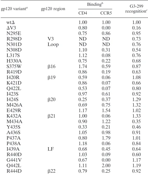
The syncytium-inducing function of the envelope glyco-proteins was disrupted to a greater extent by V3 loop insertions for primary HIV-1 isolates compared to laboratory-adapted viruses. Deletion of the V3 loop exerted an effect on gp120-gp41 association that was greater for the primary HIV-1 ADA envelope glycoproteins than for the laboratory-adapted HXBc2 envelope glycoproteins. Several studies have suggested that the V3 loops of laboratory-adapted HIV-1 envelope gly-coproteins are more exposed than the V3 loops of primary virus envelope glycoproteins; in some cases, these differences were apparent even on the monomeric gp120 glycoproteins (4, 20, 28, 46, 49, 65, 85). The decreased accessibility of the primary HIV-1 V3 loops may be a consequence of intramolecular contacts with other gp120 elements that ultimately modulate quaternary interactions on the trimer. Based on the phenotypic effects of gp120 amino acid changes on gp120-gp41 association, candidates for these gp120 elements include the V1/V2 stem (2),20-21, and 17-19. Perhaps, in the unliganded trimer, structures formed by the interaction of V3 with these elements help to create a stable binding interface with gp41, either by direct con-tact or indirectly. The proximity of V3 and21 is supported by the inclusion of these regions in the discontinuous epitope for the G3-299 monoclonal antibody, which recognizes the unliganded HIV-1 gp120 monomer better than the CD4-bound gp120 (51, 52). Changes in gp120 residues 307, 309, and 317 in the
hydro-TABLE 2. Ligand binding by HIV-1 gp120 variants
gp120 varianta
gp120 region
Bindingb
G3-299
recognitionc
CD4 CCR5
wt⌬ 1.00 1.00 1.00
⌬V3 0.80 0.00 0.16
N295E 0.75 0.86 0.95
R298D V3 ND ND 0.73
N301D Loop ND ND 0.76
N308D 1.10 0.31 0.54
L317S 1.12 0.08 0.76
H330A 0.75 0.22 0.68
S375W 16 1.74 0.59 0.87
R419D 0.86 0.19 0.63
I420R 19 0.59 0.06 1.08
K421D 0.86 0.07 0.66
Q422L 0.53 0.07 0.80
I423S 0.97 0.61 0.92
I424S 20 0.25 0.37 1.29
M426A 0.69 0.75 1.32
E429R 1.17 1.54 1.02
K432A 21 1.00 0.06 1.33
M434A 0.90 1.22 0.35
Y435S 0.33 0.21 0.46
A436S 1.05 0.98 0.91
P437A 0.80 1.79 1.01
P438A 1.18 0.06 0.84
I439A LF 0.68 0.45 0.64
R440D 1.03 0.09 0.60
G441V 0.67 0.00 1.17
Q442L 1.11 2.00 1.19
R444D 22 0.79 0.25 0.92
a
The wt⌬protein and the wt⌬mutants in the table are derived from the
HIV-1 YU2 gp120 glycoprotein. Relative to gp120, the wt⌬protein and the
mutant wt⌬proteins have a⌬82 N-terminal deletion and a⌬128-194 deletion of
the V1/V2 variable loops (58).
b
The relative values for CD4 and CCR5 binding were taken from Rizzuto et al. (58). ND, not determined.
c
Radiolabeled wild-type and mutant gp120 glycoproteins in the supernatants of 293T cells expressing the HIV-1 YU2 envelope glycoproteins were precipi-tated by the G3-299 antibody for 2 h at 4°C in the presence of Complete protease inhibitor cocktail (Roche Applied Science). Precipitates were analyzed as de-scribed in Materials and Methods. At a near-saturating concentration of G3-299 antibody for the wild-type gp120 glycoprotein, the relative ratio of the mutant gp120 glycoprotein precipitated is reported.
VOL. 84, 2010 HIV-1 gp120 V3 LOOP REGULATES TRIMER STABILITY 3157
on November 8, 2019 by guest
http://jvi.asm.org/
[image:11.585.42.283.81.367.2]phobic patch in the V3 tip and in methionine 434 in21 specif-ically disrupted the G3-299 epitope. A poorly replicating simian immunodeficiency virus altered in a single21 residue (equiva-lent to methionine 434 in HIV-1) reverted by changing the equiv-alent of V3 residue 307, further suggesting that the V3 hydropho-bic patch may be proximal to21 in the unliganded envelope glycoproteins (53). Changes in the V3 hydrophobic patch and a V1/V2 loop segment near the V1/V2 stem have recently been shown to disrupt the HIV-1 trimer-specific epitopes recognized by the broadly neutralizing PG9 and PG16 antibodies (74). This observation supports the proximity of the V3 tip and the V1/V2 stem and suggests that both of these elements reside close enough to the trimer axis to be influenced by quaternary interactions among the subunits of the unliganded HIV-1 envelope glycopro-tein spike.
The multiple intramolecular contacts required to maintain trimer integrity may impose limitations on the tolerance of primary HIV-1 envelope glycoproteins to V3 loop insertions and deletions. Despite variation in particular amino acid resi-dues, the length of the gp120 V3 loop is very well conserved among primary HIV-1 strains, with rare exceptions (42). For example, in the envelope glycoproteins of some group M HIV-1 isolates, one or two amino acid residues are inserted into the carboxy-terminal strand of the V3 loop, compared to the sequence of most HIV-1 strains. The site of these natural insertions in the V3 stem corresponds precisely to the insertion site in mutants 3 and 4. Interestingly, mutants 3 and 4 retained some function in the cell-cell fusion assay, in contrast to most of the other mutants. The V3 loops of the group O (outlier group) of HIV-1 are also longer compared to those of most
group M viruses, again due to insertions in the carboxy-termi-nal strand of the loop (42). Thus, at least some length variation in the carboxy-terminal V3 stem can be functionally tolerated, although levels of cell-cell fusion induced by mutants with changes in this region were lower than that observed for the wild-type envelope glycoproteins. Naturally occurring HIV-1 strains with V3 insertions may have evolved compensatory changes in other parts of the envelope glycoproteins.
[image:12.585.135.447.63.306.2]CD4 binding results in exposure of the V3 loop (49, 59), even in the context of monomeric gp120 (75). In a crystal structure of CD4-bound gp120 with an intact V3 region, the tip of the V3 loop is located⬃30 Å away from the gp120 core (38) (Fig. 6 and 8). Thus, any potential interaction between the V3 tip/stem and the gp120 core in the unliganded conformation is disrupted upon CD4 binding. This is consistent with the ob-servation that CD4 binding decreased the binding of gp120 by the G3-299 antibody, which recognizes a discontinuous epitope composed of V3 and21 sequences (51, 52). Moreover, mu-tant HIV-1 gp120 glycoproteins (H66A and W69L) that spon-taneously sample the CD4-bound conformation less than wild-type gp120 (39; Finzi et al., unpublished) were recognized more efficiently than the wild-type gp120 by G3-299; con-versely, a gp120 mutant (S375W) that spontaneously samples the CD4-bound conformation (83) was recognized less effi-ciently than wild-type gp120 by G3-299 (Table 1 legend). Com-bined with a recent study on CD4-induced conformational changes in the topological layers (“layers 1 and 2”) of the gp120 inner domain (Finzi et al., unpublished), our observa-tions allow a more comprehensive picture of the network of gp120 rearrangements that occur upon CD4 binding (Fig. 10).
FIG. 8. HIV-1 gp120 residues contributing to recognition by the G3-299 antibody. The ribbon diagrams of the F105-bound and CD4-bound gp120 core plus V3 loop (6, 38) are shown from the same perspective as in Fig. 6. The trimeric axis of the HIV-1 envelope glycoproteins is located on the left side of each structure, in approximately the vertical orientation (44, 48). In the F105-bound gp120 structure, the V3 loop is disordered (6). The gp120 residues in which alterations resulted in decreased recognition by the G3-299 antibody (less than 50% of the amount of the recognition observed for the wild-type HIV-1 YU2 gp120) are colored magenta (51; the present study). In the F105-bound gp120, residues that contact the F105 antibody (⬍4 Å) are colored blue (6). Serine 375 also contacts the F105 antibody (6).
on November 8, 2019 by guest
http://jvi.asm.org/
CD4-induced conformational changes in the V3 loop might alter the gp120-gp41 interaction and help prime gp41 for sub-sequent steps in the membrane fusion process.
Current models of HIV-1 gp120-CCR5 binding propose two critical points of contact: (i) between the gp120 V3 base-bridging sheet and the CCR5 N terminus and (ii) between the V3 tip and the body of CCR5 (12–14, 23, 24, 37, 38). The hydrophobic patch in the V3 tip may interact with the hydrophobic pocket thought to be formed by the membrane-spanning helices of CCR5 (19, 40, 54, 62). Although the stem that separates the V3 base and tip can tolerate both sequence variation and conformational flexibility (38, 42), changes in length appear to be more disruptive of che-mokine receptor binding. All of the V3 insertions studied herein resulted in significant decreases in CCR5 binding. Variation in
the length or conformation of the V3 stem presumably interferes with the precise spatial relationship of the two CCR5-interactive moieties on gp120, precluding high-affinity binding. This interpre-tation is consistent with the ability of a mutant CCR5 receptor with an extended N terminus to compensate partially for some of the V3 loop insertions.
Future studies should allow a more detailed understanding of the structural relationships involving the V3 loop in the unliganded HIV-1 envelope glycoprotein trimer, and the con-tribution of the V3 loop to receptor-induced conformational transitions leading to HIV-1 entry.
ACKNOWLEDGMENTS
We thank Yvette McLaughlin and Elizabeth Carpelan for manuscript preparation. We thank Michael Fung (Tanox) for the G3-299 antibody.
This study was supported by NIH grants AI24755, AI39420, and AI40895; by a Center for HIV/AIDS Vaccine Immunology grant (AI67854); by a Center for AIDS Research grant (AI24848); by an unrestricted research grant from the Bristol-Myers Squibb Founda-tion; by a gift from the late William F. McCarty-Cooper; and by funds from the International AIDS Vaccine Initiative.
REFERENCES
1.Alkhatib, G., C. Combadiere, C. C. Broder, Y. Feng, P. E. Kennedy, P. M. Murphy, and E. A. Berger.1996. CC CKR5: a RANTES, MIP-1␣, MIP-1
receptor as a fusion cofactor for macrophage-tropic HIV-1. Science272:1955–
1958.
2.Babcock, G., T. Mirzabekov, W. Wojtowicz, and J. Sodroski.2001. Ligand binding characteristics of CXCR4 incorporated into paramagnetic
proteoli-posomes. J. Biol. Chem.276:38433–38440.
3.Baik, S. S., R. W. Doms, and B. J. Doranz.1999. HIV and SIV gp120 binding
[image:13.585.45.281.69.431.2]does not predict coreceptor function. Virology259:267–273.
FIG. 9. The gp120 elements implicated in subunit association in the unliganded HIV-1 envelope glycoprotein trimer. The CD4-bound structure of the HIV-1 gp120 core (43) was fitted into the density derived from cryo-electron tomograms of the unliganded HIV-1 en-velope glycoprotein trimer (48). The V3 base is colored magenta, and the other gp120 residues are colored according to the effects of changes on gp120-gp41 association, as in Fig. 6 (bottom row). In the upper part of the figure, the viral membrane is at the top and the target cell at the bottom. The lower part of the figure shows the HIV-1 envelope glycoprotein trimers from the perspective of the target cell. The positions of the V3 loops (magenta) were approximated by ex-tending the two beta strands,12 and13, that lead into the V3 loop strands (38, 43, 44). The positions of the V1/V2 stems are indicated.
FIG. 10. Model of the conformational changes in the HIV-1 enve-lope glycoprotein induced by CD4 binding. One of the three subunits of the HIV-1 envelope glycoprotein trimer is depicted, oriented so that the viral membrane is at the top of the picture and the trimer axis is vertical. The gp120 outer domain (OD) is yellow. The HIV-1 gp120 inner domain consists of a-sandwich (red) and loops that form three topological layers (layer 1 [magenta], layer 2 [green], and layer 3 [yellow]). The-sandwich and the gp120 N and C termini (cyan) are the major gp120 elements that mediate interaction with the gp41 ectodomain (32, 55, 84). Layers 1 and 2, as well as the20-21 loop (blue) and V3 loop (purple), all contribute to stabilizing the interac-tion of gp120 with gp41 in the unliganded state (left figure). CD4 binding results in the apposition of layer 1 and layer 2; the formation of the bridging sheet from the2,3,20, and21 strands; and the projection of the V3 loop away from the gp120 core (5, 38, 43, 55; Finzi et al., unpublished). This rearrangement of gp120 slows the off-rate of CD4 (Finzi et al., unpublished), promotes chemokine receptor binding (78), and allows the gp41 ectodomain to undergo additional confor-mational changes necessary for HIV-1 entry.
VOL. 84, 2010 HIV-1 gp120 V3 LOOP REGULATES TRIMER STABILITY 3159